Article

Nanoporous Quasi-High-Entropy Alloy Microspheres

1
School of Materials Science and Engineering, Hebei University of Technology, Tianjin 300130, China
2
Key Laboratory for New Type of Functional Materials in Hebei Province, Hebei University of Technology, Tianjin 300130, China
*
Authors to whom correspondence should be addressed.
Metals 2019, 9(3), 345; https://doi.org/10.3390/met9030345
Submission received: 13 February 2019 / Revised: 17 March 2019 / Accepted: 18 March 2019 / Published: 19 March 2019
(This article belongs to the Special Issue Nanoporous Metals)

Abstract

:
High-entropy alloys (HEAs) present excellent mechanical properties. However, the exploitation of chemical properties of HEAs is far less than that of mechanical properties, which is mainly limited by the low specific surface area of HEAs synthesized by traditional methods. Thus, it is vital to develop new routes to fabricate HEAs with novel three-dimensional structures and a high specific surface area. Herein, we develop a facile approach to fabricate nanoporous noble metal quasi-HEA microspheres by melt-spinning and dealloying. The as-obtained nanoporous Cu30Au23Pt22Pd25 quasi-HEA microspheres present a hierarchical porous structure with a high specific surface area of 69.5 m2/g and a multiphase approximatively componential solid solution characteristic with a broad single-group face-centered cubic XRD pattern, which is different from the traditional single-phase or two-phase solid solution HEAs. To differentiate, these are named quasi-HEAs. The synthetic strategy proposed in this paper opens the door for the synthesis of porous quasi-HEAs related materials, and is expected to promote further applications of quasi-HEAs in various chemical fields.

1. Introduction

High-entropy alloys (HEAs) are equimolar or near-equiatomic solid solution alloys with four or more elements [1,2]. Due to the high mixing entropy, the physical and chemical properties of HEAs are quite different compared to traditional alloys. In particular, the mechanical properties of HEAs have been extensively studied and exploited. It was found that refractory NbMoTaW HEA pillars [1] presented extraordinarily high yield strengths of ~10 GPa and enhanced stability at high temperature. The CrMnFeCoNi HEA showed improved mechanical properties at cryogenic temperatures [3]. A metastable Fe50Mn30Co10Cr10 dual-phase HEA was developed to overcome the strength-ductility trade-off [4]. More and more influential work related to the fabrication, phase transformation, and mechanical properties of HEAs has been reported [5,6,7]. However, the chemical properties of HEAs have long been neglected due to the low specific surface area, limited by traditional synthesis methods. Recently, carbon-nanofiber-loaded HEA nanoparticles composite with a high specific surface area exhibited excellent selectivity and stability as ammonia oxidation catalysts [8]. To help HEAs shine in broader chemical applications, it is important to develop various HEAs with new three-dimensional (3D) structures and a high specific surface area.
Nanoporous materials usually possess a high specific surface area [9,10]. However, traditional methods for preparing porous metals cannot be mechanically duplicated to acquire nanoporous HEAs. Moreover, to our knowledge, there has still been no report on the fabrication of nanoporous HEAs. Here we report the successful synthesis of nanoporous Cu‒Au‒Pt‒Pd quasi-HEA microspheres by combining melt-spinning and dealloying processes for the first time. The as-prepared quasi-HEA presents a nearly equal atom ratio and a special multiphase approximatively componential solid solution structure with a broad single-group face-centered cubic XRD pattern. The strategy opens the door for the fabrication of various nanoporous noble metal based quasi-HEAs, and may promote the application of (quasi-)HEAs in the catalysis, sensor, and surface-enhanced Raman scattering fields.

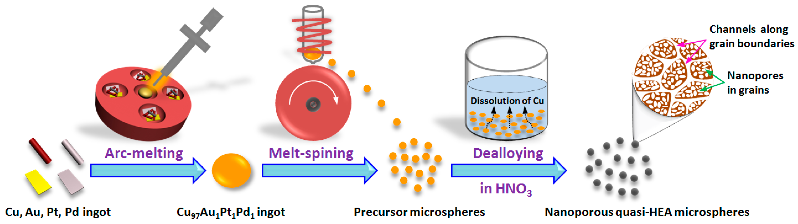
2. Materials and Methods

Cu97Au1Pt1Pd1 (at.%) ingots were first produced by arc-melting of Cu, Au, Pt, and Pd (99.99 wt.%) ingots. This starting alloy composition is designed and inspired by a previous work [11]. In this situation, a small ligament/pore structure can be obtained after dealloying. Then a melt-spinning process [12,13] was performed to obtain the precursor microspheres. In particular, to guarantee the formation of microspheres in the rapid solidification process, the tangent speed of the copper wheel is set to 3000 r/min and the gas pressure for spraying is 1.5 MPa. Finally, nanoporous quasi-HEA microspheres were fabricated by free dealloying [14,15,16] of the precursor microspheres in a 2 M HNO3 solution for 24 h. The preparation process is shown in Figure 1.
The phase composition of samples was verified by an X-ray diffractometer (XRD, D8-Advance Bruker, Karlsruhe, Germany) using Cu Kα radiation at a step rate of 10°/min. The chemical valence state of samples was examined by X-ray photoelectron spectroscopy (XPS, Thermo Fisher Scientific, Waltham, MA, USA). The morphology and chemical composition of samples were characterized by scanning electron microscopy (SEM, Nova nanoSEM 450, 2 kV, FEI, Hillsboro, OR, USA) equipped with an X-ray energy dispersive spectroscope (EDS, Oxford Instruments, Oxford, UK) and transmission electron microscopy (TEM, JEM-2010F, JEOL, Tokyo, Japan). The surface area and pore size distributions of samples were determined by using the Brunauer-Emmett-Teller (BET) or Barrett-Joyner-Halenda (BJH) method, respectively. The particle size and ligament/pore size distribution were measured by Nano Measurer V1.2, software created by Fudan University, Shanghai, China.

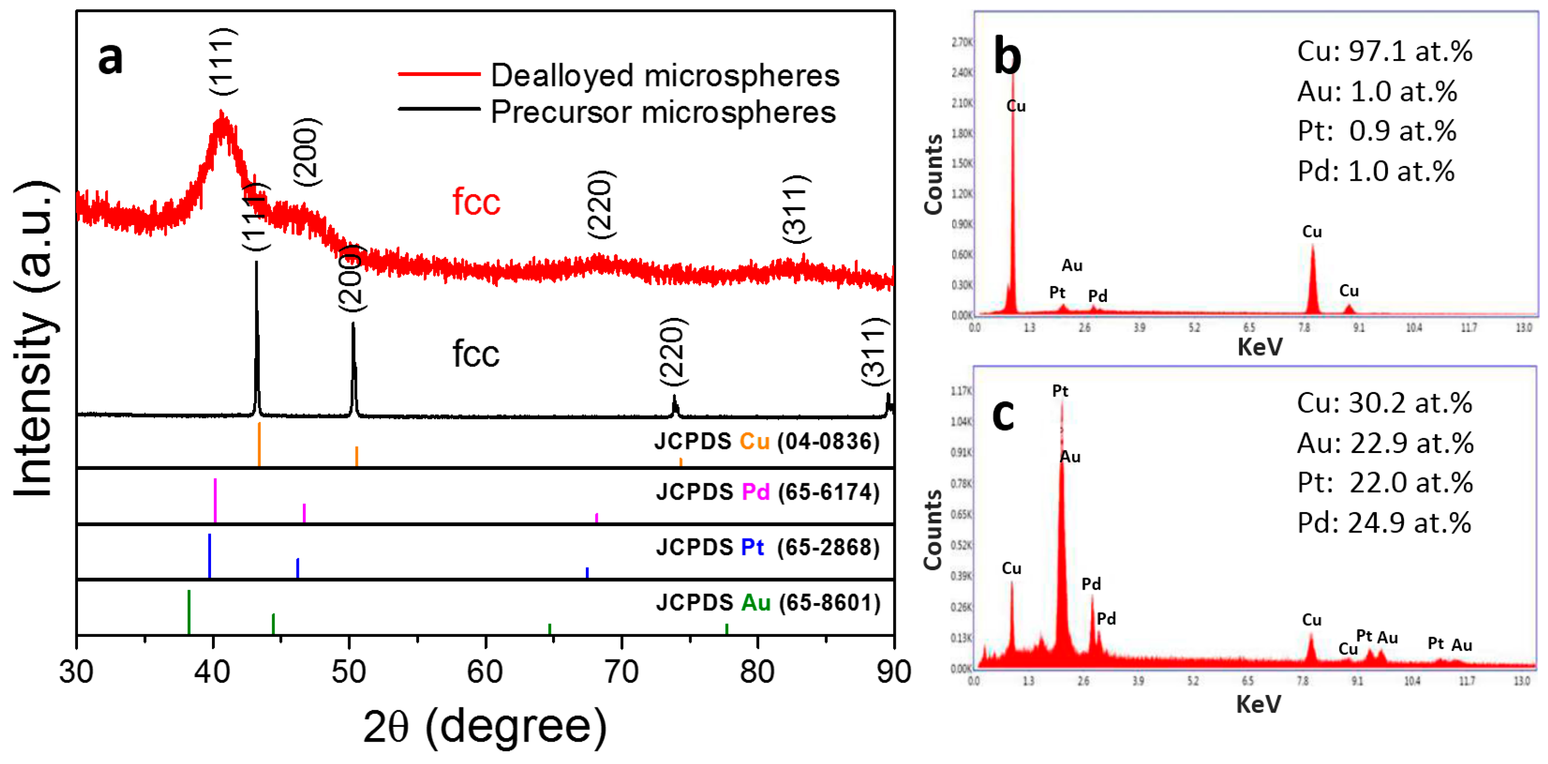
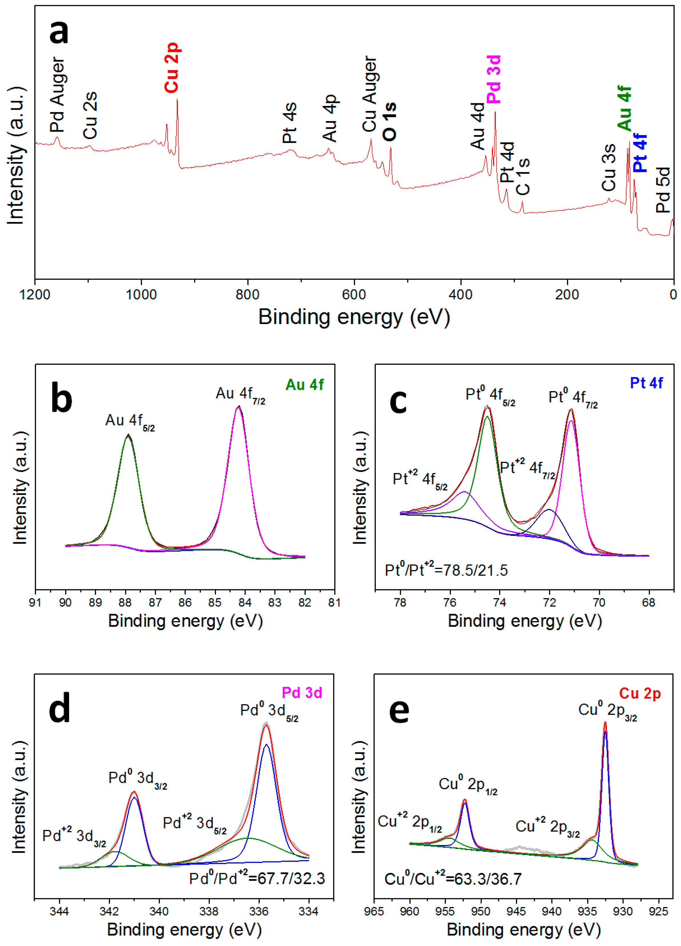
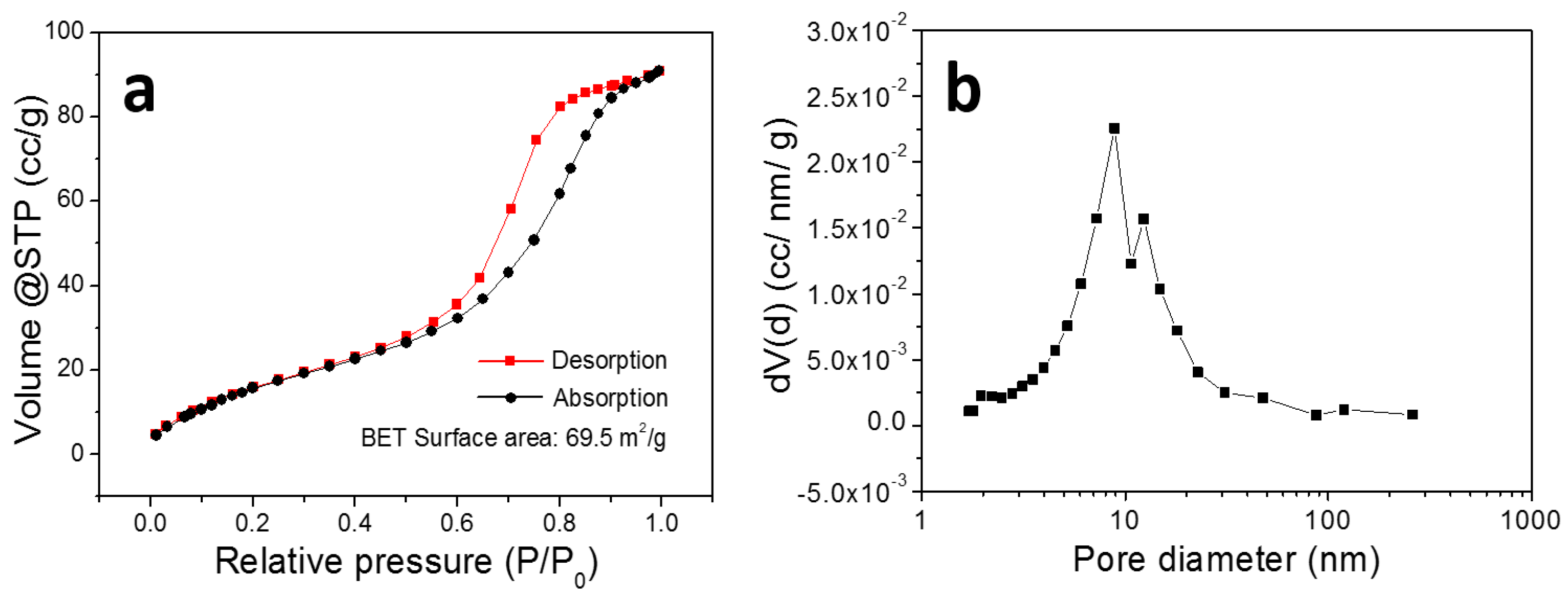
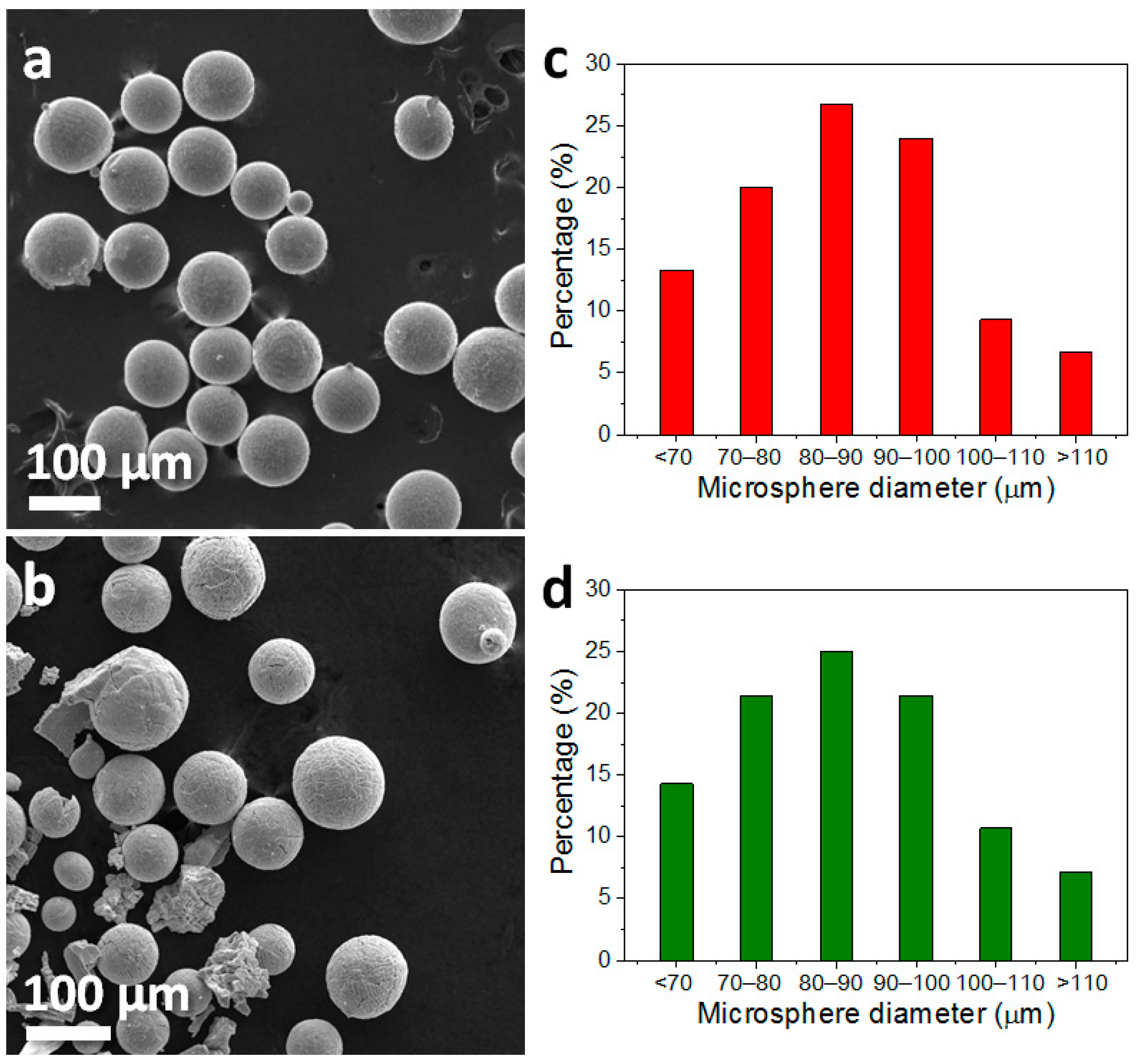
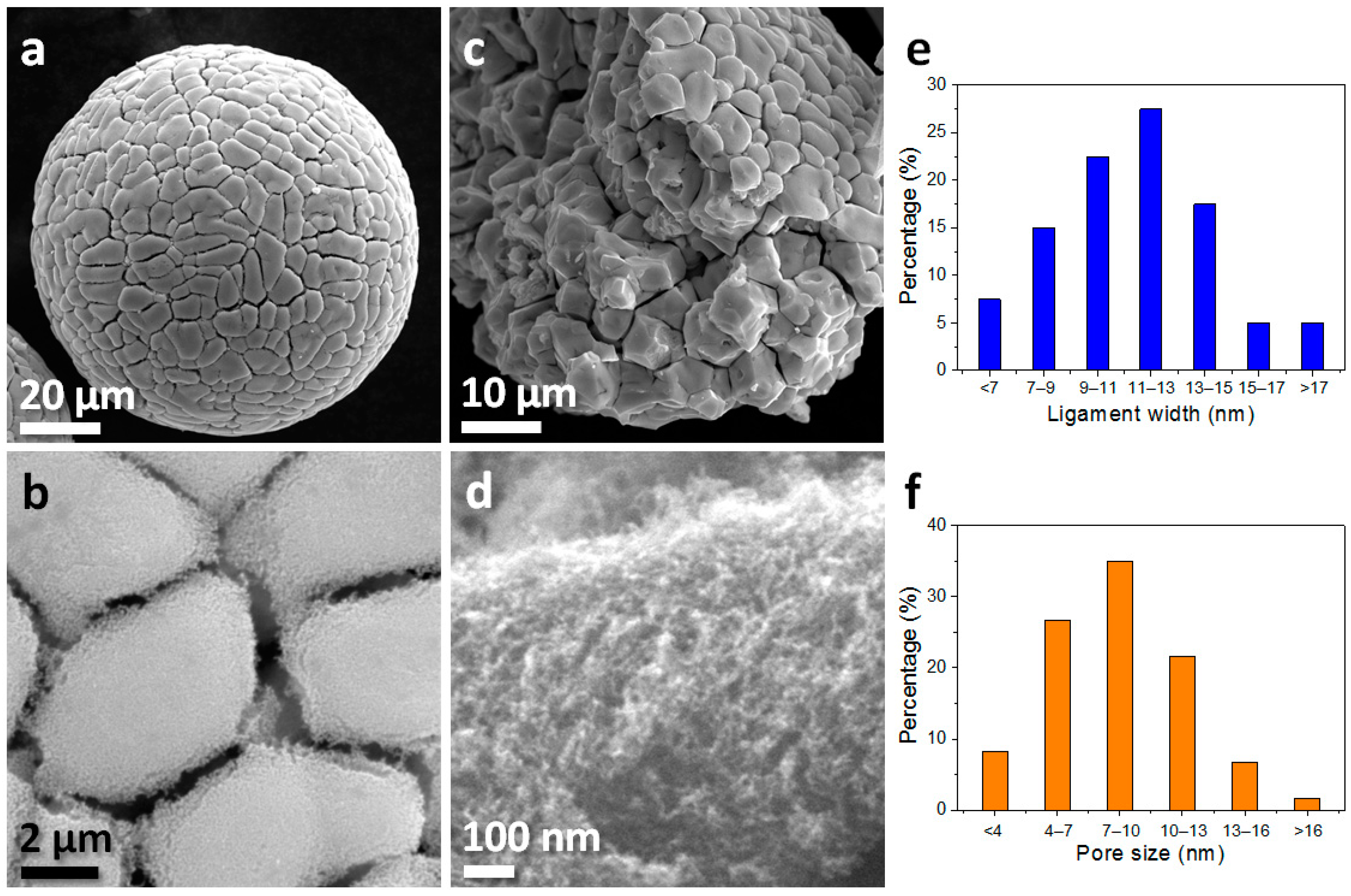
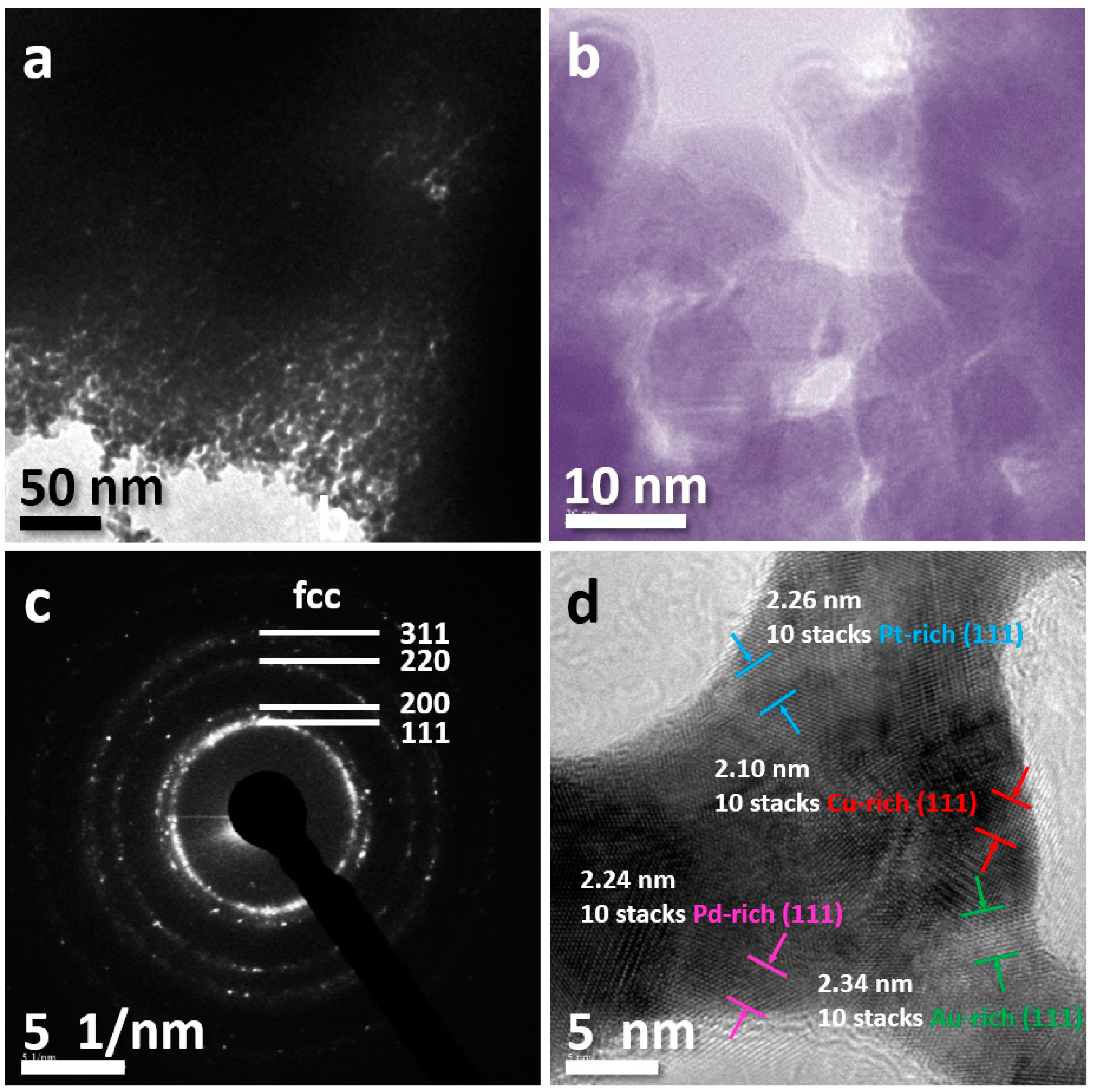
3. Results and Discussion

Figure 2a shows the XRD patterns of the microspheres before and after dealloying. Since the content of Cu is as high as 97 at.% in the precursor, the peak positions are very close to pure Cu. After dealloying, the obvious broad diffraction peaks at 40.7°, 46.8°, 68.2°, and 83.0° can be indexed as (111), (200), (220), and (311) planes of a single-group face-centered cubic (FCC) structure respectively. It is reported that multicomponent noble metal solid solutions usually present broad but not very sharp peaks [17,18] compared with the traditional crystalline XRD patterns with sharp peaks. So the solid solution structure of the as-dealloyed material can be confirmed according to the characteristic of the broad diffraction peaks. In comparison with the XRD spectra of the precursor microspheres, the diffraction peaks of the dealloyed microspheres shift to the lower diffraction angles. This is mainly due to the removal of a large amount of Cu, which is consistent with the EDS results in Figure 2b,c. According to the EDS results, as shown in Figure 2b,c, the composition of microspheres changes from Cu97.1Au1.0Pt0.9Pd1.0 (near the target ratio) to Cu30.2Au22.9Pt22.0Pd24.9 (denoted as Cu30Au23Pt22Pd25) after dealloying.
In order to predict the formation of solid solution structure in the dealloyed microspheres, typical physical parameters including ∆Smix, ∆Hmix, δ and Ω of the product as well as other reported HEAs are calculated by Equations (1)–(4) [19,20,21]:
Δ S mix = R i = 1 n c i ln c i
Δ H mix = i = 1 , i j n 4 Δ H i j mix c i c j
δ = i = 1 n c i ( 1 r i / ( i = 1 n c i r i ) ) 2
Ω = T m Δ S mix / | Δ H mix |
where ΔSmix is the mixing entropy of the alloying elements, ΔHmix is the enthalpy of mixing, δ is the atomic size difference, Ω is a special parameter defined to predict the solid solution formation, n is the total number of components in a system, R (8.31 J·K−1·mol−1) is the gas constant, ci is the molar ratio of the ith component, ri is the atomic radius of the ith component, Δ H i j mix is the enthalpy of mixing between i and j elements, and Tm is the average melting point. It was proposed [22] that the HEAs with simple solid solution structures tend to form when the following empirical rules can be satisfied simultaneously: ΔSmix > 11.3 J·K−1·mol−1, −15 kJ·mol−1 < ∆Hmix < 5 kJ·mol−1, δ < 6.6 and Ω > 1.1. Table 1 lists the above calculated physical parameters of the product and other reported HEAs [23,24,25], indicating that the composition of the product satisfies the empirical rules for the formation of HEAs.
To further investigate the chemical valence states of metal elements in the product, XPS detection was carried out (Figure 3). The XPS fully scanned spectra (Figure 3a) show predominant signals of Au, Cu, Pd, Pt, C (from the atmosphere), and O, indicating that no other impurity elements are present. As shown in Figure 3b, the two peaks located at 84.2 and 87.9 eV can be attributed to Au 4f7/2 and Au 4f5/2 of Au0, respectively [26]. The Pt 4f spectra (Figure 3c) shows two strong peaks at binding energies of 71.1 and 74.5 eV, corresponding to Pt 4f7/2 and Pt 4f5/2 of Pt0, while two weak peaks at 72.0 and 75.4 eV are attributed to Pt2+ 4f7/2 and 4f5/2, respectively [27]. For the Pd 3d5/2 spectra shown in Figure 3d, two peaks located at 335.7 and 336.5 eV can be assigned to Pd0 and Pd2+, respectively [28]. Similarly, the two peaks located at 932.5 and 934.4 eV are attributed to Cu0 and Cu2+ of Cu 2p3/2, respectively (Figure 3e) [29]. The shift of the binding energies of Au, Pt, Pd, and Cu in the product indicates the evident change in the electronic structure, probably owing to the formation of a solid solution structure [30,31]. The existence of very few divalent metals can be attributed to the oxidation of surface elements in air [29]. By calculating the peak areas of M0 and M2+, the results show the percentage of Cu0, Pd0, and Pt0 species are 63.3%, 67.7%, and 78.5%, respectively, indicating that the dealloyed microspheres are mainly in zero valence state.
When conducting further investigation into the porous structure of the as-fabricated samples, nitrogen adsorption-desorption isotherms analysis was employed (Figure 4a). In Figure 4a, a kind of type-IV isotherm characterized by a distinct hysteresis loop [32] was observed, indicating the mesoporous nature of the material. This corresponds well with the pore diameter distribution curve displayed in Figure 4b. The pore size of samples mainly ranges from 4 to 20 nm, and the peak size is about 7.9 nm. Due to the above porous structure characteristics, the specific surface area obtained by BET method reaches 69.5 m2/g.
Figure 5 shows the SEM image of the microspheres with a histogram of microsphere size distribution. One can see that the shape of the precursor is regular and spherical (Figure 5a). After dealloying, the diameter of parts of microspheres decreases (Figure 5b). The size statistics in Figure 5c show that the diameter of the precursor mainly ranges from 70 to 100 μm, with an average diameter of 86 μm. After dealloying, though the main diameter range of microsphere stays around 70‒100 μm, the ratio of microspheres with diameter less than 80 μm increases slightly.
Figure 6 shows plane and cross section view SEM images of the dealloyed microspheres. It can be seen (Figure 6a,b) that there are continuous network-like void channels with a width of ~0.6 μm formed along the grain boundaries of original precursors. That is because Cu atoms are much easier to remove from grain boundaries with abundant crystal defects [33]. These void channels can also be seen inside the microspheres (Figure 6c), indicating that they distribute throughout the whole microspheres. Moreover, highly magnified SEM images (Figure 6b,d) reveal that both the surface and internal grains show nanoporous structures after dealloying. Figure 6e,f present the ligament and pore size distribution of microspheres after dealloying, showing that the size ranges from 7 to 15 nm and from 4 to 13 nm for ligament and pore size, respectively. This result accords with the above pore size distribution curves created by the BJH method.
Further analysis by TEM (Figure 7a,b) reveals a nanoscale bicontinuous ligament/pore structure inside the grains, in which the ligament width and pore sizes are ~12 nm and ~9 nm, respectively. From the above morphological observation, a hierarchical porous structure, containing micro-sized channels along grain boundaries and nanopores inside grains, can be ascribed to the dealloyed microspheres. In addition, the selected area electron diffraction (SAED) pattern in Figure 7c can be ascribed to (111), (200), (220), and (311) diffractions of FCC structure (rather than single-phase Au, Pt, Pd, or Cu diffractions), which is consistent with the crystal planes information of the XRD result. The high-resolution TEM image in Figure 7d shows special solid solution characteristics, in which Au-rich, Pt-rich, Pd-rich, and Cu-rich solid solution areas coexist in the ligament [34,35,36,37]. The Au, Pt, Pd, and Cu elements, with FCC structures, easily form a FCC-type solid solution between each other and among three/four elements for their great miscibility, which may be the reason to form a multiphase solid solution structure. In particular, these multiphase solid solutions possess an approximate component ratio and are connected by many dislocations and defects, so that these solid solution areas cannot be completely distinguished from others. As a result, it is multiphase approximatively componential solid solutions (not four-phase separable solid solution) that create. Thus, the diffraction peaks represent a broad single-group FCC pattern rather than sharp multiple-group FCC patterns in the XRD results.
In this work, the standard equilibrium electrode potential of Cu is much lower than that of Au, Pt, and Pd [38,39,40], which offers a great driving force for the selective dissolution of Cu under free-dealloying conditions. After dealloying for 24 h, the ratio of remaining Cu and the other three elements is nearly equal, and the calculated physical parameters in Table 1 satisfy the empirical rules for forming HEAs. In particular, the as-formed nanoporous structure presents a multiphase approximatively componential solid solution characteristic, which is different from the traditional single-phase or two-phase solid solution HEAs. To differentiate, we call it quasi-HEAs. The as-fabricated nanoporous quasi-HEAs possess plentiful dislocations and defects between two neighboring solid solution areas and a high specific surface area, which may be applied in wide chemical fields [41,42,43]. The higher resolution characterization of quasi-HEAs needs to be examined in future work. For example, spherical aberration-corrected high-angle annular dark field (HAADF) STEM images of nanoporous metals could reveal surface atoms with different coordination numbers [44]. Atomically resolved elemental mapping of the surface of nanoporous electrocatalyst could uncover the surface atomic change before and after a catalytic reaction [45]. With the help of these higher resolution tests, some chemical reaction mechanisms can be clearly revealed. Thus, the exploitation and development of chemical properties of HEAs can be promoted.

4. Conclusions

We developed a facile route to fabricate nanoporous noble metal quasi-HEA microspheres by a melt-spinning and dealloying process. The material presents a hierarchical porous structure with a high specific surface area and a multiphase approximatively componential solid solution characteristic with a broad single-group FCC XRD pattern. Metallic elements such as Au, Pt, Pd, Ag, Ir, Rh, Cu, and Ni with FCC structure can be selected for developing other porous quasi-HEAs by using this route. Moreover, the work may stimulate the overall development of quasi-HEAs in various chemical applications.

Author Contributions

Data curation, L.Y. and Y.L.; Funding acquisition, Z.W.; Investigation, L.Y. and Y.L.; Methodology, Z.W. and W.Z.; Project administration, Z.W.; Resources, C.Q.; Supervision, W.Z. and C.Q.; Writing—original draft, L.Y.; Writing—review & editing, Z.W.

Funding

This work was financially supported by the Key Project of Science & Technology Research of Higher Education Institutions of Hebei Province, China (ZD2018059) and the Natural Science Foundation of Hebei Province, China (E2015202081).

Conflicts of Interest

The authors declare no conflict of interest.

References

  1. Zou, Y.; Ma, H.; Spolenak, R. Ultrastrong ductile and stable high-entropy alloys at small scales. Nat. Commun. 2015, 6, 7748. [Google Scholar] [CrossRef] [Green Version]
  2. Zhang, W.R.; Liaw, P.K.; Zhang, Y. Science and technology in high-entropy alloys. Sci. China Mater. 2018, 61, 2–22. [Google Scholar] [CrossRef] [Green Version]
  3. Gludovatz, B.; Hohenwarter, A.; Catoor, D.; Chang, E.H.; George, E.P.; Ritchie, R.O. A fracture-resistant high-entropy alloy for cryogenic applications. Science 2014, 345, 1153–1158. [Google Scholar] [CrossRef] [Green Version]
  4. Li, Z.M.; Pradeep, K.G.; Deng, Y.; Raabe, D.; Tasan, C.C. Metastable high-entropy dual-phase alloys overcome the strength-ductility trade-off. Nature 2016, 534, 227–230. [Google Scholar] [CrossRef] [PubMed]
  5. Tracy, C.L.; Park, S.; Rittman, D.R.; Zinkle, S.J.; Bei, H.B.; Lang, M.; Ewing, R.C.; Mao, W.L. High pressure synthesis of a hexagonal close-packed phase of the high-entropy alloy CrMnFeCoNi. Nat. Commun. 2017, 8, 15634. [Google Scholar] [CrossRef] [PubMed] [Green Version]
  6. Niu, C.N.; LaRosa, C.R.; Miao, J.S.; Mills, M.J.; Ghazisaeidi, M. Magnetically-driven phase transformation strengthening in high entropy alloys. Nat. Commun. 2018, 9, 1363. [Google Scholar] [CrossRef] [PubMed] [Green Version]
  7. Jo, Y.H.; Jung, S.; Choi, W.M.; Sohn, S.S.; Kim, H.S.; Lee, B.J.; Kim, N.J.; Lee, S. Cryogenic strength improvement by utilizing room-temperature deformation twinning in a partially recrystallized VCrMnFeCoNi high-entropy alloy. Nat. Commun. 2017, 8, 15719. [Google Scholar] [CrossRef] [PubMed] [Green Version]
  8. Yao, Y.G.; Huang, Z.N.; Xie, P.F.; Lacey, S.D.; Jacob, R.J.; Xie, H.; Chen, F.J.; Nie, A.M.; Pu, T.C.; Rehwoldt, M.; et al. Carbothermal shock synthesis of high-entropy-alloy nanoparticles. Science 2018, 359, 1489–1494. [Google Scholar] [CrossRef] [PubMed]
  9. Dan, Z.H.; Qu, J.H.; Yang, Y.L.; Qin, F.X.; Chang, H. Evolution of nanoporous surface layers on gas-atomized Ti60Cu39Au1 powders during dealloying. Nanomaterials 2018, 8, 581. [Google Scholar] [CrossRef] [PubMed]
  10. Wang, Z.F.; Liu, J.Y.; Qin, C.L.; Yu, H.; Xia, X.C.; Wang, C.Y.; Zhang, Y.S.; Hu, Q.F.; Zhao, W.M. Dealloying of Cu-based metallic glasses in acidic solutions: Products and energy storage applications. Nanomaterials 2015, 5, 697–721. [Google Scholar] [CrossRef]
  11. Qiu, H.J.; Gao, J.J.; Chiang, F.K.; Wen, Y.R.; Yao, A.Y.; Du, P.; Fang, G.; Wang, J.Q.; Liu, X.J. A general and scalable approach to produce nanoporous alloy nanowires with rugged ligaments for enhanced electrocatalysis. J Mater. Chem. A 2018, 6, 12541–12550. [Google Scholar] [CrossRef]
  12. Zheng, D.H.; Zhao, F.; Li, Y.Y.; Qin, C.L.; Zhu, J.S.; Hu, Q.F.; Wang, Z.F.; Inoue, A. Flexible NiO micro-rods/nanoporous Ni/metallic glass electrode with sandwich structure for high performance supercapacitors. Electrochim. Acta 2019, 297, 767–777. [Google Scholar] [CrossRef]
  13. Liu, S.Y.; Pang, F.J.; Zhang, Q.W.; Guo, R.J.; Wang, Z.F.; Wang, Y.C.; Zhang, W.Q.; Ou, J.Z. Stable nanoporous Sn/SnO2 composites for efficient electroreduction of CO2 to formate over wide potential range. Appl. Mater. Today 2018, 13, 135–143. [Google Scholar] [CrossRef]
  14. Zhao, W.M.; Fei, P.Y.; Zhang, X.M.; Zhang, Y.G.; Qin, C.L.; Wang, Z.F. Porous TiO2/Fe2O3 nanoplate composites prepared by de-alloying method for Li-ion batteries. Mater. Lett. 2018, 211, 254–257. [Google Scholar] [CrossRef]
  15. Wang, Z.F.; Zhang, X.M.; Yan, Y.H.; Zhang, Y.G.; Wang, Y.C.; Qin, C.L.; Bakenov, Z. Nanoporous GeO2/Cu/Cu2O network synthesized by dealloying method for stable Li-ion storage. Electrochim. Acta 2019, 300, 363–372. [Google Scholar] [CrossRef]
  16. Qin, C.L.; Zhang, Y.S.; Wang, Z.F.; Xiong, H.Q.; Yu, H.; Zhao, W.M. One-step synthesis of CuO@brass foil by dealloying method for low-cost flexible supercapacitor electrodes. J. Mater. Sci. Mater. Electron. 2016, 27, 9206–9215. [Google Scholar] [CrossRef]
  17. Zhang, Q.; Kusada, K.; Wu, D.S.; Yamamoto, T.; Toriyama, T.; Matsumura, S.; Kawaguchi, S.; Kubota, Y.; Kitagawa, H. Selective control of fcc and hcp crystal structures in Au–Ru solid-solution alloy nanoparticles. Nat. Commun. 2018, 9, 510. [Google Scholar] [CrossRef] [Green Version]
  18. Sarker, M.S.I.; Nakamura, T.; Sato, S. All-proportional solid-solution Rh–Pd–Pt alloy nanoparticles by femtosecond laser irradiation of aqueous solution with surfactant. J. Nanopart. Res. 2015, 17, 259. [Google Scholar] [CrossRef]
  19. Yew, J.W.; Chen, S.K.; Lin, S.J.; Gan, J.Y.; Chin, T.S.; Shun, T.T.; Tsau, C.H.; Chang, S.Y. Nanostructured high-entropy alloys with multiple principal elements: Novel alloy design concepts and outcomes. Adv. Eng. Mater. 2004, 6, 299–303. [Google Scholar]
  20. Ma, D.C.; Yao, M.J.; Pradeep, K.G.; Tasan, C.C.; Springer, H.; Raabe, D. Phase stability of non-equiatomic CoCrFeMnNi high entropy alloys. Acta. Mater. 2015, 98, 288–296. [Google Scholar] [CrossRef]
  21. Zhang, Y.; Zuo, T.T.; Tang, Z.; Gao, M.C.; Dahmen, K.A.; Liaw, P.K.; Lu, Z.P. Microstructures and properties of high-entropy alloys. Prog. Mater. Sci. 2014, 61, 1–93. [Google Scholar] [CrossRef]
  22. Miracle, D.B.; Senkov, O.N. A critical review of high entropy alloys and related concepts. Acta. Mater. 2017, 122, 448–511. [Google Scholar] [CrossRef] [Green Version]
  23. Kao, Y.F.; Chen, T.J.; Chen, S.K.; Yeh, J.W. Microstructure and mechanical property of as-cast, -homogenized, and -deformed AlxCoCrFeNi (0 ≤ x ≤ 2) high-entropy alloys. J. Alloys Compd. 2009, 488, 57–64. [Google Scholar] [CrossRef]
  24. Wu, Z.; Bei, H.; Otto, F.; Pharr, G.M.; George, E.P. Recovery, recrystallization, grain growth and phase stability of a family of FCC-structured multi-component equiatomic solid solution alloys. Intermetallics 2014, 46, 131–140. [Google Scholar] [CrossRef]
  25. Sohn, S.W.; Liu, Y.H.; Liu, J.B.; Gong, P.; Prades-Rodel, S.; Blatter, A.; Scanley, B.E.; Broadbridge, C.C.; Schroers, J. Noble metal high entropy alloys. Scr. Mater. 2017, 126, 29–32. [Google Scholar] [CrossRef] [Green Version]
  26. Ge, X.B.; Chen, L.Y.; Zhang, L.; Wen, Y.R.; Hirata, A.; Chen, M.W. Nanoporous metal enhanced catalytic activities of amorphous molybdenum sulfide for high-efficiency hydrogen production. Adv. Mater. 2014, 26, 3100–3104. [Google Scholar] [CrossRef]
  27. Wang, R.Y.; Liu, J.G.; Liu, P.; Bi, X.X.; Yan, X.L.; Wang, W.X.; Ge, X.B.; Chen, M.W.; Ding, Y. Dispersing Pt atoms onto nanoporous gold for high performance direct formic acid fuel cells. Chem. Sci. 2014, 5, 403–409. [Google Scholar] [CrossRef]
  28. Tressaud, A.; Khairoun, H.; Touhara, H.; Watanabe, N. X-ray photoelectron spectroscopy of palladium fluorides. Z. Anorg. Allg. Chem. 1986, 540, 291–299. [Google Scholar] [CrossRef]
  29. Du, Y.X.; Ni, K.; Zhai, Q.X.; Yun, Y.P.; Xu, Y.J.; Sheng, H.T.; Zhu, Y.W.; Zhu, M.Z. Facile air oxidative induced dealloying of hierarchical branched PtCu nanodendrites with enhanced activity for hydrogen evolution. Appl. Catal. A General 2018, 557, 72–78. [Google Scholar] [CrossRef]
  30. Chen, L.Y.; Guo, H.; Fujita, T.; Hirata, A.; Zhang, W.; Inoue, A.; Chen, M.W. Nanoporous PdNi bimetallic catalyst with enhanced electrocatalytic performances for electro-oxidation and oxygen reduction reactions. Adv. Funct. Mater. 2011, 21, 4364–4370. [Google Scholar] [CrossRef]
  31. Xu, C.X.; Wang, R.Y.; Chen, M.W.; Zhang, Y.; Ding, Y. Dealloying to nanoporous Au/Pt alloys and their structure sensitive electrocatalytic properties. Phys. Chem. Chem. Phys. 2010, 12, 239–246. [Google Scholar] [CrossRef]
  32. Dong, C.Q.; Kou, T.Y.; Gao, H.; Peng, Z.Q.; Zhang, Z.H. Eutectic-derived mesoporous Ni-Fe-O nanowire network catalyzing oxygen evolution and overall water splitting. Adv. Eng. Mater. 2018, 8, 1701347. [Google Scholar] [CrossRef]
  33. Liu, X.; Du, J.; Shao, Y.; Zhao, S.F.; Yao, K.F. One-pot preparation of nanoporous Ag-Cu@Ag core-shell alloy with enhanced oxidative stability and robust antibacterial activity. Sci. Rep. 2017, 7, 10249. [Google Scholar] [CrossRef]
  34. Gong, M.X.; Fu, G.T.; Chen, Y.; Tang, Y.W.; Lu, T.H. Autocatalysis and selective oxidative etching induced synthesis of platinum-copper bimetallic alloy nanodendrites electrocatalysts. ACS Appl. Mater. Inter. 2014, 6, 7301–7308. [Google Scholar] [CrossRef]
  35. Xing, Y.L.; Wang, S.B.; Fang, B.Z.; Feng, Y.F.; Zhang, S.C. Three-dimensional nanoporous Cu65Sn5/Cu composite from dealloying as anode for lithium ion batteries. Micropor. Mesopor. Mat. 2018, 261, 237–243. [Google Scholar] [CrossRef]
  36. Hong, J.W.; Kang, S.W.; Choi, B.S.; Kim, D.; Lee, S.B.; Han, S.W. Controlled synthesis of Pd-Pt alloy hollow nanostructures with enhanced catalytic activities for oxygen reduction. ACS Nano 2012, 6, 2410–2419. [Google Scholar] [CrossRef]
  37. Liu, K.; Bai, Y.C.; Zhang, L.; Yang, Z.B.; Fan, Q.K.; Zheng, H.Q.; Yin, Y.D.; Gao, C.B. Porous Au-Ag nanospheres with high-density and highly accessible hotspots for SERS analysis. Nano Lett. 2016, 16, 3675–3681. [Google Scholar] [CrossRef]
  38. Zhou, Q.X.; Qi, L.; Yang, H.X.; Xu, C.X. Hierarchical nanoporous platinum-copper alloy nanoflowers as highly active catalysts for the hydrolytic dehydrogenation of ammonia borane. J. Colloid Inerf. Sci. 2018, 513, 258–265. [Google Scholar] [CrossRef]
  39. Xu, Y.; Yiu, P.M.; Shan, G.C.; Shibayama, T.; Watanabe, S.; Ohnuma, M.; Huang, W.; Shek, C.H. 3D nanoporous gold with very low parting limit derived from Au-based metallic glass and enhanced methanol electro-oxidation catalytic peiformance induced by metal migration. ChemNanoMat 2018, 4, 88–97. [Google Scholar] [CrossRef]
  40. Shi, S.; Markmann, J.; Weissmuller, J. Actuation by hydrogen electrosorption in hierarchical nanoporous palladium. Philos. Mag. 2017, 97, 1571–1587. [Google Scholar] [CrossRef]
  41. Hou, Y.; Chen, L.Y.; Liu, P.; Kang, J.L.; Fujita, T.; Chen, M.W. Nanoporous metal based flexible asymmetric pseudocapacitors. J. Mater. Chem. A 2014, 2, 10910–10916. [Google Scholar] [CrossRef]
  42. Zhang, W.Q.; He, J.; Liu, S.Y.; Niu, W.X.; Liu, P.; Zhao, Y.; Pang, F.J.; Xi, W.; Chen, M.W.; Zhang, W.; et al. Atomic origins of high electrochemical CO2 reduction efficiency on nanoporous gold. Nanoscale 2018, 10, 8372–8376. [Google Scholar] [CrossRef]
  43. Pang, F.J.; Wang, Z.F.; Zhang, K.; He, J.; Zhang, W.Q.; Guo, C.X.; Ding, Y. Bimodal nanoporous Pd3Cu1 alloy with restrained hydrogen evolution for stable and high yield electrochemical nitrogen reduction. Nano Energy 2019, 58, 834–841. [Google Scholar] [CrossRef]
  44. Liu, P.; Guan, P.F.; Hirata, A.; Zhang, L.; Chen, L.Y.; Wen, Y.R.; Ding, Y.; Fujita, T.; Erlebacher, J.; Chen, M.W. Visualizing under-coordinated surface atoms on 3D nanoporous gold catalysts. Adv. Mater. 2016, 28, 1753–1759. [Google Scholar] [CrossRef]
  45. Li, J.; Yin, H.M.; Li, X.B.; Okunishi, E.; Shen, Y.L.; He, J.; Tang, Z.K.; Wang, W.X.; Yücelen, E.; Li, C.; et al. Surface evolution of a Pt–Pd–Au electrocatalyst for stable oxygen reduction. Nat. Energy 2017, 2, 17111. [Google Scholar] [CrossRef]
Figure 1. The synthesis process of nanoporous quasi-HEA microspheres.
Metals 09 00345 g001
Figure 2. (a) XRD patterns of the precursor microspheres before and after dealloying in 2 M HNO3 for 24 h. EDS analysis of the precursor microspheres before (b) and after (c) dealloying in 2 M HNO3 for 24 h.
Metals 09 00345 g002
Figure 3. XPS spectra of the precursor microspheres after dealloying in 2M HNO3 for 24 h: (a) fully scanned spectra; (b) Au 4f; (c) Pt 4f; (d) Pd 3d; (e) Cu 2p.
Metals 09 00345 g003
Figure 4. N2 adsorption desorption isotherms (a) and corresponding BJH pore size distribution curves (b) of nanoporous quasi-HEA microspheres.
Metals 09 00345 g004
Figure 5. SEM images of the precursor microspheres (a) and the dealloyed microspheres (b). Corresponding size statistics of microspheres before (c) and after (d) dealloying.
Metals 09 00345 g005
Figure 6. (ad) SEM images of microspheres after dealloying in 2M HNO3 for 24 h: (a,b) plane view; (c,d) cross section view. Ligament (e) and pore (f) size distribution of microspheres after dealloying.
Metals 09 00345 g006
Figure 7. TEM image (a), corresponding SAED patterns (b), magnified TEM image (c), and high-resolution TEM image (d) of the dealloyed porous microspheres.
Metals 09 00345 g007
Table 1. Calculated parameters of the quasi-HEA microspheres and other reported HEAs.
AlloyPhaseSmix (J·K−1·mol−1)Hmix (kJ·mol−1)δΩReference
Al0.375CoCrFeNiFCC13.0−8.14.12.4[23]
CoCrFeNiFCC11.6−31.36.1[24]
AuPdAgPtFCC11.6−22.27.9[25]
Cu30Au23Pt22Pd25FCC11.5−8.62.31.8This work

