Article

Effect of TiN Coating on the Structure, Mechanical Properties and Fracture of the Mg-Ca-Zn Alloy

by
Anton Khrustalyov
*,
Alexander Monogenov
,
Gulsharat Baigonakova
,
Anastasia Akhmadieva
,
Ekaterina Marchenko
and
Alexander Vorozhtsov
Faculty of Physics and Engineering, National Research Tomsk State University, 36 Lenin Ave., 634050 Tomsk, Russia
*
Author to whom correspondence should be addressed.
Metals 2022, 12(12), 2140; https://doi.org/10.3390/met12122140
Submission received: 16 November 2022 / Revised: 9 December 2022 / Accepted: 10 December 2022 / Published: 13 December 2022
(This article belongs to the Special Issue Advanced Metal Matrix Nanocomposites)

Abstract

:
This paper establishes the optimal thickness of TiN ceramic coating on the surface of Mg-Ca-Zn alloy using optical and scanning microscopy methods. X-ray diffraction analysis and tests on its mechanical properties showed that deposition of coating with a thickness of not less than 1 μm on the alloy causes a uniform distribution of the TiN phase over the magnesium alloy surface. The TiN coating also contributes to simultaneous increases in the yield strength, tensile strength, ductility and microhardness of the Mg-Ca-Zn alloy.

1. Introduction

Metals and alloys are used as implant materials in almost all areas of medicine [1,2]. Implant materials possess high physical and mechanical properties; yet, there is a problem of biochemical and biomechanical compatibility with living tissues [3,4]. Biochemical compatibility is determined by the absence of the response of living tissues to implant introduction and the ability of the body to accumulate a certain amount of implant material without serious aftereffects [3,5,6]. Biomechanical compatibility is ensured by the similarity of the mechanical behavior of the implant to the behavior of living tissue on dependence σ(ε). One of the critical problems in medicine is the creation of an implant that can subsequently dissolve inside the restored bone tissue without repeated surgical intervention and with no harm to the body. The bone structures of a living organism contain magnesium, which participates in biochemical processes and, along with calcium, enhances the restoration of damaged bone tissue [7,8]. The use of biodegradable magnesium alloys will prevent repeated surgical intervention and minimize the difference between the Young’s modulus of the bone and the implant [9]. Despite the obvious advantages of magnesium alloys over conventional materials used in medicine (titanium-based alloys, stainless steel, etc.), their use is rather limited due to their low corrosion resistance in air and physiological media [10,11,12]. Corrosion resistance to the aggressive fluids of the body, as well as the mechanical properties of the alloy, can be increased by alloying agents [13,14] and applying coatings with higher corrosion properties [15,16]. Doping with aluminum, manganese, zinc, and rare earth elements increases corrosion resistance [17,18,19], and calcium additives refine the alloy microstructure [20,21]. The combination of mechanical and corrosion properties with biocompatibility should make it possible to expand the use of magnesium alloys for the manufacture of medical implants. Hence, this study employed a magnesium-based alloying agent with Ca and Zn as the basis. The most appropriate method for producing magnesium alloys is casting [22,23], which allows you to control the amount of alloying elements and has a high performance. The casting process is also associated with the complexity of the homogenization of the structure, the occurrence of segregation zones and defects in the form of pores. Although alloying additives improve corrosion resistance, for magnesium alloys used in medicine, doping with chemical elements is not sufficient; therefore, an additional coating is applied to obtain maximum anti-corrosion properties. Titanium nitride can be regarded as a promising coating for magnesium alloys; it is widely known due to its high hardness, corrosion resistance and thermal stability [24,25,26]. A TiN coating deposited on the Mg-Ca-Zn magnesium alloy provides corrosion protection for up to 4 weeks without change in its shape and mass loss. In vitro testing of the TiN-coated sample showed its higher biocorrosion resistance, low mass loss rate (12% of the initial mass within 4 weeks) in a physiological medium and high cytocompatibility compared with the uncoated sample [27]. A complex use of alloying agents, such as Ca and Zn, and the application of an additional TiN coating will provide a uniform surface structure of the magnesium alloy and improved mechanical and corrosion properties. Further development of biodegradable magnesium-based alloys in combination with titanium nitride deposition requires more data on the structure, composition and mechanical properties, which depend on the coating thickness.
The aim of this study was to analyze the effect of the non-metallic TiN coating on the structure, mechanical properties and fracture of the cast Mg-Ca-Zn alloy. As part of the work, the tasks of obtaining materials, and studying the microstructure, microhardness and mechanical properties in the tension and fracture of Mg-Ca-Zn alloys with different thicknesses of TiN coating were solved.

2. Materials and Methods

Magnesium (99.99%), metallic zinc (99.99%) and calcium (99.99%) of high purity were used as initial materials. Magnesium 1000 g in weight was placed in a steel crucible and melted in inert argon gas supplied as a protective medium. Melting was performed in an open-type muffle furnace with easy access and possibility of melt processing. Argon blowing was carried out throughout the entire process of melt treatment before it was poured into the mold. At a melt temperature of 720 °C, zinc was introduced using a mechanical mixer at a rotation speed of 1200 rpm until complete dissolution; then, it was stirred for 60 s. After that, the melt was stored until its temperature attained 720 °C; then, calcium was introduced into magnesium with a steel bell and subsequent processing with a mechanical mixer was performed for 60 s. Pouring and crystallization of the melt were performed with simultaneous vibration using a vibrating stand with a rigidly attached steel mold. Vibration of the melt proceeded at a frequency of 60 Hz and an amplitude of 0.5 mm. The amount of zinc and calcium in magnesium was 1.5 and 0.5 wt%, respectively.
The coating was magnetron sputtered on the surface of the magnesium alloy sample. The vacuum chamber with the sample was pre-evacuated to a pressure of not less than 1 × 10⁻³ Pa using a cryopump. After that, the parts were cleaned using an ion source with a Hall electron drift in argon plasma at the following parameters: PAr = 0.08 Pa, U = 2500 V and I = 0.15 A for 15 min. During cleaning, negative pulse bias was applied to the parts: Ucm = −150 V and f = 50 kHz. After cleaning, titanium nitride was directly sputtered in a nitrogen atmosphere. PN2 = 0.15 Pa. Arc current of 100 A. Bias parameters: Ucm = −150 V and f = 50 kHz. For uniform coating sputtering, technological equipment was developed to sputter all sides of the sample.
The analyses were carried out with the equipment of Tomsk Regional Core Shared Research Facilities Center of National Research, Tomsk State University. Center was supported by the Ministry of Science and Higher Education of the Russian Federation Grant no. 075-15-2021-693 (no. 13.RFC.21.0012). The microstructure of the Mg-Ca-Zn alloy was studied using a Olympus GX71 (Olympus Scientific Solutions Americas, Waltham, MA, USA).
Electron microscopic studies of the surface microstructure and cross-section of the coated sample, as well as elemental analysis, were performed using a Tescan MIRA 3 LMU (TESCAN ORSAY HOLDING, Brno, Czech Republic) scanning electron microscope equipped with an Oxford Instruments Ultim Max 40 energy-dispersive X-ray spectrometer. The qualitative and quantitative phase composition was studied using X-ray diffraction with a Shimadzu XRD-6000 diffractometer (Shimadzu, Tsukinowa, Japan) in Cu Kα radiation in the standard mode in the symmetric Bragg–Berentano geometry (XRD). The quantitative content of phases in the coating was assessed by full profile Rietveld analysis using the POWDER CELL 2.4 software and the PDF4+ crystal structure database.
Tensile experiments were performed using an Instron 3369 (Instron European Headquarters, High Wycombe, Buckinghamshire, UK) universal electromechanical testing machine at a loading rate of 0.4 mm/min. Each magnesium alloy was subjected to 5 tensile tests. Samples were cut from castings using electroerosion cutting and flat blades with a length and width of the working part of 25 and 6 mm, respectively, a thickness of 2 mm and a rounding radius of 14 mm.
Fractographic studies of sample fractures after tensile tests were conducted using a Quanta 200 3d (FEI Company, Hillsboro, OR, USA) scanning electron microscope. The microhardness of the magnesium alloys was tested using a Metolab 501 (Metolab Company, Moscow, Russia) hardness tester with a load of 50 g imposed on the diamond indenter.

3. Results and Discussion

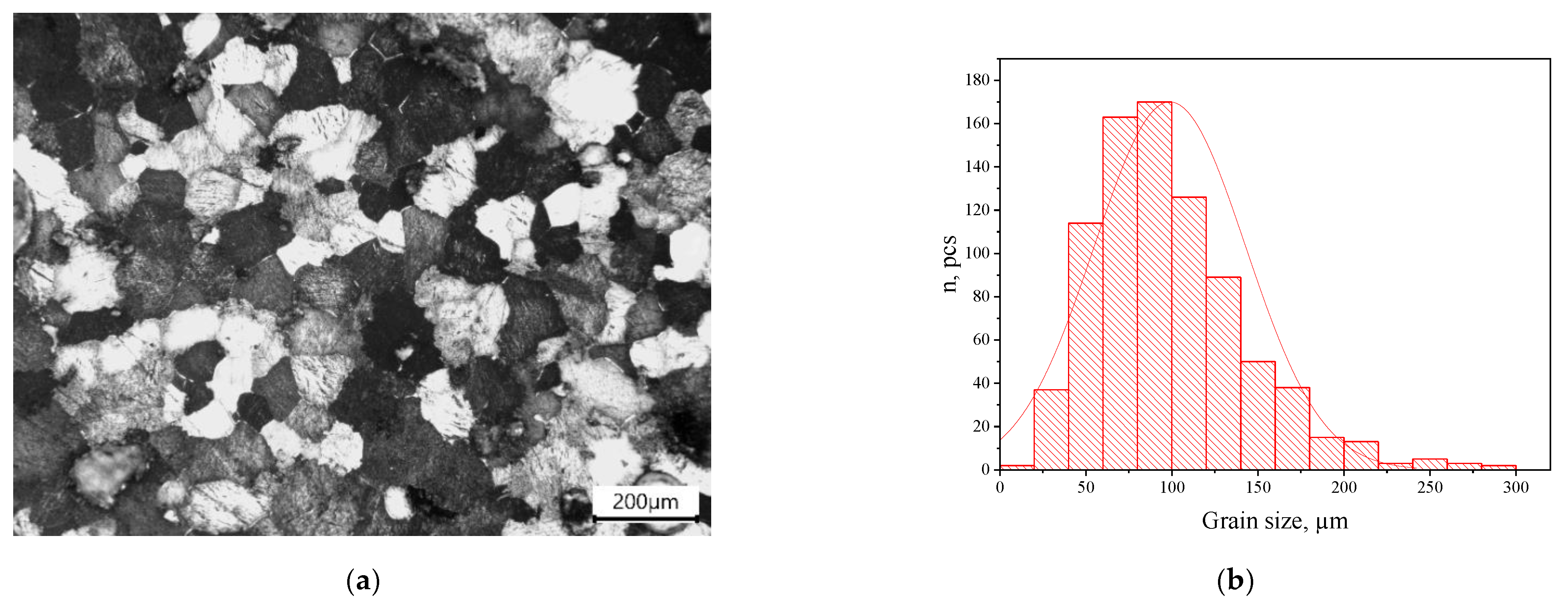
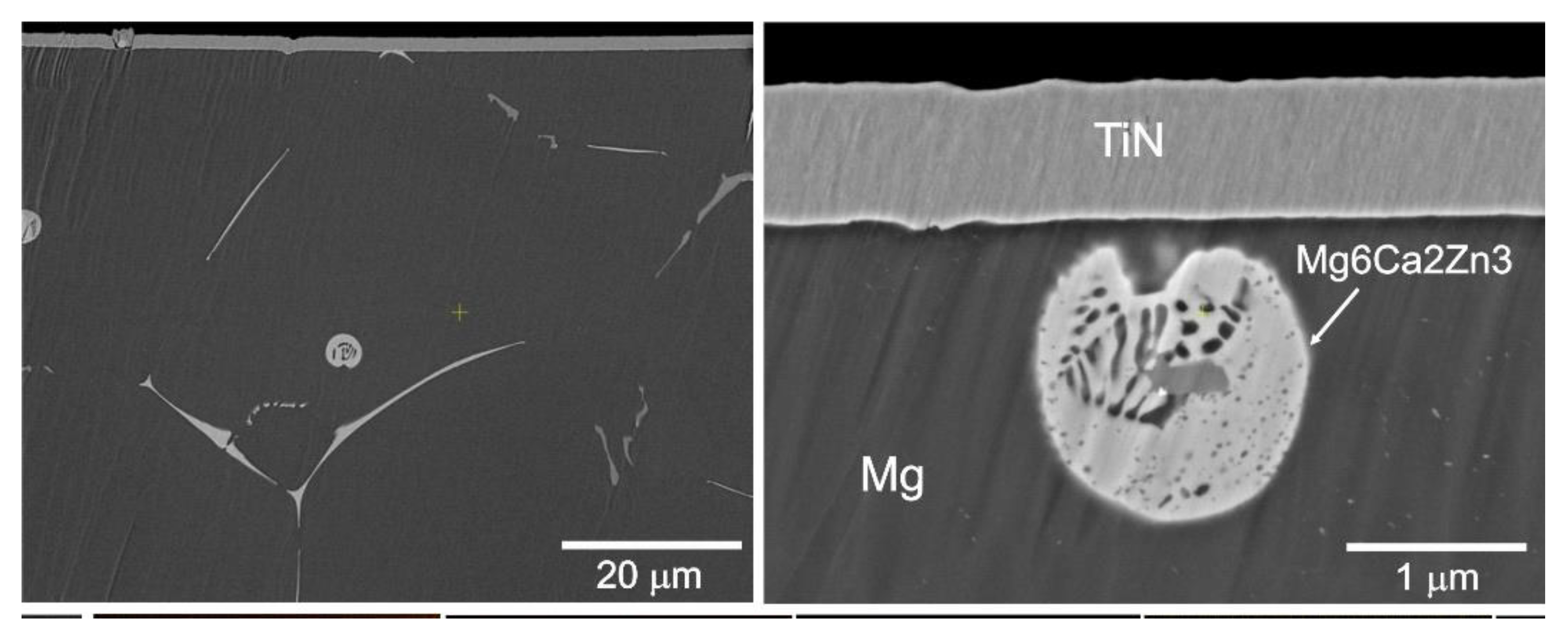
3.1. Microstructure of the Initial Mg-Ca-Zn Alloy

The mechanical properties of Mg-Ca-Zn alloys coated with titanium nitride depend on the uniformity of the microstructure and phase composition. The uncoated Mg-Ca-Zn alloy exhibits a uniform macrostructure with a unimodal grain size distribution (Figure 1). The average grain size in the uncoated alloy is 95 µm. The minimum value is about 10 µm and the maximum value attains 278 µm. The number of measurements performed is 822. The representative sample characterizes the microstructure of the Mg-Ca-Zn alloy with sufficient reliability. The Mg-Ca-Zn alloy microstructure can be classified as uniform due to a large number of grain sizes ranging from 50 to 150 µm (Figure 1b). Consider the Mg-Ca-Zn alloy with titanium nitride magnetron sputtered on the sample surface.

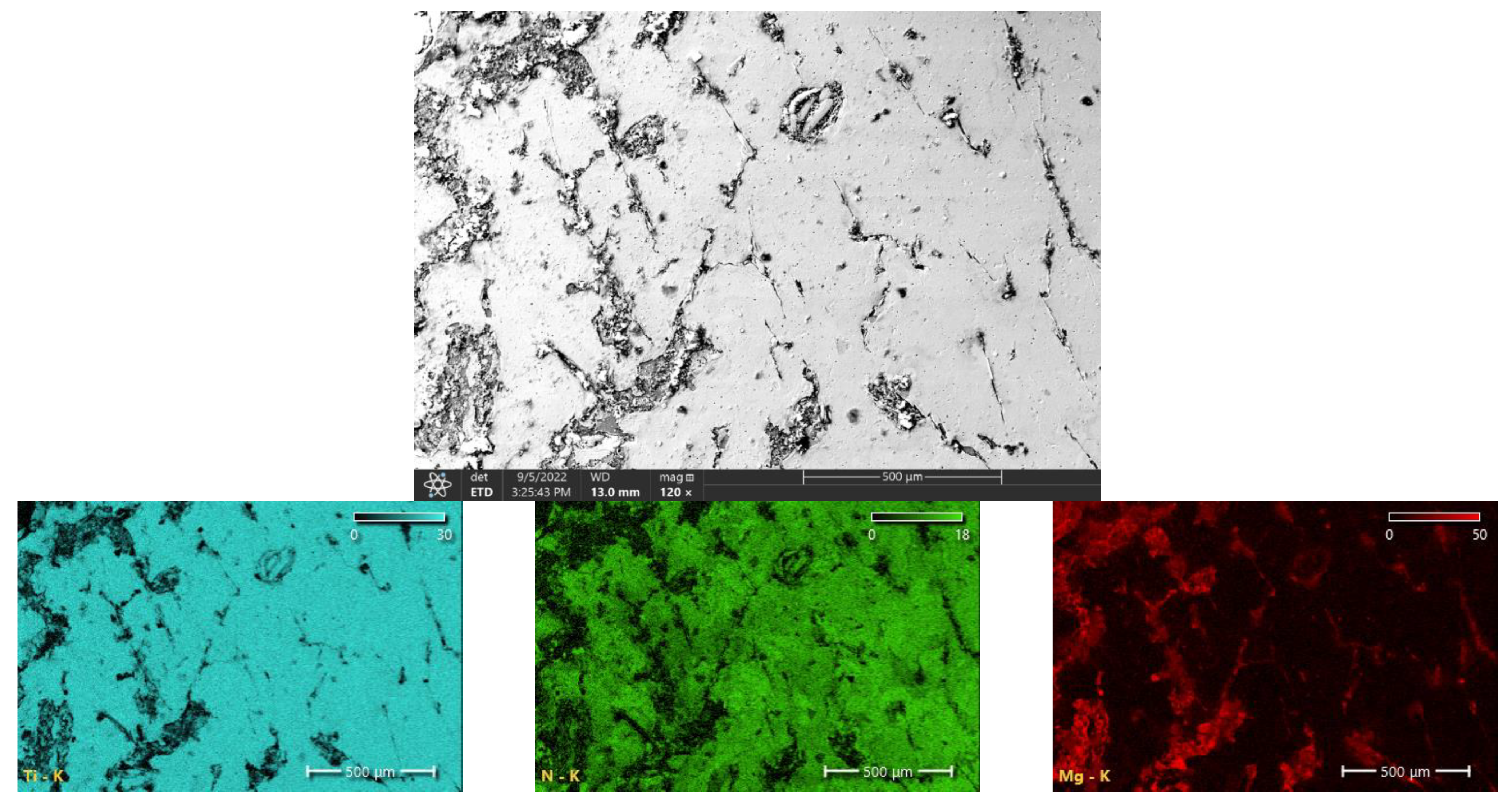
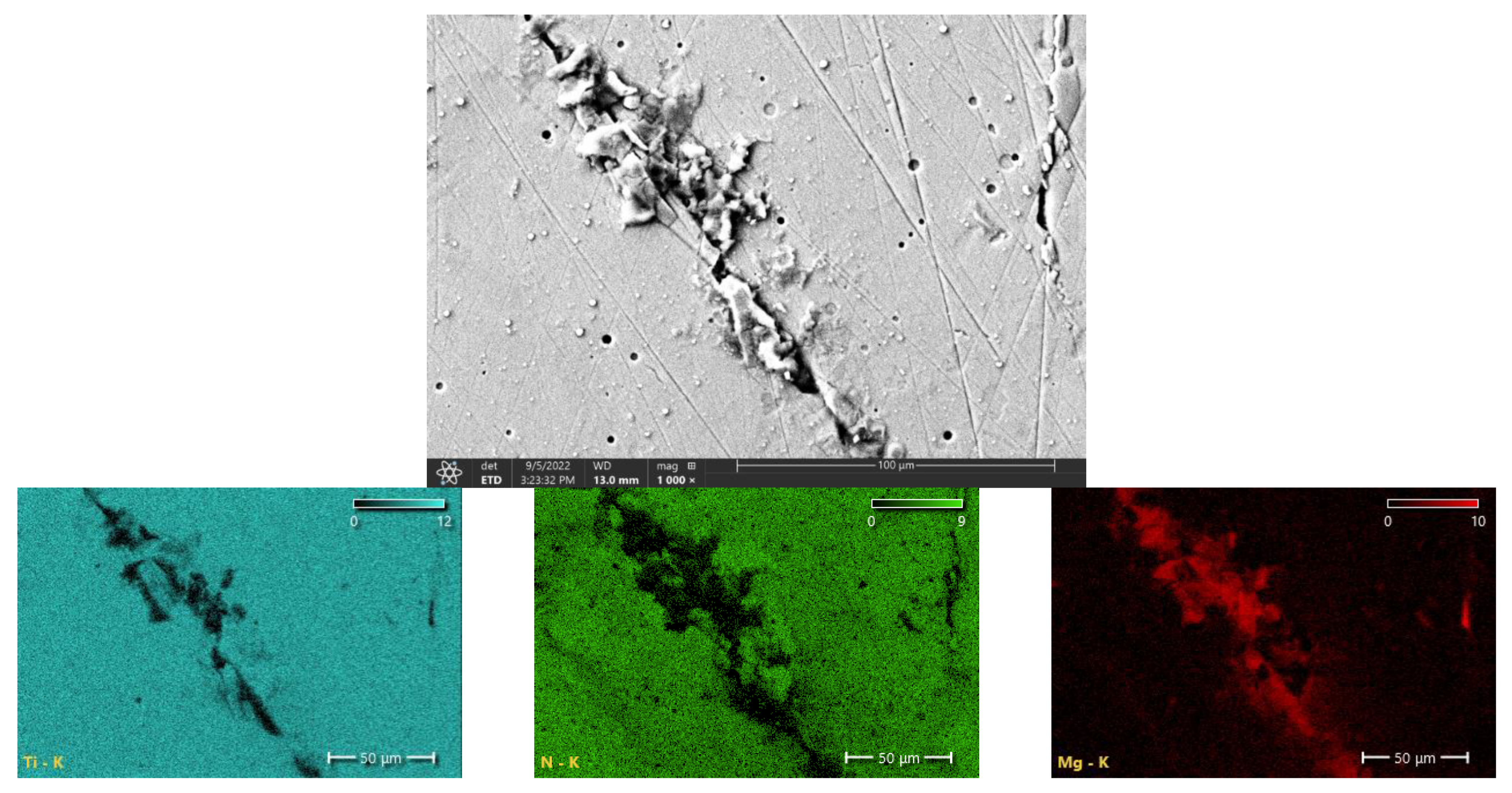
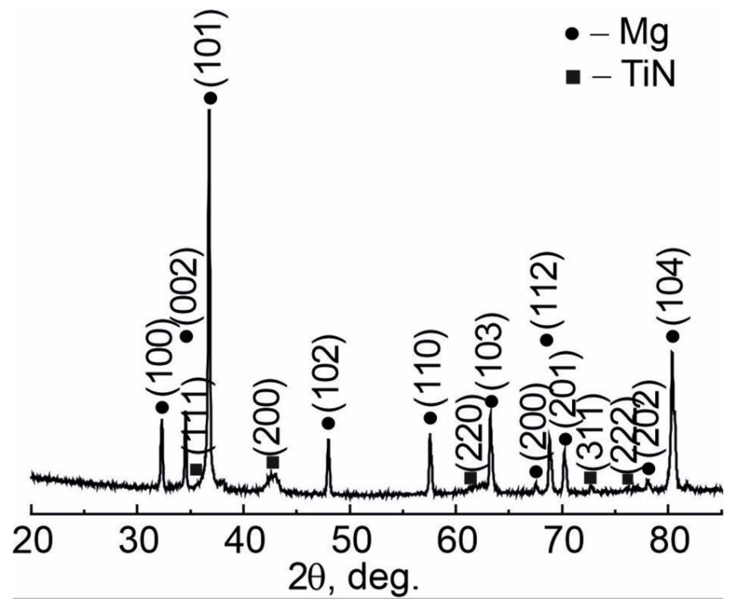
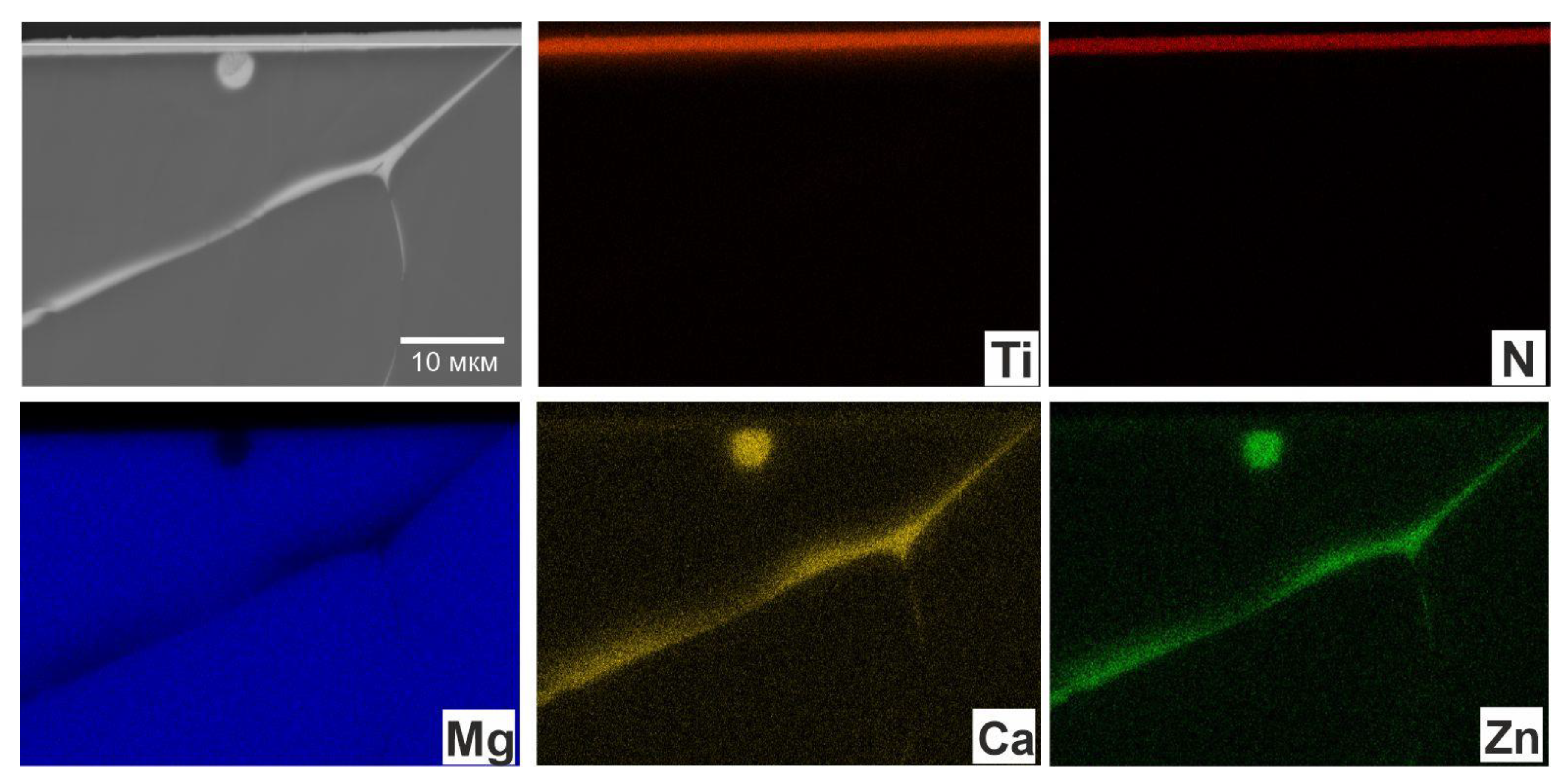
3.2. Microstructure and XRD Analysis of the Mg-Ca-Zn Alloy with a 0.5 µm Thick TiN Coating

The XRD analysis of the Mg-Ca-Zn alloy with a 0.5 μm TiN coating revealed three phases (Figure 2). The main structural component of the alloy is the pure magnesium phase, as evidenced by high-intensity structural reflections from the (100), (002), (101), (102), (110), (104), (112), (103) and (201) planes of the alloy substrate. The analysis also identified reflections from the crystalline phase of the TiN coating and secondary phases of the Mg6Ca2Zn3 matrix. The TiN coating shows a high level of internal microstresses in the crystal lattice due to its imperfect structure (Table 1).
Figure 3 and Figure 4 present the surface morphology and elemental composition of the coating. The morphology of the coating surface is not porous. The coating distribution on the magnesium alloy is nonuniform and, in particular, microcracks are identified. According to the energy-dispersive spectroscopy (EDS) analysis, these microcracks contain Mg. Thus, a 0.5 µm TiN coating is insufficient to provide a continuous uniform coating.
The XRD analysis of the Mg-Ca-Zn alloy with a 1 µm TiN coating revealed three phases: Mg and the TiN phase of the coating. The XRD pattern clearly shows high-intensity diffraction reflections from the pure Mg phase, corresponding to (101), (104), (112), (103) and (002) crystal planes (Figure 5). Structural reflections from the Mg phase are shifted towards larger Bragg angles (2θ) compared with the conventional Mg profile, which indicates a larger lattice parameter for Mg compared with the reference. The TiN coating is fully crystalline, with a preferred orientation along (200). The diffraction reflections from the TiN compound are broadened due to the nanocrystalline structure. The volume ratio of the phases varies depending on the thickness of the coating. The thicker the coatings, the smaller the proportion of the substrate phase. The main phase of Mg has reflexes, the intensity of which is significantly lower than in the previous case, which is explained by the presence of a thicker coating. The lattice parameter of the main phase increases with increasing coating thickness. With an increase in the coating thickness, the crystal lattice of the magnesium phase as a whole expands (in the projection onto the xy plane), while its height decreases. (Table 2).
Figure 6 and Figure 7 present SEM images of the surface morphology and cross-section of the coated sample. The coating surface exhibits a uniform structure with small shallow pinholes 2–5 μm in size. They do not propagate into the coating and therefore cannot form a direct channel between the substrate (Mg-Ca-Zn) and the aggressive medium. In addition to the main Ti and N elements, the element distribution map shows an insignificant amount of oxygen localized in these pinholes.
The SEM image of the coated sample cross-section shows a dense columnar structure with a thickness of 1 µm (Figure 7). Pores, internal defects and microcracks, which initiate channels between the substrate and the aggressive medium to form a galvanic cell and trigger pitting corrosion [28], were not detected over the entire cross-section of the coating. Otherwise, aggressive ions penetrated through these small channels under capillary forces can trigger anodic dissolution at the interfaces of the uncoated areas entailing the remove of the entire coating due to peeling or cracking. Voids or cracks are not detected at the interface between the coating and the Mg-Ca-Zn substrate, which indicates a very dense coating and good adhesion at the interface.
The elemental mapping image of the coating cross-section shows that the layer is composed of Ti and N (Figure 8). The EDS analysis of the substrate presented in Figure 4 shows the formation of Mg-Ca-Zn intermetallic compounds at the grain boundaries in addition to the main Mg phase.

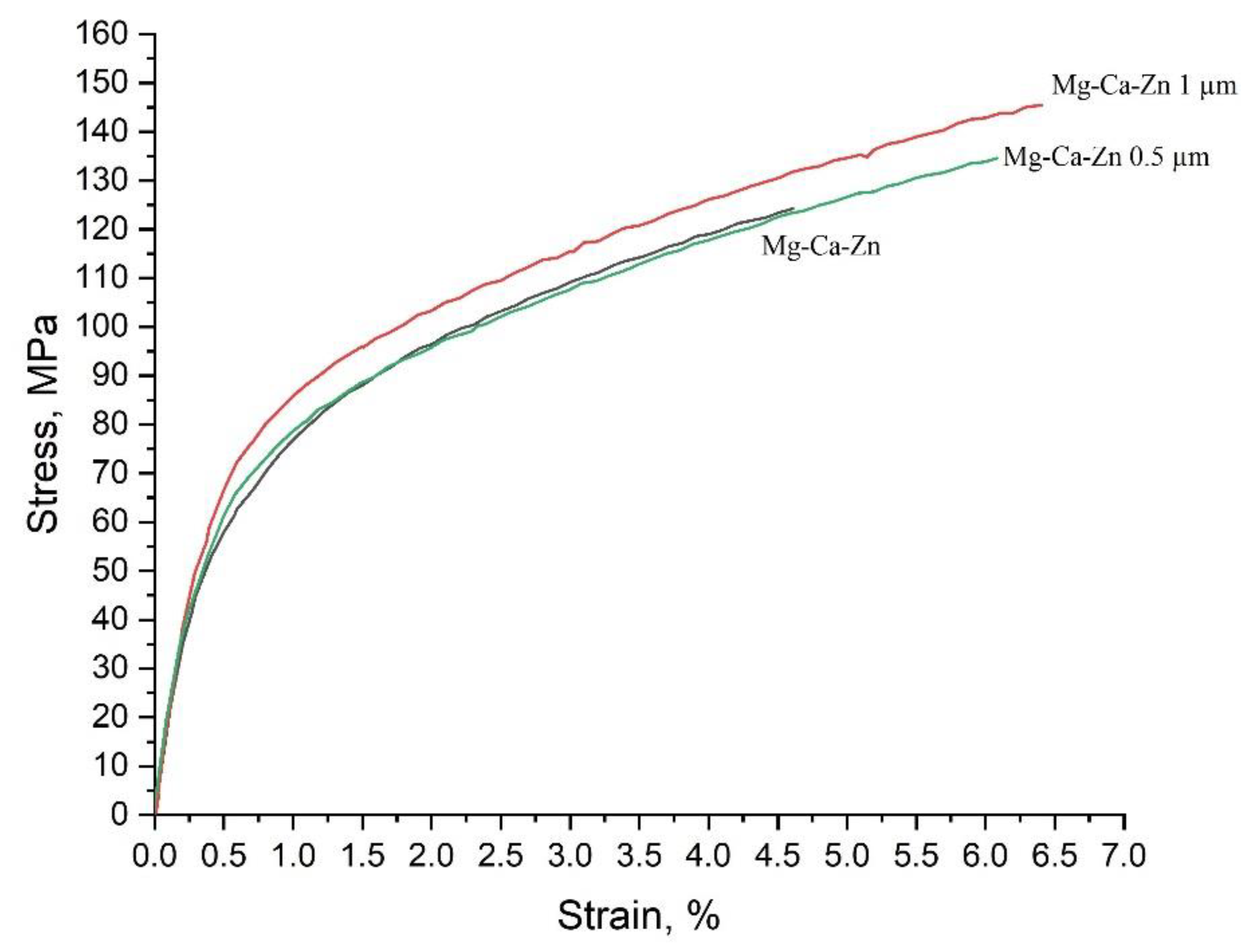
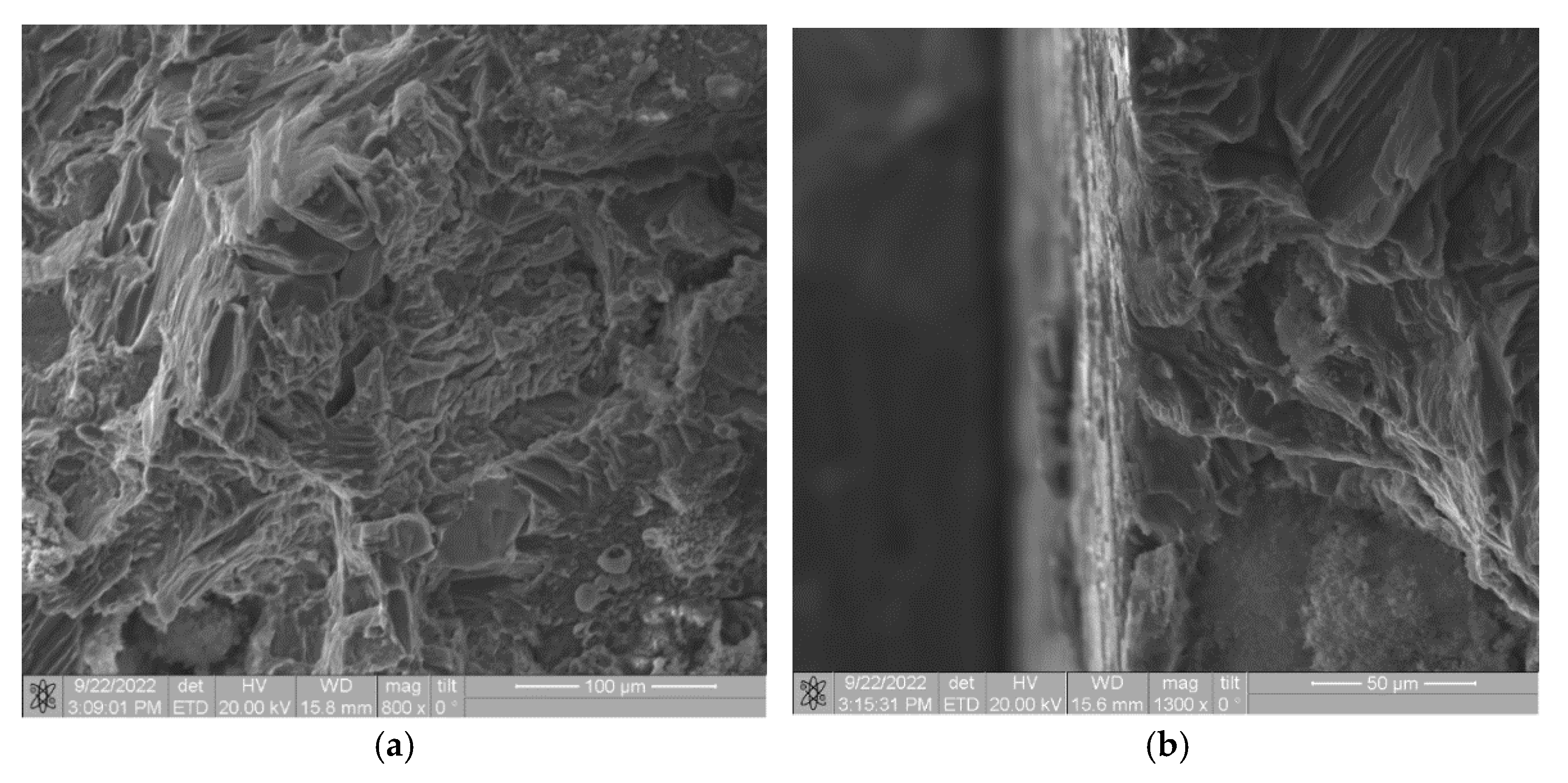
3.3. Mechanical Properties and Fracture of Mg-Ca-Zn Alloys with TiN Coatings of Different Thickness

Tensile tests performed for magnesium alloys (Figure 9, Table 1) showed that a 0.5 µm TiN coating slightly increased the yield strength from 58 to 67 MPa, the tensile strength from 121 to 138 MPa and the maximum accumulated strain up to failure from 4.6 to 6.1%. In this case, tension of the Mg-Ca-Zn + 0.5 μm TiN alloy changed its mechanical properties in a wide range compared with the initial alloy due to the uneven thickness of the TiN coating. When the coating thickness attains 1 µm, the Mg-Ca-Zn alloy exhibited more stable mechanical behavior, with the yield strength increasing from 67 to 74 MPa, tensile strength increasing from 138 to 152 MPa, and the maximum strain up to failure increasing from 6.1 to 6.4%, respectively.
The microhardness of the obtained alloys was studied with regard to its significant impact of the surface layer on the mechanical properties of the alloy (Table 3). Measurements of the surface hardness showed that a 0.5 μm thick coating significantly increased microhardness from 56 to 167 HV. However, the surface of the Mg-Ca-Zn alloy was characterized by non-uniform distribution of microhardness in the range of ±36 HV. Such distribution can be due to defects, including pores, on the surface of cast alloys that do not provide a stable TiN coating at a thickness of 0.5 μm. Analysis of the literature data and the obtained results showed that the heterogeneity, for example, of a ceramic coating, provided a large range of hardness distribution due to the effect of the plastic magnesium matrix [29,30]. The thickness of the TiN coating increased to 1 μm did not affect microhardness relative to the 0.5 μm coating, whereas its microhardness was two-fold higher than that of the initial magnesium alloy. In addition, Table 3 shows that the confidence interval of the hardness measured in the experiment for the 1 μm thick TiN coating is narrower, which eventually maximally increased the yield strength, tensile strength and tensile ductility (Figure 9).
The fractographic failure analysis of the initial Mg-Ca-Zn alloy showed uniform fracture, while cracks found on the alloy surface indicated a brittle fracture (Figure 10a). The revealed brittle fracture was due to the strain concentrated in the area of structural defects of the alloy. Figure 10b shows that on the fracture surface of the Mg-Ca-Zn alloy with a 0.5 μm thick TiN coating, the coating is non-uniformly distributed over the surface (on the right side), which causes an earlier fracture (Figure 9, Table 3). The increased thickness of the TiN coating resulted in the smoothing of the surface defects of the cast alloy, which increased fracture uniformity (Figure 10c).

4. Conclusions

1. The mechanical properties of the Mg-Ca-Zn alloy with a 1 μm TiN coating provide a 20% increase in the yield strength, tensile strength and maximum strain during tensile failure;
2. The 0.5 µm TiN coating increases microhardness of the Mg-Ca-Zn alloy by 100%, while the coating thickness increased up to 1 µm makes the surface hardness more uniform;
3. The 0.5 µm TiN coating is non-continuous and not evenly distributed. The thickness of the TiN coating increased to 1 μm leads to the formation of a uniform continuous layer, which isolates the Mg-Ca-Zn alloy matrix;
4. The XRD analysis revealed no chemical reaction of the substrate coated with titanium nitride, since no additional phases were found at the coating–substrate interface. The thickness of the TiN coating of Mg-Ca-Zn alloys increased up to 1 μm results in a two-fold increase in the level of microdistortions of the crystal lattice of the TiN coating.

Author Contributions

Conceptualization, A.K., A.M. and E.M.; methodology, A.K. and G.B.; investigation, G.B. and A.A.; writing—original draft preparation, A.K. and A.M.; writing—review and editing, A.K., E.M. and A.M.; project administration and funding acquisition, E.M.; supervision, A.V. All authors have read and agreed to the published version of the manuscript.

Funding

This work was carried out with financial support from the Ministry of Science and Higher Education of the Russian Federation (agreement no. 075-15-2021-1384).

Acknowledgments

This work was carried out with financial support from the Ministry of Science and Higher Education of the Russian Federation (agreement no. 075-15-2021-1384).

Conflicts of Interest

The authors declare no conflict of interest.

References

  1. Wu, R.Z.; Yan, Y.D.; Wang, G.X.; Murr, L.E.; Han, W.; Zhang, Z.W.; Zhang, M.L. Recent progress in magnesium–lithium alloys. Int. Mater. Rev. 2015, 60, 65–100. [Google Scholar] [CrossRef]
  2. Dobrzański, L.A.; Dobrzańska-Danikiewicz, A.D.; Dobrzański, L.B.; Dobrzańska, J. The Importance of Synthesis and Characterization of Biomedical Materials for the Current State of Medicine and Dentistry. Processes 2021, 9, 978. [Google Scholar] [CrossRef]
  3. Riaz, U.; Shabib, I.; Haider, W. The current trends of Mg alloys in biomedical applications—A review. J. Biomed. Mater. Res. Part B Appl. Biomater. 2019, 107, 1970–1996. [Google Scholar] [CrossRef] [PubMed]
  4. Huang, J.; Li, X.; Guo, Z.X. Biomechanical and biochemical compatibility in innovative biomaterials. In Biocompatibility and Performance of Medical Devices; Woodhead Publishing: Boston, UK, 2020; pp. 23–46. [Google Scholar] [CrossRef]
  5. Li, G.; Yang, H.; Zheng, Y.; Chen, X.-H.; Yang, J.-A.; Zhu, D.; Ruan, L.; Takashima, K. Challenges in the use of zinc and its alloys as biodegradable metals: Perspective from biomechanical compatibility. Acta Biomater. 2019, 97, 23–45. [Google Scholar] [CrossRef] [PubMed]
  6. Wang, J.; Pang, X.; Jahed, H. Surface protection of Mg alloys in automotive applications: A review. AIMS Mater. Sci. 2019, 6, 567–600. [Google Scholar] [CrossRef]
  7. Li, W.; Qiao, W.; Liu, X.; Bian, D.; Shen, D.; Zheng, Y.; Wu, J.; Kwan, K.Y.H.; Wong, T.M.; Cheung, K.M.C.; et al. Biomimicking Bone–Implant Interface Facilitates the Bioadaption of a New Degradable Magnesium Alloy to the Bone Tissue Microenvironment. Adv. Sci. 2021, 8, 2102035. [Google Scholar] [CrossRef]
  8. Du, H.; Wei, Z.; Liu, X.; Zhang, E. Effects of Zn on the microstructure, mechanical property and bio-corrosion property of Mg–3Ca alloys for biomedical application. Mater. Chem. Phys. 2011, 125, 568–575. [Google Scholar] [CrossRef]
  9. Staiger, M.P.; Pietak, A.M.; Huadmai, J.; Dias, G. Magnesium and its alloys as orthopedic biomaterials: A review. Biomaterials 2006, 27, 1728–1734. [Google Scholar] [CrossRef]
  10. Tsakiris, V.; Tardei, C.; Clicinschi, F.M. Biodegradable Mg alloys for orthopedic implants—A review. J. Magnes. Alloy. 2021, 9, 1884–1905. [Google Scholar] [CrossRef]
  11. Prakasam, M.; Locs, J.; Salma-Ancane, K.; Loca, D.; Largeteau, A.; Berzina-Cimdina, L. Biodegradable Materials and Metallic Implants—A Review. J. Funct. Biomater. 2017, 8, 44. [Google Scholar] [CrossRef] [Green Version]
  12. Wang, J.-L.; Xu, J.-K.; Hopkins, C.; Chow, D.H.-K.; Qin, L. Biodegradable Magnesium-Based Implants in Orthopedics—A General Review and Perspectives. Adv. Sci. 2020, 7, 1902443. [Google Scholar] [CrossRef] [PubMed] [Green Version]
  13. Atrens, A.; Shi, Z.; Mehreen, S.U.; Johnston, S.; Song, G.-L.; Chen, X.; Pan, F. Review of Mg alloy corrosion rates. J. Magnes. Alloy. 2020, 8, 989–998. [Google Scholar] [CrossRef]
  14. Castellani, C.; Lindtner, R.A.; Hausbrandt, P.; Tschegg, E.; Stanzl-tschegg, S.E.; Zanoni, G.; Beck, S.; Weinberg, A.-M. Bone–implant interface strength and osseointegration: Biodegradable magnesium alloy versus standard titanium control. Acta Biomater. 2011, 7, 432–440. [Google Scholar] [CrossRef] [PubMed]
  15. Ding, C.; Tai, Y.; Wang, D.; Tan, L.; Fu, J. Superhydrophobic composite coating with active corrosion resistance for AZ31B magnesium alloy protection. Chem. Eng. J. 2019, 357, 518–532. [Google Scholar] [CrossRef]
  16. Liu, J.; Song, Y.; Chen, J.; Chen, P.; Shan, D.; Han, E.H. The Special Role of Anodic Second Phases in the Micro-galvanic Corrosion of EW75 Mg Alloy. Electrochim. Acta 2016, 189, 190–195. [Google Scholar] [CrossRef]
  17. Jang, H.S.; Seol, D.; Lee, B.J. A comparative study on grain boundary segregation and solute clustering in Mg-Al-Zn and Mg-Zn-Ca alloys. J. Alloys Compd. 2022, 894, 162539. [Google Scholar] [CrossRef]
  18. Wang, X.J.; Xu, D.K.; Wu, R.Z.; Chen, X.B.; Peng, Q.M.; Jin, L.; Xin, Y.C.; Zhang, Z.Q.; Liu, Y.; Chen, X.H.; et al. What is going on in magnesium alloys? J. Mater. Sci. Technol. 2017, 34, 245–247. [Google Scholar] [CrossRef]
  19. Ghali, E.; Dietzel, W.; Kainer, K.U. General and localized corrosion of magnesium alloys: A critical review. J. Mater. Eng. Perform. 2004, 13, 7–23. [Google Scholar] [CrossRef]
  20. Pan, H.; Yang, C.; Yang, Y.; Dai, Y.; Zhou, D.; Chai, L.; Huang, Q.; Yang, Q.; Liu, S.; Ren, Y.; et al. Ultra-fine grain size and exceptionally high strength in dilute Mg–Ca alloys achieved by conventional one-step extrusion. Mater. Lett. 2019, 237, 65–68. [Google Scholar] [CrossRef]
  21. Sahu, M.R.; Kumar, T.S.S.; Chakkingal, U. A review on recent advancements in biodegradable Mg-Ca alloys. J. Magnes. Alloy. 2022, 10, 2094–2117. [Google Scholar] [CrossRef]
  22. Istrate, B.; Munteanu, C.; Antoniac, I.-V.; Lupescu, Ș.-C. Current Research Studies of Mg–Ca–Zn Biodegradable Alloys Used as Orthopedic Implants—Review. Crystals 2022, 12, 1468. [Google Scholar] [CrossRef]
  23. Huang, S.J.; Ali, A.N. Experimental investigations of effects of SiC contents and severe plastic deformation on the microstructure and mechanical properties of SiCp/AZ61 magnesium metal matrix composites. J. Mater. Process. Technol. 2019, 272, 28–39. [Google Scholar] [CrossRef]
  24. Tian, B.; Yue, W.; Fu, Z.; Gu, Y.; Wang, C.; Liu, J. Microstructure and tribological properties of W-implanted PVD TiN coatings on 316L stainless steel. Vacuum 2014, 99, 68–75. [Google Scholar] [CrossRef]
  25. Łępicka, M.; Grądzka-Dahlke, M.; Pieniak, D.; Pasierbiewicz, K.; Kryńska, K.; Niewczas, A. Tribological performance of titanium nitride coatings: A comparative study on TiN-coated stainless steel and titanium alloy. Wear 2019, 422, 68–80. [Google Scholar] [CrossRef]
  26. Ou, Y.X.; Lin, J.; Tong, S.; Sproul, W.D.; Lei, M.K. Structure, adhesion and corrosion behavior of CrN/TiN superlattice coatings deposited by the combined deep oscillation magnetron sputtering and pulsed dc magnetron sputtering. Surf. Coat. Technol. 2016, 293, 21–27. [Google Scholar] [CrossRef]
  27. Baigonakova, G.; Marchenko, E.; Zhukov, I.; Vorozhtsov, A. Structure, cytocompatibility and biodegradation of nanocrystalline coated Mg–Ca–Zn alloys. Vacuum 2023, 207, 111630. [Google Scholar] [CrossRef]
  28. Zhang, Z.; Zhao, Z.; Bai, P.; Li, X.; Liu, B.; Tan, J.; Wu, X. In-situ monitoring of pitting corrosion of AZ31 magnesium alloy by combining electrochemical noise and acoustic emission techniques. J. Alloys Compd. 2021, 878, 160334. [Google Scholar] [CrossRef]
  29. Habibnejad-Korayem, M.; Mahmudi, R.; Ghasemi, H.M.; Poole, W.J. Tribological behavior of pure Mg and AZ31 magnesium alloy strengthened by Al2O3 nano-particles. Wear 2010, 268, 405–412. [Google Scholar] [CrossRef]
  30. Lin, C.J.; Chen, K.C.; He, J.L. The cavitation erosion behavior of electroless Ni-P-SiC composite coating. Wear 2006, 261, 1390–1396. [Google Scholar] [CrossRef]
Figure 1. Microstructure of the Mg-Ca-Zn alloy (a) and grain size distribution (b).
Metals 12 02140 g001
Figure 2. XRD pattern of the Mg-Ca-Zn alloy with a 0.5 µm TiN coating.
Metals 12 02140 g002
Figure 3. SEM images of the surface morphology of the 0.5 µm TiN coating with element distribution maps for Ti, N and Mg (120× magnification).
Metals 12 02140 g003
Figure 4. SEM images of the surface morphology of the 0.5 µm TiN coating with element distribution maps for Ti, N and Mg (1000× magnification).
Metals 12 02140 g004
Figure 5. XRD pattern of the Mg-Ca-Zn alloy with a 1 µm TiN coating.
Metals 12 02140 g005
Figure 6. SEM image of the surface morphology of the 1 μm TiN coating of the Mg-Ca-Zn alloy with element distribution maps for Ti, N, O and C.
Metals 12 02140 g006
Figure 7. SEM image of the cross section of the Mg-Ca-Zn alloy with a 1 µm thick TiN coating.
Metals 12 02140 g007
Figure 8. EDS map of the cross section of the Mg-Ca-Zn alloy with a 1 µm TiN coating.
Metals 12 02140 g008
Figure 9. Diagrams of tension tests of magnesium alloys Mg-Ca-Zn.
Metals 12 02140 g009
Figure 10. Fracture surface of the Mg-Ca-Zn alloy (a), Mg-Ca-Zn 0.5 µm (b) and Mg-Ca-Zn 1 µm (c).
Metals 12 02140 g010aMetals 12 02140 g010b
Table 1. Calculated values of the structural parameters for the Mg-Ca-Zn alloy with a 0.5 µm TiN coating.
PhaseVolume Fraction, vol. %Lattice Parameters, Å and Unit Cell Volume, Å3CSR
Dimensions, nm
Microdistortions, Δd/d
Mg91a = 3.1775
c = 5.2826
V = 46.1902
c/a = 1.6625
591.7 × 10⁻³
Mg6Ca2Zn34a = 10.0236
c = 96067
V = 835.39
371.4 × 10⁻³
TiN5a = 4.3199
V = 80.6160
312.6 × 10⁻³
Table 2. Calculated values obtained via processing of the XRD pattern of the Mg-Ca-Zn alloy with a 1 µm TiN coating.
PhaseVolume Fraction, vol. %Lattice Parameters, Å and Unit Cell Volume, Å3CSR
Dimensions, nm
Microdistortions, Δd/d
Mg78a = 3.2047
c = 5.2052
V = 46.2959
c/a = 1.6242
581.7 × 10⁻³
TiN19a = 4.2896
V = 78.9315
212.5 × 10⁻³
Table 3. Mechanical properties of the obtained magnesium alloys.
AlloyMicrohardness, HVσ0.2, MPaσB, MPaδ, %
Mg-Ca-Zn56 ± 458 ± 4121 ± 84.6 ± 0.3
Mg-Ca-Zn 0.5131 ± 3667 ± 8138 ± 76.1 ± 0.5
Mg-Ca-Zn 1132 ± 1674 ± 2152 ± 56.4 ± 0.1
Publisher’s Note: MDPI stays neutral with regard to jurisdictional claims in published maps and institutional affiliations.

