Article

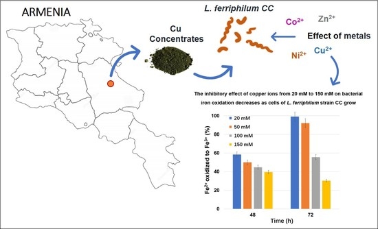
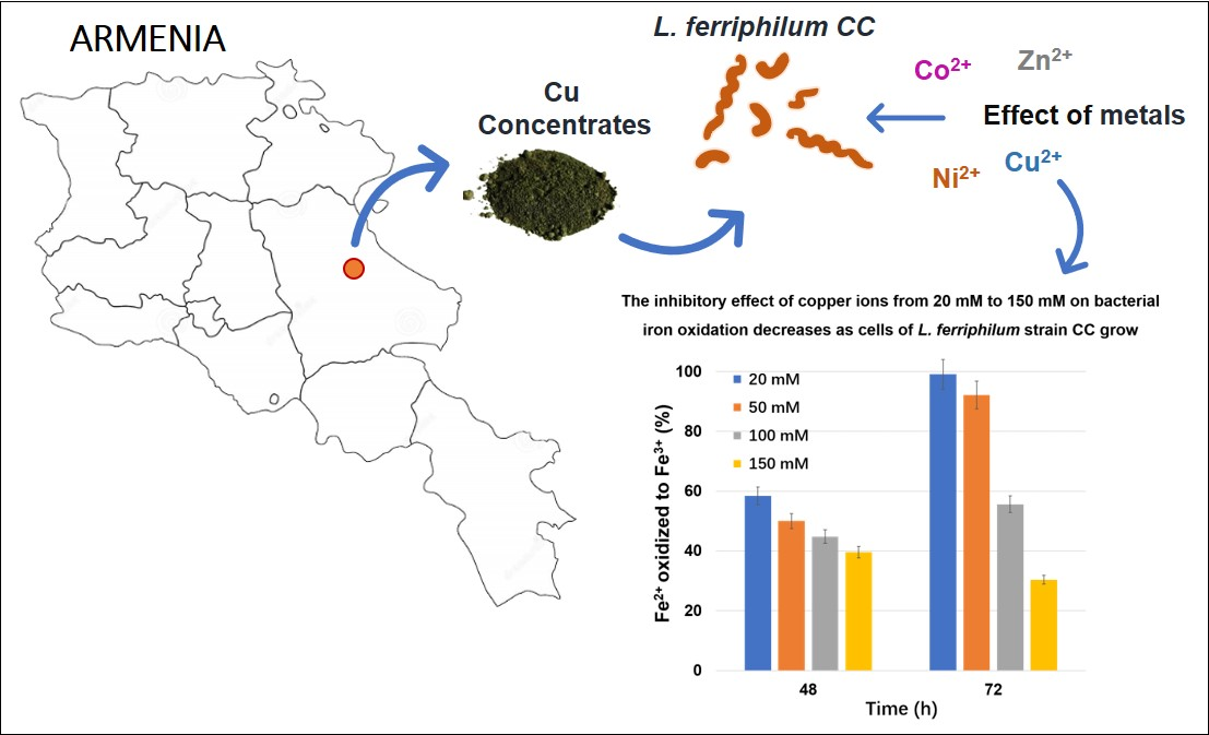
The Effect of Metal Ions on the Growth and Ferrous IronOxidation by Leptospirillum ferriphilum CC Isolated from Armenia Mine Sites

1
Department of Microbiology, SPC “Armbiotechnology” of the National Academy of Sciences of Armenia, 14 Gyurjyan Str., Yerevan 0056, Armenia
2
Key Laboratory of Marine Environmental Corrosion and Biofouling, Institute of Oceanology, Chinese Academy of Sciences, No. 7 Nanhai Road, Qingdao 266071, China
3
Center for Ocean Mega-Science, Chinese Academy of Sciences, No. 7 Nanhai Road, Qingdao 266071, China
4
Open Studio for Marine Corrosion and Protection, Pilot National Laboratory for Marine Science and Technology (Qingdao), No. 1 Wenhai Road, Qingdao 266237, China
5
Department of Applied Mathematics, Materials Science and Engineering and Electronic Technology, School of Experimental Sciences and Technology, Rey Juan Carlos University, 28935 Móstoles, Spain
*
Authors to whom correspondence should be addressed.
Metals 2021, 11(3), 425; https://doi.org/10.3390/met11030425
Submission received: 20 January 2021 / Revised: 22 February 2021 / Accepted: 27 February 2021 / Published: 5 March 2021
(This article belongs to the Special Issue Leaching/Bioleaching and Recovery of Metals)

Abstract

:
The aim of this study is to investigate the potential of newly isolated strain Leptospirillum (L.) ferriphilum CC for bioleaching of pyrite and chalcopyrite in pure or mixed culture with other iron- and/or sulfur-oxidizing bacteria. In this paper, kinetics of ferrous iron (Fe2+) oxidation by newly isolated strain Leptospirillum (L.) ferriphilum CC was studied. The effect of initial Fe2+ in the concentration range of 50–400 mM on bacterial growth and iron oxidation was studied. It was shown that microbial Fe2+ oxidation was competitively inhibited by Fe3+. The influence of copper, zinc, nickel and cobalt ions on the oxidation of Fe2+ by L. ferriphilum CC was also studied. Minimal inhibitory concentrations (MIC) for each metal ion were determined. The toxicity of the ions was found to be as follows: Co > Zn > Ni > Cu. The comparison of iron oxidation kinetic parameters of L. ferriphilum CC with other strains of L. ferriphilum indicates the high potential of strain L. ferriphilum CC for biogenic regeneration of concentrated ferric iron (Fe3+) in bioleaching processes of ores and ore concentrates. Bioleaching tests indicated that the newly isolated L. ferriphilum CC can be a prospective strain for the bioleaching of sulfide minerals in pure culture or in association with other iron- and/or sulfur-oxidizing bacteria.

Graphical Abstract

1. Introduction

Bioleaching is an environmentally friendly technology that is increasingly applied worldwide for processing of mineral raw materials and for recovery of copper, uranium and gold from low-grade ores and waste materials [1]. Bioleaching technology converts an insoluble valuable metal sulfide into a soluble form by means of microorganisms or destroys the lattice of the sulfide minerals to make the gold available for further extraction by cyanidation [2,3]. Studies have shown that Leptospirillum sp. (mainly Leptospirillum ferriphilum) are the dominant iron-oxidizing bacteria in gold-bearing arsenopyrite (FeAsS) and pyrite (FeS2) in biooxidation reactors functioning at or over 40 °C [4,5]. A high Fe3+/Fe2+ ratio, elevated temperatures (40 °C), as well as extremely low pH values (pH 1.0) are the most favorable conditions for the growth of the bacteria of the genus Leptospirillum.
It is considered that the main mechanism of microbial attack on the metal sulfides is an indirect contact mechanism. Metal sulfide is oxidized by Fe3+ and the role of microorganisms refers to the oxidation of Fe2+ and regeneration of Fe3+. The importance of microbial Fe2+ oxidation in bioleaching of sulfide minerals is well known, and widely reported in the literature [6,7]. During bioleaching processes, the sulfide minerals are chemically oxidized by Fe3+ (Equation (1)). The resulting Fe2+ is then regenerated biologically by microorganisms (Equation (2)) [7,8].
2MS + 4Fe3+ → 2M2+ + 4Fe2+ + 2S0
4Fe2+ + 4H+ + O2 → 4Fe3+ + 2H2O
Therefore, the influence of Fe3+ and Fe2+ ions on the growth of bacteria and Fe2+ oxidation activity often determines the intensity of metal bioleaching processes [9,10].
The most important microorganisms involved in the regeneration of Fe3+ iron, responsible for the oxidation of exposed sulfide minerals are Fe2+ oxidizing bacteria Acidithiobacillus (At.) ferrooxidans, Leptospirillum (L.) ferrooxidans, L. ferriphilum, Sulfobacillus (Sb.) thermosulfidooxidans, Acidimicrobium ferroooxidans. Currently, the mechanism and kinetics of Fe2+ oxidation is well studied in At. ferrooxidans. However, during recent decades, Leptospirillum species have increasingly attracted the attention of researchers as alternative iron oxidizers. This interest can be attributed to the fact that compared to the acidithiobacilli, leptospirilli are more tolerant to lower pH and higher cultivation temperature [7,11], besides leptospirilli possess higher iron oxidation activity, compared with Acidithiobacillus sp. and Sulfobacillus sp. bacteria [12]. These properties make the species of genera Leptospirillum potential candidates for the development of new technologies based on the biological Fe2+ oxidation.
Dissolution of metal sulfides results in low pH environments containing highly elevated concentrations of transition and non-transition metals. Metals at high concentrations disrupt cell membranes, alter enzymatic specificity, hinder cellular functions, and damage DNA [13]. The study of the resistance of acidophilic bacteria to metal ions is limited only to the Gram-negative bacterium At. ferrooxidans. At. ferrooxidans is resistant to high concentrations of copper (up to 800 mM CuSO4) and other metals [14,15]. L. ferrooxidans is known to be resistant to 5 mM Cu2+ in the medium [16]. There are no data on the resistance of L. ferriphilum to copper. The resistance of bacteria to copper is very important from the point of view of their application in biotechnological processes, where the concentration of copper ions can vary in the range from 15 mM to 100 mM [17].
Gram-negative bacteria have a mechanism for active removal of copper from the cytoplasm into the periplasmic space with the help of P-type ATP-ases, localized in the inner membrane [18]. Some microorganisms can pump copper from the cytoplasm directly into the extracellular space using a system of resistant nodule cell division (RND). This type of detoxification is well known and described in E. coli [19]. It has also been reported about the ability of certain bacteria to bind copper in the periplasmic space with copper chaperones [14,20].
It is shown that At. ferrooxidans ATCC 23270 can function at high copper concentrations due to approximately 10 genes in the chromosome, which are directly related to its resistance to copper. They include three genes encoding P-type ATP-ases that are related to its resistance to copper transport (copA1Af, copA2Af, and copBAf), three RND-related genes responsible for removing copper from the cell (cusAAf, cusBAf, and cusCAf) and two genes, encoding periplasmic chaperones for copper (cusFAf and copCAf) [15,21].
Some of these genes associated with copper resistance are also found in Leptospirillum spp. bacteria using metagenomic analyzes of nucleotide sequences [22].
The toxicity of zinc is associated with its ability to form complexes with cellular components [17]. It is shown that zinc ions competitively inhibit the oxidation of Fe2+ in Sb. thermosulfidooxidans [23]. The toxicity of zinc to At. ferrooxidans depends on the substrate used. At. ferrooxidans resistant to 153 mM Zn2+ whilst growing on Fe2+ is sensitive to 92 mM when growing on thiosulfate [24]. Kondrateva et al. reported the adaptation of At. ferrooxidans to 1 M of Zn2+ [25].
An example of high tolerance to Fe3+ is the competition between iron-oxidizing bacteria in leaching solutions, which according to Rawlings et al. occurs in favor of L. ferrooxidans and Sb. themosulfidooxidans [26]. It was shown that Fe3+ ions competitively inhibit the oxidation of Fe2+ in At. ferrooxidans, L. ferrooxidans, and Sb. themosulfidooxidans [23,26,27].
At. ferrooxidans isolates from a Cu2+/Ni2+ tailings environment were inhibited by >320 mM Ni2+ and adapted strains can tolerate up to 1 M Ni2+ [28]. Sampson and Phillips studied the influence of Ni2+ in the range of 0 to 150 mM on Fe2+ oxidation of a mixed culture of At. ferrooxidans and L. ferrooxidans [29]. They showed that the oxidizing ability of the mixed culture was unaffected up to 80 mM Ni2+. Increasing the concentration of Ni2+ from 80 to 150 mM, the rate of oxidation was found to be only reduced slightly.
Acidophiles are metal-tolerant by both passive and active mechanisms. Passive mechanisms include an internal positive membrane potential that creates a chemiosmotic gradient against which metal cations must move, as well as the formation of metal sulfate complexes reducing the concentration of the free metal ion. Active systems include efflux proteins that pump metals out of the cytoplasm and conversion of the metal to a less toxic form. An important strategy used by all microorganisms to survive a toxic flux of metals is the formation of biofilms. Metal sequestration takes place in biofilms since extracellular polymers, cell membranes and cell walls of the microbial community provide many cationic and anionic sites for the interaction with metal ions and their biosorption [30].
Inhibition kinetics of Fe2+ oxidation by L. ferriphilum in the presence of ferric, nickel and zinc ions were studied by Nurmi et al. 2009 [31]. The results of their study demonstrate that L. ferriphilum is tolerant to high levels of metal ions in ferrous sulfate medium. The low pH value of 1.0 prevents metal precipitation and thus the metal additions represented the bio-available levels. Precipitation of Fe3+ in the form of ferric hydroxysulfates occurs at higher pH (>1.5).
The study of kinetics of Fe2+ oxidation by L. ferriphilum is of great interest from the point of their great potential in bioleaching of metals from mineral raw materials at elevated temperature.
In this paper, the influence of Fe2+, Fe3+, as well as copper, zinc, nickel and cobalt ions on the kinetics of Fe2+ oxidation by isolated L. ferriphilum CC was comparatively studied. Microbial Fe2+ oxidation by L. ferriphilum CC was competitively inhibited by Fe3+. The toxicity of the studied ions was found to be as follows: Co > Zn > Ni > Cu. The iron oxidation kinetic studies indicate that L. ferriphilum CC possesses high capacity for biogenic regeneration of concentrated Fe3+ in bioleaching processes of ores and ore concentrates.

2. Materials and Methods

2.1. Microorganisms and Cultivation

In this study, the iron-oxidizing bacterium L. ferriphilum CC isolated from bioleaching pulp of copper concentrate in Armenia was used [12,32]. L. ferriphilum CC is deposited at the Microbial Depository Center (MDC) of the SPC “Armbiotechnology” of National Academy of Sciences (NAS) of Armenia under the number MDC 7047.
L. ferriphilum CC and At. ferrooxidans Ks [33] were grown in Mackintosh (MAC) medium [34], containing 20 g/L of FeSO4 × 7H2O (analytical grade, VWR Chemicals, Radnor, PA, USA, GPR RECTAPUR®) as an energy source [32]. In the logarithmic phase of the growth the bacterial cells were collected by centrifugation at 6000 g for 10 min using a centrifuge (Biosan LMC-56, Riga, Latvia). Biomass collected was resuspended in the same medium without Fe2+. Extracellular polymeric substances (EPS)-free cells were prepared according to Castro et al. 2014 [35].

2.2. Bioleaching of Sulfide Minerals

Pyrite (FeS2) consists of 43.8% Fe, 49% S and chalcopyrite (CuFeS2) containing 30.2% Cu, 29.7% Fe, 38% S from Shamlugh ore deposit (Armenia) ground to 43-63 µm were used for leaching experiment. Leaching experiments were carried out in 250 mL conical flasks. A volume of 50 mL Mackintosh medium without Fe2+, adjusted to pH 1.7 by H2SO4 and a bacterial suspension with 5 g of mineral grains (pyrite, chalcopyrite), was added to the flasks. Both pure cultures of L. ferriphilum CC and At. ferrooxidans Ksh and mixed cultures of both species were used in the bioleaching experiments. The initial cell number was 1.5-2 × 10⁸ cells/mL. The bioleaching experiments were carried out at 37 °C with shaking at 180 rpm in an incubator (Biosan, Latvia). Sampling was performed every 3rd day over a period of 16 days. The intensity of pyrite and chalcopyrite oxidation was estimated by the quantity of the dissolved Fe3+ and Fe2+ and Cu2+ ions in the medium.

2.3. The Influence of Fe2+/Fe3+ and Other Metal Ions on Growth and Fe2+ Oxidation

The kinetics of Fe2+ oxidation by L. ferriphilum CC was studied in Mackintosh medium with Fe2+ in the concentration range of 50–400 mM. The influence of Fe3+ ions on the growth rate and Fe2+ oxidation by L. ferriphilum CC was studied at the concentrations from 2 to 100 mM.
For metal ions Co2+, Zn2+, Ni2+, and Cu2+, appropriate sulfates (analytical grade, VWR) were used. Ferric sulphate from Puratronic® (analytical grade, Alfa Aesar, Haverhill, MA, USA) was used to prepare Fe3+ solutions. The concentration range of Cu2+, Zn2+ and Ni2+ on Fe2+ oxidation by L. ferriphilum was 10–150 mM.
Number of viable cells was determined by the method of tenfold dilution. The most probable number (MPN) of cells was calculated using the Mac-Credy tables with three replicates [36]. The maximum specific growth rate (µmax) of bacteria was determined using Monod equation, which gives the relationship between the specific growth rate (µ) and the limiting substrate concentration:
µ = µmax × S/(KS + S)
where µ is the specific grow rate (1/h), µmax is a constant (the maximum specific growth rate), KS is a constant (substrate saturation constant, mM or Monod constant) which equals the substrate concentration that supports half of the maximum growth rate, S is the concentration (mM) of the growth rate-limiting substrate. The equation of Michaelis–Menten was used to determine Vmax.
Vmax = [S]/KM + [S]
where Vmax represents the maximum oxidation rate achieved, happening at saturating substrate concentration. The value of the Michaelis constant KM is numerically equal to the substrate concentration at which the reaction rate is half of Vmax. The Lineweaver–Burk plot was used to determine KM and Vmax. The Lineweaver–Burk plot puts 1/[S] on the x-axis and 1/V on the y-axis.
The inhibition constant (Ki) was calculated from the Equation (5) [37]:
Ki = i/(Kp/Km – 1)
where Ki is inhibitory constant (mM), I is concentration of inhibitor (Fe2(SO4)3) (mM), KM is Michaelis constant. KM (mM) is that concentration of substrate at which V = Vmax. KP is Michaelis constant in the presence of Fe3+ in the medium (mM).
Influence of copper, zinc, nickel and cobalt ions on oxidation of iron by L. ferriphilum CC was studied in MAC medium with 100 mM Fe2+ in the concentration range from 10 mM to 150 mM. We investigated iron oxidation dynamics in the presence of metals for 3–4 days.

2.4. Physico–Chemical Analysis

Concentrations of Fe3+ and Fe2+ were determined by the complexometric method with EDTA [36]. Total iron ions were determined by atomic-absorption spectrophotometry AAS 1N (Carl Zeiss, Jena, Germany) using an air–propane–butane flame.
The redox potential was measured with an oxidation/reduction potentials (ORP)-electrode met BNC-connector (Pt/Ag/AgCl) of Hi2211-01 Benchtop pH/mV Meter (Hanna Instruments, Vöhringen, Germany). pH was determined with a Hi2211-01 Benchtop pH/mV Meter equipped with an Ag/AgCl electrode.
Experiments were performed in triplicate. The data were analyzed statistically by Excel using student t-test and the presented data in the text are the average values from repeated experiments with ±2% variation (in standard deviation).

3. Results and Discussion

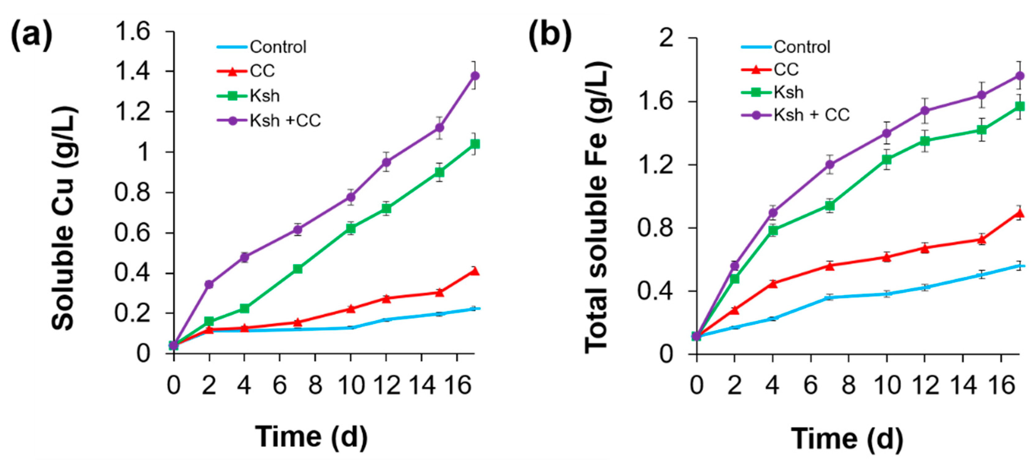
3.1. Bioleaching of Sulfide Minerals

The newly isolated iron-oxidizing bacterium L. ferriphilum CC was studied for bioleaching of sulfide minerals in comparison with iron- and sulfur-oxidizing bacteria At. ferrooxidans Ksh [37]. Results obtained showed that newly isolated strain L. ferriphilum CC is capable of oxidizing pyrite; however, At. ferrooxidans Ksh significantly exceeded L. ferriphilum in the activity of pyrite oxidation (Table 1). As has been shown previously, the oxidation of pyrite by iron-oxidizing Leptospirillum spp. bacteria is accompanied by the accumulation of sulfur on the surface of the mineral, which in turn prevents further oxidation of pyrite [38]. Taking into consideration the results mentioned above, we tested the association of L. ferriphilum with At. ferrooxidans Ksh in bioleaching of pyrite. The data given in the Table 1 show that the efficiency At. ferrooxidans Ksh in pyrite oxidation increases 1.8 times when co-cultivated with L. ferriphilum CC. It should be noted that when using the At. ferrooxidans Ksh with L. ferriphilum CC due to the high iron-oxidizing activity of L. ferriphilum CC, the extracted iron was found exclusively in the form of Fe3+, which ensured the highest ORP value (775 mV) and, therefore, a high oxidizing property of the leaching solution (Table 1).
As shown in Figure 1, isolated iron- and sulfur-oxidizing bacterium At. ferrooxidans Ksh oxidize chalcopyrite more actively than L. ferriphilum CC. However, the extraction of copper (Figure 1a) and iron (Figure 1b) from chalcopyrite by At. ferrooxidans Ksh enhances by about 1.3 and 1.2 times, respectively, in association with L. ferriphilum CC (Figure 1). It is concluded that the presence of L. ferriphilum CC in association resulted in rapid oxidation of Fe2+ and regeneration of oxidative agent Fe3+, which in turn accelerates chalcopyrite oxidation.
It is concluded, that newly isolated L. ferriphilum CC can be a prospective strain for the bioleaching of sulfide minerals in pure culture or in association with other iron- and/or sulfur-oxidizing bacteria.

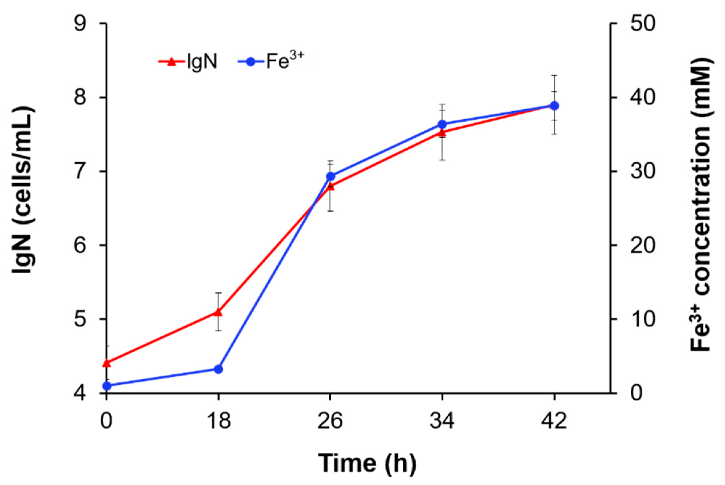
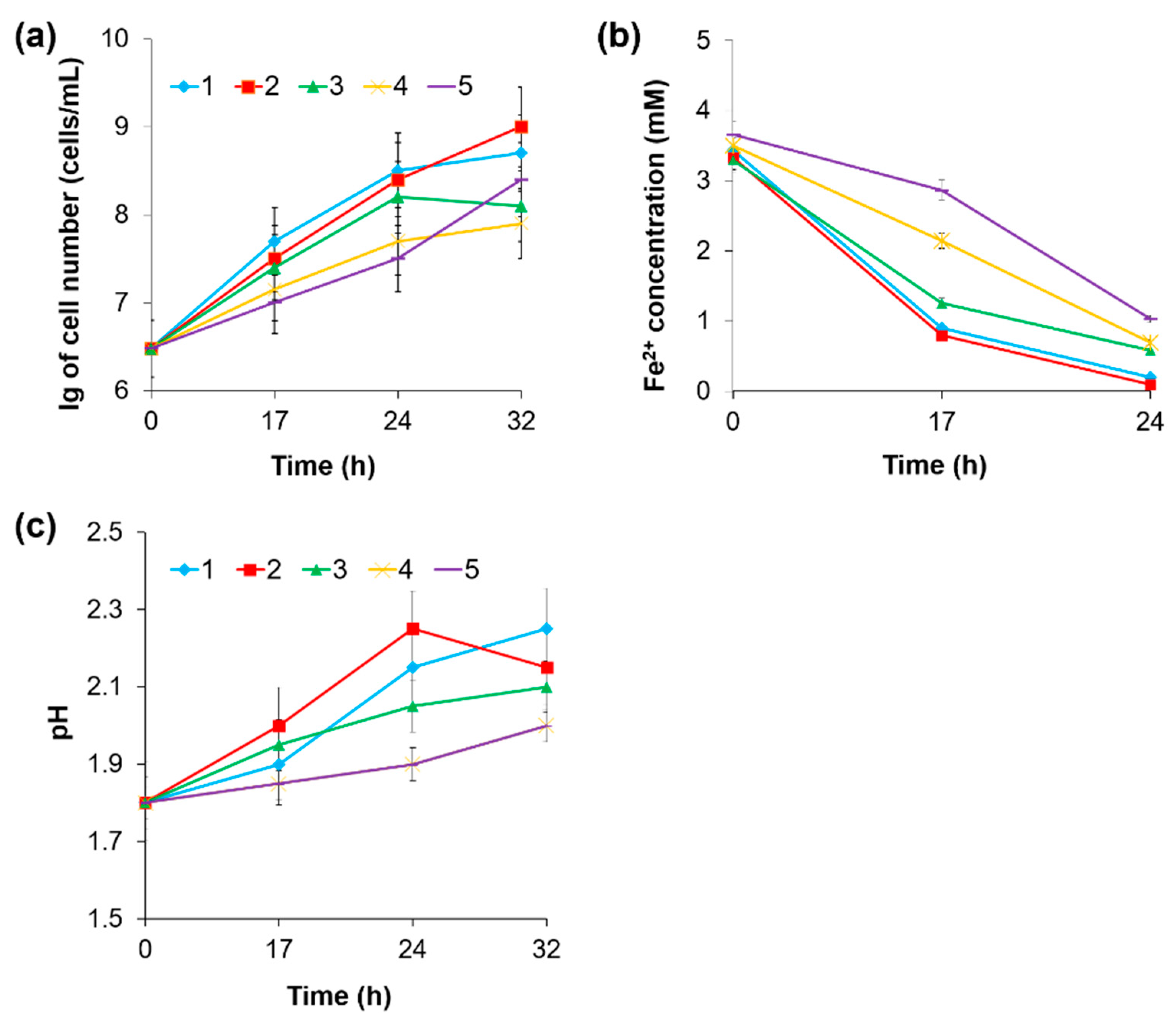
3.2. The Effect of Substrate Concentration

Fe2+ oxidation by L. ferriphilum CC was carried out on the rotary shaker. Kinetics of bacterial growth and iron oxidation is well described by Monod equation. Applying the Monod equation to the data obtained (Figure 2), growth and Fe2+ biooxidation parameters µmax and Vmax were determined as 0.48/h and 6.2 mM/h, respectively. Studies carried out showed that the growth of L. ferriphilum CC and the activity of Fe2+ oxidation depend on the concentration of the latter in the medium. Below is a quantitative characteristic of bacterial growth and Fe2+ oxidation depending on the initial concentration of Fe2+ in the medium (Table 2).
As can be seen from Table 2, maximum values of specific growth (0.41–0.48/h) and Fe2+ oxidation rates (6.0–6.2 mM/h) were detected at Fe2+ concentrations of 100–200 mM. At higher concentrations, a slow growth of bacteria and Fe2+ oxidation suppression was observed, reaching maximum values at 400 mM Fe2+. At a concentration of 50 mM Fe2+ in the medium, limitation by the substrate occurred. Thus, the optimal concentrations of Fe2+ for the growth of L. ferriphilum CC are 100–200 mM.

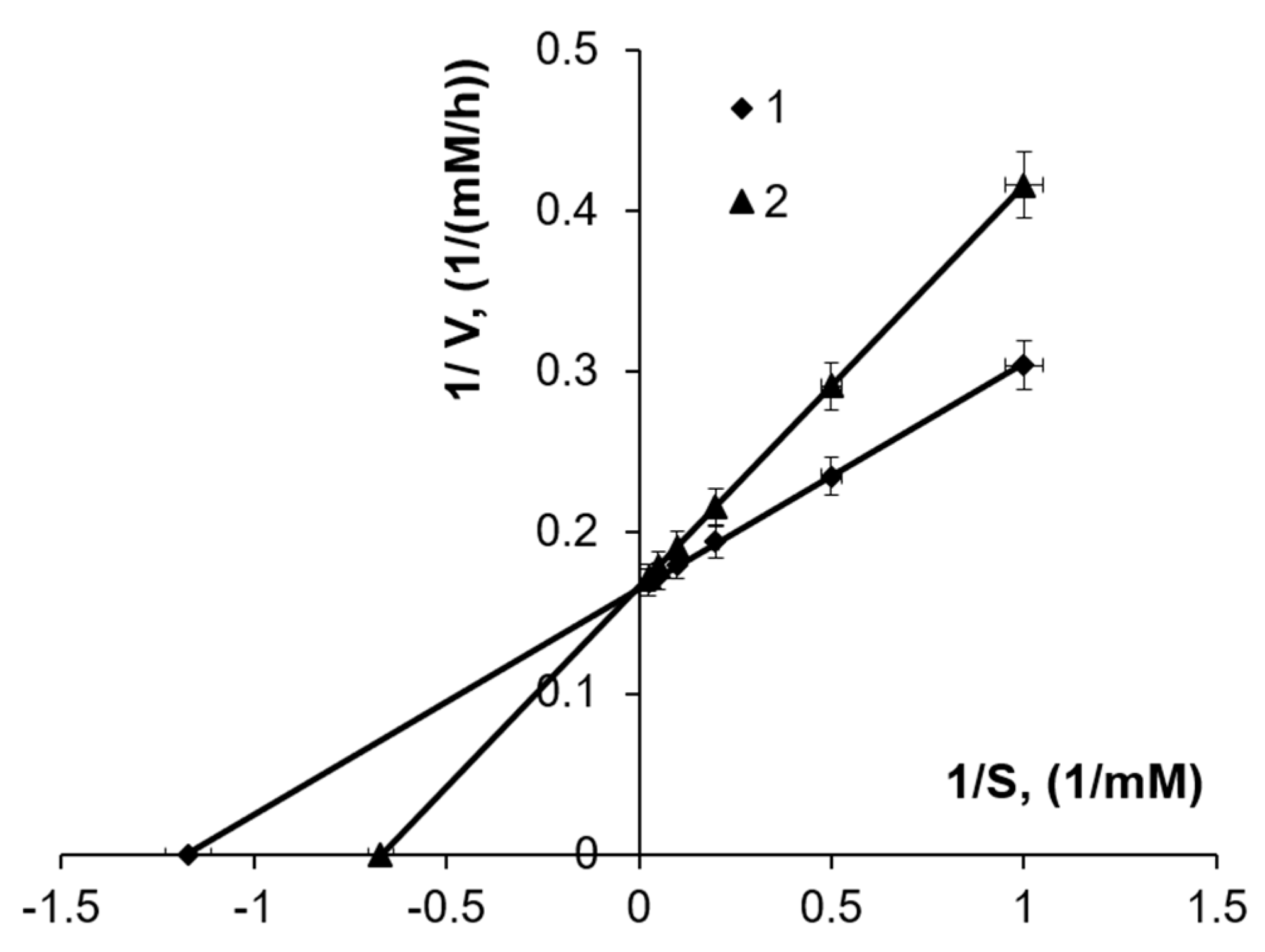
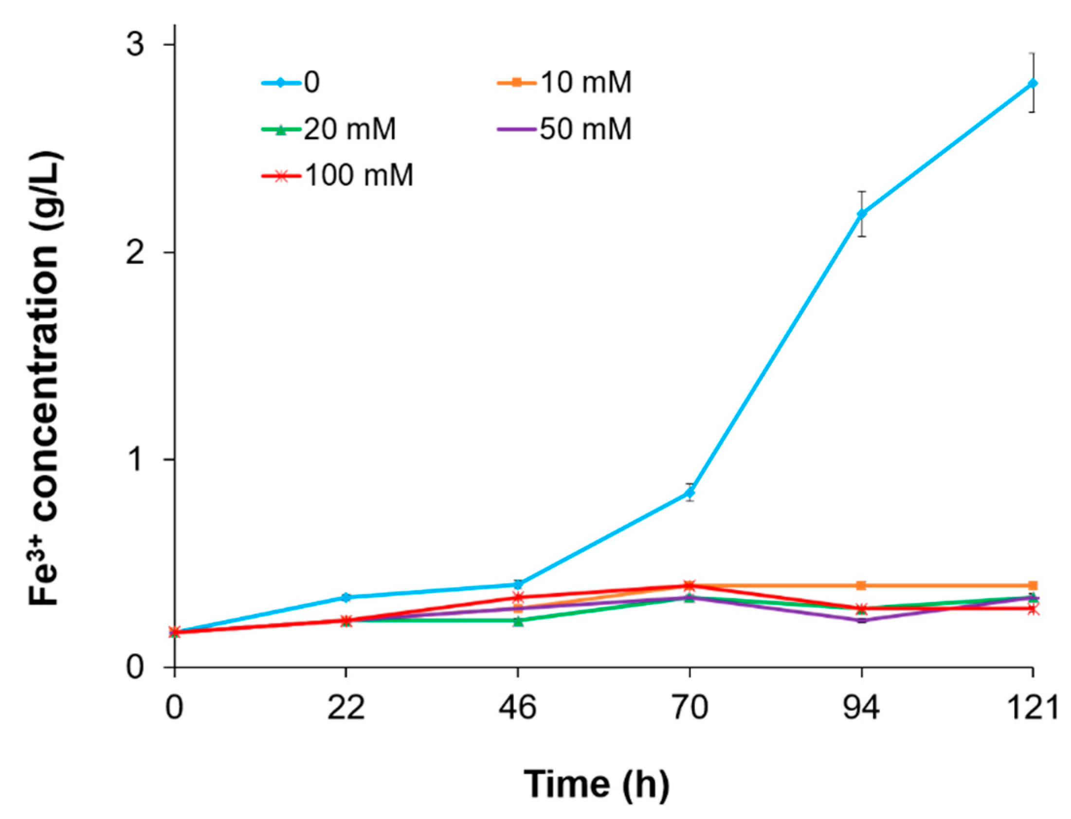
3.3. The Effect of Fe3+ Concentration

The tolerance of metal leaching bacteria to high Fe3+ concentrations is important in tank leaching applications. Fe3+, being the product of Fe2+ oxidation, accumulates in the medium as iron-oxidizing chemolithotrophic bacteria grow. Therefore, the effect of the initial concentrations of Fe3+ on the oxidation of Fe2+ by bacteria can be studied only during the first hours (10–17 h) of cultivation, as the generated amount of Fe3+ by oxidizing Fe2+ is insignificant. Results obtained are presented in Table 3.
As the presented data show, in the presence of elevated concentrations of Fe3+ ions, inhibition of the growth of L. ferriphilum CC and Fe2+ oxidation was observed. Inhibition is expressed in the decrease in the specific growth rate and Fe2+ ion oxidation rate (Table 3, Figure 3.). Furthermore, the inhibition degree increases with increased concentrations of Fe3+. The correlation between Fe2+ oxidation rate by L. ferriphilum CC and dynamics of changes in pH of the medium was detected (consumption of H+ ions and increase in pH) (Figure 3c). At low values of Fe3+, more active consumption of H+ protons have been observed, which are necessary for the oxidation of Fe2+ (Equation (2)).
The study of the kinetic parameters of the oxidation of Fe2+ has shown that the affinity of L. ferriphilum CC to Fe2+ increases in the presence of Fe3+ ions in the medium. Thus, the saturation constant (Km) for Fe2+ oxidation in L. ferriphilum CC was 0.83 mM FeSO4 in the absence of Fe3+, while it increased to 1.5 mM FeSO4 at the initial concentration of 50.0 mM Fe2(SO4)3 in the medium (Figure 4). It is concluded that like Sb. thermosulfidooxidans [23] and At. ferrooxidans [11], Fe3+ ions competitively inhibit the oxidation of Fe2+ in L. ferriphilum CC. The inhibition constant (Ki) was 61.95 mM Fe2(SO4)3.
Based on the results, the optimal concentrations of Fe2+ for the growth of L. ferriphilum CC were in the range of 100–200 mM. Higher concentrations of Fe2+ inhibited the growth of bacteria and the consequent oxidation of Fe2+. For cells of L. ferriphilum CC, the saturation constant for Fe2+ was less than that of Sb. thermosulfidooxidans (3.4–4.1 mM Fe2+) [23] and At. ferrooxidans (1.34 mM Fe2+) [39,40]. Thus, by the affinity for Fe2+, L. ferriphilum CC considerably exceeds At. ferrooxidans.
Fe3+ ions competitively inhibit the growth of L. ferriphilum CC and Fe2+ oxidation. This has been reported for other bacteria like At. ferrooxidans [27,39,41]. It should be noted that Fe3+ at a concentration of 280 mM inhibits the growth and Fe2+ oxidation activity of At. ferrooxidans [42].
Fe2+ oxidation proceeds in the presence of 200 mM Fe3+ (data not shown) allows the successful use of L. ferriphilum CC in biogenic regeneration of concentrated Fe3+ solutions for biohydrometallurgical applications.

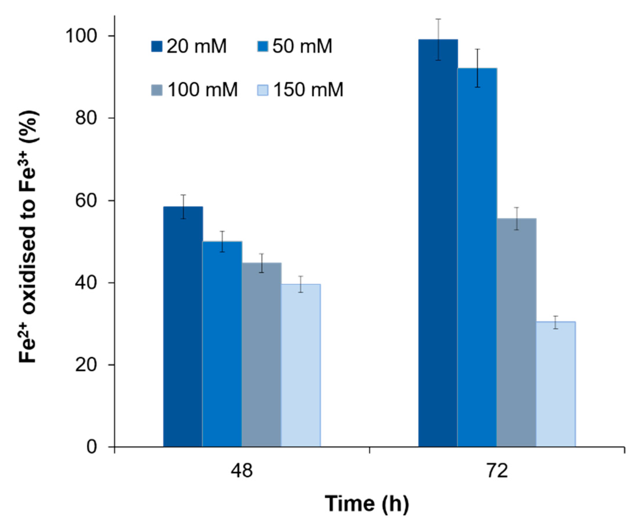
3.4. The Influence of Other Metal Ions

The influence of copper, zinc, nickel and cobalt ions on the oxidation of Fe2+ by L. ferriphilum CC was studied. The ions concentration varied from 10 to 150 mM. The studies showed that the extent of Fe2+ oxidation by L. ferriphilum CC was 58.4%, 42.8%, 53.3% and 31.3% at 20 mM copper, zinc, nickel and cobalt, respectively (Table 4). As can be seen from the presented data, zinc and cobalt ions at a concentration of 10 mM inhibited the oxidation of Fe2+ in L. ferriphilum CC by about 40% and 60%, respectively. Minimal inhibitory concentrations were 10 mM for Zn and Co and 20 mM for Cu and Ni. The toxicity of the ions was found to be as follows: Co > Zn > Ni > Cu.
Simultaneously, it has been established that as bacteria grow, the decrease in the inhibitory effect of copper ions is observed (Figure 5). Thus, with the growth of bacteria, the extent of inhibition of iron oxidation by L. ferriphilum CC decreases. Overcoming of the inhibitory effect of copper in L. ferriphilum CC was observed only at copper concentrations of 20 mM and 50 mM. At higher concentrations of copper, iron oxidation was inhibited at about the same extent and did not depend on the duration of cultivation of bacteria (Figure 5).
It is assumed that with the growth of bacteria, cells form EPS and accordingly create a less toxic and more favorable environment for their growth in the presence of copper ions. To test this hypothesis, we conducted studies with bacterial cells devoid of EPS. Properties and composition of EPS produced by L. ferriphilum CC were described previously [32,33]. The studies have shown that cells of L. ferriphilum CC devoid of EPS resulted from centrifugation at 6000 g are not able to grow and oxidize Fe2+ even in the presence of low concentrations of metals (Figure 6).
EPS produced by microorganisms make up the intercellular space and form the structure of the biofilm matrix. The main role of EPS is the mediation of the initial attachment of cells to solid substrates (sulfide minerals, etc.) and protection against undesirable environmental factors. It is assumed that during their growth, bacterial cells form biofilm consisting of EPS which significantly increases the resistance of bacteria to heavy metals. The toxicity of the ions was found to be as follows: Co > Zn > Ni > Cu.

4. Conclusions

The effect of initial Fe2+ and Fe3+ concentrations on growth and Fe2+ oxidation by a newly isolated strain L. ferriphilum CC from Armenia mine sites was studied. The highest specific growth and Fe2+ oxidation rates were detected at Fe2+ concentrations of 100–200 mM. At higher concentrations (above 200 mM), the growth of bacteria and Fe2+ oxidation suppression was observed. The maximum specific growth rate (µmax) of bacteria and half saturation constant (KS) were 6.2 mM/h, and 0.83/h, respectively. For cells of L. ferriphilum CC, the saturation constant for Fe2+ was less than that of Sb. thermosulfidooxidans (3.4–4.1 mM Fe2+) [23] and At. ferrooxidans (1.34 mM Fe2+) [39,40]. Thus, by the affinity for Fe2+, L. ferriphilum CC considerably exceeds At. ferrooxidans. It was shown that Fe2+ oxidation was competitively inhibited by Fe3+. The toxicity of the metal ions was found to be as follows: Co > Zn > Ni > Cu. Comparing the obtained results with the data in the literature, it can be concluded that L. ferriphilum CC studied is generally not inferior in their resistance to copper, zinc, nickel [26,28]. Along with the growth of bacteria the decrease in inhibitory effect of metal ions was observed. It is assumed that during their growth, bacterial cells form biofilm consisting of EPS which significantly increases the resistance of bacteria to heavy metals. The comparison of kinetic parameters obtained for L. ferriphilum with other bacteria indicates the high potential of L. ferriphilum in leaching processes of ores and concentrates for biogenic regeneration of concentrated Fe3+.

Author Contributions

Conceptualization, N.V., A.V. and R.Z.; methodology, N.V., A.V.; software, N.V.; validation, N.V., and A.V.; formal analysis, A.K. and A.V.; investigation, A.K. and A.V.; resources, N.V.; data curation, A.V. and N.V.; writing—original draft preparation, A.V., N.V. and R.Z.; writing—review and editing, R.Z. and L.C.; visualization, A.K. and A.V.; supervision, N.V. and R.Z.; project administration, N.V.; funding acquisition, R.Z. and L.C. All authors have read and agreed to the published version of the manuscript.

Funding

This research was funded by the Spanish Ministry of Economy and Competitiveness (project MAT2014-59222R).

Data Availability Statement

Not applicable.

Conflicts of Interest

The authors declare no conflict of interest. The funders had no role in the design of the study; in the collection, analyses, or interpretation of data; in the writing of the manuscript, or in the decision to publish the results.

References

  1. Schippers, A.; Hedrich, S.; Vasters, J.; Drobe, M.; Sand, W.; Willscher, S. Biomining: Metal recovery from ores with microorganisms. In Geobiotechnology I. Advances in Biochemical Engineering/Biotechnology; Schippers, A., Glombitza, F., Sand, W., Eds.; Springer: Berlin/Heidelberg, Germany, 2013; Volume 141, pp. 1–47. [Google Scholar]
  2. Johnson, D.B. The evolution, current status, and future prospects of using biotechnologies in the mineral extraction and metal recovery sectors. Minerals 2018, 8, 343. [Google Scholar] [CrossRef] [Green Version]
  3. Liao, R.; Yu, S.-C.; Wu, B.-Q.; Zhao, C.-X.; Lin, H.; Hong, M.-X.; Wu, H.-Y.; Yang, C.-R.; Zhang, Y.-S.; Xie, J.-P.; et al. Sulfide mineral bioleaching: Understanding of microbe-chemistry assisted hydrometallurgy technology and acid mine drainage environment protection. J. Central South Univ. 2020, 27, 1367–1372. [Google Scholar] [CrossRef]
  4. Schippers, A. Microorganisms involved in bioleaching and nucleic acid-based molecular methods for their identification and quantification. In Microbial Processing of Metal Sulfides; Donati, E., Sand, W., Eds.; Springer: Dordrecht, The Netherlands, 2007; pp. 3–33. [Google Scholar] [CrossRef]
  5. Fu, B.; Zhou, H.; Zhang, R.; Qiu, G. Bioleaching of chalcopyrite by pure and mixed cultures of Acidithiobacillus spp. and Leptospirillum ferriphilum. Int. Biodeterior. Biodegrad. 2008, 62, 109–115. [Google Scholar] [CrossRef]
  6. Bosecker, K. Bioleaching: Metal solubilization by microorganisms. FEMS Microbiol. Rev. 1997, 20, 591–604. [Google Scholar] [CrossRef]
  7. Rawlings, D.E. Heavy metal mining using microbes. Annu. Rev. Microbiol. 2002, 56, 65–91. [Google Scholar] [CrossRef] [PubMed]
  8. Vera, M.; Schippers, A.; Sand, W. Progress in bioleaching: Fundamentals and mechanisms of bacterial metal sulfide oxidation—part A. Appl. Microbiol. Biotechnol. 2013, 97, 7529–7541. [Google Scholar] [CrossRef]
  9. Lizama, H.M.; Suzuki, I. Synergetic competitive inhibition of ferrous iron oxidation by Thiobacillus ferrooxidans by increasing concentration of ferric iron and cells. Appl. Environmen. Microbiol. 1989, 10, 2588–2591. [Google Scholar] [CrossRef] [Green Version]
  10. Dorofeyev, A.G.; Pivovarova, T.A.; Karavaiko, G.I. Kinetics of ferrous ion oxidation by Thiobacillus ferrooxidans: The effect of products of arsenopyrite oxidation on pH of the medium. Microbiology 1990, 59, 205–212. [Google Scholar]
  11. Rawlings, D.E. Characteristics and adaptability of iron– and sulfur–oxidizing microorganisms used for the recovery of metals from minerals and their concentrates. Microb. Cell Factories 2005, 4, 13. [Google Scholar] [CrossRef] [Green Version]
  12. Vardanyan, A.; Stepanyan, S.; Vardanyan, N.; Markosyan, L.; Sand, W.; Vera, M.; Zhang, R. Study and assessment of microbial communities in natural and commercial bioleaching systems. Miner. Eng. 2015, 81, 167–172. [Google Scholar] [CrossRef]
  13. Bruins, M.R.; Kapil, S.; Oehme, F.W. Microbial resistance to metals in the environment. Ecotoxicol. Environ. Safety 2000, 45, 198–207. [Google Scholar] [CrossRef]
  14. Dopson, M.; Baker-Austin, C.; Koppineedi, P.R.; Bond, P.L. Growth in sulfidic mineral environments: Metal resistance mechanisms in acidophilic micro–organisms. Microbiology 2003, 149, 1959–1970. [Google Scholar] [CrossRef] [Green Version]
  15. Orell, A.; Navarro, C.A.; Arancibia, R.; Mobarec, J.C.; Jerez, C.A. Life in blue: Copper resistance mechanisms of bacteria and Archaea used in industrial biomining of minerals. Biotechnol. Adv. 2010, 28, 839–848. [Google Scholar] [CrossRef]
  16. Johnson, D.B.; Ghauri, M.A.; Said, M.F. Isolation and characterization of an acidophilic, heterotrophic bacterium capable of oxidizing ferrous iron. Appl. Environ. Microbiol. 1992, 58, 1423–1428. [Google Scholar] [CrossRef] [Green Version]
  17. Watling, H. The bioleaching of sulphide minerals with emphasis on copper sulphides—A review. Hydrometallurgy 2006, 84, 81–108. [Google Scholar] [CrossRef]
  18. Rensing, C.; Grass, G. Escherichia coli mechanisms of copper homeostasis in a changing environment. FEMS Microbiol. Rev. 2003, 27, 197–213. [Google Scholar] [CrossRef] [Green Version]
  19. Outten, F.W.; Huffman, D.L.; Hale, J.A.; O’Halloran, T.V. The independent cue and cus systems confer copper tolerance during aerobic and anaerobic growth in Escherichia coli. J. Biol. Chem. 2001, 276, 30670–30677. [Google Scholar] [CrossRef] [PubMed] [Green Version]
  20. Puig, S.; Thiele, D.J. Molecular mechanisms of copper uptake and distribution. Curr. Opin. Chem. Biol. 2002, 6, 171–180. [Google Scholar] [CrossRef]
  21. Navarro, C.A.; Orellana, L.H.; Mauriaca, C.; Jerez, C.A. Transcriptional and functional studies of Acidithiobacillus ferrooxidans genes related to survival in the presence of copper. Appl. Environ. Microbiol. 2009, 75, 6102–6109. [Google Scholar] [CrossRef] [Green Version]
  22. Simmons, S.L.; DiBartolo, G.; Denef, V.J.; Goltsman, D.S.A.; Thelen, M.P.; Banfield, J.F. Population genomic analysis of strain variation in Leptospirillum group II bacteria involved in acid mine drainage formation. PLoS Biol. 2008, 6, 1427–1442. [Google Scholar] [CrossRef]
  23. Vartanyan, N.S.; Karavaiko, G.I.; Pivovarova, T.A.; Dorofeyev, A.G. The resistance of Sulfobacillus thermosulfidooxidans subsp. Asporogenes against copper, zinc and nickel ions. Microbiology 1990, 59, 587–594. [Google Scholar]
  24. Trevors, J.T.; Oddie, K.M.; Belliveau, B.H. Metal resistance in bacteria. FEMS Microbiol. Rev. 1985, 1, 39–54. [Google Scholar] [CrossRef]
  25. Kondratyeva, T.F.; Muntyan, L.N.; Karavaiko, G.I. Zinc– and arsenic-resistant strains of Thiobacillus ferrooxidans have increased copy numbers of chromosomal resistance genes. Microbiology 1995, 141, 1157–1162. [Google Scholar] [CrossRef] [Green Version]
  26. Rawlings, D.E.; Tributsch, H.; Hansford, G.S. Reasons why ‘Leptospirillum’–like species rather than Thiobacillus ferrooxidans are the dominant iron–oxidizing bacteria in many commercial processes for the biooxidation of pyrite and related ores. Microbiology 1999, 145, 5–13. [Google Scholar] [CrossRef] [Green Version]
  27. Boon, M.; Ras, C.; Heijnen, J.J. The ferrous iron oxidation kinetics of Thiobacillus ferrooxidans in batch cultures. Appl. Microbiol. Biotechnol. 1999, 51, 813–819. [Google Scholar] [CrossRef]
  28. Dew, D.W.; Muhlbauer, R.; van Buuren, C. Bioleaching of copper sulfide concentrates with mesophiles and thermophiles. In Proceedings of the Alta Copper 99, Brisbane, Australia, 18–19 September 1999. [Google Scholar]
  29. Sampson, M.; Phillips, C. Influence of base metals on the oxidising ability of acidophilic bacteria during the oxidation of ferrous sulfate and mineral sulfide concentrates, using mesophiles and moderate thermophiles. Miner. Eng. 2001, 14, 317–340. [Google Scholar] [CrossRef]
  30. Harrison, J.J.; Ceri, H.; Turner, R.J. Multimetal resistance and tolerance in microbial biofilms. Nat. Rev. Genet. 2007, 5, 928–938. [Google Scholar] [CrossRef] [PubMed]
  31. Nurmi, P.; Özkaya, B.; Kaksonen, A.; Tuovinen, O.; Puhakka, J. Inhibition kinetics of iron oxidation by Leptospirillum ferriphilum in the presence of ferric, nickel and zinc ions. Hydrometallurgy 2009, 97, 137–145. [Google Scholar] [CrossRef]
  32. Vardanyan, A.; Vardanyan, N.; Khachatryan, A.; Zhang, R.; Sand, W. Adhesion to mineral surfaces by cells of Leptospirillum, Acidithiobacillus and Sulfobacillus from Armenian sulfide ores. Miner. 2019, 9, 69. [Google Scholar] [CrossRef] [Green Version]
  33. Vardanyan, N.; Badalyan, H.; Markosyan, L.; Vardanyan, A.; Zhang, R.; Sand, W. Newly isolated Acidithiobacillus sp. Ksh from Kashen copper ore: Peculiarities of EPS and colloidal exopolysaccharide. Front. Microbiol. 2020, 11, 1802. [Google Scholar] [CrossRef]
  34. Mackintosh, M.E. Nitrogen fixation by Thiobacillus ferrooxidans. J. Gen. Microbiol. 1978, 105, 215–218. [Google Scholar] [CrossRef] [Green Version]
  35. Castro, L.; Zhang, R.; Muñoz, J.A.; González, F.; Blázquez, M.L.; Sand, W.; Ballester, A. Characterization of exopolymeric substances (EPS) produced by Aeromonas hydrophila under reducing conditions. Biofouling 2014, 30, 501–511. [Google Scholar] [CrossRef]
  36. Karavaiko, G.I.; Rossi, G.; Agate, A.D.; Groudev, S.N.; Avakyan, Z.A. Biotechnology of metals. Manual/Eds. Centre for International Projects GKNT. 1988; 350. [Google Scholar]
  37. Pirt, S.J. Principles of Microbe and Cell Cultivation; Blackwell Scientific Publications: Oxford, UK; London, UK, 1975. [Google Scholar]
  38. Vardanyan, N.S.; Akopyan, V.P. Leptospirillum–like bacteria and evaluation of their role in pyrite oxidation. Microbiology 2003, 72, 438–442. [Google Scholar] [CrossRef]
  39. Kovalenko, T.V.; Karavaiko, G.I.; Piskunov, V.P. Effect of Fe3+ ions on Thiobacillus ferrooxidans oxidation of ferrous oxide at various temperatures. Microbiology 1982, 51, 156–160. [Google Scholar]
  40. Norris, P.R.; Barry, D.W.; Hinson, D. Iron and mineral oxidation by acidophilic bacteria: Affinities for iron and attachment to pyrite. In Biohydrometallurgy, Proceedings of International Symposium, Science and Technology Letters.; Norris, P.R., Kelly, D.P., Eds.; Kew: Surrey, UK, 1988; pp. 43–59. [Google Scholar]
  41. Penev, K.; Karamanev, D. Batch kinetics of ferrous iron oxidation by Leptospirillum ferriphilum at moderate to high total iron concentration. Biochem. Eng. J. 2010, 50, 54–62. [Google Scholar] [CrossRef]
  42. Curutchet, G.; Pogliani, C.; Donati, E.; Tedesco, P. Effect of iron (III) and its hydrolysis products (jarosites) on Thiobacillus ferrooxidans growth and on bacterial leaching. Biotechnol. Lett. 1992, 14, 329–334. [Google Scholar] [CrossRef]
Figure 1. Bioleaching of Cu (a) and Fe (b) from chalcopyrite by iron- and sulfur-oxidizing bacteria At. ferrooxidans Ksh and iron-oxidizing L. ferriphilum CC.
Metals 11 00425 g001
Figure 2. Dynamics of growth and iron oxidation by L. ferriphilum CC. Cells were grown in Mackintosh (MAC) medium with 70 mM Fe2+ and an initial pH of 2 at 37 °C and 200 rpm. N represents cell number (cells/mL).
Metals 11 00425 g002
Figure 3. Effect of Fe3+ on dynamics of growth of L. ferriphilum CC (a), oxidation of Fe2+ (b) and pH changes (c) at concentrations: 2 mM (1), 20 mM (2), 50 mM (3), 75 mM (4) and 100 mM Fe3+ (5).
Metals 11 00425 g003
Figure 4. Determination of Km according to Lineweaver–Burk; 1—in the absence of Fe3+, 2—in the presence of 50 mM Fe3+.
Metals 11 00425 g004
Figure 5. Effect of copper ions at different concentrations on Fe2+ oxidation by L. ferriphilum CC. Cells were grown in MAC medium with an initial pH of 2 at 37 °C and 200 rpm.
Metals 11 00425 g005
Figure 6. Dynamics of iron oxidation by cells of L. ferriphilum CC at different concentrations of Zn. The inoculum was extracellular polymeric substances (EPS)-free cells prepared according to Castro et al. 2014 [35].
Metals 11 00425 g006
Table 1. Bioleaching of iron from pyrite by L. ferriphilum and At. ferrooxidans Ksh and their association.
Bacteria Extracted Fe for 20 DayspH,
Initial/Final
Final ORP
mV
mg/L%
Fe3+Fe2+Fe Total
Control08408404.81.7/1.6600
L. ferriphilum7841456224012.81.7/1.3625
At. ferrooxidans Ksh16241792341619.51.7/1.2635
At. ferrooxidans Ksh +
L. ferriphilum CC
5824616644036.81.7/1.3775
Table 2. Growth and iron oxidation characteristics of L. ferriphilum CC at different initial concentrations of Fe2+.
Fe2+ Concentration (mM)µmax (1/h)Vmax (mM/h)
500.31 ± 0.0241.7
1000.48 ± 0.0186.2
2000.41 ± 0.0176.0
3000.28 ± 0.0264.6
4000.090 ± 0.000823.7
Table 3. Growth of L. ferriphilum CC on 100 mM Fe2+ with different initial concentrations of Fe3+.
Fe3+ Concentration (mM)µmax (1/h)Vmax (mM/h)
2.0 0.35 ± 0.0256.5
20.0 0.32 ± 0.0266.1
50.0 0.26 ± 0.0145.5
75.00.19 ± 0.0103.2
100.0 0.16 ± 0.0111.9
Table 4. The influence of metal ions on Fe2+ oxidation by L. ferriphilum CC.
Metal ions
(mM)
Fe, Oxidized in 46–48 h
Cu2+Zn2+Ni2+Co2+
g/L%g/L%g/L%g/L%
03.2 ± 0.191001.6 × 0.0231003.4 ± 0.0281003.8 ± 0.024100
101.0 ± 0.03664.31.5 ± 0.004838.8
201.9 ± 0.04958.40.67 ± 0.01542.81.8 ± 0.003653.31.2 ± 0.001731.3
501.6 ± 0.01450.00.56 ± 0.01335.71.5 ± 0.01843.30.85 ± 0.01322.6
1001.3 ± 0.004939.60.50 ± 0.005017.61.1 ± 0.04931.70.78 ± 0.008620.9
1501.2 ± 0.008036.90.84 ± 0.01125.00.74 ± 0.002919.7
Cells were grown at 37 °C and 200 rpm with an initial pH 1.8.
Publisher’s Note: MDPI stays neutral with regard to jurisdictional claims in published maps and institutional affiliations.

