Review

Cognitive Fitness: Harnessing the Strength of Exerkines for Aging and Metabolic Challenges

by
Mona Saheli
1,
Mandana Moshrefi
2,
Masoumeh Baghalishahi
1,
Amirhossein Mohkami
3,
Yaser Firouzi
4,
Katsuhiko Suzuki
5,* and
Kayvan Khoramipour
6,*
1
Department of Anatomical Sciences, Afzalipour Faculty of Medicine, Kerman University of Medical Sciences, Kerman 7616913555, Iran
2
Department of Physiology and Pharmacology, Afzalipour Faculty of Medicine, Kerman University of Medical Sciences, Kerman 7616913555, Iran
3
Department of Exercise Physiology, Faculty of Sport Sciences, Hakim Sabzevari University, Sabzevar 9617976487, Iran
4
Department of Exercise Physiology, Faculty of Sport Sciences, Shahid Bahonar University, Kerman 7616913439, Iran
5
Faculty of Sport Sciences, Waseda University, Tokorozawa 359-1192, Japan
6
Neuroscience Research Center, Institute of Neuropharmacology, Kerman University of Medical Sciences, Kerman 7619813159, Iran
*
Authors to whom correspondence should be addressed.
Sports 2024, 12(2), 57; https://doi.org/10.3390/sports12020057
Submission received: 13 December 2023 / Revised: 31 January 2024 / Accepted: 6 February 2024 / Published: 13 February 2024

Abstract

:
Addressing cognitive impairment (CI) represents a significant global challenge in health and social care. Evidence suggests that aging and metabolic disorders increase the risk of CI, yet promisingly, physical exercise has been identified as a potential ameliorative factor. Specifically, there is a growing understanding that exercise-induced cognitive improvement may be mediated by molecules known as exerkines. This review delves into the potential impact of aging and metabolic disorders on CI, elucidating the mechanisms through which various exerkines may bolster cognitive function in this context. Additionally, the discussion extends to the role of exerkines in facilitating stem cell mobilization, offering a potential avenue for improving cognitive impairment.

Graphical Abstract

1. Introduction

Cognitive impairment, also known as CI, pertains to challenges or deficits in cognitive functions, which encompass various mental processes such as memory (i.e., the capacity to retain and retrieve information) [1], attention (i.e., the ability to concentrate on specific tasks or stimuli) [2], executive function (i.e., higher-order cognitive processes such as planning, organizing, decision-making, and problem-solving) [3], language (i.e., comprehension, expression, and communication through spoken and written language) [4], perception (i.e., interpreting and comprehending sensory information from the environment) [5], and spatial navigation (the ability to perceive and navigate the physical space surrounding us) [6]. CI can be classified into various degrees, spanning from normal cognitive function to mild, moderate, and severe impairment. This classification is frequently determined by diverse assessment tools and criteria, including the Mini-Mental State Examination (MMSE) and the American Geriatrics Society criteria. Here is a summary outlining the distinct degrees of CI:
Mild CI: Mild CI is characterized by a slight decline in cognitive abilities, which may not interfere with daily life. Individuals with mild CI may have difficulty with memory, language, or visuospatial skills, but they can still perform most daily tasks.
Moderate CI: Moderate CI is marked by a more significant decline in cognitive abilities, which can impact daily life and may require assistance in various tasks. Individuals with moderate CI may have difficulty with memory, language, visuospatial skills, and executive function.
Severe CI: Severe CI is characterized by a profound decline in cognitive abilities, which can render individuals unable to perform most daily tasks. This stage of CI may require more extensive support and assistance in various aspects of daily life [7,8,9,10].
The rate of cognitive disorders in different human societies varies due to several factors, including genetic factors, ethnicity, geographical location, physical activity, gender, nutrition, and suffering from metabolic diseases. These factors collectively play a role in the prevalence of cognitive impairment. Some of these factors have a direct impact on cognitive function, while others influence the prevalence of cognitive disorders through their effects on health and lifestyle.
  • Genetic factors: genetic defects play a significant role in the development of cognitive disorders, with some genetic factors increasing the risk of cognitive impairment and dementia.
  • Ethnicity: Different ethnic groups may have varying susceptibility to cognitive disorders due to genetic factors and cultural influences. For example, individuals of African descent are at a higher risk of developing Alzheimer’s disease (AD)‚ while individuals of Asian descent may have a lower risk [11].
  • Geographical location: The prevalence of cognitive disorders can differ across regions due to factors such as access to healthcare, education, and lifestyle. For instance, cognitive disorders may be more common in urban areas with higher levels of pollution and stress [12].
  • Physical activity: Engaging in regular physical activity has been shown to reduce the risk of CI and improve cognitive function. Studies have found that engaging in light physical activity (LPA) for at least 3 h per day can reduce the chance of CI in older adults [13,14].
  • Gender: gender has been found to play a role in the prevalence of cognitive disorders, with some studies suggesting that women are at a higher risk of developing AD [15].
A study conducted in Hawaii found that a higher proportion of women than men had AD, with the highest female prevalence among Native Hawaiians and Pacific Islanders (NHPI) [14]. Another study found that 60.8% of women over 64 years old had cognitive deficits, while 36% of men in the same age group had cognitive deficits. However, it is essential to note that these percentages are based on a small sample size and may not be representative of the general population.
6.
Nutrition: Dietary factors play a crucial role in cognitive function and prevention of cognitive disorders. A diet rich in anti-inflammatory, low-sugar, and minimally processed foods, as well as a proper omega-6 to omega-3 ratio, can help maintain cognitive health [11].
7.
Metabolic diseases: suffering from metabolic diseases, such as diabetes and obesity, has been associated with an increased risk of CI and dementia [12].

1.1. Aging, Metabolic Disorders, and CI

The pathophysiological link between aging and CI encompasses intricate fundamental interactions and extends to include the correlation between metabolic disorders and CI, as discussed below [16,17,18].

1.1.1. Brain Atrophy

The process of normal aging encompasses a diverse range of changes, both functional and structural, within the brain, accompanied by declines across various cognitive domains [19,20]. The “disconnection hypothesis” seeks to elucidate the relationship between these age-associated alterations in cognition and brain structure. This hypothesis posits that disruptions in the communication pathways between different cortical regions can contribute to the observed cognitive decline [20,21]. A key factor potentially underlying this disconnection is the integrity of white matter (WM) in the brain. Existing evidence already indicates a correlation between compromised WM integrity and cognitive decline during the course of normal aging. This decline in WM integrity can be attributed to specific neural morphological alterations [22,23] that occur with aging [24]. Such morphological changes encompass a reduction in dendritic length and branching, decreased density of spines and synapses, and shifts in the distribution of spine subtypes [25,26,27,28]. These alterations are not merely structural; they have functional implications. Specifically, the loss or modification of dendritic spines and changes in their subtype distribution [29] can influence excitatory synaptic activity within neuronal networks. Such alterations in synaptic function within these networks are critical for maintaining optimal cognitive processing capabilities in normal aging individuals [25,26].

1.1.2. Neuroplasticity

Neuroplasticity, contingent upon activity, represents a pivotal characteristic of the nervous system, affording neurons the capacity to communicate and adjust connections based on prior experiences. Synaptic plasticity, in general, adheres to distinct developmental and aging trajectories in individuals without underlying health complications. However, disruptions in plasticity are implicated in various neuropsychiatric disorders, including CI. Mechanistically, synaptic plasticity operates across diverse spatial and temporal scales, spanning from microseconds to a lifetime and from microscopic synapses to the entire nervous system [30].
Physical exercise has emerged as a robust modulator of neuroplasticity, demonstrating a significant impact on the growth of new connections between cells in crucial cortical areas of the brain. This phenomenon, intricately linked to brain plasticity, extends its benefits to both healthy and diseased states. The mechanisms underlying exercise-induced neuroplasticity involve the elevation of trophic factors, modulation of cerebrovascular function, and the reduction of toxic Aβ and tau proteins, collectively contributing to the optimization of cognitive function and neuronal resilience [31]. However, we still need more studies to understand exactly how different types of exercise affect neuroplasticity.

1.1.3. Neurogenesis

Throughout a person’s lifetime‚ the hippocampus produces fresh neurons using neural stem cells (NSCs). These newly formed neurons have a crucial role in regulating mood and cognitive flexibility. Nevertheless, as one gets older, the generation of new neurons in the hippocampus decreases substantially due to age-related issues affecting NSC function. The aging brain experiences diminished neurogenesis primarily due to the significant decline in NSC activity, marked by enhanced quiescence and diminished proliferation of NSC.

1.1.4. Neurotransmitter Changes

An imbalance in various neurotransmitter pathways can contribute to CI. This imbalance involves disruptions in key neurotransmitter systems such as the monoaminergic, GABAergic, histaminergic, and cholinergic systems, each characterized by specific neurotransmitters (e.g., acetylcholine and dopamine) [31,32,33] and receptors that regulate cognitive functions [33].
In addition, the intricate balance of neurotransmitter receptors within the brain is subject to a dynamic regulatory framework [34,35]. Factors such as receptor activation, gene expression patterns, and external stimuli play pivotal roles in governing the production and degradation of these receptors [36].

1.1.5. Amyloid Plaques (Aβ) and Tau Tangles

Unusual clusters of proteins, known as amyloid plaques (made up of the Aβ protein) and tau tangles (resulting from the excessive phosphorylation of tau protein), tend to build up in the brains of those affected by CI [37]. It is worth noting that these protein aggregates can be detected in the hippocampus and cerebral cortex of certain elderlies [38] and individuals with metabolic disorders [39]. These clusters have been linked to neural harm and the deterioration of cognitive abilities [40].
Tau is a vital protein that is closely associated with microtubules and is typically found in neurons. Its main role is to maintain the stability of microtubules when everything is functioning normally. However, in pathological situations, tau tends to separate from microtubules and becomes involved in various neurological disorders, which are known for causing irreversible damage to neurons. These disorders include necrosis, apoptosis, necroptosis, pyroptosis, ferroptosis, autophagy-dependent neuronal death, and phagocytosis by microglia.
Researches indicate that the buildup of Aβ has been connected to difficulties in episodic memory, executive function, processing speed, visuospatial function, and overall cognition. The accumulation of Aβ and tau proteins can occur either independently or simultaneously in conditions such as primary age-related tauopathy and AD. Despite numerous efforts, our comprehension of the reciprocal influences of Aβ and tau accumulations on clinical characteristics remains incomplete [41].

1.1.6. Oxidative Stress and Inflammation

Cellular damage and CI can result from these mechanisms, which also play a role in neurodegeneration. CI is heavily influenced by oxidative stress, where the generation of reactive oxygen species (ROS) surpasses their removal. This imbalance is greatly responsible for chronic inflammation. In elderlies [42] and individuals with metabolic disorders [43], mitochondrial dysfunction occurs alongside reduced activity of antioxidant enzymes, leading to heightened ROS levels. ROS is responsible for harmful effects on lipids, proteins, and nucleic acids, including DNA. This damage also takes place in the mitochondria, the primary source of ROS. As a result, a detrimental cycle is initiated, resulting in depleted energy and eventual cell demise. This phenomenon is observed in both peripheral tissues and the brain [42]. Moreover, the augmented generation of ROS results in the impairment of the blood-brain barrier (BBB) function and the escalation of BBB permeability. Consequently, the defense mechanism against external toxins, pathogens, and pro-inflammatory cytokines becomes compromised, triggering an intensified activation of microglia, neuroinflammation, deposition of amyloid, and infliction of damage upon neurons [44].
Peripheral pro-inflammatory cytokines like IL-1 and TNF-α, along with macrophages, have the ability to breach the BBB and stimulate the brain’s microglia—macrophages that reside in the brain. This activation results in neuroinflammation, worsens amyloid buildup, and causes harm to neurons [45]. Additionally, inflammation throughout the body harms the BBB and causes it to become more permeable, diminishing its ability to shield against harmful external substances like toxins, pathogens, and pro-inflammatory cytokines [46]. The initiation of CI relies on the dysfunctional BBB, which plays a crucial role in clearing amyloid from the brain and transferring it into the bloodstream. The accumulation of amyloid in the brain, perceived as a significant pathology, can be attributed to the malfunctioning BBB [47].

1.1.7. Vascular Changes and Dysfunction

The brain’s blood flow can be hindered by age-related alterations in blood vessels, as well as metabolic disorders, causing a scarcity of oxygen and nutrients for brain cells. This can lead to CI and a higher susceptibility to medical conditions such as vascular dementia. Vascular aging encompasses a range of transformations in blood vessels as a person ages, including heightened rigidity, modifications in vessel walls, diminished ability for blood vessel growth, and impaired capability for endothelium-driven vasodilation. Individuals who are at risk for or already have cardiovascular disease may experience these changes associated with aging earlier than others [48]. These changes are known as early or premature vascular aging and are primarily caused by damage to the large and small vessels in the brain. This type of aging in the blood vessels is a significant contributor to CI that is commonly observed with increasing age [49].

1.1.8. Mitochondrial Dysfunction

Aging and metabolic disorders deteriorate mitochondrial function, resulting in a diminished ability to supply sufficient energy to brain cells. This decline in mitochondrial function could potentially play a key role in the development of CI [50]. The mitochondrial cascade hypothesis suggests that the pathogenesis of cognitive decline is closely linked to mitochondrial dysfunction. This is because the initial state of mitochondrial function and the rate at which it changes over time both have a significant impact on the progression of cognitive decline [51].

1.1.9. Insulin Resistance

Insulin resistance, as indicated by various studies, has been linked to both CI and type 2 diabetes. The brain heavily relies on insulin for sustaining memory and cognitive abilities at optimal levels. Any interference caused by insulin resistance may potentially disrupt these vital functions, ultimately paving the way for the onset of CI [52].

1.1.10. Brain Energy Metabolism

Memory and other cognitive processes may be affected when metabolic disorders interfere with the brain’s energy metabolism [43].

1.1.11. Advanced Glycation End Products (AGEs)

Elevated blood sugar levels have the potential to give rise to advanced glycation end products, which have the ability to amass in the brain and result in oxidative stress and inflammation. These factors are known to play a role in CI [53].

1.2. Management of CI
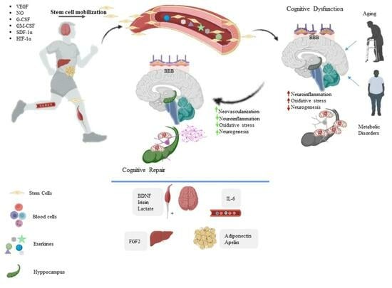
There have been multiple studies exploring various ways to combat CI. A systematic review and meta-analysis found no consistent evidence of a benefit for any pharmacologic agent in preventing and treating CI in older adults and individuals with metabolic disorders. As a result, non-pharmacological approaches are considered desirable for preventing and treating CI. One of the standout methods is engaging in physical activity and exercise, which has been touted as a secure, successful, and productive strategy that not only addresses but also prevents CI [54].
Fitness levels have been shown to have a significant impact on brain structure and function, particularly in older adults. Adequate physical activity and maintaining aerobic fitness are critical for cognitive function and brain health, especially during preadolescence, when the brain is undergoing significant changes in structure and function related to cognition, including executive control and relational memory [24,55,56,57,58]. Higher cardiorespiratory fitness levels are associated with greater gray matter volume in the prefrontal cortex and hippocampus in older adults. Also, it has been positively associated with white matter microstructure, particularly in frontal areas of the brain. Consistent positive findings suggest a promising role for physical activity in promoting hippocampal structure and function throughout the lifespan. Studies showed that cardiorespiratory fitness is positively associated with brain network integrity and measures of network integration and specialization in older adults. Cardiorespiratory fitness has also been associated with cognitive function across multiple domains, including memory, in older adults [24,56,57,59].
Physical exercise has been associated with increased neuroplasticity, neurotrophic factors, and improvements in brain function. It promotes the growth of new connections between cells in important cortical areas of the brain, which is linked to brain plasticity [36].
It benefits neuroplasticity in both health and disease stages by targeting different aspects, such as increasing trophic factors, changing cerebrovascular function, and lowering toxic Aβ and tau proteins [34].
Despite numerous studies indicating exerkines as the primary factor, the detailed mechanisms behind this phenomenon remain incompletely elucidated. Our objective was to furnish compelling evidence showcasing the beneficial impact of physical exercise on cognitive improvement (CI).

2. Exerkines and CI

The advantageous consequences of physical activity could potentially be attributed to the role of exerkines [60]. Exerkines encompass signaling molecules that are generated as a result of short-term and/or long-term exercise, operating through endocrine, paracrine, and/or autocrine routes to elicit their influences. Various factors are released by a diverse range of organs, cells, and tissues. These factors include myokines from skeletal muscle, cardiokines from the heart, hepatokines from the liver, adipokines from white adipose tissue, baptokines from brown adipose tissue, and neurokines from neurons. The subsequent section will aim to explore the potential impact of exerkines on enhancing CI (Table 1) [61].

2.1. IL-6

Initially, interleukin-6 (IL-6) was acknowledged for its notable anti-inflammatory function. However, IL-6 is a pleiotropic factor produced by various cell types, targeting numerous cell types and exerting a wide range of opposing biological effects. It possesses the ability to induce several distinct intracellular signaling pathways [62,63,64,65,66]. Exercise has sparked the idea that IL-6, which is released by skeletal muscle, may play a part in the metabolic changes and endurance enhancements brought on by physical activity. In fact, IL-6 has been designated as an energy detector, aiding in the regulation of blood sugar levels by promoting the breakdown of glucose and fatty acids. Additionally, the rise of IL-6 stimulates the growth of new neurons in the hippocampus [67,68]. All of these effects are believed to be induced when IL-6 is secreted by muscles during exercise, exhibiting anti-inflammatory properties [69].

2.2. Cathepsin B

The exercise-induced muscle secretory factor known as Cathepsin B (CTSB) is a cysteine protease that plays a role in hippocampal functions. In mice, CTSB is able to cross the blood-brain barrier (BBB) and is responsible for a significant increase in brain-derived neurotrophic factor (BDNF) and doublecortin (DCX) in the central nervous system. The neuroprotective effects of both proteins are closely tied to hippocampal neurogenesis and neuronal migration. Additionally, CTSB plays a role in mitochondrial cell death signaling by controlling the release of proapoptotic molecules [70].
Lysosomes are pivotal in the breakdown of certain detrimental proteins like Aβ and in upholding the equilibrium of intracellular protein. The malfunctioning of lysosomes has been perceived as a key flaw in neurons, resulting in the advancement of CI [36]. Prolonged physical activity raised the levels of fully developed cathepsin L/D enzymes within the cortex of mice with AD‚ indicating that exercise successfully rehabilitated the impaired lysosomal function in CI and reinstated the flow of autophagy [70]. AD leads to a decline in the levels of mature Cathepsin L and Cathepsin D, whereas the immature variants exhibit atypical surges. This suggests that the impairment of lysosomal hydrolase maturation occurs in AD. The development of maturity in lysosomal hydrolases is reliant on the transportation of vesicles to the lysosome and the subsequent process of cleavage. AD has been associated with impaired vesicular trafficking, according to prior research [71].
The relationship between exercise, CTSB, and cognitive function has been the subject of several studies. These studies concluded that aerobic exercise is more effective than resistance exercise in elevating CTSB levels and improving cognitive function [55,56,72,73,74].

2.3. Brain-Derived Neurotrophic Factor (BDNF)

BDNF, released by both the brain and muscles, belongs to the neurotrophin group, which signifies its vital function in neuronal differentiation and survival, synaptic integrity, brain plasticity, and memory. BDNF is crucial for the growth, preservation, and persistence of neurons, as well as for the adult production of new nerve cells in the hippocampus’ dentate gyrus [75]. BDNF, in addition to promoting long-term potentiation—an essential process for learning and memory—has also been found to enhance the creation of new mitochondria within hippocampal neurons [76]. It is worth mentioning that BDNF demonstrates its ability to boost mitochondrial biogenesis in these specific brain cells [70,77].
Older adults who engaged in aerobic exercise for one year, specifically 40 min of walking at 60–75% maximum heart rate reserve‚ three times a week‚ experienced a noteworthy 2% increase in the volume of their hippocampus. This increase is directly associated with improvements in aerobic fitness, as indicated by the correlation coefficient of 0.37 and 0.40 for the left and right hippocampus, respectively, in relation to VO2 max. The levels of BDNF in the serum show a correlation coefficient of 0.36 and 0.37 for the left and right hippocampus, respectively. Similarly, the correlation coefficient for performance on a spatial memory task is 0.23 and 0.29 for the left and right hippocampus, respectively [78]. Given that the hippocampus tends to diminish by 1–2% each year in elderly individuals without dementia [79], it can be inferred from the aforementioned findings that engaging in aerobic exercise for a year might potentially counteract the shrinkage of the hippocampus associated with aging by a period of 1–2 years [78].
A study reported that BDNF response to exercise is intensity-dependent, with intense exercise being the most effective. In a study on young college students, HIIT improved cognitive function to a higher degree compared to moderate-intensity continuous training (MICT), with cognitive improvements correlating with lactate release. However, a study on people with spinal cord injury (SCI) found that acute submaximal exercise did not impact plasma or serum BDNF levels or cognitive function, suggesting that the relationship between exercise intensity and BDNF in individuals with CI may be more complex [80,81,82].

2.4. Irisin

Irisin, derived from the cleavage of fibronectin type III domain-containing protein 5 (FNDC5), is a transmembrane glycoprotein with 112 amino acids. It is aptly named after Iris, the Greek goddess of messengers [83]. Following the proteolytic cleavage process separating it from FNDC5, irisin is released and operates as a myokine [84]. Although irisin was primarily discovered in skeletal muscles, subsequent findings revealed its expression in several tissues, encompassing the brain [85]. Exercise increases the production of a transcription factor called PGC-1α in muscles. This, in turn, enhances the expression of a protein called FNDC5, leading to the release of more irisin into the bloodstream through the cleavage of FNDC5. The level of irisin in the plasma is elevated in individuals who engage in aerobic training [86]. Specifically, exercise stimulates the production of FNDC5 and BDNF in the hippocampus. This mechanism is dependent on PPARγ coactivator 1α (PGC-1α). Recent studies have shown that FNDC5/irisin activates the cyclic adenosine monophosphate/protein kinase B/cAMP response element-binding protein (cAMP/PKA/CREB) pathway in both mice and human brain tissue. Importantly, the stimulation of PGC-1α by FNDC5/irisin highlights the role of irisin as a crucial molecule facilitating communication between muscles and the brain [87]. The increase in FNDC5/irisin levels is accompanied by the promotion of hippocampal neurogenesis. Exercise-induced elevation of irisin leads to cell proliferation and BDNF expression in brain tissue, contributing to the generation of new neurons [68].
Furthermore, irisin exhibits anti-inflammatory properties by reducing the secretion of cytokines IL-6 and IL-1β from astrocytes cultured in the laboratory. Astrocytes that have been exposed to irisin provide protection to neurons against the toxic effects of Aβ [88].
Recent evidence supports the potential of exercise, particularly high-intensity training, to positively impact cognitive function in individuals with CI [89,90].

2.5. Apelin

Apelin, known as the naturally occurring ligand for the APJ orphan G protein-coupled receptor, was initially discovered in 1998. Both apelin and APJ are found throughout the body and serve as crucial factors in safeguarding cells within various organs. Apelin’s significance extends to conditions like cardiovascular disease, obesity, and cancer. Furthermore, apelins are present throughout the nervous system and have been identified to possess neuroprotective properties and have a positive effect on CI [88]. A study involving eleven obese non-diabetic males demonstrated that an eight-week endurance training program resulted in an increase in apelin expression in the muscles [91].

2.6. Clusterin

Clusterin, alternatively called apoJ, is a versatile protein that functions as a natural Chaperone. It displays involvement in numerous physiological and pathological conditions, including CI. The therapeutic impact of clusterin on patients with CI arises from its ability to alter Aβ aggregation and neuroinflammation. Notably, clusterin potentially holds a significant influence in facilitating Aβ clearance across the BBB [68,92]. Engaging in a disciplined workout routine for half a year (voluntary exercise) resulted in raised levels of plasma clusterin. Interestingly, these increased levels were closely related to enhancements in stamina and aerobic ability. The elevated clusterin levels were found to play a role in suppressing the interferon and cytokine signaling pathways within brain endothelial cells, consequently mitigating both chronic and immediate neuroinflammation [93,94].
Studies found that exercise plasma collected from running mice and infused into sedentary mice reduces baseline neuroinflammatory gene expression and experimentally induced brain inflammation and that plasma proteomic analysis revealed a concerted increase in complement cascade inhibitors, including clusterin [72]. Additionally, a study found that aerobic exercise increases plasma clusterin, and there is a positive association between changes in plasma clusterin and cardiorespiratory fitness as a response to aerobic exercise in older adults with AD [95]. These findings suggest clusterin could mediate the positive effect of exercise on CI [72,90].

2.7. C-X3-C Motif Chemokine Ligand 1 (CX3CL1)

A myokine commonly referred to as CX3CL1 was discovered in previous studies [96]. Recent research has shown that mRNA levels of CX3CL1 were elevated in muscles, while its protein levels were found to be increased in plasma following both intense endurance and resistance workouts [84,97]. CX3CL1, as indicated by these discoveries, is a myokine triggered by physical activity, potentially facilitating inter-organ communication between skeletal muscles and various bodily systems. In the realm of the brain, CX3CL1 could potentially inhibit neuroinflammation by activating CX3CR1, a receptor found in microglial cells specific to the C-X3-C motif chemokine [88,98].
While the specific effect of exercise intensity on CX3CL1 in the context of CI is not directly addressed in the available literature [99], the studies provide insights into the potential impact of exercise and related peptides on neuroinflammation and immune responses [90,95,99], which are relevant to the broader understanding of the topic. Further research specifically focusing on the relationship between exercise intensity, CX3CL1, and CI is warranted to draw more definitive conclusions.

2.8. FGF2

FGF2, also called bFGF and FGF-β, is a crucial growth factor involved in the growth and multiplication of neural stem cells and their progenitors, promoting neural development. Its expression is widespread in different tissues, like the brain and muscles. Notably, engaging in aerobic exercise is shown to elevate FGF2 expression in animal models. FGF2, a neurotrophic factor, has the ability to promote the growth of new neurons and blood vessels in the brains of both adults and developing individuals [88,100].

2.9. FGF21

The hormone FGF21, a member of the FGF superfamily, was detected in mouse embryos in 2000. Its primary expression occurs in the liver, but it is also generated in different organs like muscle, adipose tissue, pancreas, and heart. FGF21 plays a pivotal role in maintaining energy balance through autocrine, paracrine, or endocrine mechanisms [101,102]. Preclinical research has shed light on the potential therapeutic application of FGF21 in managing metabolic disorders like diabetes. This is attributed to its ability to enhance insulin sensitivity, ameliorate glucose tolerance, and facilitate weight loss [103]. One notable consequence of exercise is the rise in FGF21 levels in the bloodstream, primarily driven by augmented FGF21 production in the liver. FGF21 has exhibited impressive protective qualities in numerous brain damage studies. It effectively counteracted inflammation and maintained the integrity of the blood-brain barrier by stimulating PPAR-γ activation. Additionally, it facilitated the growth of new blood vessels, effectively promoting neovascularization [104,105]. Furthermore, FGF21 has demonstrated its neuroprotective abilities, displaying anti-inflammatory and antioxidant properties. It significantly hindered the formation of amyloid plaques, neurofibrillary tangles, and overall neurodegeneration [105,106,107].

2.10. IGF-1

Exercise-induced neurogenesis is dependent on the presence of insulin-like growth factor 1 (IGF-1), which is also known as somatomedin C. This secreted peptide, structurally resembling insulin, plays a vital role in a range of physiological processes. It was further shown through a study that IGF-1 is crucial for facilitating exercise-induced neurogenesis [88].
Exercise has been shown to have a positive impact on cognitive function and IGF-1 levels. In a study conducted on female Wistar rats, resistance exercise training ameliorated MCI, and this improvement was attributed to the exercise-induced enhancement of IGF-1 protein and GST activity in the brain [108].
A systematic review of experimental studies in the elderly investigated the effects of exercise on IGF-1 levels and cognition. The review found disparities in the impact of exercise on IGF-1 levels, with some studies showing an increase, some showing no effect, and one showing a reduction in IGF-1 levels [109]. The review concluded that the type of physical exercise, protocols, and sample characteristics may explain these discrepancies. A study investigated the effect of resistance exercise intensity on the expression of IGF-1 in human skeletal muscle. The results showed that IGF-1 expression significantly increased following a higher intensity resistance exercise session compared to a lower intensity session. This suggests that the intensity of resistance exercise can impact the expression of IGF-1 in skeletal muscle [72].

2.11. LIF

Leukemia-inhibitory factor (LIF), a cytokine belonging to the interleukin-6 family, possesses versatile functions and was initially recognized for its ability to prompt macrophage differentiation. LIF holds significance in facilitating the growth, development, and viability of diverse cell varieties, which encompass neurons, myoblasts, hepatocytes, adipocytes, megakaryocyte progenitors, and myeloid cells. LIF has received acknowledgment as a myokine, demonstrating an escalated expression in reaction to exercise in both human and animal subjects [110,111]. The release of this entity from cells is regulated by a signaling peptide [112], and its ability to be secreted has been verified in experiments with human myotube cultures and mouse skeletal muscles [113]. A study by Besse-Patin et al. revealed that after a 3-h aerobic exercise session, there was up to four-fold increase in the expression of LIF mRNA in muscles, followed by a gradual decrease. A recent study discovered that static exercise led to a rise of nearly 50% in the plasma concentration of LIF, whereas dynamic exercise did not show any such increase. This indicates that the regulation of LIF expression may differ based on the type of exercise performed [114]. In addition, the application of ionomycin, an ionophore that transports Ca2 ions, resulted in increased levels of LIF mRNA and protein expression in muscle cells of humans [110]. The suggestion is that variations in Ca2+ levels after muscle contraction might have an impact on the transcription of LIF. Moreover, the PI3K-Akt pathway was found to influence LIF in human myotubes in culture, while the levels of JunB and c-Myc, which are stimulated by LIF, increased in skeletal muscles after resistance exercise [115].

2.12. VEGF

Adult neurogenesis in the dentate gyrus of the hippocampus is bolstered by vascular endothelial growth factor (VEGF), which works in part by stimulating angiogenesis, the process of generating new blood vessels. This is important because neurogenesis takes place in an environment conducive to angiogenesis, ensuring an adequate supply of oxygen and nutrients [77].
According to research, engaging in wheel running was discovered to enhance the creation of new neurons in the hippocampus in adult mice by amplifying the presence of VEGF in their blood [116]. In a separate study, it was revealed that 12 weeks of aerobic exercise in human participants—encompassing activities such as ergometer cycling‚ treadmill running, stair climbing, and The surge in cerebral blood volume (CBV) within the dentate gyrus showed a significant correlation (correlation coefficient = 0.662) with this improvement. Consequently, the boosted CBV led to improved results in a short-term memory assessment (correlation coefficient = 0.62) [117].
One study found that high-altitude exposure (3540 m asl) of Long Evans rats during early adulthood impaired spatial and visual memory but prevented CI when the rats were housed in an enriched environment with a running wheel for voluntary exercise. The study suggests that exercise can prevent CI via VEGF signaling [118].
Another study evaluated the relationship between VEGF and cognitive improvements following exercised-primed transcranial direct current stimulation in MCI and AD [48,118].

2.13. Glial-Cell-Line-Derived Neurotrophic Factor (GDNF)

GDNF, another neurotrophic factor, has been demonstrated to rescue neurons from naturally occurring cell death as well as axotomy-induced cell death. Exercise has been associated with elevated GDNF protein concentrations in skeletal muscle, which can then be released into the bloodstream, cross BBB, and reach CNS. Studies have revealed a dose-response pattern for GDNF in response to exercise [119]. For instance, low-intensity exercise resulted in an increase in GDNF in the soleus, primarily a slow-twitch muscle, while decreasing GDNF content in the extensor digitorum longus (EDL), predominantly a fast-twitch muscle. Increasing the intensity of exercise, such as using running wheels with added resistance, led to clear evidence of recruitment (e.g., hypertrophy) in fast-twitch muscles and an associated increase in GDNF content [120].

2.14. 3-Hydroxybutyrate (3OHB)

Exerkines are not exclusive to proteins. Other types of molecules that participate in regulating physiological functions are also stimulated by vigorous exercise. Following the activation and breakdown of fatty acids, the liver generates ketone bodies, particularly 3-hydroxybutyrate (3OHB). Evidence indicates that the rise in BDNF can be attributed to the presence of 3OHB subsequent to physical activity [67].
In a recent study, Marosi and colleagues showcased the potential of 3OHB in activating the BDNF gene promoter, specifically within cerebral cortical neurons. This activation was accomplished through a unique signaling pathway that relies on the collaboration between the transcription factor nuclear kappa B (NF-κB) and the histone acetyltransferase p300 [121]. Furthermore, the autophagy-dependent nature of β hydroxybutyrate has been observed to facilitate the enhancing impact of physical activity on cognitive function and memorization [122].

2.15. Lactate

Lactate has a double duty, serving as both a fuel source and a messenger in the body. Astrocytes have the ability to absorb glucose or break down glycogen for energy production through glycolysis, resulting in the release of lactate into the space surrounding cells. Neurons can subsequently utilize this extracellular lactate as a vital source of energy, especially when there is increased synaptic activity. This connection between synaptic activity and energy transfer establishes a significant metabolic relationship.
Conversely, lactate plays a significant role as a messenger molecule, triggering a cascade of signaling pathways through specialized receptors. This stimulation prompts the activation of immediate early genes and facilitates the growth of blood vessels in the brain. Amidst moderate to intense physical activity, lactate production escalates both in muscles and the bloodstream, compelling the brain to actively consume lactate originating from skeletal muscles. As a result, this process boosts aerobic glycolysis, ultimately resulting in higher levels of lactate produced by the brain.
Exercise has a notable impact on the functionality and performance of transporters and enzymes responsible for the astrocyte-neuron lactate shuttle. This enhances the effectiveness of the process. Moreover, exercise triggers the activation of the lactate receptor known as hydroxycarboxylic acid receptor 1 (HCAR1), which subsequently influences brain plasticity [123].
The effect of exercise intensity on lactate in cognition impairment has been studied in some studies. One study found that high-intensity exercise [124] improved the acute cognitive response to a higher degree when compared to moderate-intensity exercise or control [125]. The cognitive improvements correlated with lactate release, providing a plausible molecular explanation for the cognitive enhancement [90]. Another study evaluated the effects of high-intensity functional training on general cognition in older adults with CI and found that a progressive high-intensity functional training program improves general cognition. The authors suggested lactate signaling as an important mediator [73,89].

2.16. PGC1-a

PGC1-a has been linked to alterations in neurocognitive function caused by exercise. Researches indicated that participating in exercise, either resistance or endurance, could increase the blood levels of PGC1-a, which can positively impact cognitive functions. One study suggested that the PGC1-α-dependent myokine, which stimulates the brown-fat-like growth of fatty tissue, may possess neuroprotective functions in AD [101,102,103].
In particular, it has been found that exercise-induced overexpression of PGC1-a can enhance the presence of kynurenine aminotransferases, which convert kynurenine to kynureniceacid (KYNA), an endogenous antagonist of N-methyl-d-aspartate and alpha 7-nicotinic acetylcholine receptor [124]. KYNA is also the antagonist of the α7 nAChR. KYNA level changes in brains may, therefore, affect the physiological functions related to α7-nicotinic acetylcholine neurotransmission [126]. Failure to maintain physiological concentrations of brain KYNA could be one of the causative factors leading to neuropathological conditions such as AD and CI [127].

2.17. Neurotransmitters and Neuromodulators

Physical activity leads to the release of two neurotransmitters, dopamine and 5-hydroxytriptamin (5-HT), which are essential for motor control and learning. These neurotransmitters have been observed to increase in various brain areas, such as the hippocampus, prefrontal cortex (PFC), stratum, and midbrain. Their presence is crucial in the regulation of adult neurogenesis within the hippocampus [77].
Neuromodulators are substances capable of modulating or regulating the activity of neurons. Neurotransmitters are directly involved in transmitting signals between individual neurons at synapses, while neuromodulators have a more widespread and modulatory influence on the overall activity and responsiveness of neural circuits, they play a pivotal role in cognitive function by influencing processes such as learning, memory, and attention. For instance, flavonoids, a category of dietary phytochemicals, have demonstrated positive effects on CNS [128]. They contribute to enhancing cognitive function by safeguarding neurons from stress-induced injury and suppressing neuroinflammation.
Researchers suggest that physical exercise could effectively impact neuromodulators, although study results are inconclusive and warrant further investigation [129].
One study showed that exercise could increase nerve growth factor (NGF) within the septohippocampal pathway, which is responsible for the enhancement of septo-hippocampal cholinergic structure and function. Recognizing the important link between exercise and NGF levels, it becomes evident that promoting physical activity can greatly aid in the improvement of failing septo-hippocampal functioning and potentially contribute to significant recovery in individuals with neurological disorders. However, the modulation of proNGF and proBDNF, two neurotrophins, is intricate and likely influenced by various pathways. These neurotrophin variations have previously been associated with depression, stress, and anxiety [130]. Castrillon et al. suggested that an up-regulation of neuromodulator activity (e.g., by physical activity), alongside increased brain size, is a crucial aspect of human brain evolution and can help in improving cognitive function [131].

2.18. Inflammation

Exercise suppresses chronic inflammation by inducing the release of IL-6 from skeletal muscles. IL-6 then triggers the production of IL-1 receptor antagonist (IL-1ra) and IL-10, which exert anti-inflammatory effects. IL-1ra blocks inflammation mediated by IL-1, while IL-10 inhibits the production of several pro-inflammatory cytokines, including IL-1α, IL-1β, IL-8, TNF, and macrophage inflammatory protein-α. The regulation of cellular metabolism in macrophages is another vital role played by IL-10, with the objective of minimizing inflammation. It achieves this by activating the mTOR inhibitor DDIT4, which inhibits glucose absorption and eliminates damaged mitochondria exhibiting low membrane potential and elevated reactive oxygen species [77].
The provided study and related research suggest that high-intensity aquatic-based exercise has cognitive benefits for older adults, particularly those with MCI [132,133]. The effects are believed to be mediated by alterations in pro- and anti-inflammatory protein markers.
Studies emphasize the potential of aquatic exercise to reduce the risk of late-life CI and dementia by influencing immune and inflammatory pathways. However, the need for larger studies to replicate these findings and further explore the role of activity-induced inflammatory changes on late-life brain health is also highlighted. The evidence presented underscores the potential of high-intensity aquatic exercise as an effective non-pharmacological intervention to improve cognitive function and mitigate cognitive decline in older adults, especially those with MCI [133].

2.19. Oxidative Stress

Physical activity promotes the growth of new mitochondria and enhances the functionality of antioxidant enzymes, benefiting not only the body but also the brain [134]. This dual effect contributes to the reduction of oxidative stress. Additionally, exercise aids in removing amyloid, a protein associated with neurodegenerative diseases ‚ by enhancing both the blood-brain barrier and the glymphatic system. Animal models of Alzheimer’s disease have shown that engaging in wheel running can prevent microglia activation and decrease oxidative stress and inflammation in the hippocampus and cortex. This, in turn, can lead to reduced amyloid deposition and enhanced spatial learning abilities, as observed in the Morris Water Maze and Barnes Maze etc. [135]. Running on a wheel has the added benefit of diminishing the amount of triggered microglia and deposits of amyloid while simultaneously augmenting the number of dendrites, dendritic spines, and postsynaptic density protein found in the cortex and hippocampus of regular elderly mice. This correlation improved the grasping of spatial concepts in the Morris Water Maze [136]. Older adults who engage in higher levels of physical activity, as measured by a pedometer, experience a decelerated cognitive decline related to amyloid accumulation and a reduction in the volume of gray matter, which is where neuron cell bodies reside within the brain [137].
Recent findings collectively suggest that exercise intensity can influence oxidative stress [138], and higher intensities of aerobic exercise would be the best in reducing oxidative stress [90,99].

2.20. Beneficial Effect of Stem Cells

Athletes’ plasma has been found to contain increased levels of various substances‚ such as vascular endothelial growth factor (VEGF), IL-6, nitric oxide (NO), granulocyte colony-stimulating factor (G-CSF), granulocyte macrophage colony-stimulating factor (GM-CSF), stromal cell-derived factor-1α (SDF-1α), and hypoxia-inducible factor-1α (HIF-1α). This elevation in levels is known to stimulate the mobilization of endothelial progenitor cells from the bone marrow [139,140,141,142,143,144,145,146,147,148,149,150]. Enhancement of endothelial function, restoration of damaged endothelium through new cell production, and promotion of neovascularization lead to the restoration of BBB integrity, culminating in reduced levels of neuroinflammation [139,140,141,142,143,144,145,146,147,148,149,150].
Moreover, SDF-1α facilitates the attraction of neural stem/progenitor cells (NSCs) towards the site of brain injury by engaging with its receptor, chemokine receptor type 4 (CXCR4). CXCR4 is predominantly found on neural stem/progenitor cells, and cells that express CXCR4 migrate in response to the concentration gradient of SDF-1α [151,152]. NSCs are responsible for neurogenesis and brain plasticity that could replace dead neurons [153]. Consequently, the process of triggering the enlistment and relocation of NSCs to the location of the harm ultimately results in the production of fresh neural cells [154,155,156].
Monocarboxylate transporters (MCTs) play a crucial role in regulating lactate levels in the extracellular region of the brain. Hence, when MCT1 is lacking in brain endothelial cells (ECs), it results in elevated lactate levels. These increased lactate levels have a dual effect—causing heightened depletion of the NSC (neural stem cell) pool and a decline in the production of fully developed newborn neurons within the hippocampus [157]. The activity of runners’ plasma has the potential to activate MCT1 expression in the endothelial cells of the brain. This, in turn, can regulate the growth and development of neural stem cells, ultimately leading to the restoration of cognitive function [123,157].
Mesenchymal stem cells (MSCs) are a type of stem cell that has the ability to differentiate into neurons. The brain-damaged regions receive a boost in the number of progenitor and proliferating cells when MSCs are stimulated to migrate using SDF-1 and G-CSF cytokines [158]. Furthermore, the potential of MSCs to enhance cognitive function arises from their ability to exhibit anti-inflammatory properties, modulate the immune system, and prevent cell death [159]. In addition, the impact of MSCs on the bone marrow extends further, stimulating the growth of endothelial progenitor cells and circulating endothelial cells (ECs). This process ultimately leads to the remodeling of the BBB in individuals with AD [160]. Injecting runner plasma has the potential to enhance both the proliferation rate and total quantity of MSCs in bone marrow [161].

3. Exercise Prescription Guidelines

Type of Exercise: Both aerobic exercise (AE) and resistance exercise (RE) have been shown to improve cognitive function in older adults, including those with MCI. AE includes activities like walking, swimming, and cycling, while RE involves strength training with bodyweight or weights. Research indicates that high-intensity exercise, such as high-intensity functional training (HIFT) and high-intensity interval training (HIIT), may have potential effects on cognitive performance in individuals with cognitive impairment. For instance, a systematic review of randomized controlled trials found that a progressive HIFT program improved general cognition in older adults with CI, while another study compared the effects of HIIT and moderate continuous exercise training on cognitive performance in persons with multiple sclerosis (pwMS), suggesting potential benefits of high-intensity exercise on cognitive performance in this population.
Personalization: Exercise prescription should be personalized based on deficits in specific cognitive domains. For individuals who may have mobility issues, RE can be recommended as an alternative to AE.
Duration and Intensity: while specific durations are not mentioned in the search results, exercise interventions are suggested to be longer than four weeks, and the intensity should be tailored to the individual’s fitness level and cognitive abilities.
Supervision and Adherence: Effective supervision and monitoring of exercise training are important to ensure safety and efficacy. Encouraging adherence to the exercise program is crucial for maintaining cognitive function and preventing cognitive decline.
In summary, the guidelines for exercise prescription to promote cognitive function emphasize the personalization of exercise based on individual cognitive deficits, the inclusion of both aerobic and resistance exercises, and the importance of supervision and adherence to the exercise program [73,89,162,163,164].

4. Conclusions

Exerkines confer a myriad of benefits that contribute significantly to enhanced cognition. This encompasses the release of growth factors, neurotransmitters, and cytokines, along with the elevation of specific metabolites such as lactate and ketone bodies. Moreover, exerkines are intricately linked to a reduction in inflammation and oxidative stress, thereby mediating the observed cognitive improvements. Furthermore, the potential of exerkines extends to the facilitation of increased stem cell mobilization. This, in turn, can exert anti-inflammatory, immunomodulatory, and antiapoptotic effects, ultimately promoting cognitive function.
This comprehensive understanding of the intricate mechanisms involved not only underscores the pivotal role of exerkines but also lays the groundwork for developing targeted interventions to harness the full cognitive benefits of exercise.

Funding

This research was funded by Neuroscience Research Center, Kerman University of Medical Sciences.

Data Availability Statement

The original contributions presented in the study are included in the article further inquiries can be directed to the corresponding authors.

Conflicts of Interest

The authors declare no conflict of interest.

References

  1. Petersen, R.C. Mild cognitive impairment. Contin. Lifelong Learn. Neurol. 2016, 22, 404. [Google Scholar] [CrossRef] [PubMed]
  2. Posner, M.I. Attention in Cognitive Neuroscience: An Overview; The MIT Press: Cambridge, MA, USA, 1995. [Google Scholar]
  3. Corbo, I.; Casagrande, M. Higher-level executive functions in healthy elderly and mild cognitive impairment: A systematic review. J. Clin. Med. 2022, 11, 1204. [Google Scholar] [CrossRef]
  4. Roark, B.; Mitchell, M.; Hosom, J.-P.; Hollingshead, K.; Kaye, J. Spoken language derived measures for detecting mild cognitive impairment. IEEE Trans. Audio Speech Lang. Process. 2011, 19, 2081–2090. [Google Scholar] [CrossRef] [PubMed]
  5. Singleton, E.H.; Fieldhouse, J.L.; van’t Hooft, J.J.; Scarioni, M.; van Engelen, M.P.E.; Sikkes, S.A.; de Boer, C.; Bocancea, D.I.; van den Berg, E.; Scheltens, P.A.; et al. Social cognition deficits and biometric signatures in the behavioural variant of Alzheimer’s disease. Brain 2023, 146, 2163–2174. [Google Scholar] [CrossRef] [PubMed]
  6. Maier, F.; Greuel, A.; Hoock, M.; Kaur, R.; Tahmasian, M.; Schwartz, F.; Csoti, I.; Jessen, F.; Drzezga, A.; van Eimeren, T.; et al. Impaired self-awareness of cognitive deficits in Parkinson’s disease relates to cingulate cortex dysfunction. Psychol. Med. 2023, 53, 1244–1253. [Google Scholar] [CrossRef] [PubMed]
  7. Chen, P.; Cai, H.; Bai, W.; Su, Z.; Tang, Y.L.; Ungvari, G.S.; Ng, C.H.; Zhang, Q.; Xiang, Y.T. Global prevalence of mild cognitive impairment among older adults living in nursing homes: A meta-analysis and systematic review of epidemiological surveys. Transl. Psychiatry 2023, 13, 88. [Google Scholar] [CrossRef] [PubMed]
  8. Song, W.X.; Wu, W.W.; Zhao, Y.Y.; Xu, H.L.; Chen, G.C.; Jin, S.Y.; Chen, J.; Xian, S.X.; Liang, J.H. Evidence from a meta-analysis and systematic review reveals the global prevalence of mild cognitive impairment. Front. Aging Neurosci. 2023, 15, 1227112. [Google Scholar] [CrossRef]
  9. Orumiyehei, A.; Khoramipour, K.; Rezaei, M.H.; Madadizadeh, E.; Meymandi, M.S.; Mohammadi, F.; Chamanara, M.; Bashiri, H.; Suzuki, K. High-intensity interval training-induced hippocampal molecular changes associated with improvement in anxiety-like behavior but not cognitive function in rats with type 2 diabetes. Brain Sci. 2022, 12, 1280. [Google Scholar] [CrossRef]
  10. Rajizadeh, M.A.; Moslemizadeh, A.; Hosseini, M.S.; Rafiei, F.; Soltani, Z.; Khoramipour, K. Adiponectin receptor 1 could explain the sex differences in molecular basis of cognitive improvements induced by exercise training in type 2 diabetic rats. Sci. Rep. 2023, 13, 16267. [Google Scholar] [CrossRef]
  11. Kurczyńska, A.; Ślusarczyk, K. Dietary factors and controversies in dementia prevention. Sci. Pap. Witelon Coll. 2020, 4, 69–89. [Google Scholar] [CrossRef]
  12. Dominguez, L.J.; Veronese, N.; Vernuccio, L.; Catanese, G.; Inzerillo, F.; Salemi, G.; Barbagallo, M. Nutrition, physical activity, and other lifestyle factors in the prevention of cognitive decline and dementia. Nutrients 2021, 13, 4080. [Google Scholar] [CrossRef]
  13. Lautenschlager, N.T.; Cox, K.L.; Ellis, K.A. Physical activity for cognitive health: What advice can we give to older adults with subjective cognitive decline and mild cognitive impairment? Dialogues Clin. Neurosci. 2019, 21, 61–68. [Google Scholar] [CrossRef]
  14. Hsiao, C.; Wen, C.-J.; Yen, H.-Y.; Hsueh, M.-C.; Liao, Y. Association between accelerometer-measured light-intensity physical activity and cognitive function in older adults. J. Nutr. Health Aging 2022, 26, 230–235. [Google Scholar] [CrossRef]
  15. Huang, L.; Chen, H.; Gao, M.; Shen, J.; Tao, Y.; Huang, Y.; Lv, R.; Xie, R.; Lv, X.; Xu, X.; et al. Dietary factors in relation to the risk of cognitive impairment and physical frailty in Chinese older adults: A prospective cohort study. Eur. J. Nutr. 2024, 63, 267–277. [Google Scholar]
  16. Shaik, M.G.; Joshi, S.V.; Akunuri, R.; Rana, P.; Rahman, Z.; Polomoni, A.; Yaddanapudi, V.M.; Dandekar, M.P.; Srinivas, N. Small molecule inhibitors of NLRP3 inflammasome and GSK-3β in the management of traumatic brain injury: A review. Eur. J. Med. Chem. 2023, 9, 115718. [Google Scholar] [CrossRef]
  17. Burtscher, J.; Soltany, A.; Visavadiya, N.P.; Burtscher, M.; Millet, G.P.; Khoramipour, K.; Khamoui, A.V. Mitochondrial stress and mitokines in aging. Aging Cell 2023, 22, e13770. [Google Scholar] [CrossRef]
  18. Alsolami, K. Structural and Functional Changes in Cerebral Aging. Egypt. Acad. J. Biol. Sci. C Physiol. Mol. Biol. 2023, 15, 149–180. [Google Scholar] [CrossRef]
  19. Harada, C.N.; Love, M.C.N.; Triebel, K.L. Normal cognitive aging. Clin. Geriatr. Med. 2013, 29, 737–752. [Google Scholar] [CrossRef]
  20. Murman, D.L. The impact of age on cognition. In Seminars in Hearing; Thieme Medical Publishers: Leipzig, Germany, 2015; Volume 36, pp. 111–121. [Google Scholar]
  21. Bennett, I.J.; Madden, D.J. Disconnected aging: Cerebral white matter integrity and age-related differences in cognition. Neuroscience 2014, 276, 187–205. [Google Scholar] [CrossRef] [PubMed]
  22. Coelho, A.; Fernandes, H.M.; Magalhães, R.; Moreira, P.S.; Marques, P.; Soares, J.M.; Amorim, L.; Portugal-Nunes, C.; Castanho, T.; Santos, N.C. Signatures of white-matter microstructure degradation during aging and its association with cognitive status. Sci. Rep. 2021, 11, 4517. [Google Scholar] [CrossRef] [PubMed]
  23. Xiong, Y.Y.; Mok, V. Age-related white matter changes. J. Aging Res. 2011, 2011, 617927. [Google Scholar] [CrossRef]
  24. Wei, Y.-C.; Hsu, C.-C.H.; Huang, W.-Y.; Chen, Y.-L.; Lin, C.; Chen, C.-K.; Lin, C.; Shyu, Y.-C.; Lin, C.-P. White matter integrity underlies the physical-cognitive correlations in subjective cognitive decline. Front. Aging Neurosci. 2021, 13, 700764. [Google Scholar] [CrossRef]
  25. Dickstein, D.L.; Weaver, C.M.; Luebke, J.I.; Hof, P.R. Dendritic spine changes associated with normal aging. Neuroscience 2013, 251, 21–32. [Google Scholar] [CrossRef]
  26. Huang, L.; Zhou, H.; Chen, K.; Chen, X.; Yang, G. Learning-dependent dendritic spine plasticity is reduced in the aged mouse cortex. Front. Neural Circuits 2020, 14, 581435. [Google Scholar] [CrossRef]
  27. Vázquez-Hernández, N.; Martínez-Torres, N.; González-Burgos, I. Plastic changes to dendritic spines in the cerebellar and prefrontal cortices underlie the decline in motor coordination and working memory during successful aging. Behav. Brain Res. 2021, 400, 113014. [Google Scholar] [CrossRef]
  28. Feldman, M.L.; Dowd, C. Loss of dendritic spines in aging cerebral cortex. Anat. Embryol. 1975, 148, 279–301. [Google Scholar] [CrossRef]
  29. Kirch, C.; Gollo, L.L. Single-neuron dynamical effects of dendritic pruning implicated in aging and neurodegeneration: Towards a measure of neuronal reserve. Sci. Rep. 2021, 11, 1309. [Google Scholar] [CrossRef]
  30. Kinnunen, K.M.; Cash, D.M.; Poole, T.; Frost, C.; Benzinger, T.L.; Ahsan, R.L.; Leung, K.K.; Cardoso, M.J.; Modat, M.; Malone, I.B. Presymptomatic atrophy in autosomal dominant Alzheimer’s disease: A serial magnetic resonance imaging study. Alzheimer’s Dement. 2018, 14, 43–53. [Google Scholar] [CrossRef]
  31. Teleanu, R.I.; Niculescu, A.-G.; Roza, E.; Vladâcenco, O.; Grumezescu, A.M.; Teleanu, D.M. Neurotransmitters—Key Factors in Neurological and Neurodegenerative Disorders of the Central Nervous System. Int. J. Mol. Sci. 2022, 23, 5954. [Google Scholar] [CrossRef]
  32. Nimgampalle, M.; Chakravarthy, H.; Sharma, S.; Shree, S.; Bhat, A.R.; Pradeepkiran, J.A.; Devanathan, V. Neurotransmitter systems in the etiology of major neurological disorders: Emerging insights and therapeutic implications. Ageing Res. Rev. 2023, 89, 101994. [Google Scholar] [CrossRef]
  33. Xu, Y.; Yan, J.; Zhou, P.; Li, J.; Gao, H.; Xia, Y.; Wang, Q. Neurotransmitter receptors and cognitive dysfunction in Alzheimer’s disease and Parkinson’s disease. Prog. Neurobiol. 2012, 97, 1–13. [Google Scholar] [CrossRef]
  34. Borodinsky, L.N.; Belgacem, Y.H.; Swapna, I.; Sequerra, E.B. Dynamic regulation of neurotransmitter specification: Relevance to nervous system homeostasis. Neuropharmacology 2014, 78, 75–80. [Google Scholar] [CrossRef]
  35. Kittler, J.T.; Oliver, P.L. 15 Genomic and Post-Genomic Tools for Studying Synapse Biology. In The Dynamic Synapse: Molecular Methods in Ionotropic Receptor Biology; CRC Press/Taylor & Francis: Boca Raton, FL, USA, 2006; p. 279. [Google Scholar]
  36. Hansen, J.Y.; Shafiei, G.; Markello, R.D.; Smart, K.; Cox, S.M.; Nørgaard, M.; Beliveau, V.; Wu, Y.; Gallezot, J.-D.; Aumont, É. Mapping neurotransmitter systems to the structural and functional organization of the human neocortex. Nat. Neurosci. 2022, 25, 1569–1581. [Google Scholar] [CrossRef]
  37. Hines, A.D.; McGrath, S.; Latham, A.S.; Kusick, B.; Mulligan, L.; Richards, M.L.; Moreno, J.A. Activated gliosis, accumulation of amyloid β, and hyperphosphorylation of tau in aging canines with and without cognitive decline. Front. Aging Neurosci. 2023, 15, 1128521. [Google Scholar] [CrossRef]
  38. Pais, R.; Ruano, L.; Carvalho, O.P.; Barros, H. Global cognitive impairment prevalence and incidence in community dwelling older adults—A systematic review. Geriatrics 2020, 5, 84. [Google Scholar] [CrossRef]
  39. Sánchez-Tapia, M.; Mimenza-Alvarado, A.; Granados-Domínguez, L.; Flores-López, A.; López-Barradas, A.; Ortiz, V.; Pérez-Cruz, C.; Sánchez-Vidal, H.; Hernández-Acosta, J.; Ávila-Funes, J.A. The Gut Microbiota–Brain Axis during Aging, Mild Cognitive Impairment and Dementia: Role of Tau Protein, β-Amyloid and LPS in Serum and Curli Protein in Stool. Nutrients 2023, 15, 932. [Google Scholar] [CrossRef]
  40. Gaikwad, S.; Senapati, S.; Haque, M.A.; Kayed, R. Senescence, brain inflammation, and oligomeric tau drive cognitive decline in Alzheimer’s disease: Evidence from clinical and preclinical studies. Alzheimer’s Dement. 2023, 20, 709–727. [Google Scholar] [CrossRef]
  41. Shimada, H.; Kitamura, S.; Shinotoh, H.; Endo, H.; Niwa, F.; Hirano, S.; Kimura, Y.; Zhang, M.-R.; Kuwabara, S.; Suhara, T. Association between Aβ and tau accumulations and their influence on clinical features in aging and Alzheimer’s disease spectrum brains: A [11C] PBB3-PET study. Alzheimer’s Dement. Diagn. Assess. Dis. Monit. 2017, 6, 11–20. [Google Scholar] [CrossRef]
  42. Tönnies, E.; Trushina, E. Oxidative stress, synaptic dysfunction, and Alzheimer’s disease. J. Alzheimer’s Dis. 2017, 57, 1105–1121. [Google Scholar] [CrossRef]
  43. Jeong, H.; Lee, C.; Lee, M.J.; Jung, Y. Therapeutic strategies to improve liver regeneration after hepatectomy. Exp. Biol. Med. 2023, 248, 1313–1318. [Google Scholar] [CrossRef]
  44. Song, K.; Li, Y.; Zhang, H.; An, N.; Wei, Y.; Wang, L.; Tian, C.; Yuan, M.; Sun, Y.; Xing, Y. Oxidative stress-mediated blood-brain barrier (BBB) disruption in neurological diseases. Oxidative Med. Cell. Longev. 2020, 2020, 4356386. [Google Scholar] [CrossRef]
  45. Heppner, F.L.; Ransohoff, R.M.; Becher, B. Immune attack: The role of inflammation in Alzheimer disease. Nat. Rev. Neurosci. 2015, 16, 358–372. [Google Scholar] [CrossRef]
  46. Galea, I. The blood–brain barrier in systemic infection and inflammation. Cell. Mol. Immunol. 2021, 18, 2489–2501. [Google Scholar] [CrossRef]
  47. Iadecola, C. Neurovascular risk factors and dysfunction in aging and dementia. Veins Lymphat. 2022, 11. [Google Scholar] [CrossRef]
  48. Ghebre, Y.T.; Yakubov, E.; Wong, W.T.; Krishnamurthy, P.; Sayed, N.; Sikora, A.G.; Bonnen, M.D. Vascular aging: Implications for cardiovascular diseases and therapy. Transl. Med. 2016, 6, 183. [Google Scholar] [CrossRef]
  49. Inoue, Y.; Shue, F.; Bu, G.; Kanekiyo, T. Pathophysiology and probable etiology of cerebral small vessel disease in vascular dementia and Alzheimer’s disease. Mol. Neurodegener. 2023, 18, 46. [Google Scholar] [CrossRef]
  50. Tobias-Wallingford, H.; Coppotelli, G.; Ross, J.M. Mitochondria in Ageing and Diseases: Partie Deux. Int. J. Mol. Sci. 2023, 24, 10359. [Google Scholar] [CrossRef]
  51. Ashleigh, T.; Swerdlow, R.H.; Beal, M.F. The role of mitochondrial dysfunction in Alzheimer’s disease pathogenesis. Alzheimer’s Dement. 2023, 19, 333–342. [Google Scholar] [CrossRef]
  52. Ramya, C.; Rajalakshmi, R.; Vijayashankar, U.; Bhanukumar, M. Role of glycemic status and insulin resistance indices on cognition. Biomedicine 2023, 43, 718–721. [Google Scholar]
  53. Portero-Otin, M.; de la Maza, M.P.; Uribarri, J. Dietary Advanced Glycation End Products: Their Role in the Insulin Resistance of Aging. Cells 2023, 12, 1684. [Google Scholar] [CrossRef]
  54. Lu, Y.; Bu, F.Q.; Wang, F.; Liu, L.; Zhang, S.; Wang, G.; Hu, X.Y. Recent advances on the molecular mechanisms of exercise-induced improvements of cognitive dysfunction. Transl. Neurodegener. 2023, 12, 9. [Google Scholar] [CrossRef]
  55. Erickson, K.I.; Hillman, C.H.; Kramer, A.F. Physical activity, brain, and cognition. Curr. Opin. Behav. Sci. 2015, 4, 27–32. [Google Scholar] [CrossRef]
  56. Bidzan-Bluma, I.; Lipowska, M. Physical activity and cognitive functioning of children: A systematic review. Int. J. Environ. Res. Public Health 2018, 15, 800. [Google Scholar] [CrossRef]
  57. Kim, B.R.; Kwon, H.; Chun, M.Y.; Park, K.D.; Lim, S.M.; Jeong, J.H.; Kim, G.H. White matter integrity is associated with the amount of physical activity in older adults with super-aging. Front. Aging Neurosci. 2020, 12, 549983. [Google Scholar] [CrossRef]
  58. Luan, X.; Liu, J.; Luo, X. Examining the Link between physical activity and cognitive function: A parallel mediation model of Health and Wellbeing among Adolescents. Front. Psychol. 2022, 13, 764842. [Google Scholar] [CrossRef]
  59. Chaddock-Heyman, L.; Mackenzie, M.J.; Zuniga, K.; Cooke, G.E.; Awick, E.; Roberts, S.; Erickson, K.I.; McAuley, E.; Kramer, A.F. Higher cardiorespiratory fitness levels are associated with greater hippocampal volume in breast cancer survivors. Front. Hum. Neurosci. 2015, 9, 465. [Google Scholar] [CrossRef]
  60. Gupta, R.; Khan, R.; Cortes, C.J. Forgot to exercise? Exercise derived circulating myokines in alzheimer’s disease: A perspective. Front. Neurol. 2021, 12, 649452. [Google Scholar] [CrossRef]
  61. Chow, L.S.; Gerszten, R.E.; Taylor, J.M.; Pedersen, B.K.; Van Praag, H.; Trappe, S.; Febbraio, M.A.; Galis, Z.S.; Gao, Y.; Haus, J.M. Exerkines in health, resilience and disease. Nat. Rev. Endocrinol. 2022, 18, 273–289. [Google Scholar] [CrossRef]
  62. Jones, S.A.; Jenkins, B.J. Recent insights into targeting the IL-6 cytokine family in inflammatory diseases and cancer. Nat. Rev. Immunol. 2018, 18, 773–789. [Google Scholar] [CrossRef]
  63. Murakami, M.; Kamimura, D.; Hirano, T. Pleiotropy and specificity: Insights from the interleukin 6 family of cytokines. Immunity 2019, 50, 812–831. [Google Scholar] [CrossRef]
  64. Kishimoto, T. Interleukin-6: Discovery of a pleiotropic cytokine. Arthritis Res. Ther. 2006, 8, 1–6. [Google Scholar] [CrossRef]
  65. Tanaka, T.; Narazaki, M.; Kishimoto, T. IL-6 in inflammation, immunity, and disease. Cold Spring Harb. Perspect. Biol. 2014, 6, a016295. [Google Scholar] [CrossRef]
  66. Linge, I.; Tsareva, A.; Kondratieva, E.; Dyatlov, A.; Hidalgo, J.; Zvartsev, R.; Apt, A. Pleiotropic Effect of IL-6 produced by B-lymphocytes during early phases of adaptive immune responses against TB infection. Front. Immunol. 2022, 13, 750068. [Google Scholar] [CrossRef] [PubMed]
  67. Khoramipour, K.; Hekmatikar, A.A.; Sotvan, H. An overview of Fatmax and MFO in exercise. Razi J. Med. Sci. 2020, 27, 49–59. [Google Scholar]
  68. Valenzuela, P.L.; Castillo-García, A.; Morales, J.S.; de la Villa, P.; Hampel, H.; Emanuele, E.; Lista, S.; Lucia, A. Exercise benefits on Alzheimer’s disease: State-of-the-science. Ageing Res. Rev. 2020, 62, 101108. [Google Scholar] [CrossRef]
  69. García-Hermoso, A.; Ramírez-Vélez, R.; Díez, J.; González, A.; Izquierdo, M. Exercise training-induced changes in exerkine concentrations may be relevant to the metabolic control of type 2 diabetes mellitus patients: A systematic review and meta-analysis of randomized controlled trials. J. Sport Health Sci. 2023, 12, 147–157. [Google Scholar] [CrossRef]
  70. Burtscher, J.; Millet, G.P.; Place, N.; Kayser, B.; Zanou, N. The muscle-brain axis and neurodegenerative diseases: The key role of mitochondria in exercise-induced neuroprotection. Int. J. Mol. Sci. 2021, 22, 6479. [Google Scholar] [CrossRef]
  71. Bunk, J.; Prieto Huarcaya, S.; Drobny, A.; Dobert, J.P.; Walther, L.; Rose-John, S.; Arnold, P.; Zunke, F. Cathepsin D variants associated with neurodegenerative diseases show dysregulated functionality and modified α-synuclein degradation properties. Front. Cell Dev. Biol. 2021, 9, 581805. [Google Scholar]
  72. Zhang, M.; Jia, J.; Yang, Y.; Zhang, L.; Wang, X. Effects of exercise interventions on cognitive functions in healthy populations: A systematic review and meta-analysis. Ageing Res. Rev. 2023, 92, 102116. [Google Scholar] [CrossRef]
  73. Sanders, L.M.; Hortobagyi, T.; la Bastide-van Gemert, S.; van der Zee, E.A.; van Heuvelen, M.J. Dose-response relationship between exercise and cognitive function in older adults with and without cognitive impairment: A systematic review and meta-analysis. PLoS ONE 2019, 14, e0210036. [Google Scholar] [CrossRef]
  74. Kumar, M.; Srivastava, S.; Muhammad, T. Relationship between physical activity and cognitive functioning among older Indian adults. Sci. Rep. 2022, 12, 2725. [Google Scholar] [CrossRef] [PubMed]
  75. Anacker, C.; Hen, R. Adult hippocampal neurogenesis and cognitive flexibility—Linking memory and mood. Nat. Rev. Neurosci. 2017, 18, 335–346. [Google Scholar] [CrossRef] [PubMed]
  76. Kandel, E.R.; Dudai, Y.; Mayford, M.R. The molecular and systems biology of memory. Cell 2014, 157, 163–186. [Google Scholar] [CrossRef] [PubMed]
  77. Chen, C.; Nakagawa, S. Physical activity for cognitive health promotion: An overview of the underlying neurobiological mechanisms. Ageing Res. Rev. 2023, 86, 101868. [Google Scholar] [CrossRef]
  78. Erickson, K.I.; Voss, M.W.; Prakash, R.S.; Basak, C.; Szabo, A.; Chaddock, L.; Kim, J.S.; Heo, S.; Alves, H.; White, S.M. Exercise training increases size of hippocampus and improves memory. Proc. Natl. Acad. Sci. USA 2011, 108, 3017–3022. [Google Scholar] [CrossRef]
  79. Raz, N.; Lindenberger, U.; Rodrigue, K.M.; Kennedy, K.M.; Head, D.; Williamson, A.; Dahle, C.; Gerstorf, D.; Acker, J.D. Regional brain changes in aging healthy adults: General trends, individual differences and modifiers. Cereb. Cortex 2005, 15, 1676–1689. [Google Scholar] [CrossRef]
  80. Jeon, Y.K.; Ha, C.H. The effect of exercise intensity on brain derived neurotrophic factor and memory in adolescents. Environ. Health Prev. Med. 2017, 22, 27. [Google Scholar] [CrossRef]
  81. Tarassova, O.; Ekblom, M.M.; Moberg, M.; Lövdén, M.; Nilsson, J. Peripheral BDNF response to physical and cognitive exercise and its association with cardiorespiratory fitness in healthy older adults. Front. Physiol. 2020, 11, 1080. [Google Scholar] [CrossRef]
  82. Walsh, E.I.; Smith, L.; Northey, J.; Rattray, B.; Cherbuin, N. Towards an understanding of the physical activity-BDNF-cognition triumvirate: A review of associations and dosage. Ageing Res. Rev. 2020, 60, 101044. [Google Scholar] [CrossRef] [PubMed]
  83. Boström, P.; Wu, J.; Jedrychowski, M.P.; Korde, A.; Ye, L.; Lo, J.C.; Rasbach, K.A.; Boström, E.A.; Choi, J.H.; Long, J.Z. A PGC1-α-dependent myokine that drives brown-fat-like development of white fat and thermogenesis. Nature 2012, 481, 463–468. [Google Scholar] [CrossRef] [PubMed]
  84. Panati, K.; Suneetha, Y.; Narala, V. Irisin/FNDC5—An updated review. Eur. Rev. Med. Pharmacol. Sci. 2016, 20, 689–697. [Google Scholar]
  85. Aydin, S.; Kuloglu, T.; Aydin, S.; Kalayci, M.; Yilmaz, M.; Cakmak, T.; Albayrak, S.; Gungor, S.; Colakoglu, N.; Ozercan, İ.H. A comprehensive immunohistochemical examination of the distribution of the fat-burning protein irisin in biological tissues. Peptides 2014, 61, 130–136. [Google Scholar] [CrossRef]
  86. Jedrychowski, M.P.; Wrann, C.D.; Paulo, J.A.; Gerber, K.K.; Szpyt, J.; Robinson, M.M.; Nair, K.S.; Gygi, S.P.; Spiegelman, B.M. Detection and quantitation of circulating human irisin by tandem mass spectrometry. Cell Metab. 2015, 22, 734–740. [Google Scholar] [CrossRef]
  87. Lourenco, M.V.; Frozza, R.L.; de Freitas, G.B.; Zhang, H.; Kincheski, G.C.; Ribeiro, F.C.; Gonçalves, R.A.; Clarke, J.R.; Beckman, D.; Staniszewski, A. Exercise-linked FNDC5/irisin rescues synaptic plasticity and memory defects in Alzheimer’s models. Nat. Med. 2019, 25, 165–175. [Google Scholar] [CrossRef]
  88. Lee, B.; Shin, M.; Park, Y.; Won, S.-Y.; Cho, K.S. Physical exercise-induced myokines in neurodegenerative diseases. Int. J. Mol. Sci. 2021, 22, 5795. [Google Scholar] [CrossRef]
  89. Brown, B.M.; Frost, N.; Rainey-Smith, S.R.; Doecke, J.; Markovic, S.; Gordon, N.; Weinborn, M.; Sohrabi, H.R.; Laws, S.M.; Martins, R.N. High-intensity exercise and cognitive function in cognitively normal older adults: A pilot randomised clinical trial. Alzheimer’s Res. Ther. 2021, 13, 33. [Google Scholar] [CrossRef]
  90. Mandolesi, L.; Polverino, A.; Montuori, S.; Foti, F.; Ferraioli, G.; Sorrentino, P.; Sorrentino, G. Effects of physical exercise on cognitive functioning and wellbeing: Biological and psychological benefits. Front. Psychol. 2018, 9, 509. [Google Scholar] [CrossRef]
  91. Besse-Patin, A.; Montastier, E.; Vinel, C.; Castan-Laurell, I.; Louche, K.; Dray, C.; Daviaud, D.; Mir, L.; Marques, M.; Thalamas, C. Effect of endurance training on skeletal muscle myokine expression in obese men: Identification of apelin as a novel myokine. Int. J. Obes. 2014, 38, 707–713. [Google Scholar] [CrossRef] [PubMed]
  92. Merino-Zamorano, C.; Fernández-de Retana, S.; Montañola, A.; Batlle, A.; Saint-Pol, J.; Mysiorek, C.; Gosselet, F.; Montaner, J.; Hernández-Guillamon, M. Modulation of Amyloid-β 1–40 Transport by ApoA1 and ApoJ Across an in vitro Model of the Blood-Brain Barrier. J. Alzheimer’s Dis. 2016, 53, 677–691. [Google Scholar] [CrossRef] [PubMed]
  93. De Miguel, Z.; Betley, M.J.; Willoughby, D.; Lehallier, B.; Olsson, N.; Bonanno, L.; Fairchild, K.J.; Contrepois, K.; Elias, J.E.; Rando, T.A. Exercise conditioned plasma dampens inflammation via clusterin and boosts memory. bioRxiv 2019. [Google Scholar] [CrossRef]
  94. De Miguel, Z.; Khoury, N.; Betley, M.J.; Lehallier, B.; Willoughby, D.; Olsson, N.; Yang, A.C.; Hahn, O.; Lu, N.; Vest, R.T. Exercise plasma boosts memory and dampens brain inflammation via clusterin. Nature 2021, 600, 494–499. [Google Scholar] [CrossRef] [PubMed]
  95. Lin, T.-W.; Tsai, S.-F.; Kuo, Y.-M. Physical exercise enhances neuroplasticity and delays Alzheimer’s disease. Brain Plast. 2018, 4, 95–110. [Google Scholar] [CrossRef]
  96. Peake, J.; Della Gatta, P.; Suzuki, K.; Nieman, D. Cytokine expression and secretion by skeletal muscle cells: Regulatory mechanisms and exercise effects. Exerc. Immunol. Rev. 2015, 21, 8–25. [Google Scholar] [PubMed]
  97. Cervenka, I.; Agudelo, L.Z.; Ruas, J.L. Kynurenines: Tryptophan’s metabolites in exercise, inflammation, and mental health. Science 2017, 357, eaaf9794. [Google Scholar] [CrossRef]
  98. Strömberg, A.; Olsson, K.; Dijksterhuis, J.P.; Rullman, E.; Schulte, G.; Gustafsson, T. CX3CL1—A macrophage chemoattractant induced by a single bout of exercise in human skeletal muscle. Am. J. Physiol.-Regul. Integr. Comp. Physiol. 2016, 310, R297–R304. [Google Scholar] [CrossRef]
  99. Lee, C.C.; Wu, D.Y.; Chen, S.y.; Lin, Y.P.; Lee, T.M. Exercise intensities modulate cognitive function in spontaneously hypertensive rats through oxidative mediated synaptic plasticity in hippocampus. J. Cell. Mol. Med. 2021, 25, 8546–8557. [Google Scholar] [CrossRef]
  100. Singh, M.; Kakkar, A.; Sharma, R.; Kharbanda, O.; Monga, N.; Kumar, M.; Chowdhary, S.; Airan, B.; Mohanty, S. Synergistic effect of BDNF and FGF2 in efficient generation of functional dopaminergic neurons from human mesenchymal stem cells. Sci. Rep. 2017, 7, 10378. [Google Scholar] [CrossRef]
  101. Fisher, F.M.; Maratos-Flier, E. Understanding the physiology of FGF21. Annu. Rev. Physiol. 2016, 78, 223–241. [Google Scholar] [CrossRef]
  102. Rahmaty, S.; Dehghan, P.; Khoramipour, K.; Saboory, M. The effect of listening to brain waves’ relaxing and exciting music during intense endurance training on blood cortisol levels of adult men. Am. J. Sports Sci. Med. 2015, 3, 77–81. [Google Scholar]
  103. Lewis, J.E.; Ebling, F.J.; Samms, R.J.; Tsintzas, K. Going back to the biology of FGF21: New insights. Trends Endocrinol. Metab. 2019, 30, 491–504. [Google Scholar] [CrossRef]
  104. Sa-Nguanmoo, P.; Tanajak, P.; Kerdphoo, S.; Jaiwongkam, T.; Wang, X.; Liang, G.; Li, X.; Jiang, C.; Pratchayasakul, W.; Chattipakorn, N. FGF21 and DPP-4 inhibitor equally prevents cognitive decline in obese rats. Biomed. Pharmacother. 2018, 97, 1663–1672. [Google Scholar] [CrossRef]
  105. Basereh, A.; Ebrahim, K.; Hovanloo, F.; Dehghan, P.; Khoramipour, K. Effect of blood flow restriction deal during isometric exercise on growth hormone and testosterone active males. Sport Physiol. 2017, 9, 51–68. [Google Scholar]
  106. Jiang, Y.; Lin, L.; Liu, N.; Wang, Q.; Yuan, J.; Li, Y.; Chung, K.K.; Guo, S.; Yu, Z.; Wang, X. FGF21 protects against aggravated blood-brain barrier disruption after ischemic focal stroke in diabetic db/db male mice via cerebrovascular PPARγ activation. Int. J. Mol. Sci. 2020, 21, 824. [Google Scholar] [CrossRef]
  107. Sun, Y.; Wang, Y.; Chen, S.-T.; Chen, Y.-J.; Shen, J.; Yao, W.-B.; Gao, X.-D.; Chen, S. Modulation of the astrocyte-neuron lactate shuttle system contributes to neuroprotective action of fibroblast growth factor 21. Theranostics 2020, 10, 8430. [Google Scholar] [CrossRef] [PubMed]
  108. Tsai, C.-L.; Wang, C.-H.; Pan, C.-Y.; Chen, F.-C. The effects of long-term resistance exercise on the relationship between neurocognitive performance and GH, IGF-1, and homocysteine levels in the elderly. Front. Behav. Neurosci. 2015, 9, 23. [Google Scholar] [CrossRef] [PubMed]
  109. Rebelo, M.A.; Padovan, C.M.; Pereira, A.C.; Moraes, C.d. Moderate-intensity exercise training improves long-term memory in fructose-fed rats. Mot. Rev. Educ. Fís. 2020, 26, e10200081. [Google Scholar] [CrossRef]
  110. Broholm, C.; Mortensen, O.H.; Nielsen, S.; Akerstrom, T.; Zankari, A.; Dahl, B.; Pedersen, B.K. Exercise induces expression of leukaemia inhibitory factor in human skeletal muscle. J. Physiol. 2008, 586, 2195–2201. [Google Scholar] [CrossRef]
  111. Jia, D.; Cai, M.; Xi, Y.; Du, S. Interval exercise training increases LIF expression and prevents myocardial infarction-induced skeletal muscle atrophy in rats. Life Sci. 2018, 193, 77–86. [Google Scholar] [CrossRef]
  112. Hill, E.J.; Vernallis, A.B. Polarized secretion of leukemia inhibitory factor. BMC Cell Biol. 2008, 9, 53. [Google Scholar] [CrossRef]
  113. Broholm, C.; Pedersen, B.K. Leukaemia inhibitory factor-an exercise-induced myokine. Exerc. Immunol. Rev. 2010, 16, 77–85. [Google Scholar]
  114. Kapilevich, L.V.; Zakharova, A.N.; Kabachkova, A.V.; Kironenko, T.A.; Orlov, S.N. Dynamic and static exercises differentially affect plasma cytokine content in elite endurance-and strength-trained athletes and untrained volunteers. Front. Physiol. 2017, 8, 35. [Google Scholar] [CrossRef]
  115. Barha, C.K.; Falck, R.S.; Best, J.R.; Nagamatsu, L.S.; Hsiung, G.Y.R.; Sheel, A.W.; Hsu, C.L.; Kramer, A.F.; Voss, M.W.; Erickson, K.I.; et al. LIF is a contraction-induced myokine stimulating human myocyte proliferation. J. Appl. Physiol. 2011, 111, 251–259. [Google Scholar]
  116. Rich, B.; Scadeng, M.; Yamaguchi, M.; Wagner, P.D.; Breen, E.C. Skeletal myofiber vascular endothelial growth factor is required for the exercise training-induced increase in dentate gyrus neuronal precursor cells. J. Physiol. 2017, 595, 5931–5943. [Google Scholar] [CrossRef]
  117. Pereira, A.C.; Huddleston, D.E.; Brickman, A.M.; Sosunov, A.A.; Hen, R.; McKhann, G.M.; Sloan, R.; Gage, F.H.; Brown, T.R.; Small, S.A. An in vivo correlate of exercise-induced neurogenesis in the adult dentate gyrus. Proc. Natl. Acad. Sci. USA 2007, 104, 5638–5643. [Google Scholar] [CrossRef] [PubMed]
  118. Hohman, T.J.; Bell, S.P.; Jefferson, A.L.; Initiative, A.s.D.N. The role of vascular endothelial growth factor in neurodegeneration and cognitive decline: Exploring interactions with biomarkers of Alzheimer disease. JAMA Neurol. 2015, 72, 520–529. [Google Scholar] [CrossRef] [PubMed]
  119. Bonanni, R.; Cariati, I.; Tarantino, U.; D’Arcangelo, G.; Tancredi, V. Physical exercise and health: A focus on its protective role in neurodegenerative diseases. J. Funct. Morphol. Kinesiol. 2022, 7, 38. [Google Scholar] [CrossRef] [PubMed]
  120. VanGyseghem, J.M.; Spitsbergen, J.M. Voluntary Exercise Increases Gdnf Protein Content and Endplate Area in Hindlimb Muscle of Male and Female Rats. SSRN 2023. [Google Scholar] [CrossRef]
  121. Marosi, K.; Kim, S.W.; Moehl, K.; Scheibye-Knudsen, M.; Cheng, A.; Cutler, R.; Camandola, S.; Mattson, M.P. 3-Hydroxybutyrate regulates energy metabolism and induces BDNF expression in cerebral cortical neurons. J. Neurochem. 2016, 139, 769–781. [Google Scholar] [CrossRef]
  122. Khoury, R. Transfusion of Plasma from Young Exercise Mice Ameliorates Aging-Associated Cognitive Impairments on Learning and Memory through Activation of Autophagy. Ph.D. Thesis, Lebanese American University, Beirut, Lebanon, 2022. [Google Scholar]
  123. Xue, X.; Liu, B.; Hu, J.; Bian, X.; Lou, S. The potential mechanisms of lactate in mediating exercise-enhanced cognitive function: A dual role as an energy supply substrate and a signaling molecule. Nutr. Metab. 2022, 19, 52. [Google Scholar] [CrossRef]
  124. Sudo, M.; Costello, J.T.; McMorris, T.; Ando, S. The effects of acute high-intensity aerobic exercise on cognitive performance: A structured narrative review. Front. Behav. Neurosci. 2022, 16, 957677. [Google Scholar] [CrossRef]
  125. Ben Ayed, I.; Castor-Guyonvarch, N.; Amimour, S.; Naija, S.; Aouichaoui, C.; Ben Omor, S.; Tabka, Z.; El Massioui, F. Acute exercise and cognitive function in alzheimer’s disease. J. Alzheimer’s Dis. 2021, 82, 749–760. [Google Scholar] [CrossRef]
  126. Han, Q.; Cai, T.; Tagle, D.A.; Li, J. Structure, expression, and function of kynurenine aminotransferases in human and rodent brains. Cell. Mol. Life Sci. 2010, 67, 353–368. [Google Scholar] [CrossRef]
  127. Lu, J.; Zhang, Y.; Hao, Q.; Zhou, H.; Zong, Y. IDO-Kynurenine pathway mediates NLRP3 inflammasome activation-induced postoperative cognitive impairment in aged mice. Int. J. Neurosci. 2023, 1–11. [Google Scholar] [CrossRef] [PubMed]
  128. Spencer, J.P. Flavonoids: Modulators of brain function? Br. J. Nutr. 2008, 99, ES60–ES77. [Google Scholar] [CrossRef] [PubMed]
  129. Krugers, H.J.; Karst, H.; Joels, M. Interactions between noradrenaline and corticosteroids in the brain: From electrical activity to cognitive performance. Front. Cell. Neurosci. 2012, 6, 15. [Google Scholar] [CrossRef] [PubMed]
  130. Budde, H.; Velasques, B.; Ribeiro, P.; Soya, H. Neuromodulation of Exercise: Impact on Different Kinds of Behavior. Front Neurosci. 2020, 14, 455. [Google Scholar] [CrossRef] [PubMed]
  131. Castrillon, G.; Epp, S.; Bose, A.; Fraticelli, L.; Hechler, A.; Belenya, R.; Ranft, A.; Yakushev, I.; Utz, L.; Sundar, L. An energy costly architecture of neuromodulators for human brain evolution and cognition. Sci. Adv. 2023, 9, eadi7632. [Google Scholar] [CrossRef] [PubMed]
  132. Fedor, A.; Garcia, S.; Gunstad, J. The effects of a brief, water-based exercise intervention on cognitive function in older adults. Arch. Clin. Neuropsychol. 2015, 30, 139–147. [Google Scholar] [CrossRef] [PubMed]
  133. Louras, P.; Savettiere, A.; McNerney, W.; Fairchild, K. Effects of High-Intensity Aquatic Exercise on Inflammation and Cognition in Older Adults with MCI. Innov. Aging 2023, 7 (Suppl. S1), 1025. [Google Scholar] [CrossRef]
  134. de Souza, R.F.; de Moraes, S.R.A.; Augusto, R.L.; de Freitas Zanona, A.; Matos, D.; Aidar, F.J.; da Silveira Andrade-da, B.L. Endurance training on rodent brain antioxidant capacity: A meta-analysis. Neurosci. Res. 2019, 145, 1–9. [Google Scholar] [CrossRef]
  135. Lu, Y.; Dong, Y.; Tucker, D.; Wang, R.; Ahmed, M.E.; Brann, D.; Zhang, Q. Treadmill exercise exerts neuroprotection and regulates microglial polarization and oxidative stress in a streptozotocin-induced rat model of sporadic Alzheimer’s disease. J. Alzheimer’s Dis. 2017, 56, 1469–1484. [Google Scholar] [CrossRef] [PubMed]
  136. He, X.-F.; Liu, D.-X.; Zhang, Q.; Liang, F.-Y.; Dai, G.-Y.; Zeng, J.-S.; Pei, Z.; Xu, G.-Q.; Lan, Y. Voluntary exercise promotes glymphatic clearance of amyloid beta and reduces the activation of astrocytes and microglia in aged mice. Front. Mol. Neurosci. 2017, 10, 144. [Google Scholar] [CrossRef] [PubMed]
  137. Rabin, J.S.; Klein, H.; Kirn, D.R.; Schultz, A.P.; Yang, H.-S.; Hampton, O.; Jiang, S.; Buckley, R.F.; Viswanathan, A.; Hedden, T. Associations of physical activity and β-amyloid with longitudinal cognition and neurodegeneration in clinically normal older adults. JAMA Neurol. 2019, 76, 1203–1210. [Google Scholar] [CrossRef]
  138. Thirupathi, A.; Wang, M.; Lin, J.K.; Fekete, G.; István, B.; Baker, J.S.; Gu, Y. Effect of different exercise modalities on oxidative stress: A systematic review. BioMed Res. Int. 2021, 2021, 1947928. [Google Scholar] [CrossRef]
  139. Li, Y.; Zhou, S.; Li, J.; Sun, Y.; Hasimu, H.; Liu, R.; Zhang, T. Quercetin protects human brain microvascular endothelial cells from fibrillar β-amyloid1–40-induced toxicity. Acta Pharm. Sin. B 2015, 5, 47–54. [Google Scholar] [CrossRef]
  140. Yang, Z.; Wang, J.-M.; Chen, L.; Luo, C.; Tang, A.; Tao, J. Acute exercise-induced nitric oxide production contributes to upregulation of circulating endothelial progenitor cells in healthy subjects. J. Hum. Hypertens. 2007, 21, 452–460. [Google Scholar] [CrossRef]
  141. Möbius-Winkler, S.; Hilberg, T.; Menzel, K.; Golla, E.; Burman, A.; Schuler, G.; Adams, V. Time-dependent mobilization of circulating progenitor cells during strenuous exercise in healthy individuals. J. Appl. Physiol. 2009, 107, 1943–1950. [Google Scholar] [CrossRef] [PubMed]
  142. Cubbon, R.M.; Murgatroyd, S.R.; Ferguson, C.; Bowen, T.S.; Rakobowchuk, M.; Baliga, V.; Cannon, D.; Rajwani, A.; Abbas, A.; Kahn, M. Human exercise-induced circulating progenitor cell mobilization is nitric oxide-dependent and is blunted in South Asian men. Arterioscler. Thromb. Vasc. Biol. 2010, 30, 878–884. [Google Scholar] [CrossRef]
  143. Van Craenenbroeck, E.M.; Bruyndonckx, L.; Van Berckelaer, C.; Hoymans, V.Y.; Vrints, C.J.; Conraads, V.M. The effect of acute exercise on endothelial progenitor cells is attenuated in chronic heart failure. Eur. J. Appl. Physiol. 2011, 111, 2375–2379. [Google Scholar] [CrossRef]
  144. Silva, J.F.R.d.; Rocha, N.G.; Nóbrega, A.C.L.d. Mobilization of endothelial progenitor cells with exercise in healthy individuals: A systematic review. Arq. Bras. Cardiol. 2012, 98, 182–191. [Google Scholar]
  145. Krüger, K.; Pilat, C.; Schild, M.; Lindner, N.; Frech, T.; Muders, K.; Mooren, F. Progenitor cell mobilization after exercise is related to systemic levels of G-CSF and muscle damage. Scand. J. Med. Sci. Sports 2015, 25, e283–e291. [Google Scholar] [CrossRef]
  146. Chang, E.; Paterno, J.; Duscher, D.; Maan, Z.N.; Chen, J.S.; Januszyk, M.; Rodrigues, M.; Rennert, R.C.; Bishop, S.; Whitmore, A.J. Exercise induces SDF-1 mediated release of endothelial progenitor cells with increased Vasculogenic function. Plast. Reconstr. Surg. 2015, 135, 340e. [Google Scholar] [CrossRef]
  147. Ribeiro, F.; Ribeiro, I.P.; Gonçalves, A.C.; Alves, A.J.; Melo, E.; Fernandes, R.; Costa, R.; Sarmento-Ribeiro, A.B.; Duarte, J.A.; Carreira, I.M. Effects of resistance exercise on endothelial progenitor cell mobilization in women. Sci. Rep. 2017, 7, 17880. [Google Scholar] [CrossRef]
  148. Souza, L.V.; De Meneck, F.; Fernandes, T.; Oliveira, E.M.; Franco, M.d.C. Physical activity intervention improved the number and functionality of endothelial progenitor cells in low birth weight children. Nutr. Metab. Cardiovasc. Dis. 2020, 30, 60–70. [Google Scholar] [CrossRef]
  149. Souza, L.V.; De Meneck, F.; Oliveira, V.; Higa, E.M.; Akamine, E.H.; do Carmo Franco, M. Beneficial impact of moderate to vigorous physical activity program on circulating number and functional capacity of endothelial progenitor cells in children: The crucial role of nitric oxide and VEGF-A. Pediatr. Exerc. Sci. 2019, 31, 322–329. [Google Scholar] [CrossRef]
  150. Sylviana, N.; Goenawan, H.; Susanti, Y.; Lesmana, R.; Megantara, I. Effect of different intensities aerobic exercise to cardiac angiogenesis regulation on Wistar rats. Pol. J. Vet. Sci. 2022, 25, 119–128. [Google Scholar]
  151. Addington, C.; Dharmawaj, S.; Heffernan, J.; Sirianni, R.; Stabenfeldt, S. Hyaluronic acid-laminin hydrogels increase neural stem cell transplant retention and migratory response to SDF-1α. Matrix Biol. 2017, 60, 206–216. [Google Scholar] [CrossRef]
  152. Deng, Y.; Guo, F.; Han, X.; Huang, X. Repetitive transcranial magnetic stimulation increases neurological function and endogenous neural stem cell migration via the SDF-1α/CXCR4 axis after cerebral infarction in rats. Exp. Ther. Med. 2021, 22, 1037. [Google Scholar] [CrossRef]
  153. Brewster, J. Applications and Challenges of Neural Stem Cell Therapy. Senior Honors Thesis, Liberty University, Lynchburg, VA, USA, 2022. [Google Scholar]
  154. Liu, W.; Xu, B.; Xue, W.; Yang, B.; Fan, Y.; Chen, B.; Xiao, Z.; Xue, X.; Sun, Z.; Shu, M. A functional scaffold to promote the migration and neuronal differentiation of neural stem/progenitor cells for spinal cord injury repair. Biomaterials 2020, 243, 119941. [Google Scholar] [CrossRef]
  155. Wong, R.S.; Cheong, S.-K. Therapeutic potentials of neural stem cells in Alzheimer’s disease. Malays. J. Pathol. 2020, 42, 157–170. [Google Scholar]
  156. Fu, C.-H.; Iascone, D.M.; Petrof, I.; Hazra, A.; Zhang, X.; Pyfer, M.S.; Tosi, U.; Corbett, B.F.; Cai, J.; Lee, J. Early seizure activity accelerates depletion of hippocampal neural stem cells and impairs spatial discrimination in an Alzheimer’s disease model. Cell Rep. 2019, 27, 3741–3751.e4. [Google Scholar] [CrossRef]
  157. Scandella, V.; Knobloch, M. Sensing the environment: Extracellular lactate levels control adult neurogenesis. Cell Stem Cell 2019, 25, 729–731. [Google Scholar] [CrossRef]
  158. Komaki, A.; Shahidi, S.; Hashemi-Firouzi, N.; Rafat, Z.; Keymoradzadeh, A.; Golipoor, Z. Combined Effect of Co-administration of Stromal Cell-Derived Factor-1 and Granulocyte-Colony Stimulating Factor on Rat Model of Alzheimer’s Disease. Front. Behav. Neurosci. 2022, 16, 796230. [Google Scholar] [CrossRef]
  159. Farahzadi, R.; Fathi, E.; Vietor, I. Mesenchymal stem cells could be considered as a candidate for further studies in cell-based therapy of Alzheimer’s disease via targeting the signaling pathways. ACS Chem. Neurosci. 2020, 11, 1424–1435. [Google Scholar] [CrossRef]
  160. Emmons, R.; Niemiro, G.M.; Owolabi, O.; De Lisio, M. Acute exercise mobilizes hematopoietic stem and progenitor cells and alters the mesenchymal stromal cell secretome. J. Appl. Physiol. 2016, 120, 624–632. [Google Scholar] [CrossRef]
  161. Marędziak, M.; Śmieszek, A.; Chrząstek, K.; Basinska, K.; Marycz, K. Physical activity increases the total number of bone-marrow-derived mesenchymal stem cells, enhances their osteogenic potential, and inhibits their adipogenic properties. Stem Cells Int. 2015, 2015, 379093. [Google Scholar] [CrossRef]
  162. Barha, C.K.; Falck, R.S.; Best, J.R.; Nagamatsu, L.S.; Hsiung, G.-Y.R.; Sheel, A.W.; Hsu, C.L.; Kramer, A.F.; Voss, M.W.; Erickson, K.I. Reshaping the path of mild cognitive impairment by refining exercise prescription: A study protocol of a randomized controlled trial to understand the “what,” “for whom,” and “how” of exercise to promote cognitive function. Trials 2022, 23, 766. [Google Scholar] [CrossRef]
  163. Quigley, A.; MacKay-Lyons, M.; Eskes, G. Effects of exercise on cognitive performance in older adults: A narrative review of the evidence, possible biological mechanisms, and recommendations for exercise prescription. J. Aging Res. 2020, 2020, 1407896. [Google Scholar] [CrossRef]
  164. Gajewski, P.D.; Golka, K.; Hengstler, J.G.; Kadhum, T.; Digutsch, J.; Genç, E.; Wascher, E.; Getzmann, S. Does physical fitness affect cognitive functions differently across adulthood? An advantage of being older. Front. Psychol. 2023, 14, 1134770. [Google Scholar] [CrossRef]
Table 1. The effects of exerkines on CI.
ExerkinesEffects on Cognitive Impairments
Interleukin-6 (IL-6)Stimulates the growth of new neurons in the hippocampus
Cathepsin B (CTSB)Neuroprotective effects, especially in hippocampal neurogenesis and neuronal migration
Brain-derived neurotrophic factor (BDNF)Neuronal differentiation and survival, synaptic integrity, brain plasticity, memory, and cognitive flexibility
Glial-derived neurotrophic factor (GDNF)Gliogenesis
IrisinCell proliferation and BDNF expression in brain tissue, contributing to the generation of new neurons, protection to neurons against the toxic effects of Aβ
Fractalkine (FNDC5/irisin)Activates (cAMP/PKA/CREB) in brain, promotion of hippocampal neurogenesis, improve cognitive function
ApelinNeuroprotective
ClusterinFacilitating Aβ clearance across the BBB
C-X3-C Motif Chemokine Ligand 1 (CX3CL1)Implicated in memory-associated synaptic plasticity in the rat hippocampus
Fibroblast growth factor 2 (FGF2)Promote the growth of new neurons and blood vessels in the brain
Fibroblast growth factor 21 (FGF21)Hindered the formation of amyloid plaques, neurofibrillary tangles, and overall neurodegeneration
Insulin-like Growth Factor-1 (IGF-1)Neurogenesis
Leukemia-inhibitory factor (LIF)Growth and development of neurons
Vascular endothelial growth factor (VEGF)Neurogenesis in the dentate gyrus of the hippocampus, angiogenesis, improved results in a short-term memory assessment
3-Hydroxybutyrate (3OHB)Rise in BDNF can be attributed to the presence of 3OHB subsequent to physical activity, enhancing cognitive function and memorization by the effect of the autophagy-dependent nature of β-hydroxybutyrate
LactateFuel source of brain, impact on the functionality and performance of transporters and enzymes responsible for the astrocyte-neuron lactate shuttle, brain plasticity
Peroxisome proliferator-activated receptor-gamma coactivator (PGC)-1αNeuroprotective
Anti -inflammatory cytokinesIL-6 triggers the production of IL-1 receptor antagonist (IL-1ra) and IL-10, which exert anti-inflammatory effects. IL-1ra blocks inflammation mediated by IL-1, while IL-10 inhibits the production of several pro-inflammatory cytokines, including IL-1α, IL-1β, IL-8, TNF, and macrophage inflammatory protein-α. The regulation of cellular metabolism in macrophages is another vital role played by IL-10
Disclaimer/Publisher’s Note: The statements, opinions and data contained in all publications are solely those of the individual author(s) and contributor(s) and not of MDPI and/or the editor(s). MDPI and/or the editor(s) disclaim responsibility for any injury to people or property resulting from any ideas, methods, instructions or products referred to in the content.

