Article

The Compensatory Response of Photosystem II Photochemistry to Short-Term Insect Herbivory Is Suppressed Under Water Deficit

by
Julietta Moustaka
1,
Ilektra Sperdouli
2,
Stefanos S. Andreadis
2,
Nikoletta Stoikou
2,
Kleoniki Giannousi
3,
Catherine Dendrinou-Samara
3 and
Michael Moustakas
1,*
1
Department of Botany, Aristotle University of Thessaloniki, 54124 Thessaloniki, Greece
2
Institute of Plant Breeding and Genetic Resources, Hellenic Agricultural Organisation-Demeter (ELGO-Demeter), 57001 Thessaloniki, Greece
3
Laboratory of Inorganic Chemistry, Department of Chemistry, Aristotle University of Thessaloniki, 54124 Thessaloniki, Greece
*
Author to whom correspondence should be addressed.
Insects 2025, 16(9), 984; https://doi.org/10.3390/insects16090984
Submission received: 16 July 2025 / Revised: 14 September 2025 / Accepted: 18 September 2025 / Published: 21 September 2025
(This article belongs to the Collection Plant Responses to Insect Herbivores)

Simple Summary

The tomato leafminer (Tuta absoluta) can cause extensive damage to tomato plants (Solanum lycopersicum). After 20 min of feeding by T. absoluta larvae, a differential response mechanism of photosystem II (PSII) was observed in well-watered and mildly drought-stressed plants. In well-watered plants, whole-leaf PSII photochemistry compensated for the decreased PSII photochemistry at the feeding zone by increasing its light energy use efficiency. In contrast, in mildly drought-stressed plants, this compensatory capability was suppressed. “Plant health status” seems to be critical in the response of PSII photochemistry to insect herbivory. It is concluded that, as a result of increased drought stress episodes, increased crop damage by insect herbivory is likely to occur.

Abstract

Photosystem II (PSII) is very sensitive to both biotic and abiotic stress conditions, mirroring global climate changes. Crop production worldwide faces rising hazards from the increased duration, frequency, and intensity of drought stress episodes as a result of climate change, and its effects, when combined with biotic stress, are becoming more noticeable. In the present work, we examined PSII responses of well-watered (WW) tomato plants or mildly drought-stressed (MDS) plants to 20 min of Tuta absoluta larvae feeding. The effective quantum yield of PSII photochemistry (ΦPSII) of the whole leaf in WW plants, after 20 min of larvae feeding, compensated for the reduction in ΦPSII observed at the feeding area. In contrast, the reduced ΦPSII at the feeding areas of MDS plants, after 20 min of larvae feeding, was not compensated at the whole-leaf level because of the drought stress. The increased ΦPSII and electron transport rate (ETR) at the whole-leaf level in WW plants was attributed to the increased fraction of open PSII reaction centers (qp), since there was no difference in the efficiency of the open PSII reaction centers (Fv′/Fm′) before and after feeding. Therefore, the response of PSII photochemistry in WW plants to short-term biotic stress resulted in an overcompensation reaction, which developed a whole-leaf photosynthetic enhancement. However, short-term biotic stress in combination with mild abiotic stress resulted in decreased PSII photochemistry. It is concluded that increased crop damage is likely to occur due to the global climate-change-induced drought episodes, influencing insect herbivory.

1. Introduction

Sustaining and improving crop photosynthesis under abiotic and biotic stresses is critical for agricultural production [1]. Under such adverse environmental conditions, plants experience a sequence of alterations at biochemical and molecular levels [2]. Such deviations are more noticeable under herbivory and drought, two major environmental stressors that can trigger functional consequences to crops [1,3]. However, the combined effects of drought and herbivory do not always result in a simple additive response, but rather, they may elicit complex and interconnected physiological and molecular mechanisms [2,4]. Drought-sensitive crops like tomato (Solanum lycopersicum L.), which is the most extensively grown vegetable worldwide [5], are particularly susceptible to diminished water availability as a result of climate change [6,7,8].
Plants are subjected to drought stress when soil moisture becomes insufficient or when transpiration rates are elevated [9,10,11,12]. This water scarcity disrupts the plant’s osmotic balance, hinders photosynthetic efficiency, impairs growth, and affects the overall energy homeostasis, ultimately leading to reduced agricultural productivity [13,14,15]. Among the various environmental challenges linked to climate change, drought stands out as a major limiting factor for global crop yield [16,17]. Additionally, climate change has significant implications for plant–herbivore dynamics. Elevated temperatures, increased atmospheric CO2 concentrations, and water stress can intensify herbivore feeding on plants and influence insect development patterns [3]. Combining these issues, the growing resistance of pests to chemical control agents poses a further threat to sustainable crop production [18,19,20,21].
Drought stress conditions can modify plant metabolism and defense reactions, resulting in changes in the synthesis and emission of volatile organic compounds [22,23,24]. During drought stress, tomato suppresses the emission of specific herbivore-induced plant volatiles (HIPVs) that enhance the defense responses against herbivores [25]. Likewise, water limitation in Vicia faba alters volatile organic compounds (VOCs) emission, increasing attractiveness to parasitoids [26]. Nevertheless, these reactions can differ significantly between plant species and insects [27], as revealed in Brassica oleracea L., where drought-stressed plants attracted more moths for oviposition, while the behavior of other parasitoids was not affected [28]. Drought stress decreases photosynthetic efficiency and changes the biosynthesis of defense-related compounds [29,30], compromising plants’ ability to defend against herbivores and to attract natural enemies [27,31,32,33,34]. Still, herbivorous insects have been reported to perform better on drought-stressed host plants [34]. However, drought stress can induce the synthesis of stress-specific volatile organic compounds, such as those connected with oxidative stress and stress signal responses to insect herbivory [35,36].
Plants utilize both constitutive and inducible defense mechanisms, along with additional response strategies, to counter herbivore attacks [37,38]. In response to these, herbivores have developed specific adaptations that enable them to bypass these plant defenses [37,38]. The interaction between plants and insect herbivores is regulated by complex molecular pathways, which also influence subsequent compensatory mechanisms in the plant [39,40]. Gaining insight into the broad spectrum of plant responses to herbivory requires us to analyze how physical damage affects physiological processes, particularly photosynthesis [38,41,42,43]. Reports on leaf-level photosynthetic responses following herbivory vary widely, including increases, no observable changes, or reductions in photosynthetic capacity [42,44,45].
Conventional methods for estimating productivity loss resulting from herbivory in agriculture often fail to account for the impact on photosynthesis in the remaining intact leaf tissue [38,46]. A detailed analysis of the photosynthetic response allows for a more accurate understanding of how undamaged areas are physiologically affected [38]. Chlorophyll fluorescence has been widely used to assess the functionality of the photosynthetic apparatus and to evaluate plant tolerance to both biotic and abiotic stresses [15,39,45,47,48]. Since biotic and abiotic stresses alter light energy utilization, chlorophyll fluorescence is effective in detecting associated impairments in photosynthesis, as it is a noninvasive, cheap, and accurate method [7,8,38,39,45,49,50]. However, photosynthetic activity is spatially heterogeneous across the leaf surface, limiting the standard point-based chlorophyll fluorescence measurements [12,47,51,52,53,54]. The advent of chlorophyll fluorescence imaging has addressed this limitation by enabling spatially resolved analysis of photosynthetic heterogeneity at the whole-leaf level [12,54,55]. Insect herbivory, along with other biotic stresses, is known to alter photosynthetic activity, most often reported to reducing it, although compensatory responses can also occur [38,42,44,45,49,50]. Photosynthesis consists of two main components: the electron transport chain and carbon fixation [56]. Since the light reactions of photosynthesis offer the energy required for the synthesis of compounds used in plant defense, they have to be incorporated into the plant’s reaction to herbivory [57]. Photosystem II (PSII) is a multi-subunit complex that converts solar energy to chemical energy and, by oxidizing water, produces the atmosphere’s oxygen, thus sustaining life on Earth [15,58,59,60]. PSII is considered to be vulnerable to both biotic and abiotic stress conditions [61,62].
Tuta absoluta (Meyrick) (Lepidoptera: Gelechiidae) is characterized as one of the most damaging phytophagous pests of tomato production worldwide, causing serious problems in both greenhouse conditions and open fields by diminishing crop yield [63,64,65]. Although plant–insect interactions have been broadly studied, limited investigations have been conducted on how the photosynthetic mechanism responds to insect herbivore feeding [49,57]. Yet, the response mechanism of PSII photochemistry in well-watered compared to drought-stressed plants to insect herbivory remains to be elucidated. Here, we studied how mild drought stress influences the response of the photosynthetic mechanism of the most extensively grown vegetable worldwide to short-term insect herbivore feeding of the most damaging phytophagous pest of tomato. Our aim was to assess if global climate change, with the associated increased drought stress episodes, is going to influence plant–insect interactions and increase the crop damage by insect herbivory.

2. Materials and Methods

2.1. Plant Material and Growth Conditions

Tomato (Solanum lycopersicum L. cv Ecstasy) plants grew in 5 L round plastic pots that contained peat moss (Terrahum, Fytoprostasia, Chalkida, Greece) and perlite (Geoflor, Perlite Hellas, Volos, Greece) (1:1 v/v) in an insect-proof greenhouse, with a 23 ± 1/20 ± 1 °C (day/night) temperature, a 14 h photoperiod with photosynthetic a photon flux density (PPFD) of 250 ± 10 μmol quanta m−2 s−1, and a day/night relative humidity of 70 ± 5/80 ± 5%.

2.2. Tuta Absoluta

Tuta absoluta (Meyrick) (Lepidoptera: Gelechiidae) individuals used in this experiment originated from a colony maintained in the Entomology Lab of the Institute of Plant Breeding and Genetic Resources, Hellenic Agricultural Organization—Demeter (Thermi, Greece), as described previously [49]. Approximately 100 adults of T. absoluta were transferred with a simple mouth-operated aspirator (BioQuip Products, Compton, CA, USA) into pop-up breeding cages (40 × 40 × 60 cm) with a single vinyl window and zip closure (Raising Butterflies, Salt Lake City, UT, USA), containing three-to-four insect-free tomato plants, and were allowed to oviposit for 24–48 h [49]. After being allowed to oviposit for 24–48 h, the infested leaves were cautiously detached and positioned inside smaller breeding cages (30 × 30 × 30 cm) to permit larval development to the third instar. Third-instar larvae (L3) used in the study were starved for 24–30 h prior the experiments.

2.3. Experimental Design

Tomatoes used for the experiments were at the flowering growth stage 61 according to the BBCH numerical scale, a system for the uniform coding of growth stages [66]. The leaflet, of a well-watered or mildly drought-stressed plant, was enclosed in the measurement chamber of the fluorometer and the photosynthetic efficiency was measured before herbivore feeding. Subsequently, one third-instar larva (L3) was added in a leaflet of either a well-watered or mildly drought-stressed plant, and a cap was placed over to act as an enclosure [38]. After 20 min of feeding, the larva was removed, and the PSII function was measured immediately.

2.4. Water Stress Treatments

Tomato plants in the greenhouse were either adequately watered every 2 days and considered well-watered (WW) plants (control plants) or mildly drought-stressed (MDS) plants that were not watered by withholding the irrigation until the soil water content reduced to almost half that of the control plants.

2.5. Soil and Leaf Water Water Content Determination

The soil volumetric water content (SWC) was measured with a ProCheck device connected to the 5TE sensor (Decagon Devices, Pullman, WA, USA) [67]. Leaf water content of tomato leaflets was evaluated using the electronic moisture balance (MOC120H, Shimadzu, Tokyo, Japan) [68].

2.6. Chlorophyll Fluorescence Analysis

Chlorophyll fluorescence measurements were performed with the Imaging-PAM Fluorometer M-Series MINI-Version (Heinz Walz GmbH, Effeltrich, Germany), as described in detail previously [69]. All leaves were dark-adapted for 20 min before measuring Fo and Fm (the minimum and maximum, respectively, chlorophyll a fluorescence). Fm′ (maximum chlorophyll a fluorescence in the light) was acquired with saturating pulses (SPs) every 20 s for 5 min after application of actinic light (AL), while Fo′ (minimum chlorophyll a fluorescence in the light) was computed as Fo′ = Fo/(Fv/Fm + Fo/Fm′) [70]. Steady-state photosynthesis (Fs) was measured after 5 min of illumination time with the AL of 250 μmol photons m−2 s−1, corresponding to the growth light intensity (GLI), and with 1000 μmol photons m−2 s−1 AL, as a high light intensity (HLI). The chlorophyll fluorescence parameters, which are described in detail in Table S1, were estimated by using the software Win V2.41a (Heinz Walz GmbH, Effeltrich, Germany).

2.7. Statistics

Statistical analysis was performed with R software (version 4.3.1, R Core Team, 2023). Statistically significant differences were evaluated by two-way ANOVA for each parameter, with treatments (well-watered or mildly drought-stressed) and insect herbivory (before and after feeding) as factors, followed by Tukey’s post hoc test. The data were checked for normality and homogeneity of variance with the Shapiro–Wilk test and Levene’s test, respectively. Values were considered significantly different at p < 0.05. A linear regression analysis was also performed.

3. Results

3.1. Leaf Water Content and Soil Water Content

The soil water content of mildly drought-stressed (MDS) tomato plants decreased by 57% compared to well-watered (WW, control) plants. However, despite this high decrease in soil water content, the leaf water content decreased by only 3% (Table 1).

3.2. Impact of Herbivore Feeding on the Maximum Efficiency of Photosystem II Photochemistry and the Efficiency of the the Oxygen-Evolving Complex

The maximum efficiency of PSII photochemistry (Fv/Fm) in WW tomatoes decreased after 20 min of larvae feeding (Figure 1a), but MDS did not result in any significant difference in the Fv/Fm ratio compared to before feeding, or to 20 min of larvae feeding in well-watered plants (Figure 1a). The response pattern of the efficiency of the oxygen-evolving complex (Fv/Fo) (Figure 1b) was similar to that of the Fv/Fm ratio (Figure 1a), suggesting a close correlation between them.

3.3. Impact of Herbivore Feeding on Light Energy Use Efficiency

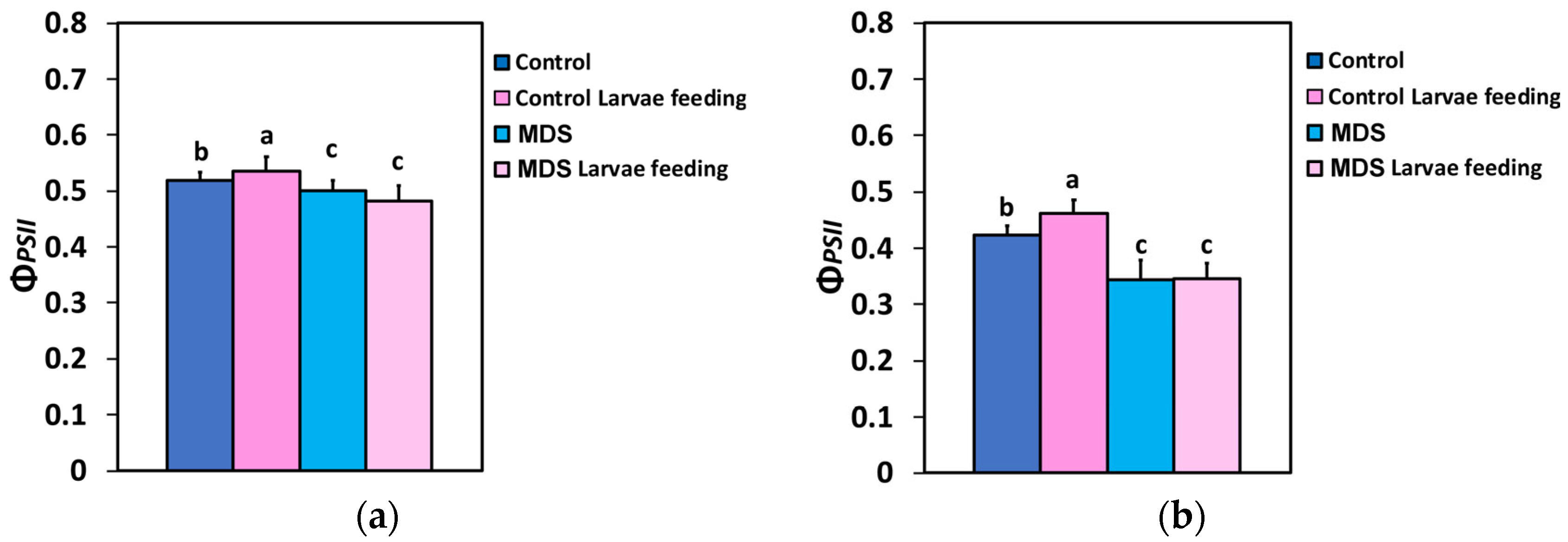
The allocation of absorbed light energy at PSII, for (i) beneficial use for photochemistry (ΦPSII), (ii) heat dissipation loss (ΦNPQ), or (iii) loss by a nonregulatory way (ΦNO) [71], was measured before and after 20 min of larvae feeding in WW or MDS tomatoes. The effective quantum yield of PSII photochemistry (ΦPSII) at both the growth light intensity (GLI, 250 μmol photons m−2 s−1) and the high light intensity (HLI, 1000 μmol photons m−2 s−1) increased after 20 min of larvae feeding in the WW tomatoes (Figure 2a,b). At MDS, ΦPSII decreased compared to controls, but larvae feeding had no effect compared to drought stressed tomatoes before feeding (Figure 2a,b).
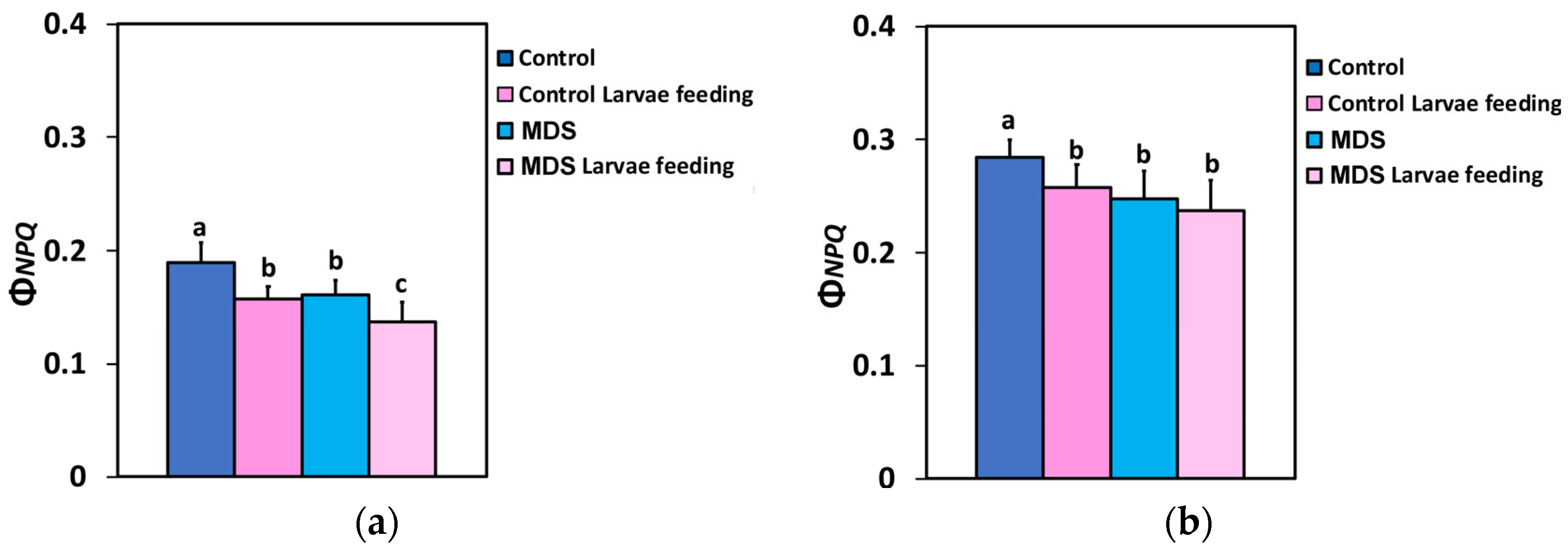
Τhe quantum yield of regulated non-photochemical energy loss in PSII (ΦNPQ) decreased after 20 min of larvae feeding in well-watered tomatoes, at both 250 (GLI)- and 1000 (HLI) μmol photons m−2 s−1 light intensity (Figure 3a,b). Twenty min of larvae feeding in MDS tomatoes did not result in any significant difference in ΦNPQ at the HLI, compared to before feeding or to that of 20 min feeding in WW plants (Figure 3b), while at the GLI ΦNPQ decreased further by larvae feeding in MDS tomatoes, compared to before feeding (Figure 3a).
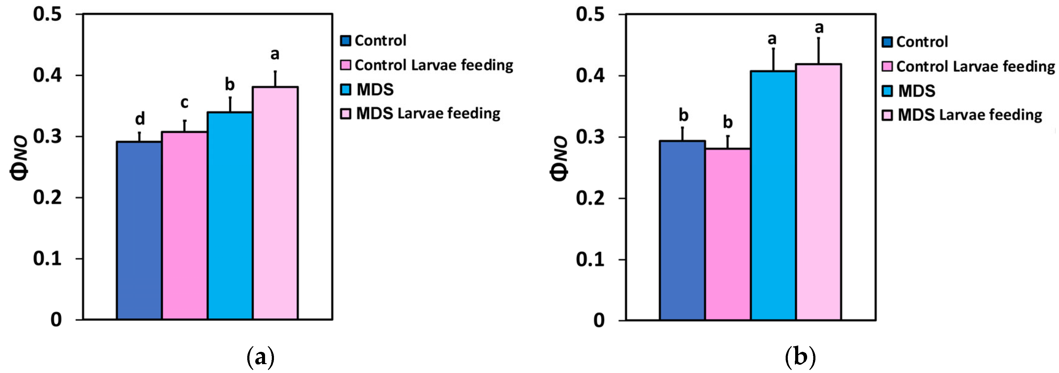
The quantum yield of non-regulated energy loss in PSII (ΦNO) at the GLI increased after larvae feeding, compared to controls (Figure 4a), but remained at the level of the controls at HLI (Figure 4b). ΦNO increased by MDS at both light intensities (Figure 4a,b), but ΦNO after larvae feeding in MDS tomatoes increased further only at the GLI (Figure 4a). At the HLI ΦNO after larvae feeding in MDS tomatoes was maintained at the pre-feeding level (Figure 4b). Thus, larvae feeding increased ΦNO in both WW and MDS tomatoes only under the low light intensity that corresponded to the GLI.

3.4. Impact of Herbivore Feeding on the Electron Transport Rate and the Photoprotective Mechanism

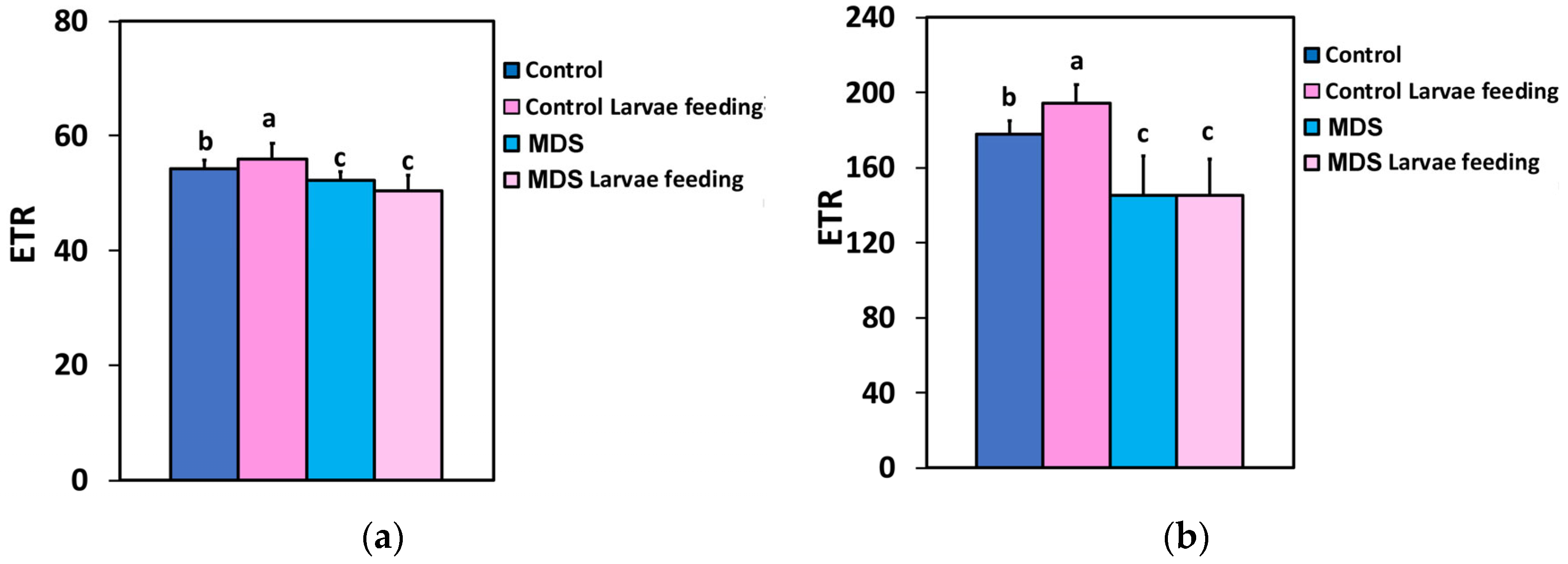
The electron transport rate (ETR), at both the GLI and the HLI, increased after 20 min of larvae feeding in WW tomatoes, while the ETR decreased in the MDS tomatoes compared to controls. However, larvae feeding on MDS tomatoes had no effect, at both the GLI and the HLI (Figure 5a,b).
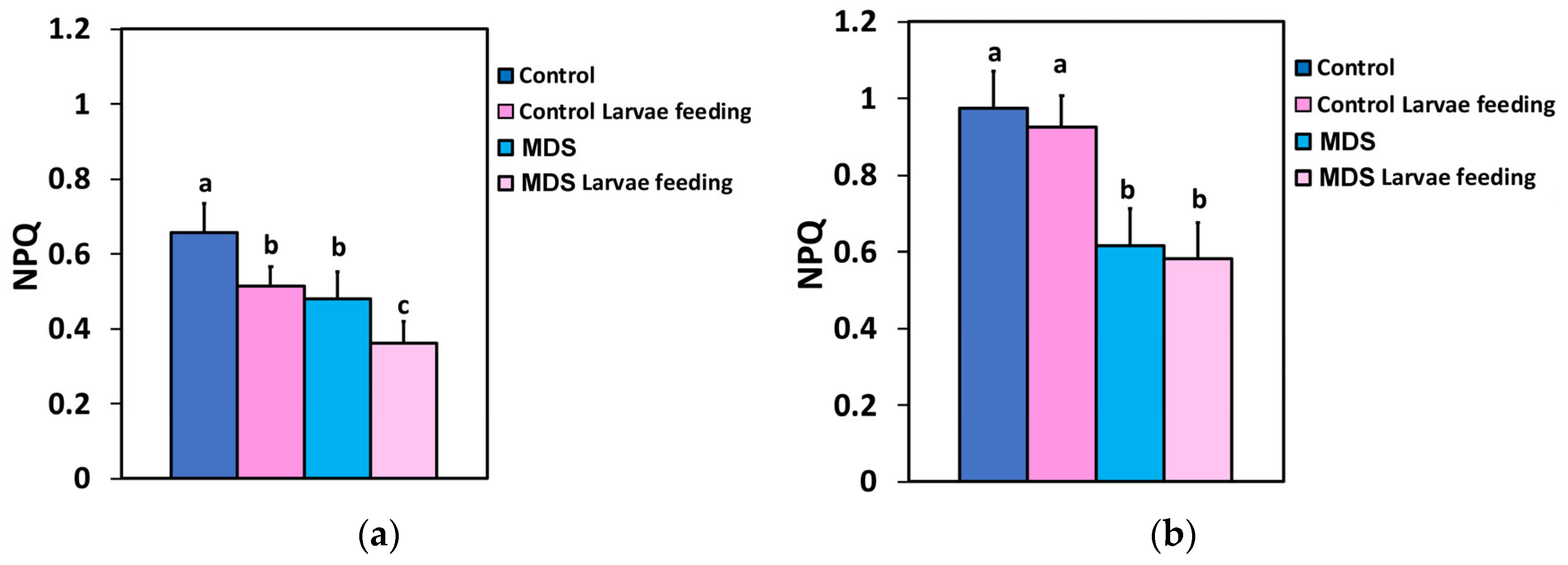
The photoprotective heat dissipation by the non-photochemical quenching (NPQ) mechanism decreased at the GLI after 20 min of larvae feeding in WW tomatoes (Figure 6a), but at the HLI it was unaffected (Figure 6b). In drought-stressed tomatoes NPQ decreased compared to controls at both GLI and HLI (Figure 6a,b), while larvae feeding decreased NPQ further at the GLI (Figure 6a), but at the HLI it remained the same as before feeding (Figure 6b).

3.5. The Fraction of Open PSII Reaction Centers and Their Efficiency Before and After Herbivore Feeding

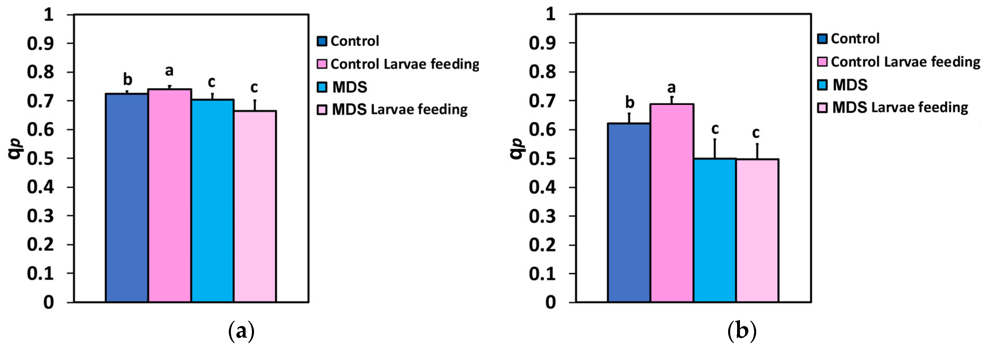
The fraction of open PSII rection centers (qp), at both the GLI and the HLI, increased after 20 min of larvae feeding in the WW tomatoes. In MDS tomatoes, qp decreased, compared to controls, but larvae feeding had no effect, compared to drought-stressed tomatoes before feeding, at both the GLI and HLI (Figure 7a,b).
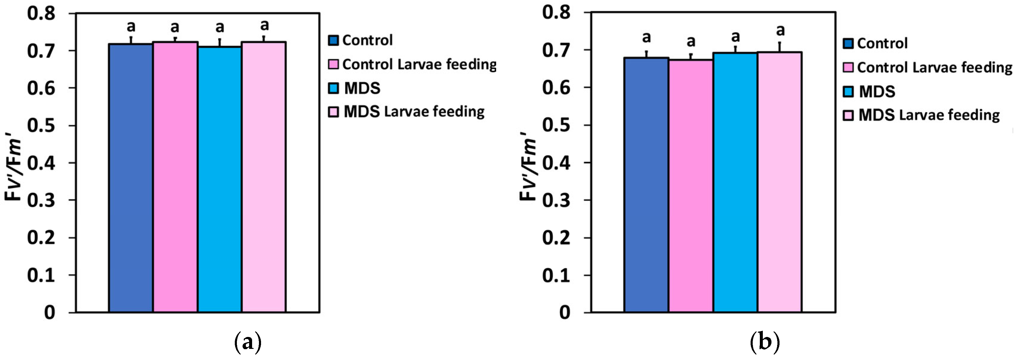
The efficiency of the excitation energy capture by the open PSII reaction centers (Fv′/Fm′), at both the GLI and the HLI, did not differ between the treatments, showing a remarkable resistance to both biotic and abiotic stressors (Figure 8a,b).

3.6. The Excess Excitation Energy and the Excitation Pressure at PSII Before and After Herbivore Feeding

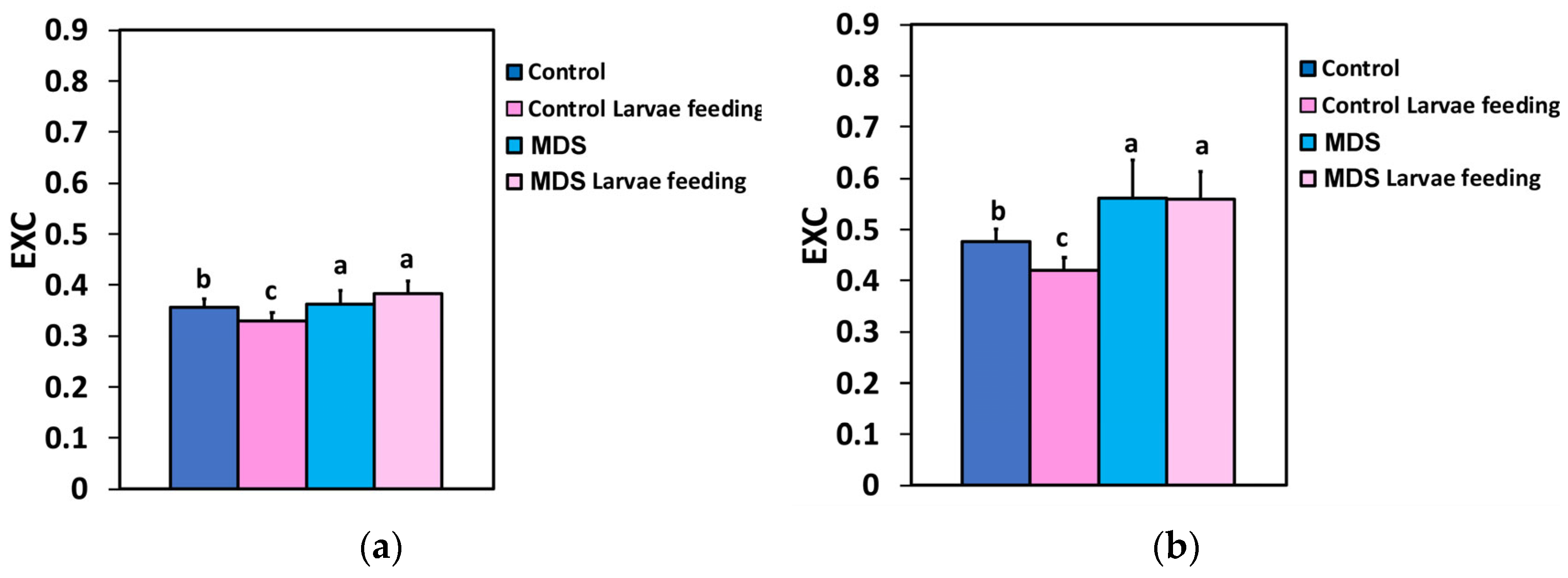
The excess excitation energy (EXC), at both the GLI and the HLI, decreased after 20 min of larvae feeding in well-watered tomatoes, while drought stress increased EXC compared to controls. The larvae feeding had no effect on EXC, compared to drought-stressed tomatoes before feeding, at both the GLI and the HLI (Figure 9a,b).
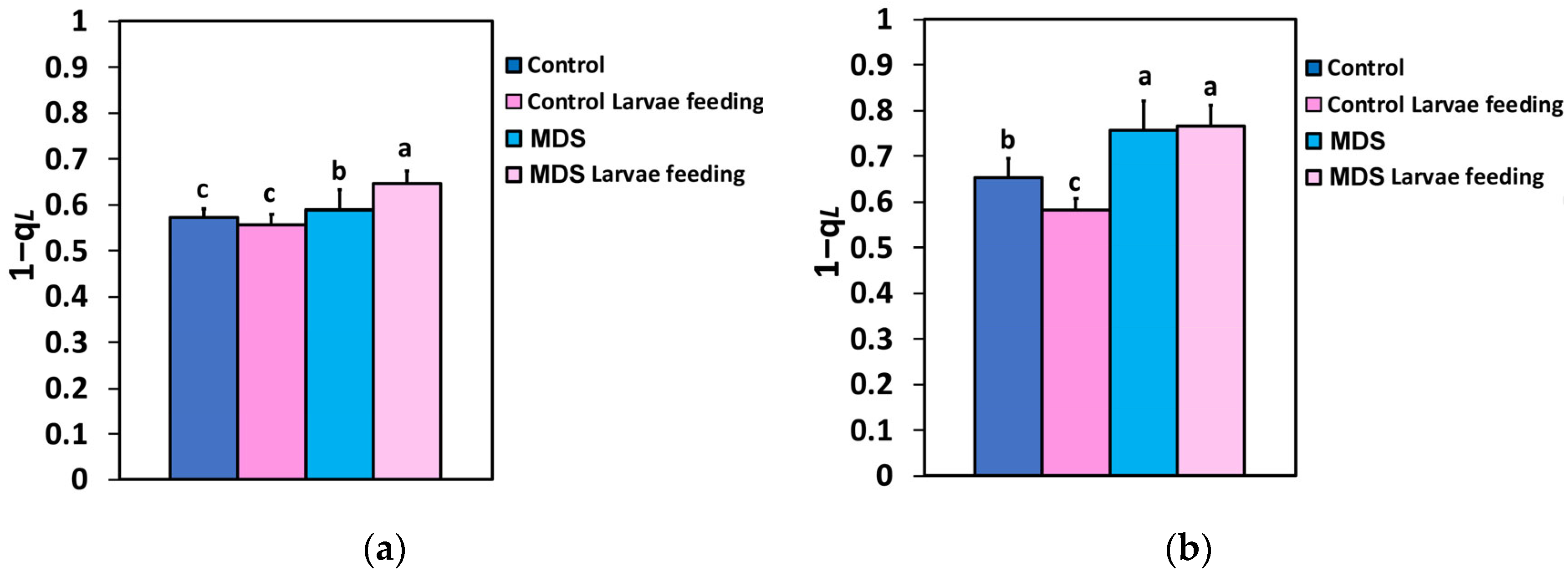
The excitation pressure at PSII (1-qL) did not change after 20 min of larvae feeding in well-watered tomatoes at the GLI (Figure 10a), while at the HLI in well-watered tomatoes, it decreased after 20 min of larvae feeding (Figure 10b). Drought stress increased the excitation pressure at PSII at both the GLI and the HLI, compared to the well-watered tomatoes (Figure 10a,b). However, in MDS tomatoes after larvae feeding, the excitation pressure (1-qL) increased further only at 250 μmol photons m−2 s−1 light intensity (Figure 10a), while at 1000 μmol photons m−2 s−1 light intensity, it remained at pre-feeding level (Figure 10b).

3.7. Correlation of the Efficiency of the Oxygen-Evolving Complex with the Maximum Efficiency of PSII

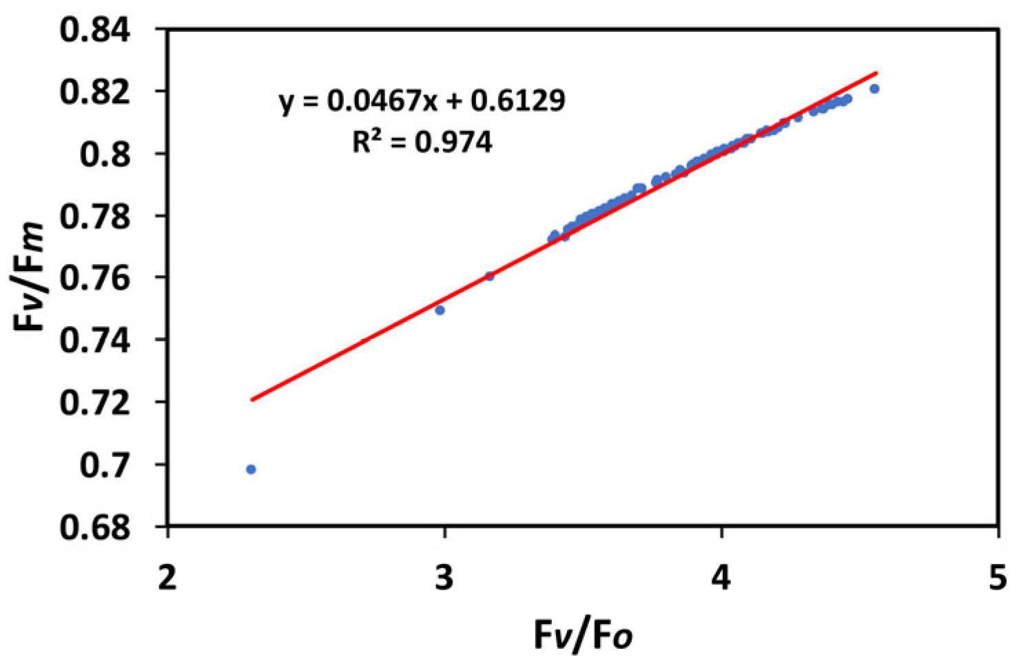
The patterns of the maximum efficiency of photosystem II photochemistry (Fv/Fm) and of the efficiency of the oxygen-evolving complex (Fv/Fo) were closely related (Figure 1a,b), as it was documented by the significant positive correlation that they showed in the regression analysis (R2 = 0.974, p < 0.001) (Figure 11).

3.8. Correlation of the Excess Excitation Energy with the Effective Quantum Yield of PSII

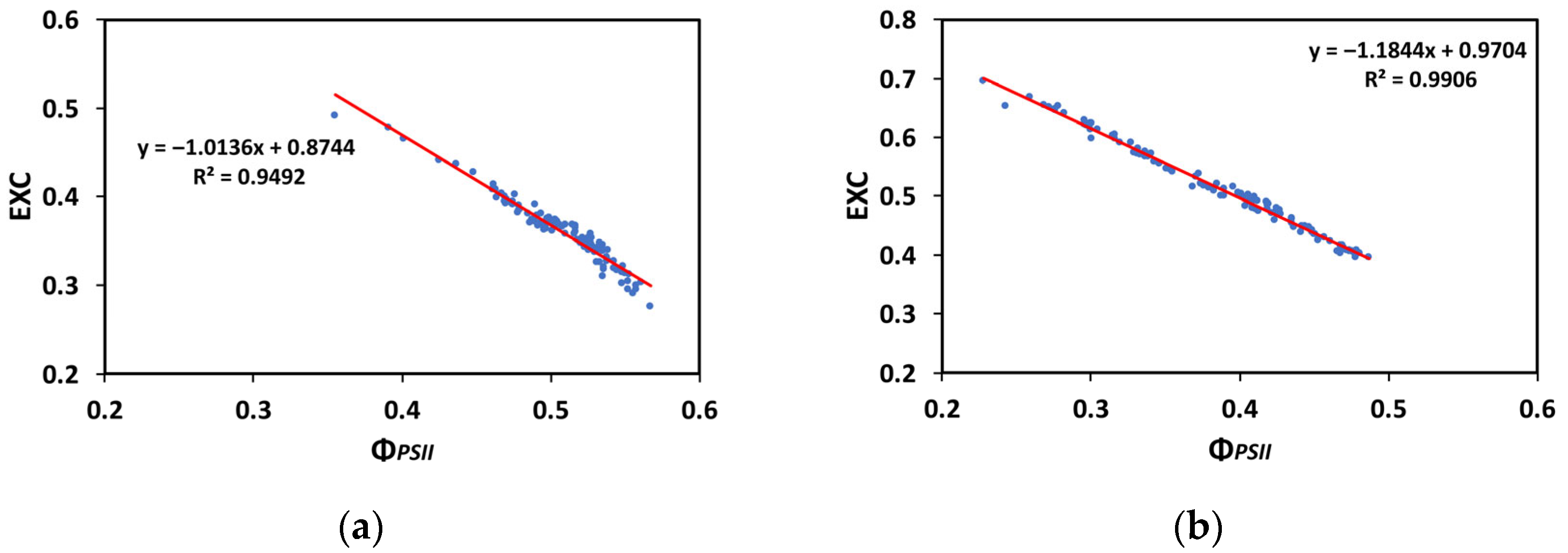
A significant negative correlation was found between the excess excitation energy at PSII (EXC) and the quantum yield of PSII photochemistry (ΦPSII) (Figure 12a,b). A decreased ΦPSII was corelated with an increased excess excitation energy at both the GLI (R2 = 0.9492, p < 0.001, Figure 12a) and the HLI (R2 = 0.9906, p < 0.001, Figure 12b).

3.9. Correlation of the Open PSII Reaction Centers with the Effective Quantum Yield of PSII

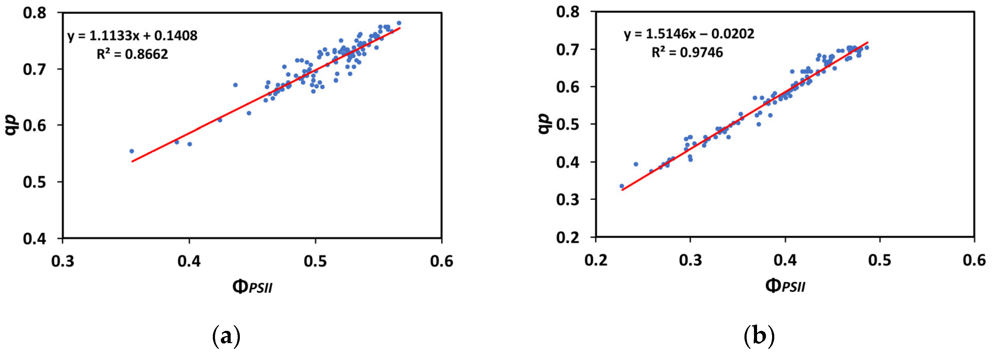
A significant positive correlation between the redox state of quinone A (QA), representing the portion of the open PSII reaction centers (qp), and the effective quantum yield of PSII photochemistry (ΦPSII), at both the GLI (R2 = 0.8662, p < 0.001, Figure 13a) and the HLI (R2 = 0.9746, p < 0.001, Figure 13b), was discovered, suggesting that the efficiency of the excitation energy capture by the open PSII reaction centers (Fv′/Fm′) was not significantly influenced by the treatments and had a negligible impact on the increase or decrease in the quantum yield of PSII photochemistry.

3.10. The Spatiotemporal Heterogeneity of PSII Function Before and After Herbivore Feeding

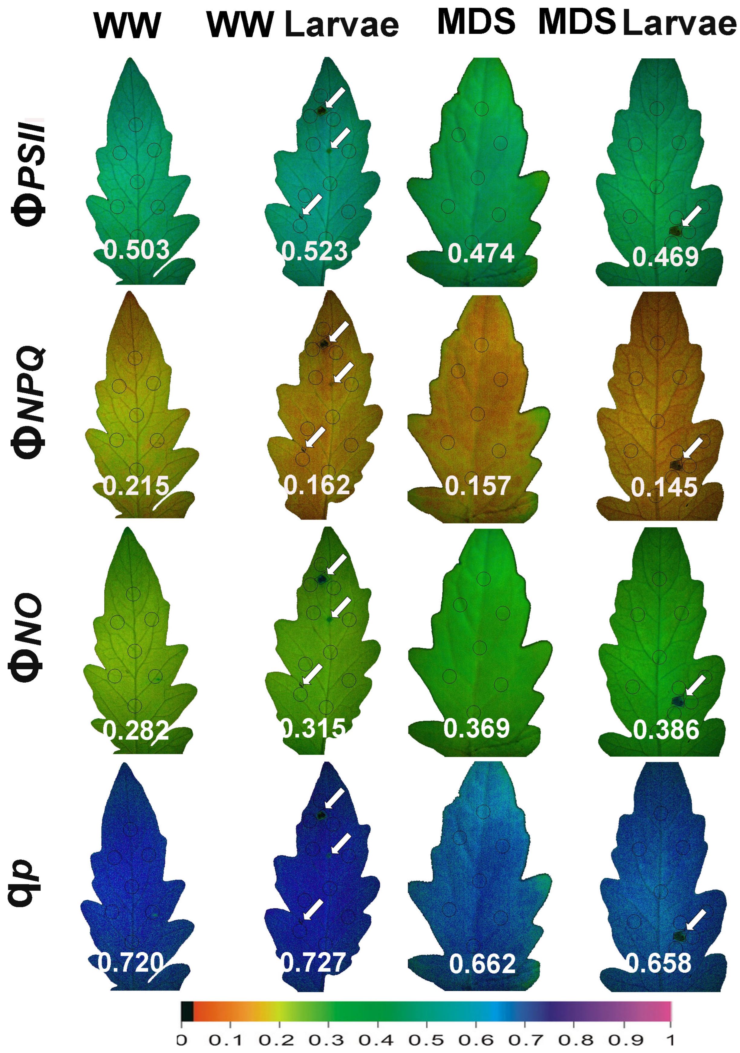
Representative color pictures of the whole area of the tomato leaves for the parameters ΦPSII, ΦNPQ, ΦNO, and qp (captured at the growth light intensity, GLI) of WW and MDS tomatoes, before and after 20 min of larvae feeding, are shown in Figure 14. After 20 min of larvae feeding, in WW tomatoes the effective quantum yield of PSII photochemistry (ΦPSII) increased at the whole-leaf level despite the decreased ΦPSII at the feeding areas (shown by white arrows in Figure 14). At the same time, the significantly decreased quantum yield of heat dissipation (ΦNPQ) resulted in increased values of the quantum yield of non-regulated energy loss in PSII (ΦNO), compared to before feeding. In MDS tomatoes, it decreased.
ΦPSII and ΦNPQ caused a higher increase in ΦNO. Larvae feeding in MDS tomatoes reduced further both the ΦPSII and ΦNPQ, triggering a higher increase in ΦNO.

4. Discussion

Crops are exposed to a range of abiotic and biotic stresses, either separately or simultaneously, which can vary significantly across space and time [72,73]. In response to these stressors, plants undergo various metabolic alterations that are particularly pronounced under conditions of drought and herbivory—two predominant abiotic and biotic stressors—that can lead to functional impacts such as diminished plant vigor and yield [3,4,73,74,75]. However, plants can react to the disruption of their homeostasis, induced by any abiotic or biotic stress factor, by exhibiting adaptive responses, often resulting in a compensatory reaction characterized as hormesis [59,73,76,77,78,79]. The term “hormesis” denotes an overcompensatory physiological adjustment following a disruption of homeostasis, a phenomenon that has been observed across a wide range of organisms, irrespective of the nature of the stressor or the specific metabolic pathways involved [59,76,77,78,79,80,81,82,83,84,85,86,87,88,89].
Understanding the molecular mechanisms that initiate hormesis in plants is key to enhancing crop productivity [87,90]. The effectiveness of hormesis largely depends on factors such as the selected dose range, exposure duration, and the experimental setup [59,80,82,85,90,91,92]. Twenty min of feeding induced an enhancement of PSII photochemistry in WW plants resembling a hormetic response, which was not observed after feeding in MDS tomatoes. It is well recognized that PSII hormetic responses are evident only when studies are carefully and appropriately designed [59,90]. Therefore, the effect of longer-duration feeding may not have the same response.
The effective quantum yield of PSII photochemistry (ΦPSII) (Figure 2a,b), and, respectively, the electron transport rate (ETR) (Figure 5a,b), of the whole leaf in WW plants after 20 min of larvae feeding overcompensated for the decreased ΦPSII observed at the feeding area, compared to the pre-feeding whole-leaf level (Figure 14). This increased ΦPSII at both the GLI and the HLI after herbivore feeding in WW plants seems to be a hormetic response to the disruption of homeostasis. In contrast, ΦPSII and ETR in MDS plants after 20 min of larvae feeding did not compensate at the whole-leaf level for the reduced ΦPSII because of drought stress (Figure 14). The increased ΦPSII and ETR at the whole-leaf level in WW plants after 20 min of larvae feeding was the result of an increased fraction of open PSII reaction centers (qp) (Figure 7a,b), since there was no difference in the efficiency of the open PSII reaction centers (Fv′/Fm′) before and after feeding (Figure 8a,b). Plants have to accomplish defense strategies to adjust their PSII function when they experience herbivore attacks [38].
The decreased maximum efficiency of PSII photochemistry (Fv/Fm) after 20 min of larvae feeding on WW plants suggests possible photoinhibition. Photoinhibition appears when the light energy absorbed by the leaves is neither used for photosynthesis nor dissipated harmlessly as heat by the photoprotective mechanism of non-photochemical quenching (NPQ), or alternatively, when the primary cause is the excitation of Mn in the oxygen-evolving complex (OEC) by photons [93,94,95,96,97,98,99]. Thus, photoinhibition can appear by dual mechanisms [95,97,100]. In our experiments the observed photoinhibition after 20 min of larvae feeding on WW plants led to a reduced efficiency of the OEC, as estimated from the ratio Fv/Fo [101,102,103,104,105,106,107]. The superior maximum efficiency of PSII photochemistry (Fv/Fm) in the WW leaves (Figure 1a) was in agreement with the parallel response pattern of the OEC in the same leaves (Figure 1b). It is well documented that a lower OEC efficiency also indicates a decrease in Fv/Fm [108,109]. Consistent with this, a significant positive correlation between the OEC efficiency and the maximum efficiency of PSII photochemistry (Fv/Fm) was revealed by regression analysis (Figure 11). Photoinhibition has been shown to be correlated with reduced efficiency of the OEC [110,111,112,113] and is frequently connected with crop production [114].
Despite the decrease in the Fv/Fm ratio after 20 min of larvae feeding on WW plants (Figure 1a), the effective quantum yield of PSII photochemistry (ΦPSII), at both the GLI and the HLI, increased after larvae feeding in the WW tomatoes (Figure 2a,b), thus corroborating the view that the observed photoinhibition was not related to reduced light energy use efficiency, but to reduced efficiency of the OEC [110,111,112,113]. Opposing our results, Hu et al. [1] recently reported that herbivory-induced PSII photoinhibition in tomatoes, observed by a reduction in Fv/Fm, was associated not only with a decrease in non-photochemical quenching (NPQ) but also with a decrease in the effective quantum yield of PSII photochemistry (ΦPSII) after larval feeding. Contradictory results likely stem from the diversity of experimental approaches, embracing diverse time-point measurements [46,115,116], and/or to the absence of spatiotemporal measurements [38,39,117,118]. Also, the plant species examined (e.g., potato, tomato) or the insect tested (e.g., Tuta absoluta, Spodoptera exigua) can lead to different conclusions [38,49].
To prevent photoinhibition, primarily of PSII, though PSI can also be affected to a lesser extent, plants have evolved regulatory mechanisms to manage excess excitation energy [119,120,121,122]. The increased fraction of open PSII reaction centers after 20 min of larvae feeding on WW plants (Figure 7a,b) that caused an increased ΦPSII resulted in decreased excess excitation energy (EXC) at the whole-leaf level (Figure 9a,b). This was also documented by the relationship between the excess excitation energy (EXC) and the effective quantum yield of PSII photochemistry (ΦPSII) (Figure 12a,b). On the other hand, the increased excess excitation energy in the MDS leaves, before and after larvae feeding on these leaves (Figure 9a,b), was attributed to the fact that the absorbed light energy was not used for photochemistry (Figure 2a,b), nor it was safely dissipated by the NPQ process as heat (Figure 6a,b). “Excess excitation energy” that is reaching the PSII and is not used for photochemistry must be dissipated non-radiatively as heat (NPQ) to safeguard the photosynthetic apparatus [123,124,125] and avoid any photodamage [126,127]. Photodamage of the photosynthetic apparatus [128] can lead to photoinactivation [129,130], decreasing photosynthetic activity and causing photoinhibition [131,132].
Under most environmental stress conditions, the amount of light energy absorbed by PSII and PSI exceeds the capacity for photochemical utilization, leading to elevated production of reactive oxygen species (ROS), including singlet oxygen (1O2), superoxide anion radicals (O2•−), and hydrogen peroxide (H2O2) [125,133,134,135,136,137]. When the amount of absorbed light energy exceeds the amount that can be used for photochemistry, then PSII excitation pressure increase [132]. Excitation pressure is defined as the relative measure of the reduction state of QA, representing the percentage of closed PSII reaction centers. In our findings, 20 min of larvae feeding decreased the excitation pressure at PSII (1-qL) at the HLI in WW tomatoes, suggesting that a molecular mechanism for lessening the biotic stress effects may exist under HLI and herbivore feeding.
The small decline in leaf water content, despite the high decrease in soil water content (Table 1), suggests an osmotic adjustment [138,139,140]. One mechanism of osmotic adjustment is the accumulation of compatible solutes, or compatible osmolytes [138,139]. Under drought stress, the induction of proline, soluble sugars, and anthocyanin synthesis contributes to osmotic adjustment, helping plant cells acclimate to water stress by retaining the water content [138,139]. However, despite the small decline in leaf water content during MDSm, the effective quantum yield of PSII photochemistry (ΦPSII) decreased (Figure 2a,b). This decrease in ΦPSII could not be balanced by ΦNPQ (Figure 3a,b), and, as a result, ΦNO increased (Figure 4a,b).
Larvae feeding in WW tomatoes induced a more oxidized redox state of quinone A (QA), representing a higher fraction of open PSII reaction centers (qp), and enhanced PSII photochemistry, compared to before feeding. In contrast, MDS and larvae feeding in MDS tomatoes triggered a decrease in the fraction of open PSII reaction centers (qp) and reduced PSII photochemistry, compared to WW tomatoes. Drought stress and herbivore feeding resulted in an increased quantum yield of non-regulated energy loss in PSII (ΦNO) at the GLI, while at HLI ΦNO increased only by drought stress (Figure 4a,b). ΦNO comprises chlorophyll fluorescence internal conversions and intersystem crossing, causing 1O2 generation through the triplet state of chlorophyll (3chl*) [8,123,141,142]. Thus, at HLI, drought stress results in a higher photodamage risk than does the insect herbivory. This drought-stress-induced photodamage is related to the rise in ROS production [10,143]. It can be suggested that the molecular mechanisms for lessening biotic stress effects, such as decreasing ROS generation, may exist under herbivore feeding at HLI. On the other hand, the quantum yield of regulated energy dissipation (ΦNPQ) seems to have no role in counteracting the rise in ROS production under herbivore feeding at HLI (Figure 3b). Herbivore feeding has been reported to induce a compensatory photosynthetic response [38,39,144,145]. The effects of drought stress on herbivore survival have been reported to range from strongly positive (50% increase) to strongly negative (80% decrease), while they were found to be extremely different among closely related plant species and between co-occurring insect herbivores [146]. Plant responses can be unusually specific to the herbivore species or even to the age-related stage of the herbivore [147,148].
The spatial leaf heterogeneity that was observed under all treatments, in all chlorophyll fluorescence parameters, demonstrates the advantage of using chlorophyll fluorescence imaging technique instead of the standard point-based chlorophyll fluorescence measurements [12,15,54].

5. Conclusions

In brief, our results show that in well-watered (WW) plants, photosynthetic efficiency was only locally suppressed at the feeding area of Tuta absoluta, while at the whole-leaf level photosynthetic efficiency improved, indicating a compensatory response. In contrast, in mildly drought-stressed (MDS) plants, photosynthetic efficiency decreased at both the feeding area and the whole leaf after 20 min of larvae feeding. “Plant health status” seems to be critical in the response of PSII photochemistry to insect herbivory. Since drought stress suppresses PSII compensatory response to insect herbivory, it seems that the increased duration, frequency, and intensity of drought stress episodes due to climate change will likely result in higher yield loss attributed to the higher susceptibility of tomatoes to insect pests.
The enhancement of PSII photochemistry under short-term herbivore feeding in the WW plants seems to be a hormetic response to the disruption of homeostasis triggered by defense strategies that adjust PSII function in response to herbivore attack. In contrast, under MDS these defense strategies could not be activated in response to herbivory. Therefore, under projected climate change scenarios, with the increased drought stress episodes which are estimated to appear, and thus to influence plant–insect interactions, a decreased photosynthetic efficiency is likely to occur, attributed to the interaction of drought stress and insect herbivory. However, based on our findings, drought stress results in higher photodamage risk than does insect herbivory, so future research breeding programs must focus on developing drought-resilient crops rather than herbivore-resistant crops.
Future research should explore PSII responses under longer durations of herbivore feeding in WW and MDS crops in field experiments to uncover any similarities in PSII responses with the present study.

Supplementary Materials

The following supporting information can be downloaded at https://www.mdpi.com/article/10.3390/insects16090984/s1. Table S1: Definitions of the chlorophyll fluorescence parameters used in the experiments.

Author Contributions

Conceptualization, M.M.; methodology, J.M., I.S., S.S.A., N.S., K.G., C.D.-S., and M.M.; validation, J.M. and M.M.; formal analysis, J.M., I.S., S.S.A., N.S., K.G., C.D.-S., and M.M.; investigation, J.M., I.S., S.S.A., N.S., K.G., C.D.-S., and M.M.; resources, K.G., C.D.-S., and M.M.; data curation, J.M., I.S., S.S.A., N.S., K.G., C.D.-S., and M.M.; writing—original draft preparation, J.M. and M.M.; writing—review and editing, J.M., I.S., S.S.A., N.S., K.G., C.D.-S. and M.M.; visualization, J.M., I.S., and M.M.; supervision, M.M.; project administration, C.D.-S. and M.M. All authors have read and agreed to the published version of the manuscript.

Funding

This research project was implemented in the framework of H.F.R.I call “Basic research Financing (Horizontal support of all Sciences)” under the National Recovery and Resilience Plan “Greece 2.0” funded by the European Union—NextGenerationEU (H.F.R.I. Project Number: 15453).

Data Availability Statement

All data supporting the findings of this study are available within the paper and within its Supplementary Data published online.

Conflicts of Interest

The authors declare no conflicts of interest. The funders had no role in the design of the study; in the collection, analyses, or interpretation of data; in the writing of the manuscript; or in the decision to publish the results.

Abbreviations

The following abbreviations are used in this manuscript:
1-qLThe fraction of closed PSII reaction centres based on the “lake” model
1O2Singlet oxygen
3chlTriplet state of chlorophyll
FmMaximum chlorophyll a fluorescence in the light-adapted leaf
FoMinimum chlorophyll a fluorescence in the dark-adapted leaf
FoMinimum chlorophyll a fluorescence in the light-adapted leaf
FsSteady-state photosynthesis
Fv′/FmEfficiency of the open PSII reaction centers
Fv/FmMaximum efficiency of PSII photochemistry
Fv/FoEfficiency of the oxygen-evolving complex on the donor side of PSII
GLIGrowth light intensity
H2O2Hydrogen peroxide
HIPVsHerbivore-induced plant volatiles
HLIHigh light intensity
L3Third-instar larvae
LHCIILight-harvesting complexes of PSII
MDSMildly drought-stressed
NPQNon-photochemical quenching (dissipation of excitation energy as heat)
O2•−Superoxide anion radical
OECOxygen-evolving complex
PPFDPhotosynthetic photon flux density
PSIIPhotosystem II
QAQuinone A
qpPhotochemical quenching (fraction of open PSII reaction centers representing also the redox state of quinone A)
RCsReaction centers
ROSReactive oxygen species
SDsStandard deviations
SPsSaturating pulses
VOCsVolatile organic compounds
WWWell-watered
ΦNOQuantum yield of non-regulated energy loss in PSII
ΦNPQQuantum yield of regulated non-photochemical energy loss in PSII
ΦPSIIEffective quantum yield of PSII photochemistry

References

  1. Hu, C.; Thomas, H.R.; Wei, C.; Wu, S.; Zhu, C.; Zhou, Y.; Foyer, C.H.; Yu, J. Herbivory-triggered JA signaling suppresses photosynthesis by inducing photoinhibition in tomato. Plant Commun. 2025, 6, 101237. [Google Scholar] [CrossRef]
  2. Cao, H.; Ding, R.; Du, T.; Kang, S.; Tong, L.; Chen, J.; Gao, J. A meta-analysis highlights the cross-resistance of plants to drought and salt stresses from physiological, biochemical, and growth levels. Physiol. Plant. 2024, 176, e14282. [Google Scholar] [CrossRef] [PubMed]
  3. Hamann, E.; Blevins, C.; Franks, S.J.; Jameel, M.I.; Anderson, J.T. Climate change alters plant–herbivore interactions. New Phytol. 2021, 229, 1894–1910. [Google Scholar] [CrossRef]
  4. Gautam, M.; Kariyat, R. Drought and herbivory drive physiological and phytohormonal changes in soybean (Glycine max Merril): Insights from a meta-analysis. Plant Cell Environ. 2025, in press. [CrossRef] [PubMed]
  5. Adejumo, M.A.; Olowolaju, E.D.; Okunlola, G.O.; Akinyemi, D.S. Symptom expression and severity indices of Solanum lycopersicum L. and Solanum melongena L. at different growth stages under water deficit. Discov. Plants 2025, 2, 171. [Google Scholar] [CrossRef]
  6. Sperdouli, I.; Mellidou, I.; Moustakas, M. Harnessing chlorophyll fluorescence for phenotyping analysis of wild and cultivated tomato for high photochemical efficiency under water deficit for climate change resilience. Climate 2021, 9, 154. [Google Scholar] [CrossRef]
  7. Moustaka, J.; Sperdouli, I.; Moustakas, M. Light energy use efficiency in photosystem ΙΙ of tomato is related to leaf age and light intensity. Crops 2024, 4, 623–635. [Google Scholar] [CrossRef]
  8. Moustaka, J.; Sperdouli, I.; Panteris, E.; Adamakis, I.D.S.; Moustakas, M. Aspirin foliar spray-induced changes in light energy use efficiency, chloroplast ultrastructure, and ROS generation in tomato. Int. J. Mol. Sci. 2025, 26, 1368. [Google Scholar] [CrossRef]
  9. Nilson, S.E.; Assmann, S.M. The control of transpiration. Insights from Arabidopsis. Plant Physiol. 2007, 143, 19–27. [Google Scholar] [CrossRef]
  10. Sperdouli, I.; Moustakas, M. A better energy allocation of absorbed light in photosystem II and less photooxidative damage contribute to acclimation of Arabidopsis thaliana young leaves to water deficit. J. Plant Physiol. 2014, 171, 587–593. [Google Scholar] [CrossRef]
  11. Zhao, T.; Dai, A. The magnitude and causes of global drought changes in the twenty-first century under a low–severe emissions scenario. J. Clim. 2015, 28, 4490–4512. [Google Scholar] [CrossRef]
  12. Moustaka, J.; Moustakas, M. Early-stage detection of biotic and abiotic stress on plants by chlorophyll fluorescence imaging analysis. Biosensors 2023, 13, 796. [Google Scholar] [CrossRef]
  13. Zhu, J.K. Abiotic stress signaling and responses in plants. Cell 2016, 167, 313–324. [Google Scholar] [CrossRef] [PubMed]
  14. Hou, X.; Zhang, W.; Du, T.; Kang, S.; Davies, W.J. Responses of water accumulation and solute metabolism in tomato fruit to water scarcity and implications for main fruit quality variables. J. Exp. Bot. 2020, 71, 1249–1264. [Google Scholar] [CrossRef] [PubMed]
  15. Moustakas, M.; Sperdouli, I.; Moustaka, J. Early drought stress warning in plants: Color pictures of photosystem II photochemistry. Climate 2022, 10, 179. [Google Scholar] [CrossRef]
  16. Hussain, M.; Farooq, S.; Hasan, W.; Ul-Allah, S.; Tanveer, M.; Farooq, M.; Nawaz, A. Drought stress in sunflower: Physiological effects and its management through breeding and agronomic alternatives. Agr. Water Manag. 2018, 201, 152–166. [Google Scholar] [CrossRef]
  17. Muktadir, M.A.; Adhikari, K.N.; Ahmad, N.; Merchant, A. Chemical composition and reproductive functionality of contrasting faba bean genotypes in response to water deficit. Physiol. Plant. 2021, 172, 540–551. [Google Scholar] [CrossRef]
  18. Powles, S.B.; Yu, Q. Evolution in action: Plants resistant to herbicides. Ann. Rev. Plant Biol. 2010, 61, 317–347. [Google Scholar] [CrossRef]
  19. Culliney, T. Crop losses to arthropods. In Integrated Pest Management; Pimentel, D., Peshin, R., Eds.; Springer: Dordrecht, Germany, 2014; pp. 201–225. [Google Scholar] [CrossRef]
  20. Lucas, J.A.; Hawkins, N.J.; Fraaije, B.A. The evolution of fungicide resistance. Adv. Appl. Microbiol. 2015, 90, 29–92. [Google Scholar]
  21. Sparks, T.C.; Storer, N.; Porter, A.; Slater, R.; Nauen, R. Insecticide resistance management and industry: The origins and evolution of the Insecticide Resistance Action Committee (IRAC) and the mode of action classification scheme. Pest. Manag. Sci. 2021, 77, 2609–2619. [Google Scholar] [CrossRef]
  22. Gutbrodt, B.; Dorn, S.; Mody, K. Drought stress affects constitutive but not induced herbivore resistance in apple plants. Arthropod-Plant Interact. 2012, 6, 171–179. [Google Scholar] [CrossRef]
  23. Shivaramu, S.; Jayanthi, P.D.K.; Kempraj, V.; Anjinappa, R.; Nandagopal, B.; Chakravarty, A.K. What signals do herbivore-induced plant volatiles provide conspecific herbivores? Arthropod-Plant Interact. 2017, 11, 815–823. [Google Scholar] [CrossRef]
  24. Damodaram, K.J.P.; Gadad, H.S.; Parepally, S.K.; Vaddi, S.; Hunashikatti, R.H.; Bhat, R.M. Low moisture stress influences plant volatile emissions affecting herbivore interactions in tomato, Solanum lycopersicum. Ecol. Entomol. 2021, 46, 637–650. [Google Scholar] [CrossRef]
  25. Lin, P.A.; Paudel, S.; Bin Zainuddin, N.; Tan, C.W.; Helms, A.; Ali, J.G.; Felton, G.W. Low water availability enhances volatile-mediated direct defences but disturbs indirect defences against herbivores. J. Ecol. 2022, 110, 2759–2771. [Google Scholar] [CrossRef]
  26. Salerno, G.; Frati, F.; Marino, G.; Ederli, L.; Pasqualini, S.; Loreto, F.; Colazza, S.; Centritto, M. Effects of water stress on emission of volatile organic compounds by Vicia faba, and consequences for attraction of the egg parasitoid Trissolcus basalis. J. Pest Sci. 2017, 90, 635–647. [Google Scholar] [CrossRef]
  27. He, X.; Wang, Y.; Munawar, A.; Zhu, J.; Zhong, J.; Zhang, Y.; Guo, H.; Zhu, Z.; Baldwin, I.T.; Zhou, W. Manipulating stomatal aperture by silencing StSLAC1 affects potato plant–herbivore–parasitoid tritrophic interactions under drought stress. New Phytol. 2025, 245, 2133–2149. [Google Scholar] [CrossRef] [PubMed]
  28. Weldegergis, B.T.; Zhu, F.; Poelman, E.H.; Dicke, M. Drought stress affects plant metabolites and herbivore preference but not host location by its parasitoids. Oecologia 2015, 177, 701–713. [Google Scholar] [CrossRef]
  29. Claeys, H.; Dirk, I. The agony of choice: How plants balance growth and survival under water-limiting conditions. Plant Physiol. 2013, 162, 1768–1779. [Google Scholar] [CrossRef]
  30. Copolovici, L.; Astrid, K.; Triinu, R.; Niinemets, Ü. Volatile organic compound emissions from Alnus glutinosa under interacting drought and herbivory stresses. Environ. Exp. Bot. 2014, 100, 55–63. [Google Scholar] [CrossRef]
  31. Griesser, M.; Weingart, G.; Schoedl-Hummel, K.; Neumann, N.; Becker, M.; Varmuza, K.; Liebner, F.; Schuhmacher, R.; Forneck, A. Severe drought stress is affecting selected primary metabolites, polyphenols, and volatile metabolites in grapevine leaves (Vitis vinifera cv. Pinot noir). Plant Physiol. Biochem. 2015, 88, 17–26. [Google Scholar] [CrossRef]
  32. Caser, M.; Chitarra, W.; D’Angiolillo, F.; Perrone, I.; Demasi, S.; Lovisolo, C.; Pistelli, L.; Pistelli, L. Drought stress adaptation modulates plant secondary metabolite production in Salvia dolomitica Codd. Ind. Crops Prod. 2019, 129, 85–96. [Google Scholar] [CrossRef]
  33. Rahman, S.; Rostás, M.; Vosteen, I. Drought aggravates plant stress by favouring aphids and weakening indirect defense in a sugar beet tritrophic system. J. Pest Sci. 2025, 98, 549–564. [Google Scholar] [CrossRef]
  34. Zhao, C.; Han, W.H.; Xiong, Y.D.; Ji, S.X.; Du, H.; Chi, Y.J.; Chen, N.; Wu, H.; Liu, S.S.; Wang, X.W. Drought suppresses plant salicylic acid defence against herbivorous insects by down-regulating the expression of ICS1 via NAC transcription factor. Plant Stress 2025, in press. [CrossRef]
  35. Tattini, M.; Velikova, V.; Vickers, C.; Brunetti, C.; Di Ferdinando, M.; Trivellini, A.; Fineschi, S.; Agati, G.; Ferrini, F.; Loreto, F. Isoprene production in transgenic tobacco alters isoprenoid, non-structural carbohydrate and phenylpropanoid metabolism, and protects photosynthesis from drought stress. Plant Cell Environ. 2014, 37, 1950–1964. [Google Scholar] [CrossRef] [PubMed]
  36. Lopes, J.M.L.; Nascimento, L.S.Q.; Souza, V.C.; Matos, E.M.; Fortini, E.A.; Grazul, R.M.; Santos, M.O.; Soltis, D.E.; Soltis, P.S.; Otono, W.C.; et al. Water stress modulates terpene biosynthesis and morphophysiology at different ploidal levels in Lippia alba (Mill.) N. E. Brown (Verbenaceae). Protoplasma 2024, 261, 227–243. [Google Scholar] [CrossRef] [PubMed]
  37. Mauch-Mani, B.; Baccelli, I.; Luna, E.; Flors, V. Defense priming: An adaptive part of induced resistance. Annu. Rev. Plant Biol. 2017, 68, 485–512. [Google Scholar] [CrossRef]
  38. Moustaka, J.; Meyling, N.V.; Hauser, T.P. Induction of a compensatory photosynthetic response mechanism in tomato leaves upon short time feeding by the chewing insect Spodoptera exigua. Insects 2021, 12, 562. [Google Scholar] [CrossRef]
  39. Thomson, V.P.; Cunningham, S.A.; Ball, M.C.; Nicotra, A.B. Compensation for herbivory by Cucumis sativus through increased photosynthetic capacity and efficiency. Oecologia 2003, 134, 167–175. [Google Scholar] [CrossRef]
  40. Erb, M.; Reymond, P. Molecular interactions between plants and insect herbivores. Annu. Rev. Plant Biol. 2019, 29, 527–557. [Google Scholar] [CrossRef]
  41. Peterson, R.K.D.; Higley, L.G. Biotic Stress and Yield Loss, 1st ed.; CRC Press: Boca Raton, FL, USA, 2001; ISBN 9780849311451. [Google Scholar]
  42. Delaney, K.J. Injured and uninjured leaf photosynthetic responses after mechanical injury on Nerium oleander leaves, and Danaus plexippus herbivory on Asclepias curassavica leaves. Plant Ecol. 2008, 199, 187–200. [Google Scholar] [CrossRef]
  43. Welter, S.C. Arthropod impact on plant gas exchange. In Insect-Plant Interactions; Bernays, E.A., Ed.; CRC Press: Boca Raton, FL, USA, 2019; pp. 135–164. ISBN 13: 978-0-429-29091-6. [Google Scholar]
  44. Delaney, K.J.; Higley, L.G. An insect countermeasure impacts plant physiology: Midrib vein cutting, defoliation and leaf photosynthesis. Plant Cell Environ. 2006, 29, 1245–1258. [Google Scholar] [CrossRef]
  45. Retuerto, R.; Fernández-Lema, B.; Obeso, J.R. Changes in photochemical efficiency in response to herbivory and experimental defoliation in the dioecious tree Ilex aquifolium. Int. J. Plant Sci. 2006, 167, 279–289. [Google Scholar] [CrossRef]
  46. Zangerl, A.R.; Hamilton, J.G.; Miller, T.J.; Crofts, A.R.; Oxborough, K.; Berenbaum, M.R.; DeLucia, E.H. Impact of folivory on photosynthesis is greater than the sum of its holes. Proc. Natl. Acad. Sci. USA 2002, 99, 1088–1091. [Google Scholar] [CrossRef]
  47. Lenk, S.; Chaerle, L.; Pfündel, E.E.; Langsdorf, G.; Hagenbeek, D.; Lichtenthaler, H.K.; Van Der Straeten, D.; Buschmann, C. Multispectral fluorescence and reflectance imaging at the leaf level and its possible applications. J. Exp. Bot. 2007, 58, 807–814. [Google Scholar] [CrossRef] [PubMed]
  48. Saglam, A.; Chaerle, L.; Van Der Straeten, D.; Valcke, R. Promising monitoring techniques for plant science: Thermal and chlorophyll fluorescence imaging. In Photosynthesis, Productivity, and Environmental Stress, 1st ed.; Ahmad, P., Ahanger, M.A., Alyemeni, M.N., Alam, P., Eds.; John Wiley & Sons Ltd.: Hoboken, NJ, USA, 2020; pp. 241–266. [Google Scholar]
  49. Sperdouli, I.; Andreadis, S.; Moustaka, J.; Panteris, E.; Tsaballa, A.; Moustakas, M. Changes in light energy utilization in photosystem II and reactive oxygen species generation in potato leaves by the pinworm Tuta absoluta. Molecules 2021, 26, 2984. [Google Scholar] [CrossRef] [PubMed]
  50. Sperdouli, I.; Andreadis, S.S.; Adamakis, I.S.; Moustaka, J.; Koutsogeorgiou, E.I.; Moustakas, M. Reactive oxygen species initiate defence responses of potato photosystem II to sap-sucking insect feeding. Insects 2022, 13, 409. [Google Scholar] [CrossRef] [PubMed]
  51. Rolfe, S.A.; Scholes, J.D. Chlorophyll fluorescence imaging of plant-pathogen interactions. Protoplasma 2010, 247, 163–175. [Google Scholar] [CrossRef]
  52. Gorbe, E.; Calatayud, A. Applications of chlorophyll fluorescence imaging technique in horticultural research: A review. Sci. Hortic. 2012, 138, 24–35. [Google Scholar] [CrossRef]
  53. Guidi, L.; Calatayud, A. Non-invasive tools to estimate stress-induced changes in photosynthetic performance in plants inhabiting Mediterranean areas. Environ. Exp. Bot. 2014, 103, 42–52. [Google Scholar] [CrossRef]
  54. Moustakas, M.; Calatayud, A.; Guidi, L. Chlorophyll fluorescence imaging analysis in biotic and abiotic stress. Front. Plant Sci. 2021, 12, 658500. [Google Scholar] [CrossRef]
  55. Pérez-Bueno, M.L.; Pineda, M.; Barón, M. Phenotyping plant responses to biotic stress by chlorophyll fluorescence imaging. Front. Plant Sci. 2019, 10, 1135. [Google Scholar] [CrossRef]
  56. Niyogi, K.K.; Wolosiuk, R.A.; Malkin, R. Photosynthesis. In Biochemistry & Molecular Biology of Plants, 2nd ed.; Buchanan, B.B., Gruissem, W., Jones, R.L., Eds.; John Wiley & Sons, Ltd.: Hoboken, NJ, USA, 2015; pp. 508–566. [Google Scholar]
  57. Lu, Y.; Yao, J. Chloroplasts at the crossroad of photosynthesis, pathogen infection and plant defence. Int. J. Mol. Sci. 2018, 19, 3900. [Google Scholar] [CrossRef]
  58. Barber, J. Photosynthetic energy conversion: Natural and artificial. Chem. Soc. Rev. 2009, 38, 185–196. [Google Scholar] [CrossRef]
  59. Moustakas, M.; Moustaka, J.; Sperdouli, I. Hormesis in photosystem II: A mechanistic approach. Curr. Opin. Toxicol. 2022, 29, 57–64. [Google Scholar] [CrossRef]
  60. Imaizumi, K.; Ifuku, K. Photosystem II: Commonality and diversity with emphasis on the extrinsic subunits. Plant Cell Physiol. 2025, in press. [CrossRef] [PubMed]
  61. Zlobin, I.E.; Ivanov, Y.V.; Kartashov, A.V.; Sarvin, B.A.; Stavrianidi, A.N.; Kreslavski, V.D.; Kuznetsov, V.V. Impact of weak water deficit on growth, photosynthetic primary processes and storage processes in pine and spruce seedlings. Photosynth. Res. 2019, 139, 307–323. [Google Scholar] [CrossRef] [PubMed]
  62. Moustaka, J.; Ouzounidou, G.; Sperdouli, I.; Moustakas, M. Photosystem II is more sensitive than photosystem I to Al3+ induced phytotoxicity. Materials 2018, 11, 1772. [Google Scholar] [CrossRef]
  63. Pereyra, P.C.; Sanchez, N.E. Effect of two solanaceous plants on developmental and population parameters of the tomato leaf miner, Tuta absoluta (Meyrick) (Lepidoptera: Gelechiidae). Neotrop. Entomol. 2006, 35, 671–676. [Google Scholar] [CrossRef]
  64. Ghoneim, K. Predatory insects and arachnids as potential biological control agents against the invasive tomato leafminer, Tuta absoluta Meyrick (Lepidoptera: Gelechiidae), in perspective and prospective. J. Entomol. Zool. Stud. 2014, 2, 52–71. [Google Scholar]
  65. Heidari, N.; Sedaratian-Jahromi, A.; Ghane-Jahromi, M.; Zalucki, M.P. How bottom-up effects of different tomato cultivars affect population responses of Tuta absoluta (Lep.: Gelechiidae): A case study on host plant resistance. Arthropod-Plant Interact. 2020, 14, 181–192. [Google Scholar] [CrossRef]
  66. Fonseca Cardoso, E.; Lopes, A.R.; Dotto, M.; Pirola, K.; Moreno Giarola, C. Phenological growth stages of Gaúcho tomato based on the BBCH scale. Com. Sci. 2021, 12, e3490. [Google Scholar]
  67. Moustakas, M.; Panteris, E.; Moustaka, J.; Aydın, T.; Bayçu, G.; Sperdouli, I. Modulation of photosystem II function in celery via foliar-applied salicylic acid during gradual water deficit stress. Int. J. Mol. Sci. 2024, 25, 6721. [Google Scholar] [CrossRef] [PubMed]
  68. Sperdouli, I.; Moustakas, M. Spatio-temporal heterogeneity in Arabidopsis thaliana leaves under drought stress. Plant Biol. 2012, 14, 118–128. [Google Scholar] [CrossRef] [PubMed]
  69. Moustaka, J.; Panteris, E.; Adamakis, I.D.S.; Tanou, G.; Giannakoula, A.; Eleftheriou, E.P.; Moustakas, M. High anthocyanin accumulation in poinsettia leaves is accompanied by thylakoid membrane unstacking, acting as a photoprotective mechanism, to prevent ROS formation. Environ. Exp. Bot. 2018, 154, 44–55. [Google Scholar] [CrossRef]
  70. Oxborough, K.; Baker, N.R. Resolving chlorophyll a fluorescence images of photosynthetic efficiency into photochemical and non-photochemical components—Calculation of qP and Fv’/Fm’ without measuring Fo’. Photosynth. Res. 1997, 54, 135–142. [Google Scholar] [CrossRef]
  71. Kramer, D.M.; Johnson, G.; Kiirats, O.; Edwards, G.E. New fluorescence parameters for the determination of QA redox state and excitation energy fluxes. Photosynth. Res. 2004, 79, 209–218. [Google Scholar] [CrossRef]
  72. Balestrini, R.; Chitarra, W.; Ghirardo, A.; Nardini, A.; Nerva, L. A stressful life: How plants cope with multiple biotic and abiotic adverse factors. Plant Stress 2022, 5, 100095. [Google Scholar] [CrossRef]
  73. Moustakas, M. Molecular mechanisms of plant abiotic stress tolerance. Int. J. Mol. Sci. 2025, 26, 2731. [Google Scholar] [CrossRef]
  74. Coolen, S.; Proietti, S.; Hickman, R.; Davila Olivas, N.H.; Huang, P.P.; Van Verk, M.C.; Van Pelt, J.A.; Wittenberg, A.H.; De Vos, M.; Prins, M.; et al. Transcriptome dynamics of Arabidopsis during sequential biotic and abiotic stresses. Plant J. 2016, 86, 249–267. [Google Scholar] [CrossRef]
  75. Brodribb, T.J.; Powers, J.; Cochard, H.; Choat, B. Hanging by a thread? Forests and drought. Science 2020, 368, 261–266. [Google Scholar] [CrossRef]
  76. Calabrese, E.J. Evidence that hormesis represents an ‘‘overcompensation’’ response to a disruption in homeostasis. Ecotoxicol. Environ. Saf. 1999, 42, 135–137. [Google Scholar] [CrossRef]
  77. Sperdouli, I.; Ouzounidou, G.; Moustakas, M. Hormesis responses of photosystem II in Arabidopsis thaliana under water deficit stress. Int. J. Mol. Sci. 2023, 24, 9573. [Google Scholar] [CrossRef]
  78. Sperdouli, I.; Giannousi, K.; Moustaka, J.; Antonoglou, O.; Dendrinou-Samara, C.; Moustakas, M. Responses of tomato photosystem II photochemistry to pegylated zincdoped ferrite nanoparticles. Nanomaterials 2025, 15, 288. [Google Scholar] [CrossRef]
  79. Agathokleous, E.; Feng, Z.; Iavicoli, I.; Calabrese, E.J. The two faces of nanomaterials: A quantification of hormesis in algae and plants. Environ Int. 2019, 131, 105044. [Google Scholar] [CrossRef] [PubMed]
  80. Agathokleous, E.; Kitao, M.; Harayama, H. On the non-monotonic, hormetic photoprotective response of plants to stress. Dose-Response 2019, 17, 1559325819838420. [Google Scholar] [CrossRef] [PubMed]
  81. Agathokleous, E.; Calabrese, E.J. Hormesis: The dose response for the 21st century: The future has arrived. Toxicology 2019, 425, 152249. [Google Scholar] [CrossRef] [PubMed]
  82. Agathokleous, E.; Kitao, M.; Calabrese, E.J. Hormesis: Highly generalizable and beyond laboratory. Trends Plant Sci. 2020, 25, 1076–1086. [Google Scholar] [CrossRef]
  83. Agathokleous, E.; Feng, Z.; Peñuelas, J. Chlorophyll hormesis: Are chlorophylls major components of stress biology in higher plants? Sci. Total Environ. 2020, 726, 138637. [Google Scholar] [CrossRef]
  84. Małkowski, E.; Sitko, K.; Szopiński, M.; Gieroń, Ż.; Pogrzeba, M.; Kalaji, H.M.; Zieleźnik-Rusinowska, P. Hormesis in plants: The role of oxidative stress, auxins and photosynthesis in corn treated with Cd or Pb. Int. J. Mol. Sci. 2020, 21, 2099. [Google Scholar] [CrossRef]
  85. Agathokleous, E. The rise and fall of photosynthesis: Hormetic dose response in plants. J. For. Res. 2021, 32, 889–898. [Google Scholar] [CrossRef]
  86. Calabrese, E.J.; Agathokleous, E. Accumulator plants and hormesis. Environ. Pollut. 2021, 274, 116526. [Google Scholar] [CrossRef] [PubMed]
  87. Jalal, A.; de Oliveira Junior, J.C.; Ribeiro, J.S.; Fernandes, G.C.; Mariano, G.G.; Trindade, V.D.R.; Reis, A.R. Hormesis in plants: Physiological and biochemical responses. Ecotoxicol. Environ. Saf. 2021, 207, 111225. [Google Scholar] [CrossRef] [PubMed]
  88. Wei, C.; Jiao, Q.; Agathokleous, E.; Liu, H.; Li, G.; Zhang, J.; Fahad, S.; Jiang, Y. Hormetic effects of zinc on growth and antioxidant defense system of wheat plants. Sci. Total Environ. 2022, 807, 150992. [Google Scholar] [CrossRef] [PubMed]
  89. Agathokleous, E.; Sonne, C.; Benelli, G.; Calabrese, E.J.; Guedes, R.N.C. Low-dose chemical stimulation and pest resistance threaten global crop production. Sci. Total Environ. 2023, 878, 162989. [Google Scholar] [CrossRef]
  90. Moustakas, M.; Sperdouli, I.; Adamakis, I.-D.S.; Şaş, B.; İşgören, S.; Moustaka, J.; Morales, F. Mechanistic approach on melatonin-induced hormesis of photosystem II function in the medicinal plant Mentha spicata. Plants 2023, 12, 4025. [Google Scholar] [CrossRef]
  91. Agathokleous, E.; Kitao, M.; Calabrese, E.J. Hormesis: A compelling platform for sophisticated plant science. Trends Plant Sci. 2019, 24, 318–327. [Google Scholar] [CrossRef]
  92. Erofeeva, E.A. Environmental hormesis of non-specific and specific adaptive mechanisms in plants. Sci. Total Environ. 2022, 804, 150059. [Google Scholar] [CrossRef]
  93. Kato, M.C.; Hikosaka, K.; Hirotsu, N.; Makino, A.; Hirose, T. The excess light energy that is neither utilized in photosynthesis nor dissipated by photoprotective mechanisms determines the rate of photoinactivation in photosystem II. Plant Cell Physiol. 2003, 44, 318–325. [Google Scholar] [CrossRef]
  94. Hakala, M.; Tuominen, I.; Keränen, M.; Tyystjärvi, T.; Tyystjärvi, E. Evidence for the role of the oxygen-evolving manganese complex in photoinhibition of Photosystem II. Biochim. Biophys. Acta-Bioenerg. 2005, 1706, 68–80. [Google Scholar] [CrossRef]
  95. Ohnishi, N.; Allakhverdiev, S.I.; Takahashi, S.; Higashi, S.; Watanabe, M.; Nishiyama, Y.; Murata, N. Two-step mechanism of photodamage to photosystem II: Step 1 occurs at the oxygen-evolving complex and step 2 occurs at the photochemical reaction center. Biochemistry 2005, 44, 8494–8499. [Google Scholar] [CrossRef]
  96. Tyystjärvi, E. Photoinhibition of Photosystem II and photodamage of the oxygen evolving manganese cluster. Coord. Chem. Rev. 2008, 252, 361–376. [Google Scholar] [CrossRef]
  97. Oguchi, R.; Terashima, I.; Chow, W.S. The involvement of dual mechanisms of photoinactivation of photosystem II in Capsicum annuum L. plants. Plant Cell Physiol. 2009, 50, 1815–1825. [Google Scholar] [CrossRef]
  98. Campbell, D.A.; Tyystjärvi, E. Parameterization of photosystem II photoinactivation and repair. Biochim. Biophys. Acta-Bioenerg. 2012, 1817, 258–265. [Google Scholar] [CrossRef] [PubMed]
  99. Zavafer, A.; Koinuma, W.; Chow, W.S.; Cheah, M.H.; Mino, H. Mechanism of photodamage of the oxygen evolving Mn cluster of photosystem II by excessive light energy. Sci. Rep 2017, 7, 7604. [Google Scholar] [CrossRef]
  100. Oguchi, R.; Terashima, I.; Kou, J.; Chow, W.S. Operation of dual mechanisms that both lead to photoinactivation of photosystem II in leaves by visible light. Physiol. Plant. 2011, 142, 47–55. [Google Scholar] [CrossRef]
  101. Govindachary, S.; Bukhov, N.G.; Joly, D.; Carpentier, R. Photosystem II inhibition by moderate light under low temperature in intact leaves of chilling-sensitive and -tolerant plants. Physiol. Plant. 2004, 121, 322–333. [Google Scholar] [CrossRef]
  102. Pellegrini, E.; Carucci, M.G.; Campanella, A.; Lorenzini, G.; Nali, C. Ozone stress in Melissa officinalis plants assessed by photosynthetic function. Environ. Exp. Bot. 2011, 73, 94–101. [Google Scholar] [CrossRef]
  103. Siddiqui, H.; Ahmed, K.B.M.; Hayat, S. Comparative effect of 28-homobrassinolide and 24-epibrassinolide on the performance of different components influencing the photosynthetic machinery in Brassica juncea L. Plant Physiol. Biochem. 2018, 129, 198–212. [Google Scholar] [CrossRef]
  104. Mosadegh, H.; Trivellini, A.; Lucchesini, M.; Ferrante, A.; Maggini, R.; Vernieri, P.; Mensuali Sodi, A. UV-B physiological changes under conditions of distress and eustress in sweet basil. Plants 2019, 8, 396. [Google Scholar] [CrossRef]
  105. Gohari, G.; Farhadi, H.; Panahirad, S.; Zareei, E.; Labib, P.; Jafari, H.; Mahdavinia, G.; Hassanpouraghdam, M.B.; Ioannou, A.; Kulak, M.; et al. Mitigation of salinity impact in spearmint plants through the application of engineered chitosan-melatonin nanoparticles. Int. J. Biol. Macromol. 2023, 224, 893–907. [Google Scholar] [CrossRef]
  106. Kalisz, A.; Kornaś, A.; Skoczowski, A.; Oliwa, J.; Jurkow, R.; Gil, J.; Sękara, A.; Sałata, A.; Caruso, G. Leaf chlorophyll fluorescence and reflectance of oakleaf lettuce exposed to metal and metal (oid) oxide nanoparticles. BMC Plant Biol. 2023, 23, 329. [Google Scholar] [CrossRef]
  107. Zia, A.; Farrag, E.S.; Mahmoud, S.Y. Dieback of royal poinciana (Delonix regia) trees induced by Alternaria tenuissima and its impact on photochemical efficiency of photosystem II. Physiol. Mol. Plant Pathol. 2024, 133, 102357. [Google Scholar] [CrossRef]
  108. Tóth, S.Z.; Nagy, V.; Puthur, J.T.; Kovács, L.; Garab, G. The physiological role of ascorbate as photosystem II electron donor: Protection against photoinactivation in heat-stressed leaves. Plant Physiol. 2011, 156, 382–392. [Google Scholar] [CrossRef]
  109. Széles, E.; Kuntam, S.; Vidal-Meireles, A.; Nagy, V.; Nagy, K.; Ábrahám, Á.; Kovács, L.; Tóth, S.Z. Single-cell microfluidics in combination with chlorophyll a fluorescence measurements to assess the lifetime of the Chlamydomonas PSBO protein. Photosynthetica 2023, 61, 417–424. [Google Scholar] [CrossRef] [PubMed]
  110. Callahan, F.E.; Becker, D.W.; Cheniae, G.M. Studies on the photo-inactivation of the water-oxidizing enzyme. II. Characterization of weak light photoinhibition of PSII and its light-induced recovery. Plant Physiol. 1986, 82, 261–269. [Google Scholar] [CrossRef] [PubMed]
  111. Chen, G.X.; Kazimir, J.; Cheniae, G.M. Photoinhibition of hydroxylamine-extracted photosystem II membranes: Studies of the mechanism. Biochemistry 1992, 31, 11072–11083. [Google Scholar] [CrossRef] [PubMed]
  112. Anderson, J.M.; Park, Y.I.; Chow, W.S. Unifying model for the photoinactivation of photosystem II in vivo: A hypothesis. Photosynth. Res. 1998, 56, 1–13. [Google Scholar] [CrossRef]
  113. Sarvikas, P.; Hakala, M.; Pätsikkä, E.; Tyystjärvi, T.; Tyystjärvi, E. Action spectrum of photoinhibition in leaves of wild type and npq1-2 and npq4-1 mutants of Arabidopsis thaliana. Plant Cell Physiol. 2006, 47, 391–400. [Google Scholar] [CrossRef]
  114. Hamdani, S.; Khan, N.; Perveen, S.; Qu, M.; Jiang, J.; Govindjee; Zhu, X.G. Changes in the photosynthesis properties and photoprotection capacity in rice (Oryza sativa) grown under red, blue, or white light. Photosynth. Res. 2019, 139, 107–121. [Google Scholar] [CrossRef]
  115. Aldea, M.; Hamilton, J.G.; Resti, J.P.; Zangerl, A.R.; Berenbaum, M.R.; Frank, T.D.; DeLucia, E.H. Comparison of photosynthetic damage from arthropod herbivory and pathogen infection in understory hardwood saplings. Oecologia 2006, 149, 221–232. [Google Scholar] [CrossRef]
  116. Tang, J.Y.; Zielinski, R.E.; Zangerl, A.R.; Crofts, A.R.; Berenbaum, M.R.; DeLucia, E.H. The differential effects of herbivory by first and fourth instars of Trichoplusia ni (Lepidoptera: Noctuidae) on photosynthesis in Arabidopsis thaliana. J. Exp. Bot. 2006, 57, 527–536. [Google Scholar] [CrossRef] [PubMed]
  117. Ozaki, K.; Saito, H.; Yamamuro, K. Compensatory photosynthesis as a response to partial debudding in Ezo spruce, Picea jezoensis, seedlings. Ecol. Res. 2004, 19, 225–231. [Google Scholar] [CrossRef]
  118. Turnbull, T.L.; Adams, M.A.; Warren, C.R. Increased photosynthesis following partial defoliation of field-grown Eucalyptus globulus seedlings is not caused by increased leaf nitrogen. Tree Physiol. 2007, 27, 1481–1492. [Google Scholar] [CrossRef] [PubMed]
  119. Murata, N.; Takahashi, S.; Nishiyama, Y.; Allakhverdiev, S.I. Photoinhibition of photosystem II under environmental stress. Biochim. Biophys. Acta-Bioenerg. 2007, 1767, 414–421. [Google Scholar] [CrossRef]
  120. Ruban, A.V. Light harvesting control in plants. FEBS Lett. 2018, 592, 3030–3039. [Google Scholar] [CrossRef]
  121. Sun, H.; Shi, Q.; Zhang, S.-B.; Huang, W. Coordination of cyclic electron flow and water–water cycle facilitates photoprotection under fluctuating light and temperature stress in the epiphytic orchid Dendrobium officinale. Plants 2021, 10, 606. [Google Scholar] [CrossRef]
  122. Sperdouli, I.; Moustaka, J.; Ouzounidou, G.; Moustakas, M. Leaf age-dependent photosystem II photochemistry and oxidative stress responses to drought stress in Arabidopsis thaliana are modulated by flavonoid accumulation. Molecules 2021, 26, 4157. [Google Scholar] [CrossRef]
  123. Müller, P.; Li, X.P.; Niyogi, K.K. Non-photochemical quenching. A response to excess light energy. Plant Physiol. 2001, 125, 1558–1566. [Google Scholar] [CrossRef]
  124. Ruban, A.V. Nonphotochemical chlorophyll fluorescence quenching: Mechanism and effectiveness in protecting plants from photodamage. Plant Physiol. 2016, 170, 1903–1916. [Google Scholar] [CrossRef]
  125. Moustakas, M.; Sperdouli, I.; Adamakis, I.D.S. Reactive oxygen species in chloroplasts and chloroplast antioxidants under abiotic stress. Front. Plant Sci. 2023, 14, 1208247. [Google Scholar] [CrossRef]
  126. Ruban, A.V.; Murchie, E.H. Assessing the photoprotective effectiveness of non-photochemical chlorophyll fluorescence quenching: A new approach. Biochim. Biophys. Acta-Bioenerg. 2012, 1817, 977–982. [Google Scholar] [CrossRef]
  127. Foo, C.C.; Burgess, A.J.; Retkute, R.; Tree-Intong, P.; Ruban, A.V.; Murchie, E.H. Photoprotective energy dissipation is greater in the lower, not the upper, regions of a rice canopy: A 3D analysis. J. Exp. Bot. 2020, 71, 7382–7392. [Google Scholar] [CrossRef]
  128. Zavafer, A.; Iermak, I.; Cheah, M.H.; Chow, W.S. Two quenchers formed during photodamage of phostosystem II and the role of one quencher in preemptive photoprotection. Sci. Rep. 2019, 9, 17275. [Google Scholar] [CrossRef] [PubMed]
  129. Flexas, J.; Hendrickson, L.; Chow, W.S. Photoinactivation of photosystem II in high light-acclimated grapevines. Funct. Plant Biol. 2001, 28, 755–764. [Google Scholar] [CrossRef]
  130. Derks, A.; Schaven, K.; Bruce, D. Diverse mechanisms for photoprotection in photosynthesis. Dynamic regulation of photosystem II excitation in response to rapid environmental change. Biochim. Biophys. Acta-Bioenerg. 2015, 1847, 468–485. [Google Scholar] [CrossRef] [PubMed]
  131. Zavafer, A.; Chow, W.S.; Cheah, M.H. The action spectrum of Photosystem II photoinactivation in visible light. J. Photochem. Photobiol. B 2015, 152, 247–260. [Google Scholar] [CrossRef]
  132. Zuo, G. Non-photochemical quenching (NPQ) in photoprotection: Insights into NPQ levels required to avoid photoinactivation and photoinhibition. New Phytol. 2025, 246, 1967–1974. [Google Scholar] [CrossRef] [PubMed]
  133. Apel, K.; Hirt, H. Reactive oxygen species: Metabolism, oxidative stress, and signal transduction. Annu. Rev. Plant Biol. 2004, 55, 373–399. [Google Scholar] [CrossRef]
  134. Gill, S.S.; Tuteja, N. Reactive oxygen species and antioxidant machinery in abiotic stress tolerance in crop plants. Plant Physiol. Biochem. 2010, 48, 909–930. [Google Scholar] [CrossRef]
  135. Dietz, K.J.J.; Turkan, I.; Krieger-Liszkay, A. Redox- and reactive oxygen species dependent signaling into and out of the photosynthesizing chloroplast. Plant Physiol. 2016, 171, 1541–1550. [Google Scholar] [CrossRef]
  136. Takagi, D.; Takumi, S.; Hashiguchi, M.; Sejima, T.; Miyake, C. Superoxide and singlet oxygen produced within the thylakoid membranes both cause photosystem I photoinhibition. Plant. Physiol. 2016, 171, 1626–1634. [Google Scholar] [CrossRef]
  137. Moustakas, M. Plant photochemistry, reactive oxygen species, and photoprotection. Photochem 2022, 2, 5–8. [Google Scholar] [CrossRef]
  138. Sperdouli, I.; Moustakas, M. Interaction of proline, sugars, and anthocyanins during photosynthetic acclimation of Arabidopsis thaliana to drought stress. J. Plant Physiol. 2012, 169, 577–585. [Google Scholar] [CrossRef] [PubMed]
  139. Blum, A. Osmotic adjustment is a prime drought stress adaptive engine in support of plant production. Plant Cell Environ. 2017, 40, 4–10. [Google Scholar] [CrossRef] [PubMed]
  140. Grandin-Courbet, A.; Morvan-Bertrand, A.; Dehail, M.; Hennequart, F.; Prud’homme, M.P. Laminaria digitata extract improved leaf meristem protection under drought and nitrogen uptake after rehydration through hormesis-based chemical priming in Lolium perenne. Plant Cell Environ. 2025, 48, 6674–6690. [Google Scholar] [CrossRef] [PubMed]
  141. Kasajima, I.; Ebana, K.; Yamamoto, T.; Takahara, K.; Yano, M.; Kawai-Yamada, M.; Uchimiya, H. Molecular distinction in genetic regulation of nonphotochemical quenching in rice. Proc. Natl. Acad. Sci. USA 2011, 108, 13835–13840. [Google Scholar] [CrossRef]
  142. Gawroński, P.; Witoń, D.; Vashutina, K.; Bederska, M.; Betliński, B.; Rusaczonek, A.; Karpiński, S. Mitogen-activated protein kinase 4 is a salicylic acid-independent regulator of growth but not of photosynthesis in Arabidopsis. Mol. Plant 2014, 7, 1151–1166. [Google Scholar] [CrossRef]
  143. Vitale, L.; Vitale, E.; Costanzo, G.; De Maio, A.; Arena, C. Photo-protective mechanisms and the role of poly (ADP-ribose) polymerase activity in a facultative CAM plant exposed to long-term water deprivation. Plants 2020, 9, 1192. [Google Scholar] [CrossRef]
  144. McNaughton, S.J. Compensatory plant growth as a response to herbivory. Oikos 1983, 40, 329–336. [Google Scholar] [CrossRef]
  145. Karban, R.; Myers, J.H. Induced plant responses to herbivory. Annu. Rev. Ecol. Syst. 1989, 20, 331–348. [Google Scholar] [CrossRef]
  146. Carvajal Acosta, A.N.; Agrawal, A.A.; Mooney, K. Plant water-use strategies as mediators of herbivore drought response: Ecophysiology, host plant quality and functional traits. J. Ecol. 2022, 111, 687–700. [Google Scholar] [CrossRef]
  147. Barrett, L.G.; Heil, M. Unifying concepts and mechanisms in the specificity of plant–enemy interactions. Trends Plant Sci. 2012, 17, 282–292. [Google Scholar] [CrossRef]
  148. Kessler, A. Detecting the enemy or being manipulated by your attacker? Herbivore-derived elicitors of plant responses: An introduction to a Virtual Issue. New Phytol. 2025, 247, 431–435. [Google Scholar] [CrossRef]
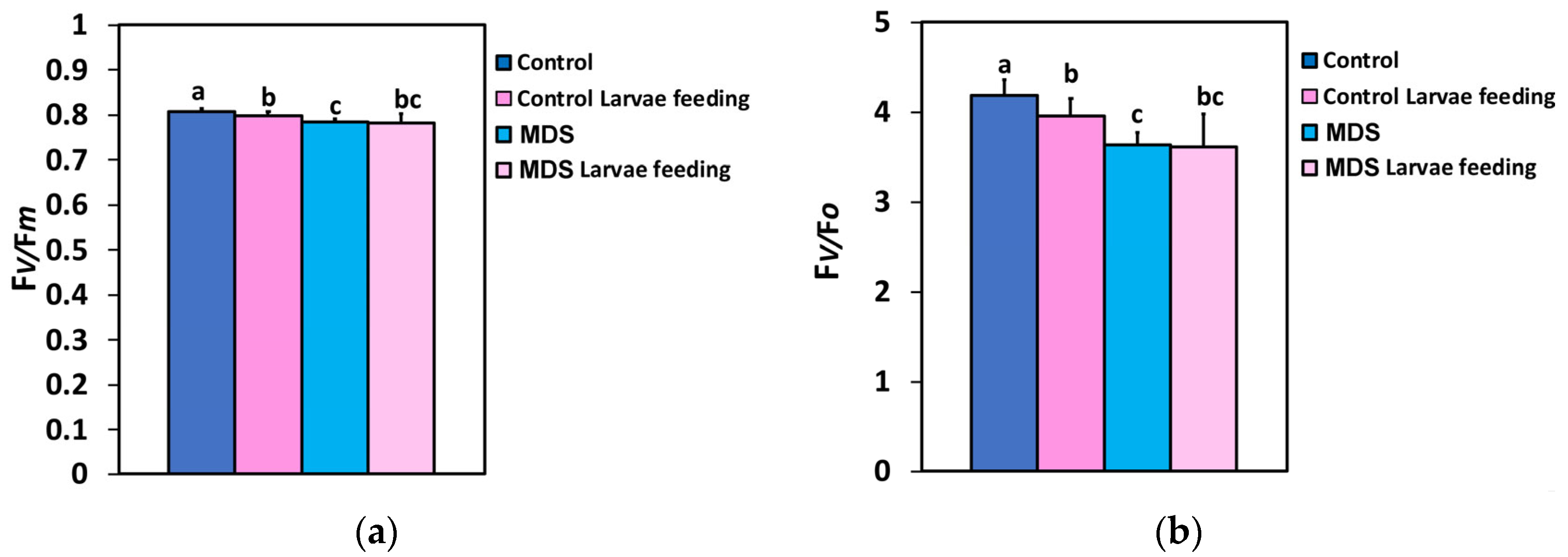
Figure 1. The maximum efficiency of PSII photochemistry (Fv/Fm) (a), and the efficiency of the oxygen-evolving complex (Fv/Fo) (b) before and after 20 min of larvae feeding, in well-watered (WW, control) or mildly drought-stressed (MDS) tomatoes. Standard deviations (SD) are shown by bars. Different lower-case letters reveal significant differences at p < 0.05 (n = 6).
Insects 16 00984 g001
Figure 2. The effective quantum yield of PSII photochemistry (ΦPSII) at actinic light (AL) of 250 μmol photons m−2 s−1, corresponding to the growth light intensity (GLI) (a), and at actinic light (AL) of 1000 μmol photons m−2 s−1, corresponding to a high light intensity (HLI) (b), measured before and after 20 min of larvae feeding, in well-watered (WW, control) or mildly drought-stressed (MDS) tomatoes. Standard deviations (SD) are shown by bars. Different lower-case letters reveal significant differences at p < 0.05 (n = 6).
Insects 16 00984 g002
Figure 3. Τhe quantum yield of regulated non-photochemical energy loss in PSII (ΦNPQ) at 250 μmol photons m−2 s−1 AL (a) and at 1000 μmol photons m−2 s−1 AL (b), before and after 20 min of larvae feeding, in well-watered (WW, control) or mildly drought-stressed (MDS) tomatoes. Standard deviations (SD) are shown by bars. Different lower-case letters reveal significant differences at p < 0.05 (n = 6).
Insects 16 00984 g003
Figure 4. The quantum yield of non-regulated energy loss in PSII (ΦNO) at 250 μmol photons m−2 s−1 AL (a) and at 1000 μmol photons m−2 s−1 AL (b), before and after 20 min of larvae feeding, in well-watered (WW, control) or mildly drought-stressed (MDS) tomatoes. Standard deviations (SD) are shown by bars. Different lower-case letters reveal significant differences at p < 0.05 (n = 6).
Insects 16 00984 g004
Figure 5. The electron transport rate (ETR) at 250 μmol photons m−2 s−1 AL (a) and at 1000 μmol photons m−2 s−1 AL (b), before and after 20 min of larvae feeding, in well-watered (WW, control) or mildly drought-stressed (MDS) tomatoes. Standard deviations (SD) are shown by bars. Different lower-case letters reveal significant differences at p < 0.05 (n = 6).
Insects 16 00984 g005
Figure 6. The non-photochemical quenching (NPQ) at 250 μmol photons m−2 s−1 AL (a) and at 1000 μmol photons m−2 s−1 AL (b), before and after 20 min of larvae feeding, in well-watered (WW, control) or mildly drought-stressed (MDS) tomatoes. Standard deviations (SD) are shown by bars. Different lower-case letters reveal significant differences at p < 0.05 (n = 6).
Insects 16 00984 g006
Figure 7. The portion of open PSII rection centers (RCs) (qp) that reveal the redox state of quinone A (QA), at 250 μmol photons m−2 s−1 AL (a) and at 1000 μmol photons m−2 s−1 AL (b), before and after 20 min of larvae feeding, in well-watered (WW, control) or mildly drought-stressed (MDS) tomatoes. Standard deviations (SD) are shown by bars. Different lower-case letters reveal significant differences at p < 0.05 (n = 6).
Insects 16 00984 g007
Figure 8. The efficiency of the open PSII RCs (Fv′/Fm′) at 250 μmol photons m−2 s−1 AL (a) and at 1000 μmol photons m−2 s−1 AL (b), before and after 20 min of larvae feeding, in well-watered (WW, control) or mildly drought-stressed (MDS) tomatoes. Standard deviations (SD) are shown by bars. Different lower-case letters reveal significant differences at p < 0.05 (n = 6).
Insects 16 00984 g008
Figure 9. The excess excitation energy at PSII (EXC) at 250 μmol photons m−2 s−1 AL (a) and at 1000 μmol photons m−2 s−1 AL (b), before and after 20 min of larvae feeding, in well-watered (WW, control) or mildly drought-stressed (MDS) tomatoes. Standard deviations (SD) are shown by bars. Different lower-case letters reveal significant differences at p < 0.05 (n = 6).
Insects 16 00984 g009
Figure 10. The excitation pressure at PSII (1-qL) at 250 μmol photons m−2 s−1 AL (a) and at 1000 μmol photons m−2 s−1 AL (b), before and after 20 min of larvae feeding, in well-watered (WW, control) or mildly drought-stressed (MDS) tomatoes. Standard deviations (SD) are shown by bars. Different lower-case letters reveal significant differences at p < 0.05 (n = 6).
Insects 16 00984 g010
Figure 11. The relationship of the maximum efficiency of PSII photochemistry (Fv/Fm) with the efficiency of the oxygen-evolving complex (Fv/Fo), before and after 20 min of larvae feeding, in well-watered (WW, control) or mildly drought-stressed (MDS) tomatoes, based on the data of Figure 1a,b. Blue dots represent the paired measurements of the variables while the red line is the regression line that illustrates the relationship between the two variables.
Insects 16 00984 g011
Figure 12. The relationship between the excess excitation energy (EXC) and the effective quantum yield of PSII photochemistry (ΦPSII) at 250 μmol photons m−2 s−1 (a) and at 1000 μmol photons m−2 s−1 AL (b), before and after 20 min of larvae feeding, in well-watered (control) or mildly drought-stressed tomatoes, based on the data of Figure 2a,b and Figure 9a,b. Blue dots represent the paired measurements of the variables while the red line is the regression line that illustrates the relationship between the two variables.
Insects 16 00984 g012
Figure 13. The relationship between the redox state of quinone A (QA) and the effective quantum yield of PSII photochemistry (ΦPSII) at 250 μmol photons m−2 s−1 (a) and at 1000 μmol photons m−2 s−1 AL (b), before and after 20 min of larvae feeding, in well-watered (control) or mildly drought-stressed tomatoes, based on the data of Figure 2a,b and Figure 7a,b. Blue dots represent the paired measurements of the variables while the red line is the regression line that illustrates the relationship between the two variables.
Insects 16 00984 g013
Figure 14. Representative color-coded images of the light energy partitioning at PSII, to photochemistry (ΦPSII), heat dissipation (ΦNPQ), or nonregulatory lost (ΦNO), and the respective color-coded images of the portion of open PSII rection centers (RCs) (qp), before and after 20 min of larvae feeding, in well-watered (WW, control) or mildly drought-stressed (MDS) tomatoes. The areas of interest (AOIs) measured at the leaf surface are shown by circles, while the whole leaflet (average) value is given in white. White arrows point out the feeding areas. The color code on the bottom of the images shows pixel values ranging from 0.0 to 1.0.
Insects 16 00984 g014
Table 1. The leaf water content of well-watered tomato plants and mildly drought-stressed tomatoes and the soil water content of their respective pots (n = 10 ± SD).
ParameterWell-Watered Plants Mildly Drought-Stressed
Leaf Water Content 186.19 ± 0.9783.99 ± 0.08
Soil Water Content 20.51 ± 0.030.22 ± 0.05
1 expressed %; 2 expressed in m3 m−3.
Disclaimer/Publisher’s Note: The statements, opinions and data contained in all publications are solely those of the individual author(s) and contributor(s) and not of MDPI and/or the editor(s). MDPI and/or the editor(s) disclaim responsibility for any injury to people or property resulting from any ideas, methods, instructions or products referred to in the content.