


Simple Summary
Chagas disease is a serious illness caused by the parasite Trypanosoma cruzi and transmitted by kissing bugs. Some of these insects, including species of the Triatoma rubrovaria group, occur in southern Brazil and may pose a risk of spreading the disease to humans. Scientists have long classified these insects into different species based on their morphological characters, but it is unclear whether they are molecularly distinct. The correct taxonomical assignment of kissing bugs is relevant to correctly determine their bionomic parameters to design purposeful vector control strategies. Here, we analyzed the genetic material of 84 specimens that morphologically belong to five species collected in the wild. Surprisingly, we found that those individuals were molecularly very similar to each other. This suggests that these insects may not be separate species, or that they are still in the early stages of evolving into different groups. Our findings highlight the need for a taxonomical revision of this group and further studies to determine the true diversity of these insects and assess their role in spreading Chagas disease.
Abstract
The Triatoma rubrovaria subcomplex, comprising several triatomine species, plays a significant role in the transmission of Chagas disease in southern Brazil. Despite morphological distinctions among these species, their genetic differentiation remains poorly understood, particularly in sympatric regions. This study investigates the genetic diversity and phylogenetic relationships through DNA sequencing analysis of five sympatric species within the T. rubrovaria subcomplex (T. rubrovaria, T. carcavalloi, T. klugi, T. circummaculata, and T. pintodiasi), using a 542-bp fragment of the mitochondrial cytochrome b (mtCytb) gene. A total of 84 specimens were collected from six municipalities in Rio Grande do Sul, Brazil, and analyzed alongside laboratory-reared specimens and sequences from the GenBank. Bayesian phylogenetic reconstructions, haplotype networks, and population structure analyses revealed a lack of clear genetic differentiation among the five species, with overlapping intra- and interspecific divergences and shared haplotypes. These findings suggest either a single species exhibiting phenotypic plasticity or a group of incipient species with ongoing gene flow. This study highlights the need for a taxonomic revision and suggests that this group could serve as a valuable model for further genomic research to elucidate potential aspects of phenotypic plasticity and/or sympatric speciation in triatomines.
1. Introduction
In 1909, the Brazilian medical researcher Carlos Justiniano Ribeiro Chagas announced the discovery of a new disease known as Chagas disease, characterizing the symptoms, the protozoan Trypanosoma cruzi as the etiological agent, and the triatomines (Hemiptera: Reduviidae) as vectors [1]. One hundred and sixteen years after the discovery, the global burden of T. cruzi infections remains, with an estimated 6 to 7 million cases primarily concentrated in 21 Latin American countries [2]. Even though Chagas disease is listed as one of the 30 candidate diseases for elimination by 2030 [3], vector transmission persists in endemic areas where sylvatic native vector species reinvade insecticide-treated dwellings and colonize peridomestic environments [4].
The initiative of Southern Cone countries in 1991 was responsible for a remarkable decline of T. cruzi transmission to humans through blood transfusion and virtual elimination of non-native populations of the primary vector Triatoma infestans (Klug 1834) [5]. In Brazil, the infestation of domiciles has been sharply reduced since 2008, and the last residual foci of this vector species were recorded in 2014 in Rio Grande do Sul [6]. In this area, however, the elimination of this primary vector has led to an increased occurrence of native species near human dwellings, including Triatoma sordida (Stål, 1859) and Triatoma rubrovaria (Blanchard, 1843) [7,8].
Triatoma rubrovaria is highly competent to transmit T. cruzi through its feces [9], but even though it has eclectic blood feeding habits, it is not frequently found with human blood in the gut (1.3–8.0%) [10,11]. This species is currently grouped according to morphological similarities of adults, geographical location and its positioning into a monophyletic subcomplex (T. rubrovaria subcomplex), together with other eight species: Triatoma carcavalloi (Jurberg, Rocha, and Lent, 1998), T. circummaculata (Stål, 1859), T. klugi (Carcavallo, Jurberg, Quaresma, and Galvão, 2001), T. pintodiasi (Jurberg, Cunha, and Rocha, 2013), T. oliveirai (Neiva, Pinto, and Quaresma, 1939), T. guasayana (Wygodzinsky and Abalos, 1949), T. patagonica (Del Ponte, 1929), and T. limai (Del Ponte, 1929) [12,13]. The first five species and T. rubrovaria are sympatric in southern Brazil, and adults are distinguishable by the shape and color patterns of the head (ocelli, clypeus, anteclypeus), thorax (anterolateral angles, lateral carinae, submedian carinae, discal tubercles, scutellum, and stridulatory sulcus), abdomen (dorsal, ventral, and posterior views of the female external genitalia), and male genitalia (pygophore, phallosoma, and paramers) [14,15,16,17].
Recent phylogenetic reconstructions based on mitochondrial markers, including a fragment of the most widely used marker in triatomines, the cytochrome b (mtCytb), confirmed the monophyly of the subcomplex, and evidenced three major clusters reciprocally monophyletic, composed of (i) T. guasayana occupying the most external cluster, (ii) T. patagonica and T. rubrovaria, and (iii) a third cluster with four species subdivided in two subclusters, one with T. pintodiasi and T. circummaculata, and another with T. carcavalloi and T. klugi [17]. These species, however, were represented by only 2–5 specimens maintained in laboratory conditions. Considering they live in sympatry in southern Brazil (excepting T. guasayana and T. patagonica) and there are still rare solid examples of sympatric speciation in nature, especially in triatomines [18], we sought to determine whether the morphological and molecular differentiation observed among the five sympatric species is consistent in the field with an increased sampling, thereby confirming the presence of reproductive barriers indicative of speciation.
2. Materials and Methods
A total of 84 specimens of the T. rubrovaria subcomplex was captured in six municipalities of Rio Grande do Sul, southern Brazil, distanced 70–150 km from each other (Caçapava do Sul, Encruzilhada do Sul, Lavras do Sul, Cachoeira do Sul, Canguçu, and São Jerônimo), in sylvatic areas 50–100 m distant from houses, where sheep and cattle are frequently found in the pasture. Our active search consisted in looking for triatomines under rocks on the ground and in rudimentary walls of overlapping rocks delimiting the lands (Table A1), since these microhabitats are commonly used as shelters by those insect species [7], and capturing them using stainless steel entomological tweezers. The specimens were identified using dichotomous keys [14,16,19]. Unfortunately, immature samples were damaged during sample preparation/transportation, leaving only the head and part of the thorax and legs intact, and thus it was not possible to identify them to the species level. In this case, the taxonomic identification was based on the relative lengths of the proboscis segments and led to the differentiation of samples into two morphogroups. In T. rubrovaria, T. carcavalloi, and T. klugi, the first segment is longer than the second, which in turn is longer than the third (1 > 2 > 3). In contrast, T. circummaculata and T. pintodiasi have the third segment as the longest, followed by the first and then the second (3 > 1 > 2) [14,16,19]. In addition, laboratory-reared adult specimens of T. rubrovaria, T. circummaculata, and T. carcavalloi were included in the analysis.
One or two legs of each specimen were dissected, and their DNA extracted individually with the QIAamp DNA Mini kit (Qiagen, Hilden, Germany), following the manufacturer’s protocol. Genomic DNA was amplified by PCR using primers (CYTB7432F and CYTB7433R) flanking a 663-bp region of the mitochondrial cytochrome b gene [20]. PCR reactions were carried out in a 50 μL final volume, containing 5 µL DNA (20 to 25 ng), 5 µL 1XGoTaq® Flexi Buffer (Promega, Madison, WI, USA), 1.6 µL 25 mM MgCl2, 0.5 µL GoTaq® Hot Start PCR enzyme (2.5 U), 0.6 µL 100mM dNTPs (Life Technologies, Carlsbad, CA, USA), 1.2 µL each primer at 10 mM, and 33.1 µL ultrapure water. Thermal cycling was carried out in the GeneAmp PCR System 9700 (Life Technologies, Carlsbad, USA), with the following conditions: Hot Start (3 min, 94 °C), followed by 35 cycles of denaturation (30 s, 94 °C), annealing (45 s, 56 °C) and extension (45 s, 72 °C), and a final step (7 min, 72 °C). Both forward and reverse strands were submitted to DNA Sanger sequencing using the BigDye Terminator v3.1 Cycle Sequencing Kit (Applied Biosystems, Carlsbad, CA, USA), following the manufacturer’s protocol, and run on an ABI 3730 XL automated sequencer (Applied Biosystems, Carlsbad, USA) at the PDTIS/FIOCRUZ Genomic Sequencing Platform (Rio de Janeiro, Brazil).
DNA sequence chromatograms of both DNA strands were visually inspected in the SeqMan Lasergene 7.0 (DNASTAR Inc., Madison, WI, USA) for possible background noises or double peaks, and edited to produce a consensus sequence for each sample. Nineteen sequences from five species belonging to the T. rubrovaria subcomplex, three sequences of T. guasayana, and three of T. sordida (which is closely related to T. guasayana [12,21]) were retrieved from GenBank https://www.ncbi.nlm.nih.gov/genbank/ (accessed on 10 December 2024) and used in the analyses (Table A1). Unfortunately, we did not include sequences of T. limai and T. oliveirai, species not collected and whose sequences are not present in GenBank, and T. patagonica, whose sequences available in the public database presented very short sizes (<400-bp). Sequences were aligned with ClustalW [22] and pairwise divergences were calculated in the MEGA X program [23], using the Kimura 2-parameter model of nucleotide substitution.
Bayesian phylogenetic trees were reconstructed with the BEAST v2.6.4 package [24]. Trees were sampled from three independent runs every 10,000 generations from 107 MCMC iterations, discarding the 20% first trees. The birth-and-death model of speciation was imposed, and the nucleotide substitution model was selected using the bModelTest package [25]. The convergence of parameters and proper mixing were inspected by checking if the effective sample sizes (ESSs) were sufficiently large (ESSs ≥ 104 for all parameters). Reliability of the recovered clades was assessed through posterior probability (PP) values. A haplotype network was constructed in popART 1.7, using the median-joining model. Clustering analyses were conducted in the R 4.2.3 environment [26]. Principal Component Analysis (PCA) was performed with the adegenet package [27] and plotted with ggplot2 [28]. Hierarchical Bayesian Population Structure analysis (hBAPS) was performed with the rhierbaps [29] and phytools packages [30] and plotted using the ggplot2 package [28]. Divergence plot was generated using the ggplot2 [28], ggpubr [31], and ggrides [32] packages.
3. Results
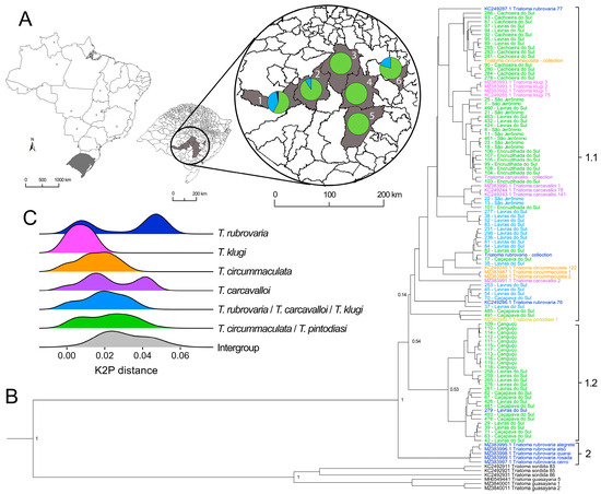
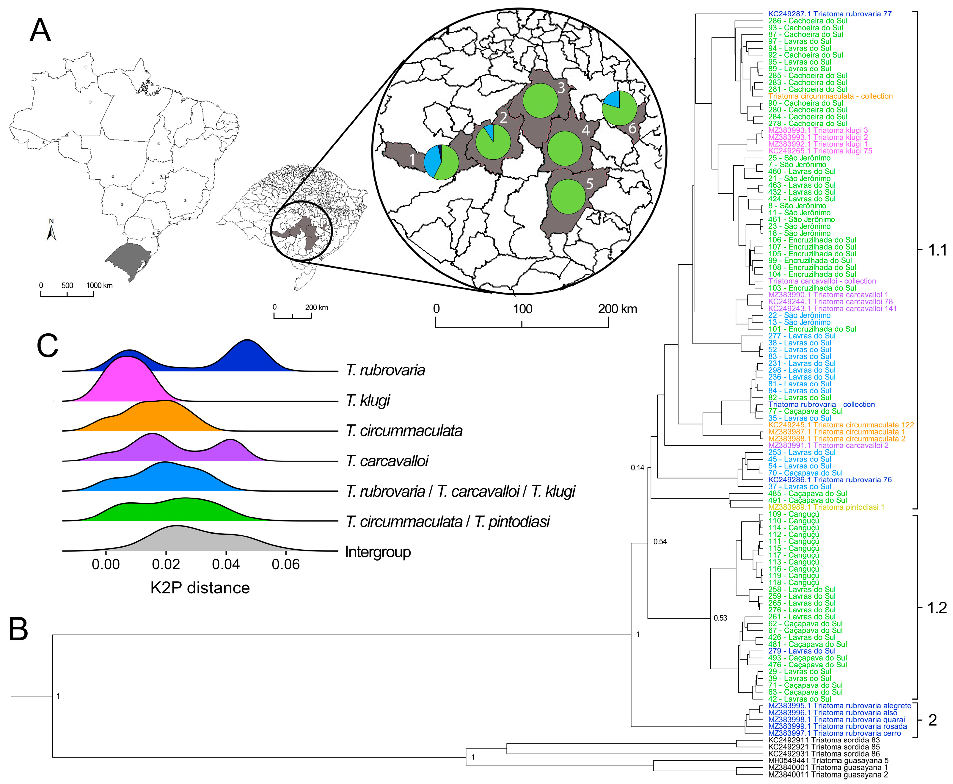
Morphological analyses of 83 nymphs (N1–N5) collected in the field allowed the identification of 16 nymphs as a T. klugi/T. rubrovaria/T. carcavalloi morphogroup and 67 nymphs as a T. circummaculata/T. pintodiasi morphogroup (Figure 1A). A single adult was collected (ID number 279) and identified as T. rubrovaria (Table A1). Both morphogroups were found in sympatry in three localities (Caçapava do Sul, Lavras do Sul, and São Jerônimo), while only specimens of the T. circummaculata/T. pintodiasi morphogroup were found in the other three localities (Cachoeira do Sul, Canguçu, and Encruzilhada do Sul; Figure 1A).
Figure 1.
Taxonomic identification, phylogenetics, and molecular distance-based analyses of the T. rubrovaria subcomplex. (A) Map of Brazil, highlighting the state of Rio Grande do Sul with the municipalities where collections took place indicated in gray. 1—Lavras do Sul; 2—Caçapava do Sul; 3—Cachoeira do Sul; 4—Encruzilhada do Sul; 5—Canguçu; 6—São Jerônimo. The proportion of nymphs identified as belonging to the morphogroups T. rubrovaria/T. carcavalloi/T. klugi (blue) or T. circummaculata/T. pintodiasi (green), and the T. rubrovaria adult (dark blue), in each municipality are shown in pie charts. (B) Bayesian phylogenetic reconstruction based on 542-bp of the mtCytb. The locations where samples were collected are listed after sample IDs (Table A1). The numbers near node trees indicate the posterior probability (PP) values. The colors of the tip labels correspond to the taxonomic classification of samples and are consistent with the Figure 1C labels: dark blue—T. rubrovaria; pink—T. klugi; orange—T. circummaculata; purple—T. carcavalloi; blue—T. rubrovaria/T. carcavalloi/T. klugi morphogroup; green—T. circummaculata/T. pintodiasi morphogroup. Moreover, the sequence of T. pintodiasi is shown in yellow and the outgroups in black. The brackets on the right represent the hBAPS results as two clusters (1 and 2), of which the first has two subclusters (1.1 and 1.2). (C) Frequency of pairwise K2P distances within each group.
Bayesian phylogenetic reconstruction based on 542-bp of the mtCytb evidenced a clear separation of T. guasayana from the other sequences comprising the T. rubrovaria subcomplex, evidencing its genetic relatedness to T. sordida (Figure 1B). Regarding the other species from the T. rubrovaria subcomplex, it is possible to observe two well-supported clusters (Clusters 1.1/1.2 and Cluster 2; PP = 1) that separate five sequences of T. rubrovaria generated in da Silva et al. [17] from all other samples used in this study, including those deposited in GenBank. These clusters diverged by an average of 4.7% (3.0–6.2%). The major cluster containing 101 sequences was divided into two weak-supported subclusters (PP = 0.55) with short branches, indicative of low polymorphism between sequences. Cluster 1.1 grouped 73 samples of all five sympatric species and the two morphogroups, while Cluster 1.2 was composed of 28 sequences from three localities (Caçapava do Sul, Canguçu, and Lavras do Sul), of which all but one sample derived from the T. circummaculata/T. pintodiasi morphogroup.
Hierarchical BAPS confirmed the separation of the dataset into two subclusters in Cluster 1 (1.1 and 1.2), besides Cluster 2, which was composed of the five sequences of T. rubrovaria from da Silva et al. [17]. Pairwise distance analysis (Figure 1C) revealed an overlap of the divergences within and between the morphogroups. The mean intraspecific divergence of sequences for the T. circummaculata/T. pintodiasi group was 2.2% (0–5.6%), and those within the T. rubrovaria/T. carcavalloi/T. klugi group was 1.8% (0–4.9%), while sequences from the different morphogroups had a mean divergence of 2.6% (0–4.1%). A secondary phylogenetic reconstruction (Figure A1) including only the reference sequences confirmed the separation of the five T. rubrovaria sequences from da Silva et al. [17] from all other sequences, and evidenced a paraphyletic pattern across all species, except for T. klugi, which was monophyletic, but with low statistical support (PP < 0.6).
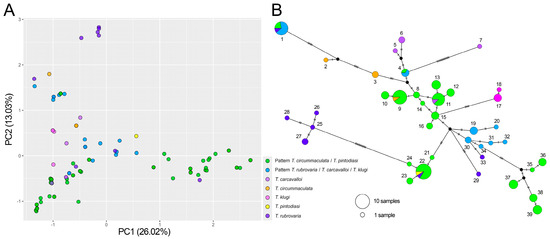
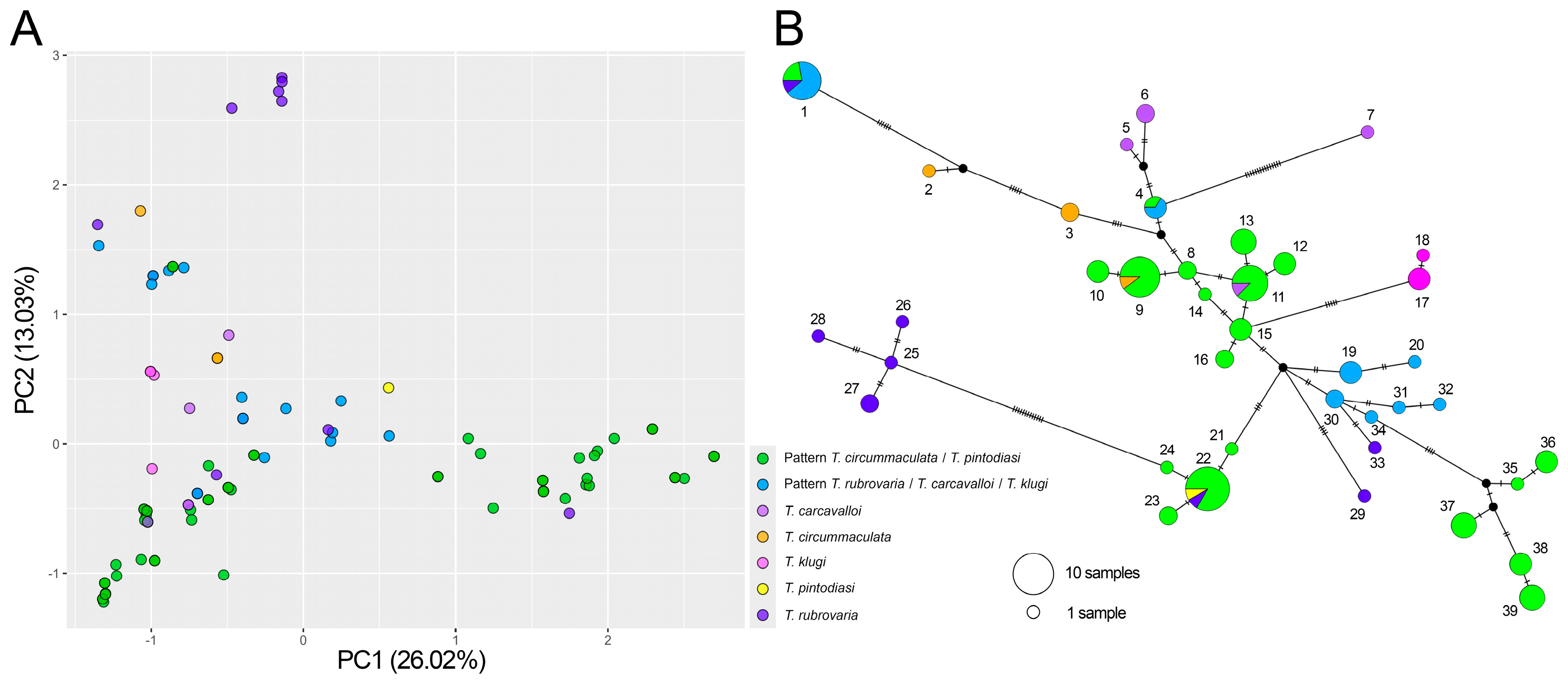
Indeed, PCA did not reveal a clear separation between the morphogroups, of which T. circummaculata/T. pintodiasi seemed to be the most heterogeneous (Figure 2A). Network analysis (Figure 2B) revealed 39 haplotypes, without a clear separation between the morphogroups. Two haplotypes (1 and 4) were shared with both morphogroups. One haplotype (11) was shared between the T. circummaculata/T. pintodiasi morphogroup and T. carcavalloi, and another haplotype (22) was shared between the T. circummaculata/T. pintodiasi morphogroup and T. rubrovaria.
Figure 2.
Molecular clustering and structure analyses of the five sympatric species that belong to the T. rubrovaria subcomplex. (A) Principal component analysis based on the genetic similarity matrix of mtCytb sequences. Color codes for the different morphogroups and species are indicated. (B) Haplotype network using the median-joining method. Dashes along the lines indicate the number of mutational steps (>1) that separate the haplotypes. Circle sizes are proportional to the haplotype frequency. In haplotypes shared between species or morphogroups, circles are drawn as pie charts to represent the proportion of samples from each group.
Phylogenetic analysis, considering only samples morphologically identified at the species level failed to retrieve reciprocally monophyletic groups that could represent T. rubrovaria, T. circummaculata, and T. carcavalloi (Figure A1). Moreover, intra and interspecific divergences also overlap. For instance, intraspecific divergences of T. carcavalloi (0–5.2%, mean = 2.3%) and T. rubrovaria (0–5.6%, mean = 3.3) were nearly the same as or even greater than the interspecific divergences (1–6.2%, mean = 3.9%).
4. Discussion
The mtCytb gene is the most widely used marker for molecular taxonomy in triatomines and has been shown to distinguish closely related species in Rhodnius (Stål, 1859) [20,33,34], Triatoma (Laporte, 1832) [35,36], and Panstrongylus (Berg, 1879) [37] genera. In the case of T. rubrovaria subcomplex, our results evidenced a lack of genetic differentiation among five members when increasing field sampling in sympatric areas. The paraphyletic assemblage in the phylogenetic reconstruction, coupled to low genetic divergence and shared haplotypes among different species, indicates that they either represent a single species with phenotypic plasticity or comprise a group of incipient species with few or no barriers for gene flow. Future population genetics studies with fast-evolving markers, such as microsatellites, or with single nucleotide polymorphisms (SNPs) will be essential to clarify this issue.
Phenotypic plasticity enables organisms to express different phenotypes in response to varying environmental conditions, allowing them to adapt without immediate genetic changes [38]. This plasticity can decouple genetic and phenotypic differentiation, as phenotypes may reflect environmental influences rather than underlying genetic divergence [39]. Phenotypic plasticity has already been observed in Rhodnius nasutus (Stål, 1859) and Rhodnius neglectus (Lent, 1954), in which their body colors matched with the fibers and fronds of their respective palm habitats [5,18,40]. The presence of at least six and two different morphotypes for these species, respectively, supports the idea that they possess genetic traits enabling a range of phenotypes, with natural selection shaping their expression based on environmental factors [18]. There is no evidence, however, of sympatric members of the T. rubrovaria subcomplex occupying different niches or with distinct host preferences in sylvatic settings [7]. Microscale ecology studies such as the identification of blood meal sources and microbiota composition could shed light on possible differences on the bionomy of the species.
Phenotypic divergence in closely related species that occur in sympatry may also indicate the occurrence of incipient differentiation that might result in speciation [41]. Genomic studies have revealed that differentiation is restricted to genomic regions of low recombination, usually near centromeres, on early stages of speciation with gene flow [42], and theoretical models predict that sympatric speciation is facilitated when traits under divergent selection influence assortative mating [43]. Members of the T. rubrovaria subcomplex represent an excellent model for studying speciation in triatomines. Crossbreeding and genome-wide association studies hold the potential to clarify the mode and pattern of speciation in this group, shedding light on genomic hotspots associated with sympatric speciation. From a public health perspective, determining whether the subcomplex constitutes a single or multiple species is crucial for accurately assessing their vector competence and capacity, and thus mapping areas of risk for Chagas disease transmission.
Intriguingly, five sequences of laboratory-reared T. rubrovaria previously published [17], collected in areas 200–460 km away from the areas sampled in this study (Alegrete and Quaraí, Rio Grande do Sul, Brazil), clustered in a well-supported monophyletic clade that diverged 3.0–6.2% (mean = 4.7%) from the other sequences. This percentage of genetic divergence is expected between triatomine sister species [18,20,35,36] and raises the possibility of having allopatric populations of T. rubrovaria in Rio Grande do Sul. Unfortunately, it was not possible to include T. oliveirai in the analysis, although it is sympatric with the five species studied in this region. However, it is considered rare, having been naturally recorded only twice in burrows of the Brazilian guinea pig, Cavia aperea (Erxleben, 1777). [14]. Moreover, the partial preservation of immature specimens constrained the scope of our morphological analysis, particularly for characters located on the abdomen and posterior segments, which are often crucial for accurate taxonomic resolution at the species level. This limitation highlights the importance of proper handling and transportation protocols in entomological surveys. Nevertheless, the reference collection in our laboratory, which includes well-preserved specimens of the identified species, played a crucial role in supporting and confirming the morphological identification based on the available structures. A broader sampling of all species of the T. rubrovaria subcomplex would help map their correct geographical distributions and areas of allopatry and sympatry, thus shedding light on historical processes that have shaped the observed genetic diversity.
Author Contributions
Conceptualization, T.V., C.B. and M.G.P.; methodology, A.R.C., L.B.M., T.V., S.C., J.O. and M.G.P.; formal analysis, A.R.C., L.B.M., J.O. and M.G.P.; resources, C.B. and M.G.P.; data curation, A.R.C., L.B.M. and M.G.P.; writing—original draft preparation, A.R.C., L.B.M. and M.G.P.; writing—review and editing, A.R.C., L.B.M., T.V., S.C., J.O., C.B. and M.G.P.; supervision, C.B. and M.G.P.; funding acquisition C.B. All authors have read and agreed to the published version of the manuscript.
Funding
This work was funded by Fiocruz and Faperj (E26/201.213-2022). L.B.M. and J.O. were funded by Capes (Financial Code 001).
Data Availability Statement
All sequences were deposited in GenBank under the accession numbers PV184590-PV184676.
Acknowledgments
We would like to thank Cleonara Bedin and her team from the Department of Health of Rio Grande do Sul, Brazil, for technical support in field collections. We also thank Cleber Galvão (Instituto Oswaldo Cruz, Fiocruz, Brazil) for kindly provided colony-reared specimens of T. rubrovaria, T. carcavalloi and T. circummaculata.
Conflicts of Interest
The authors declare no conflicts of interest.
Abbreviations
The following abbreviations are used in this manuscript:
| mtCytb | Mitochondrial Cytochrome b |
| PP | Posterior Probability |
| PCA | Principal Component Analysis |
| hBAPS | Hierarchical Bayesian Analysis of Population Structure |
| ESS | Effective Sample Size |
| K2P | Kimura 2-Parameter |
| MCMC | Markov Chain Monte Carlo |
Appendix A
Figure A1.
Bayesian phylogenetic reconstruction based on reference sequences from [17]. The numbers near node trees indicate posterior probabilities above 0.7.
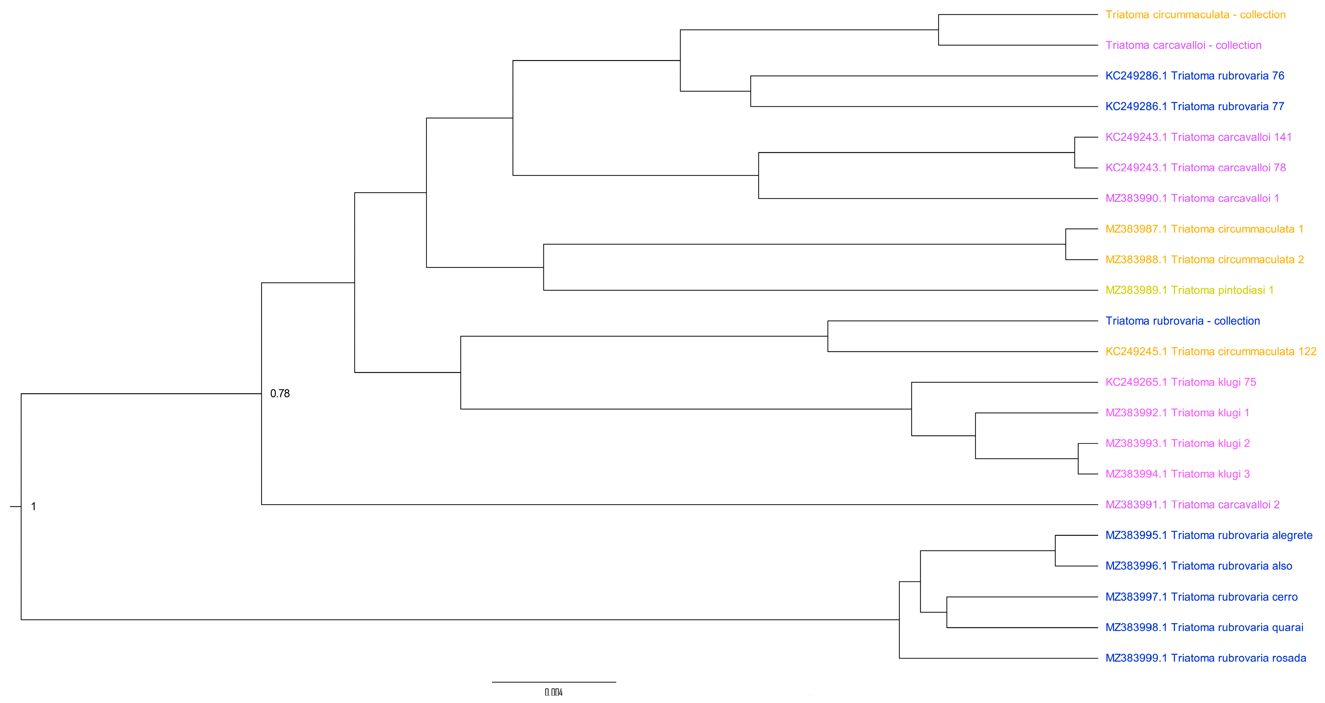
Table A1.
Samples and reference sequences used in this study.
References
- Chagas, C. Nova tripanozomiaze humana: Estudos sobre a morfolojia e o ciclo evolutivo do Schizotrypanum cruzi n. gen., n. sp., ajente etiolojico de nova entidade morbida do homem. Mem. Inst. Oswaldo Cruz. 1909, 1, 159–218. [Google Scholar] [CrossRef] [Scilit]
- World Health Organization. Chagas Disease (American Trypanosomiasis). 2024. Available online: https://www.who.int/news-room/fact-sheets/detail/chagas-disease-(american-trypanosomiasis) (accessed on 10 January 2025).
- PAHO (Pan-American Health Organization). 2024. Available online: https://www.paho.org/en/news/16-10-2023-paho-pushes-elimination-more-30-communicable-diseases-americas-2030 (accessed on 10 January 2025).
- de Arias, A.R.; Monroy, C.; Guhl, F.; Sosa-Estani, S.; Santos, W.S.; Abad-Franch, F. Chagas disease control-surveillance in the Americas: The multinational initiatives and the practical impossibility of interrupting vector-borne Trypanosoma cruzi transmission. Mem. Inst. Oswaldo Cruz. 2022, 117, e210130. [Google Scholar] [CrossRef] [Scilit]
- Dias, J.C.P. Southern Cone Initiative for the elimination of domestic populations of Triatoma infestans and the interruption of transfusion Chagas disease: Historical aspects, present situation, and perspectives. Mem. Inst. Oswaldo Cruz. 2007, 102 (Suppl. S1), 11–18. [Google Scholar] [CrossRef] [Scilit] [PubMed]
- Bedin, C.; Wilhelms, T.; Villela, M.M.; Silva, G.C.C.; Riffel, A.P.K.; Sackis, P.; Mello, F. Residual foci of Triatoma infestans infestation: Surveillance and control in Rio Grande do Sul, Brazil, 2001–2018. Rev. Soc. Bras. Med. Trop. 2021, 54, e0530-2020. [Google Scholar] [CrossRef] [Scilit]
- Almeida, C.E.; Vinhaes, M.C.; Almeida, J.R.; de Silveira, A.C.; Costa, J. Monitoring the domiciliary and peridomiciliary invasion process of Triatoma rubrovaria in the State of Rio Grande do Sul, Brazil. Mem. Inst. Oswaldo Cruz. 2000, 95, 761–768. [Google Scholar] [CrossRef] [Scilit]
- Souza, C.B.; de Santos, G.B.; dos Bedin, C.; Bergamin, M.; de Mello, F.; Villela, M.M. Triatoma rubrovaria (Hemiptera, Reduviidae, Triatominae) in the Pampa biome, Brazil: A retrospective study of its occurrence and abundance. Rev. Soc. Bras. Med. Trop. 2023, 65, e38. [Google Scholar] [CrossRef] [Scilit]
- Verly, T.; Costa, S.; Lima, N.; Mallet, J.; Odêncio, F.; Pereira, M.; Moreira, C.J.; Britto, C.; Pavan, M.G. Vector competence and feeding-excretion behavior of Triatoma rubrovaria (Blanchard, 1843) (Hemiptera: Reduviidae) infected with Trypanosoma cruzi TcVI. PLoS Neglected Tropical Diseases. 2020, 14, e0008712. [Google Scholar] [CrossRef] [Scilit] [PubMed]
- Salvatella, R.; Calegari, L.; Puime, A.; Basmadjian, Y.; Rosa, R.; Guerrero, J.; Martinez, M.; Mendaro, G.; Briano, D.; Montero, C.; et al. Perfil alimentario de Triatoma rubrovaria (Blanchard, 1843) (Hemiptera, Triatominae) en ámbitos peridomiciliarios, de una localidad rural de Uruguay. Rev. Soc. Bras. Med. Trop. São Paulo 1994, 36, 311–320. [Google Scholar] [CrossRef] [Scilit]
- Almeida, C.E.; Duarte, R.; Nascimento, R.G.d.; Pacheco, R.S.; Costa, J. Triatoma rubrovaria (Blanchard, 1843) (Hemiptera, Reduviidae, Triatominae) II: Trophic resources and ecological observations of five populations collected in the State of Rio Grande do Sul, Brazil. Mem. Inst. Oswaldo Cruz. 2002, 97, 1127–1131. [Google Scholar] [CrossRef] [Scilit]
- Schofield, C.J.; Galvão, C. Classification, evolution, and species groups within the Triatominae. Acta Tropica. 2009, 110, 88–100. [Google Scholar] [CrossRef] [Scilit]
- Pita, S.; Lorite, P.; Nattero, J.; Galvão, C.; Alevi, K.C.C.; Teves, S.C.; Azeredo-Oliveira, M.T.V.; Panzera, F. New arrangements on several species subcomplexes of Triatoma genus based on the chromosomal position of ribosomal genes (Hemiptera-Triatominae). Infect. Genet. Evol. 2016, 43, 225–231. [Google Scholar] [CrossRef] [Scilit]
- Lent, H.; Wygodzinsky, P. Revision of the Triatominae (Hemiptera, Reduviidae), and their significance as vectors of Chagas’ disease. Bull. Am. Mus. Nat. Hist. 1979, 163, 125–520. [Google Scholar]
- Jurberj, J.; Carcavallo, U.R.; Lent, H. Panstrongylus sherlocki sp.n. do estado da Bahia, Brasil (Hemiptera, Reduviidae, Triatominae). Entomol. Vectores. 2001, 8, 261–274. [Google Scholar]
- Jurberg, J.; Cunha, V.; Cailleaux, S.; Raigorodschi, R.; Lima, M.S.; Rocha, D.d.S.; Moreira, F.F.F. Triatoma pintodiasi sp. nov. do subcomplexo T. rubrovaria (Hemiptera, Reduviidae, Triatominae). Rev. Pan-Amaz. Saúde 2013, 4, 43–56. [Google Scholar] [CrossRef] [Scilit]
- da Silva, L.A.; Belintani, T.; de Paiva, V.F.; Nascimento, J.D.; Rimoldi, A.; Gardim, S.; Rocha, C.S.; de Mello, F.; Obara, M.T.; de Oliveira, J.; et al. Integrative taxonomy and phylogenetic systematics of the Triatoma rubrovaria subcomplex (Hemiptera, Triatominae). Acta Trop. 2023, 237, 106699. [Google Scholar] [CrossRef] [Scilit] [PubMed]
- Pavan, M.G.; Lazoski, C.; Monteiro, F.A. Speciation Processes in Triatominae. In Triatominae—The Biology of Chagas Disease Vectors. Entomology in Focus; Guarneri, A., Lorenzo, M., Eds.; Springer: Cham, Switzerland, 2021. [Google Scholar] [CrossRef] [Scilit]
- Jurberg, J.; Barbosa, H.S.; Galvão, C.; Rocha, D.d.S.; Silva, M.B.A. Descrição de ovos e ninfas de Triatoma klugi (Hemiptera, Reduviidae, Triatominae). Iheringia Ser. Zool. 2010, 100, 43–54. [Google Scholar] [CrossRef] [Scilit]
- Monteiro, F.A.; Barrett, T.V.; Fitzpatrick, S.; Cordon-Rosales, C.; Feliciangeli, D.; Beard, C.B. Molecular phylogeography of the Amazonian Chagas disease vectors Rhodnius prolixus and R. robustus. Mol. Ecol. 2003, 12, 997–1006. [Google Scholar] [CrossRef] [Scilit]
- Justi, S.A.; Russo, C.A.M.; Mallet, J.R.d.S.; Obara, M.T.; Galvão, C. Molecular phylogeny of Triatomini (Hemiptera: Reduviidae: Triatominae). Parasites Vectors. 2014, 7, 149. [Google Scholar] [CrossRef] [Scilit]
- Thompson, J.D.; Higgins, D.G.; Gibson, T.J. CLUSTAL W: Improving the sensitivity of progressive multiple sequence alignment through sequence weighting, position-specific gap penalties and weight matrix choice. Nucleic Acids Res. 1994, 22, 4673–4680. [Google Scholar] [CrossRef] [Scilit]
- Kumar, S.; Stecher, G.; Li, M.; Knyaz, C.; Tamura, K. MEGA X: Molecular evolutionary genetics analysis across computing platforms. Mol. Biol. Evol. 2018, 35, 1547–1549. [Google Scholar] [CrossRef] [Scilit]
- Bouckaert, R.; Vaughan, T.G.; Barido-Sottani, J.; Duchêne, S.; Fourment, M.; Gavryushkina, A.; Heled, J.; Jones, G.; Kühnert, D.; de Maio, N.; et al. BEAST 2.5: An advanced software platform for Bayesian evolutionary analysis. PLoS Comput. Biol. 2019, 15, e1006650. [Google Scholar] [CrossRef] [Scilit]
- Bouckaert, R.R.; Drummond, A.J. bModelTest: Bayesian phylogenetic site model averaging and model comparison. BMC Evol. Biol. 2017, 17, 42. [Google Scholar] [CrossRef] [Scilit] [PubMed]
- R Core Team. A Language and Environment for Statistical Computing; R Foundation for Statistical Computing: Vienna, Austria, 2023; Available online: https://www.R-project.org/ (accessed on 15 February 2024).
- Jombart, T. adegenet: A R package for the multivariate analysis of genetic markers. Bioinformatics 2008, 24, 1403–1405. [Google Scholar] [CrossRef] [Scilit]
- Wickham, H. ggplot2 Elegant Graphics for Data Analysis, 2016th ed.; Springer: Berlin/Heidelberg, Germany, 2016. [Google Scholar]
- Cheng, L.; Thomas, R.C.; Jukka, S.; David, M.A.; Jukka, C. Hierarchical and Spatially Explicit Clustering of DNA Sequences with BAPS Software. Mol. Biol. Evol. 2013, 30, 1224–1228. [Google Scholar] [CrossRef] [Scilit]
- Revell, L.J. Phytools: An R package for phylogenetic comparative biology (and other things). Methods Ecol. Evol. 2011, 3, 217–223. [Google Scholar] [CrossRef] [Scilit]
- Kassambara, A. ggpubr R Package: ggplot2-Based Publication Ready Plots. 2023. Available online: https://www.sthda.com/english/articles/24-ggpubr-publication-ready-plots/ (accessed on 22 February 2024).
- Wilke, C. Introduction to ggridges. 2024. Available online: https://wilkelab.org/ggridges/articles/introduction.html/ (accessed on 22 February 2024).
- Abad-Franch, F.; Pavan, M.G.; Jaramillo-O, N.; Palomeque, F.S.; Dale, C.; Chaverra, D.; Monteiro, F.A. Rhodnius barretti, a new species of Triatominae (Hemiptera: Reduviidae) from Western Amazonia. Mem. Inst. Oswaldo Cruz. 2013, 108 (Suppl. S1), 92–99. [Google Scholar] [CrossRef] [Scilit] [PubMed]
- Abad-Franch, F.; Monteiro, F.A.; Pavan, M.G.; Patterson, J.S.; Bargues, M.D.; Zuriaga, M.Á.; Aguilar, M.; Beard, C.B.; Mas-Coma, S.; Miles, M.A. Under pressure: Phenotypic divergence and convergence associated with microhabitat adaptations in Triatominae. Parasites Vectors 2021, 14, 195. [Google Scholar] [CrossRef] [Scilit] [PubMed]
- Monteiro, F.A.; Donnelly, M.J.; Beard, C.B.; Costa, J. Nested clade and phylogeographic analyses of the Chagas disease vector Triatoma brasiliensis in Northeast Brazil. Mol. Phylogenet. Evol. 2004, 32, 46–56. [Google Scholar] [CrossRef] [Scilit]
- Monteiro, F.A.; Peretolchina, T.; Lazoski, C.; Harris, K.; Dotson, E.M.; Abad-Franch, F.; Tamayo, E.; Pennington, P.M.; Monroy, C.; Cordon-Rosales, C.; et al. Phylogeographic pattern and extensive mitochondrial DNA divergence disclose a species complex within the Chagas disease vector Triatoma dimidiata. PLoS ONE 2013, 8, e70974. [Google Scholar] [CrossRef] [Scilit]
- Pita, S.; Gómez-Palacio, A.; Lorite, P.; Dujardin, J.P.; Chavez, T.; Villacís, A.G.; Galvão, C.; Panzera, Y.; Calleros, L.; Pereyra-Mello, S.; et al. Multidisciplinary approach detects speciation within the kissing bug Panstrongylus rufotuberculatus populations (Hemiptera, Heteroptera, Reduviidae). Mem. Inst. Oswaldo Cruz. 2021, 116, e210259. [Google Scholar] [CrossRef] [Scilit]
- Schmid, M.; Guillaume, F. The role of phenotypic plasticity on population differentiation. Heredity 2017, 119, 214–225. [Google Scholar] [CrossRef] [Scilit] [PubMed]
- Crispo, E. Modifying effects of phenotypic plasticity on interactions among natural selection, adaptation and gene flow. J. Evol. Biol. 2008, 21, 1460–1469. [Google Scholar] [CrossRef] [Scilit] [PubMed]
- Pavan, M.G.; Rivas, G.B.S.; Dias, F.B.S.; Gurgel-Gonçalves, R. Looks can be deceiving: Cryptic species and phenotypic variation in Rhodnius spp., Chagas disease vectors. In Evolutionary Biology: Biodiversification from Genotype to Phenotype; Springer International Publishing: Berlin/Heidelberg, Germany, 2015; pp. 345–372. [Google Scholar] [CrossRef] [Scilit]
- González, C.; Ornelas, J.F.; Gutiérrez-Rodríguez, C. Selection and geographic isolation influence hummingbird speciation: Genetic, acoustic and morphological divergence in the wedge-tailed sabrewing (Campylopterus curvipennis). BMC Evol. Biol. 2011, 11, 38. [Google Scholar] [CrossRef] [Scilit]
- Kautt, A.F.; Kratochwil, C.F.; Nater, A.; Machado-Schiaffino, G.; Olave, M.; Henning, F.; Torres-Dowdall, J.; Härer, A.; Hulsey, C.D.; Franchini, P.; et al. Contrasting signatures of genomic divergence during sympatric speciation. Nature 2020, 588, 106–111. [Google Scholar] [CrossRef] [Scilit]
- Nosil, P.; Feder, J.L.; Flaxman, S.M.; Gompert, Z. Tipping points in the dynamics of speciation. Nat. Ecol. Evol. 2017, 1, 0001. [Google Scholar] [CrossRef] [Scilit] [PubMed]
Disclaimer/Publisher’s Note: The statements, opinions and data contained in all publications are solely those of the individual author(s) and contributor(s) and not of MDPI and/or the editor(s). MDPI and/or the editor(s) disclaim responsibility for any injury to people or property resulting from any ideas, methods, instructions or products referred to in the content. |
© 2025 by the authors. Licensee MDPI, Basel, Switzerland. This article is an open access article distributed under the terms and conditions of the Creative Commons Attribution (CC BY) license (https://creativecommons.org/licenses/by/4.0/).