Article

Venom IMP-L2 from the Ectoparasitoid Scleroderma guani Regulates the IIS/TOR Pathway in Tenebrio molitor

1
Key Laboratory of Forest Disaster Warning and Control of Yunnan Province, Southwest Forestry University, Kunming 650224, China
2
Key Laboratory for Forest Resources Conservation and Utilization in the Southwest Mountains of China, Ministry of Education, Southwest Forestry University, Kunming 650224, China
*
Author to whom correspondence should be addressed.
Insects 2025, 16(8), 763; https://doi.org/10.3390/insects16080763
Submission received: 18 June 2025 / Revised: 21 July 2025 / Accepted: 22 July 2025 / Published: 24 July 2025
(This article belongs to the Section Insect Molecular Biology and Genomics)

Simple Summary

Most ectoparasitoids delay host development by secreting venom before or during oviposition, thereby ensuring the development of parasitoid eggs. Our previous study found that the venom of the ectoparasitoid Scleroderma guani is rich in imaginal morphogenesis factor-Late2 (IMP-L2). In many insects, the IIS/TOR signaling pathway regulates their growth and development. IMP-L2 in Drosophila can inhibit the activation of IIS by binding to insulin-like peptides (ILPs) of the IIS signaling pathway, thereby prolonging the lifespan of Drosophila. However, whether the IIS/TOR signaling pathway plays a role in the growth and development of the host Tenebrio molitor and how S. guani regulates host IIS/TOR signaling through parasitic behavior and venom IMP-L2 remains unclear. In this study, we found that the IIS/TOR signaling pathway is involved in the growth and development of T. molitor, and the parasitic behavior of S. guani inhibits the TOR signal transduction of T. molitor. IMP-L2 inhibited TOR and IIS/TOR signaling in the early and late stages of injection, respectively. When IMP-L2 was knocked down, S. guani would accelerate the death of T. molitor after parasitizing pupae. This study shows that the venom IMP-L2 of S. guani may help the egg and larval development of S. guani by inhibiting the host IIS/TOR signaling pathway.

Abstract

Parasitoid venom significantly influences host physiology and development. Our previous research identified high levels of insulin-binding protein IMP-L2 in the venom of Scleroderma guani. IMP-L2 may inhibit the insulin/insulin-like growth factor signaling (IIS) cascade by competitively binding insulin-like peptides (ILPs) with insulin receptor (InR). However, how to regulate IIS transduction is unclear. We speculate that venom-derived IMP-L2 may bind ILPs to inhibit IIS transduction. Consequently, we investigated the regulation of the IIS/TOR pathway by venom-derived IMP-L2. An expression analysis of IIS/TOR pathway genes across various developmental stages of Tenebrio molitor demonstrated that this pathway governs the entire developmental process. By examining gene expression before and after parasitism, we determined that S. guani predominantly inhibits TOR pathway signaling in T. molitor post-parasitism. Bioinformatics and expression analyses revealed that IMP-L2 is critically involved in Hymenoptera insects, exhibiting high expression in the venom apparatus, and is upregulated in response to S. guani parasitism factors. Additionally, recombinant IMP-L2 was produced via eukaryotic expression. Finally, the recombinant IMP-L2 was found to inhibit the TOR and IIS/TOR signaling pathways at early (6 h) and late (24 h) stages post-injection. Knockdown of IMP-L2 in S. guani parasitized T. molitor pupae, resulting in accelerated death of T. molitor. During parasitism, S. guani may suppress host growth and development by modulating the IIS/TOR signaling pathway through venom-derived IMP-L2, potentially affecting host lifespan.

1. Introduction

Parasitic Hymenoptera represent one of the most diverse and abundant groups of natural enemies within the class Insecta [1]. Currently, it is estimated that over one million species of parasitic wasps play a critical role in the biological control of agricultural pests [2,3]. Endoparasitoids and ectoparasitoids employ distinct strategies, typically utilizing their parasitic factors to inhibit or evade host immune responses while also regulating host growth and development [3]. The parasitic factors associated with these parasitoids include venom, poly DNA viruses (PDVs), virus-like particles (VLPs), and teratocytes [4]. Among these, venom is the most prevalent and extensively studied parasitic factor, containing a complex mixture of proteins and non-protein active compounds [1,5], many of which remain unidentified. In most ectoparasites, the primary function of venom is to induce paralysis or lethargy and developmental arrest in the host, particularly during the larval or nymphal stages [6]. Most parasitoids administer venom into the host prior to or during oviposition to manipulate host physiology, thereby facilitating the development of parasitoid eggs [7].
In insects, the insulin/insulin-like growth factor and target of rapamycin signaling pathways (IIS/TOR) coordinate metabolic processes by integrating nutrient and hormonal signals, thereby influencing developmental outcomes [8,9,10]. Each signaling molecule within the IIS/TOR pathway plays a vital regulatory role [11,12]. Insulin-like peptides (ILPs) are predominantly produced in the medial neurosecretory cells of the brain, as well as in the ovary, fat bodies, glial cells, and midgut [13,14,15,16,17,18]. Ablation of ILP-secreting neuroendocrine cells can lead to significant reductions in insect body size and manifestations resembling diabetes [19]. ILP secretion activates the IIS cascade by binding to and activating insulin receptor 1 (InR1), which in turn activates the insulin receptor substrate protein (CHICO). This InR1-mediated signaling transduces to phosphatidylinositol 3-kinase (PI3K), the phosphoinositide-dependent protein kinase 1 (PDK) and protein kinase B (AKT), inhibiting the forkhead box class O transcription factor (FOXO), a crucial downstream target of IIS, thereby regulating energy metabolism, reproduction, and longevity in insects [20,21,22,23,24]. The phosphatase and tensin homolog (PTEN) downregulates PI3K/PDK signaling, ultimately inhibiting the IIS pathway [25,26]. Additionally, the second insulin receptor, InR2, can inhibit InR1, and the co-expression of InR1 and InR2 allows FOXO to regulate transcription within the nucleus [23]. Overexpression of FOXO may impede early larval growth and development, resulting in smaller larvae [27,28], while the absence of FOXO is associated with delayed adult development and reduced body size [29]. Therefore, FOXO is essential for the regulation of insect growth and development. Moreover, AKT can inhibit the negative regulators of the TOR pathway, tuberous sclerosis complex 1 and 2 (TSC1/2), thereby activating downstream ras homolog enhanced in brain (RHEB)/TOR signaling [30,31]. Elevated intracellular amino acid concentrations also enhance TOR activity via RHEB [30,32]. Active TOR promotes cell growth by inhibiting the translational repressor 4e-binding protein (4EBP) and stimulating ribosomal protein S6 kinase (S6K), thereby facilitating translation and ribosome biogenesis [22]. Activation of 4EBP and inhibition of S6K1 can extend insect lifespan [33]. Furthermore, under high amino acid conditions, TOR signaling can activate GATA activator subtypes, leading to the activation of ILP1, ILP2, ILP3, ILP5, and ILP8 in Aedes aegypti, thus stimulating the IIS pathway [34]. Simultaneously, TOR also regulates AKT expression, while S6K modulates CHICO expression and PDK phosphorylation, thereby influencing IIS signal transduction [35,36,37].
An insulin-like peptide (DILPs) binding protein, imaginal morphogenesis factor-Late2 (IMP-L2), has been identified in Drosophila [38]. IMP-L2 belongs to the immunoglobulin superfamily and has been shown to bind human insulin-like growth factors I and II (IGF-I and IGF-II) as well as insulin with high affinity [39]. Initially considered an ortholog of insulin-like growth factor binding proteins (IGFBPs) [40,41], recent findings indicate that the structure of IMP-L2 and its hormone binding characteristics do not significantly correlate with human IGFBPs [42], leaving this classification controversial. Overexpression of IMP-L2 in Drosophila leads to a twofold increase in mRNA levels of DILP2, DILP3, and DILP5 [43]. Concurrently, secreted IMP-L2 binds to native DILP-2 and DILP-5, acting as a negative regulator of IIS activity and extending Drosophila lifespan [43,44,45].
The expression of IMP-L2 is influenced by nutritional status; under conditions of malnutrition, its expression in the fat body increases, thereby inhibiting overall IIS activity and enabling the organism to withstand starvation [45,46]. Fruit flies deficient in IMP-L2 not only exhibit larger body sizes but also resist size reduction under nutritional scarcity [46]. Notably, the regulation of nutrient-dependent IMP-L2 expression is mediated through ecdysone signaling rather than the TOR nutrient sensor in the fat body [46]. Elevated levels of ecdysone stimulate the release of IMP-L2 from the fat body [46]. Additionally, IMP-L2 expression in the intestine and specific neurons contributes to the regulation of the IIS. Enhanced expression of IMP-L2 in the intestine can inhibit IIS and promote lifespan extension, whereas its expression in certain neurons is crucial for maintaining normal IIS activity in the brain and associated glands [43,47,48]. Furthermore, overexpression of IMP-L2 can induce a 100% diapause response via the neuroendocrine cell driver c929 [49].
In summary, parasitic wasps secrete venom that influences host physiology, thereby affecting host growth and development. The IIS/TOR signaling pathway is known to regulate these processes; however, there is currently no research addressing the role of the IIS/TOR signaling pathway in the growth and development of T. molitor. Additionally, IMP-L2, derived from non-venom sources across various tissues, negatively regulates the IIS/TOR signaling pathway by binding to insulin-like peptides (ILPs). However, the regulatory mechanism is not clear. Previous studies have identified a high concentration of IMP-L2 in the venom of S. guani [50], but it has only been reported in the venoms of Nasonia vitripennis and Eumenes pomiformis [51,52]. Notably, the impact of venom-derived IMP-L2 on the host IIS/TOR signaling pathway remains unexplored.
This study aims to investigate the expression characteristics of the IIS/TOR signaling pathway throughout the developmental stages of T. molitor and elucidate how this pathway regulates the growth and development of the species. Additionally, the research will examine how parasitism influences the IIS/TOR signaling pathway in T. molitor, determining whether parasitic wasps alter host growth and development through modulation of this signaling route. Furthermore, we will analyze the sequence, tissue expression, and parasitic responses of IMP-L2 to clarify its evolutionary biology and expression patterns. Finally, the study will investigate the effects of venom-derived IMP-L2 on the IIS/TOR signaling pathway in T. molitor, providing a theoretical framework for understanding the influence of venom IMP-L2 on the growth and development of this beetle species.

2. Materials and Methods

2.1. Insects

Following the methodology outlined by Zhu [53], T. molitor were maintained on wheat bran under natural light conditions and at room temperature, with hydration provided through Chinese cabbage. Adult S. guani were nourished with a 20% honey–water solution, and the subsequent generation of S. guani was propagated by allowing adults to parasitize fresh pupae of T. molitor.

2.2. The Expression of IIS/TOR Pathway Related Genes in Different Developmental Stages of T. molitor

A total of 100 early-stage (2-day-old) eggs, 100 mid-stage (4-day-old) eggs, and 100 late-stage (6-day-old) eggs, along with 50 newly hatched larvae (one instar larvae), old larvae (7–8 instar larvae), last instar larvae (14 instar larvae), prepupae, early pupae (white pupae), mid-stage pupae (yellow pupae), late pupae (black pupae), and seven adults at various ages (1, 4, 8, 12, 16, 20, and 24 days post-eclosion) were collected and subjected to disinfection. RNA extraction and cDNA synthesis for each sample were performed according to Wang’s protocol [54]. Primers targeting cDNA sequences of genes related to the IIS/TOR pathway were designed (Table S1). Ribosomal protein L32 (rpl) was utilized as an internal reference, and RT-qPCR was conducted using SYBR® Premix Ex Taq II (Takara Biomedical Technology Co., Ltd., Beijing, China) in a 20 μL reaction volume. The RT-qPCR conditions included an initial denaturation at 95 °C for 1 min, followed by 45 cycles of denaturation at 95 °C for 15 s and annealing at 60 °C for 60 s. The experiment was replicated three times.

2.3. Regulation of Parasitism on Genes Related to IIS/TOR Signaling Pathway in T. molitor

Healthy T. molitor pupae, weighing between 0.9 and 1.4 mg, were sterilized using absorbent cotton dipped in 75% alcohol under sterile conditions. S. guani and T. molitor pupae were placed in finger-shaped glass tubes, which were sealed with cotton balls to facilitate the observation of S. guani parasitism on T. molitor pupae. Timing commenced when the tail of S. guani penetrated the pupae, after which the parasitized pupae were individually transferred to a constant temperature incubator. Pupae parasitized by one female and one male were collected at 6 h and 24 h, respectively, and RNA and cDNA were extracted from each sample according to Wang’s protocol [54]. Unparasitized pupae from the same time period served as controls. The reference gene for RT-qPCR, along with the primers for the IIS/TOR pathway genes, reaction system, and procedures were as described in Section 2.2. The entire experiment was replicated three times.

2.4. Cloning and Sequence Analysis of IMP-L2 from S. guani

Following the aforementioned method, RNA and cDNA from S. guani were extracted, and the full-length coding sequence (cds) of IMP-L2 was amplified using RT-PCR primers listed in Table S1. The amplified product was then ligated into the commercial vector pEASY®-T1 Simple Cloning Kit (TransGen Biotech Co., Ltd., Beijing, China) and transformed into competent E. coli DH5α cells. Positive transformants were sent to Platinum Biotechnology Co., Ltd. (Shanghai, China) for sequencing. The resulting sequences were processed using DNAstar Lasergene 11.0 software (DNASTAR Inc., Madison, WI, USA), and the assembled nucleic acid sequences were analyzed for homology through a BlastX search in NCBI (https://blast.ncbi.nlm.nih.gov, accessed on 16 September 2024). Subsequent analyses, including translation, signal peptide prediction, multiple sequence alignment, phylogenetic tree construction, and physical and chemical properties assessment, were performed using DNAMAN (Lynnon Biosoft Inc., San Ramon, CA, USA), SignalP 6.0 (https://services.healthtech.dtu.dk/services/SignalP-6.0/, accessed on 16 September 2024), CLUSTAL (https://www.ebi.ac.uk/jdispatcher/, accessed on 16 September 2024), Mega 7.0 (1000 neighbor-joining method) (Temple University, Philadelphia, PA, USA), ProtParam (https://web.expasy.org/protparam/, accessed on 16 September 2024), and Scansite 4.0 (https://scansite4.mit.edu/#scanProtein, accessed on 16 September 2024) for domain prediction.

2.5. Expression Analysis of IMP-L2 in S. guani Venom Organs, Developmental Stages, and Parasitic Process

Various tissues (venom apparatus and abdominal residues) were collected from S. guani across four developmental stages (15 periods: one egg period; four larval periods; three pupae periods; seven adult periods), including both parasitized and unparasitized samples, under an anatomical microscope. Female wasps that had mated for the same duration were placed in a finger-shaped tube containing fresh pupae of T. molitor. Wasps exhibiting piercing behavior and that paralyzed the host were classified as parasitized; those that did not exhibit this behavior were deemed unparasitized. RNA and cDNA extraction for each sample followed Wang’s method [54]. For the different developmental stages of S. guani, 18S rRNA served as the reference gene for the IMP-L2 gene, while 5.8S rRNA was used as the reference in abdominal residues and venom organs in both parasitic and non-parasitic conditions. The primers for IMP-L2, 18S rRNA, and 5.8S rRNA used in RT-qPCR are detailed in Table S1, with the reaction system and procedures referenced in Section 2.2. A total of 80 venom apparatus and abdominal residues were analyzed, along with 50 wasps from different developmental stages, respectively, and 40 female wasps from each of the parasitized and unparasitized wasps. The entire experiment was conducted in triplicate.

2.6. Construction of the Expression Vector pFast-bac1-IMP-L2 and Recombinant Baculovirus Expression Vector (Bacmid)

Based on the cloned IMP-L2 gene sequence of S. guani venom, the target gene was synthesized by PAS (PCR-based Accurate Synthesis) method. The target gene was digested by BamH I and Hind III and ligated to the pFast-bac1 vector with His tag to obtain the plasmid pFast-bac1-IMP-L2. The upstream/downstream primer of its digestion in Table S1.
The obtained recombinant plasmid was transferred into E. coli TOP10 competent cells, and the positive clones were selected and sent to Nanjing Zhong ding Biotechnology Co., Ltd. (Nanjing, China) for sequencing verification.
The sequencing-confirmed 200 ng pFast-bac1-IMP-L2 vector and pFast-bac1 empty vector were, respectively, added to DH10Bac E. coli competent cells, gently mixed, and placed on ice for 30 min. After a 42 °C heat shock for 90 s, they were quickly placed on the ice for 5 min. After that, a 900 μL SOC medium was added to the EP tube of activated competent cells and shaken in a shaker for 4 h at 37 °C at 225 rpm. The LB plate (containing gentamicin 7 ug/mL, kanamycin 50 ug/mL, tetracycline 10 ug/mL, and then an added 10 μL 24 mg/mL IPTG and 40 μL 20 mg/mL x-gal) was smeared with 100 μL of the bacterial solution and cultured at 37 °C for 48 h. Positive clones were chosen and stored at −80 °C.

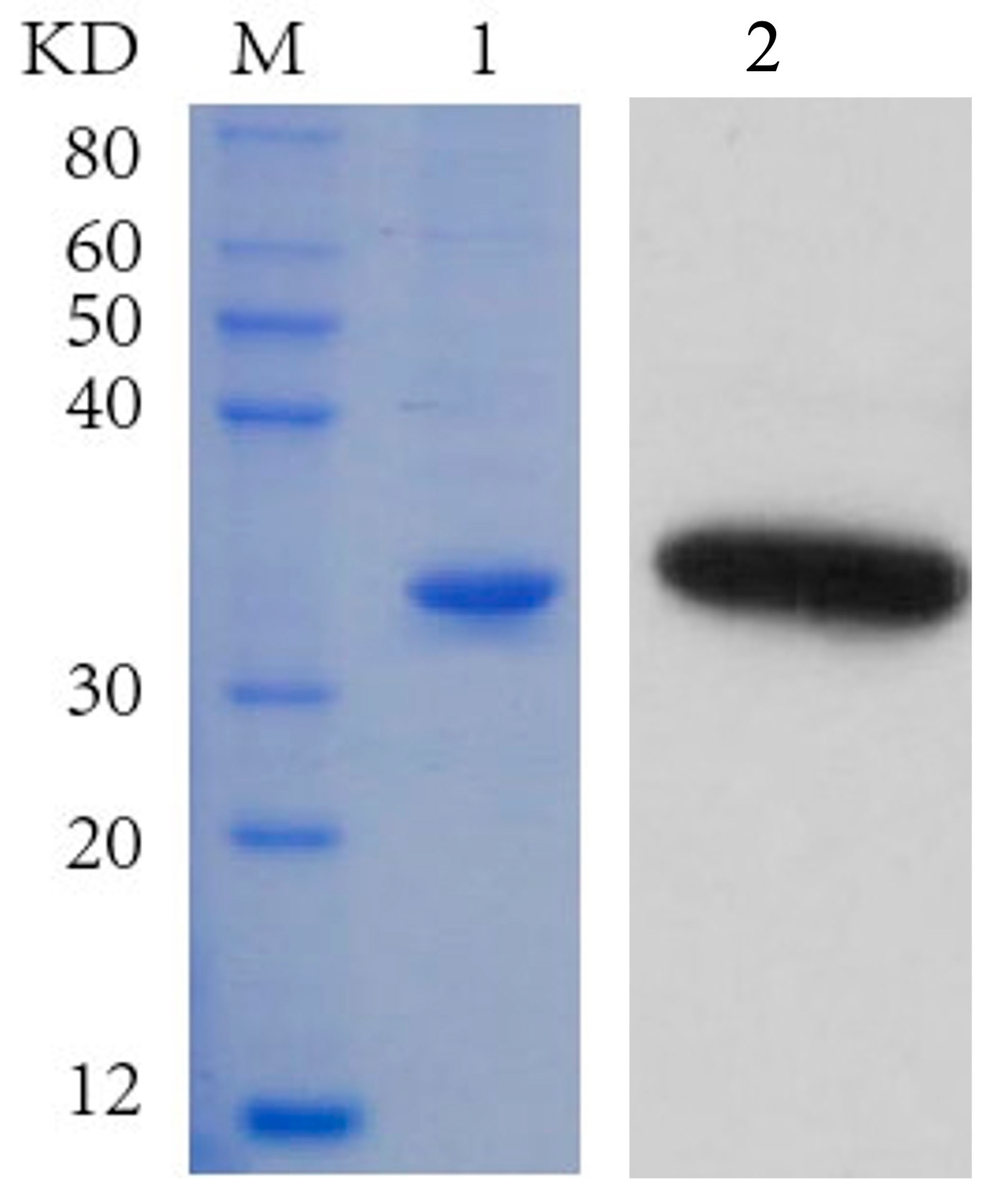
2.7. Expression and Purification of the IMP-L2 Protein

A total of 2 mL of Sf9 cells (Spodoptera frugiperda) at a concentration of 9 × 10⁵ cells per mL were transferred to each well of a six-well plate and cultured at 27 °C for 1 h. Subsequently, 1 µg of bacmid (pFast-bac1-IMP-L2 or pFast-bac1, approximately 5 µL) was diluted in 100 µL of Grace’s medium, devoid of both antibiotics and fetal bovine serum (FBS). Additionally, 6 µL of Cellfectin Reagent (Thermo Fisher Scientific Inc., Wilmington, MA, USA) was diluted with 100 µL of incomplete Grace’s medium, also devoid of antibiotics and FBS. The two solutions were combined, gently mixed, and incubated at room temperature for 15 to 45 min.
Next, 800 µL of the incomplete Grace’s culture medium was added to the mixture containing the bacmid and Cellfectin Reagent, which was then gently mixed and distributed into each well. The cells in the six-well plate were incubated at the same temperature for 5 h, after which the mixture was removed. Two mL of complete medium was added to each well, and the incubation continued for 72 h or until signs of viral infection were observed.
Upon observing signs of infection, the culture supernatant was transferred to a 15 mL centrifuge tube and centrifuged at 1000 rpm for 5 min at 4 °C to eliminate cell debris, yielding the cell supernatant containing the P1 virus. A suitable volume of this supernatant was inoculated into a 50 mL shake flask, and the supernatant was collected 72 h post-infection.
The cell supernatant was diluted 100-fold and dialyzed overnight against PBS buffer (pH 7.4) (Sangon Biotech Co., Ltd., Shanghai, China). The dialyzed supernatant was subsequently incubated with a balanced Ni column in a rotary incubator at 4 °C for 3–4 h. Following this, the incubated sample was gradually applied to a low-pressure chromatography system equipped with a Ni-IDA-Sepharose CL-6B affinity chromatography column (Sangon Biotech Co., Ltd., Shanghai, China), using a binding/wash buffer (pH 7.9–8.1) (Sangon Biotech Co., Ltd., Shanghai, China) at a flow rate of 0.5 mL/min until the OD280 value of the effluent returned to baseline.
The protein solution was then eluted with Ni-IDA elution buffer (pH 7.9–8.1) (Sangon Biotech Co., Ltd., Shanghai, China), collected at a flow rate of 1 mL/min, and transferred to a dialysis bag, where it was dialyzed overnight at 4 °C with PBS buffer (pH 7.4) (Sangon Biotech Co., Ltd., Shanghai, China) to obtain purified IMP-L2 and His-tag proteins.

2.8. SDS-PAGE and Western Blot Analysis

The purified protein solution was analyzed using 10% SDS-PAGE, and the results were visualized following staining with Coomassie Brilliant Blue and subsequent decolorization. For Western blot analysis, 10 µL of the dialysis-purified protein sample was loaded into the wells of the polyacrylamide gel. The concentrated gel was run at 90 V initially, followed by a separation gel run at 200 V. After electrophoresis, the gel was removed, and protein transfer to the membrane was performed at 100 V for 1.5 h. The membrane was washed with PBS four times for 5 min each.
Next, the membrane was incubated in 5% skim milk powder solution (blocking solution) at 37 °C for 1 h to block nonspecific binding. The 30 mL blocking solution containing His-Tag (2A8) antibody (1:5000; Abmart Co., Ltd., Shanghai, China) against the His-tag protein was then diluted in the blocking solution and applied to the membrane for 1 h at 37 °C. After this incubation, the membrane was washed four times, with each wash lasting 5 min. The goat anti-mouse IgG (1:10,000; Abmart Co., Ltd., Shanghai, China) was similarly diluted in the blocking solution and incubated with the membrane at 37 °C for 1 h.
Finally, after rinsing the membrane, enhanced chemiluminescence (ECL) imaging was conducted, and the resulting gel image served as the experimental outcome.

2.9. The Regulation of Recombinant IMP-L2 Protein on the Genes Related to the IIS/TOR Signaling Pathway of T. molitor

The expressed recombinant IMP-L2 protein was diluted to a concentration of 0.125 µg/µL (the content of IMP-L2 in a venom reservoir is 0.125 μg) in PBS (pH 7.4). Disinfection of T. molitor pupae was performed prior to experimentation. Following the duration of parasitic treatment, 2 µL of recombinant IMP-L2 was extracted using a 10 µL micro-injector (Gaoge Industry and Trade Co., Ltd., Shanghai, China) and injected into the penultimate abdominal segment of the pupae. A small amount of petroleum jelly was applied to the injection site, and the treated pupae were subsequently placed in a temperature-controlled incubator.
Following the protocol outlined in Section 2.3, the injected pupae were paired with one male and one female at 6 h and 24 h post-injection, respectively. The reference genes for RT-qPCR, along with primers for IIS/TOR pathway genes, reaction system, and procedures, were based on the methodology described in Section 2.2. His-tag protein was injected as a control, with a total of 20 heads subjected to injection for each treatment group. The entire experiment was conducted in triplicate.

2.10. Synthesis of dsRNA and RNA Interference

Specific primers (Table S1) containing the T7 promoter were designed based on the cDNA sequences of IMP-L2 (Genbank: PP108620) and GFP (from the pEGFP-1 plasmid preserved in the laboratory). A 2× Phanta Max Master Mix (Dye Plus) (Vazyme Biotech Co., Ltd., Nanjing, China) was used for PCR amplification. The amplified products were purified by SteadyPure Agarose Gel DNA Purification Kit (Accurate Biotechnology (Hunan) Co., Ltd., Changsha, China). The purified products were used to synthesize dsIMP-L2 (double-stranded IMP-L2 RNA) and dsGFP (double-stranded GFP RNA) by T7 RiboMAX TM Express RNAi System (Promega Biotech Co., Ltd., Beijing, China). The concentration of the synthesized double-stranded RNA was detected by NanoDrop One (Thermo Fisher Scientific Inc., Wilmington, MA, USA). The dsRNA was diluted to 5000 ng/uL with RNase Free H2O and placed at −80 °C.
The newly emerged female S. guani was used for RNA interference experiments. First, 200 nL (1 ug) dsRNA was injected at a rate of 20 nL/s with Nanoject II Programmable Nanoliter Injector (Drummond Scientific Company, Broomall, PA, USA). After injection, S. guani was reared in an incubator at 28 ± 2 °C and relative humidity of 60–70% (fed with honey water). After 24 h of injection, Trizol was used to extract RNA from the experimental group and the control group. The extracted total RNA was used to synthesize cDNA with HiScript III RT SuperMix for qPCR (+gDNA wiper) (Vazyme Biotech Co., Ltd., Nanjing, China). RT-qPCR primers were designed, and 5.8S rRNA gene was used as the internal reference gene (Table S1). The expression level of IMP-L2 was verified by RT-qPCR. The dsIMP-L2 was used as the experimental group and dsGFP was used as the control group. Each group consisted of 3–5 S. guani, and the experiment included three biological replicates.

2.11. Records of Survival Pupae of T. molitor

The dsGFP and dsIMP-L2 were injected into 30 newly emerged S. guani females, respectively, and a treated S. guani parasitized an early T. molitor pupae. After observation of the parasitic behavior, the S. guani was removed, and the parasitized T. molitor pupae were placed in an incubator at 28 ± 2 °C and relative humidity of 60–70%. The time from parasitism to death of T. molitor pupae was recorded (the successfully parasitized pupae could not eclosion).

2.12. Statistical Analyses

Real-time fluorescence quantitative data were analyzed using the 2(−∆∆CT) method [55]. Data analysis was performed with IBM SPSS Statistics 20.0 (SPSS Inc., Chicago, IL, USA), employing one-way ANOVA and the Tukey’s multiple-comparisons test to assess significant differences in the relative expression of IIS/TOR signaling pathway genes or IMP-L2 gene across various developmental stages of T. molitor or S. guani. Differences in the relative expression levels of IMP-L2 among different tissues of S. guani, as well as between parasitized versus and unparasitized conditions, were evaluated using Student’s t-test. Additionally, significant differences in the relative expression of T. molitor IIS/TOR signaling pathway genes between S. guani under parasitized and unparasitized conditions at 6 h and 24 h were also assessed using Student’s t-test. The comparative expression levels of T. molitor IIS/TOR signaling pathway genes between recombinant IMP-L2 and His-tag injections at 6 h and 24 h were similarly analyzed. The interference efficiency of dsGFP and dsIMP-L2 after interference was also compared using the same analysis. Statistical significance was determined at p < 0.05. Graphical representations were created using GraphPad Prism 9.5 (GraphPad Software Inc., San Diego, CA, USA) and Photoshop CS6 (Adobe Systems Inc., San Jose, CA, USA).

3. Results

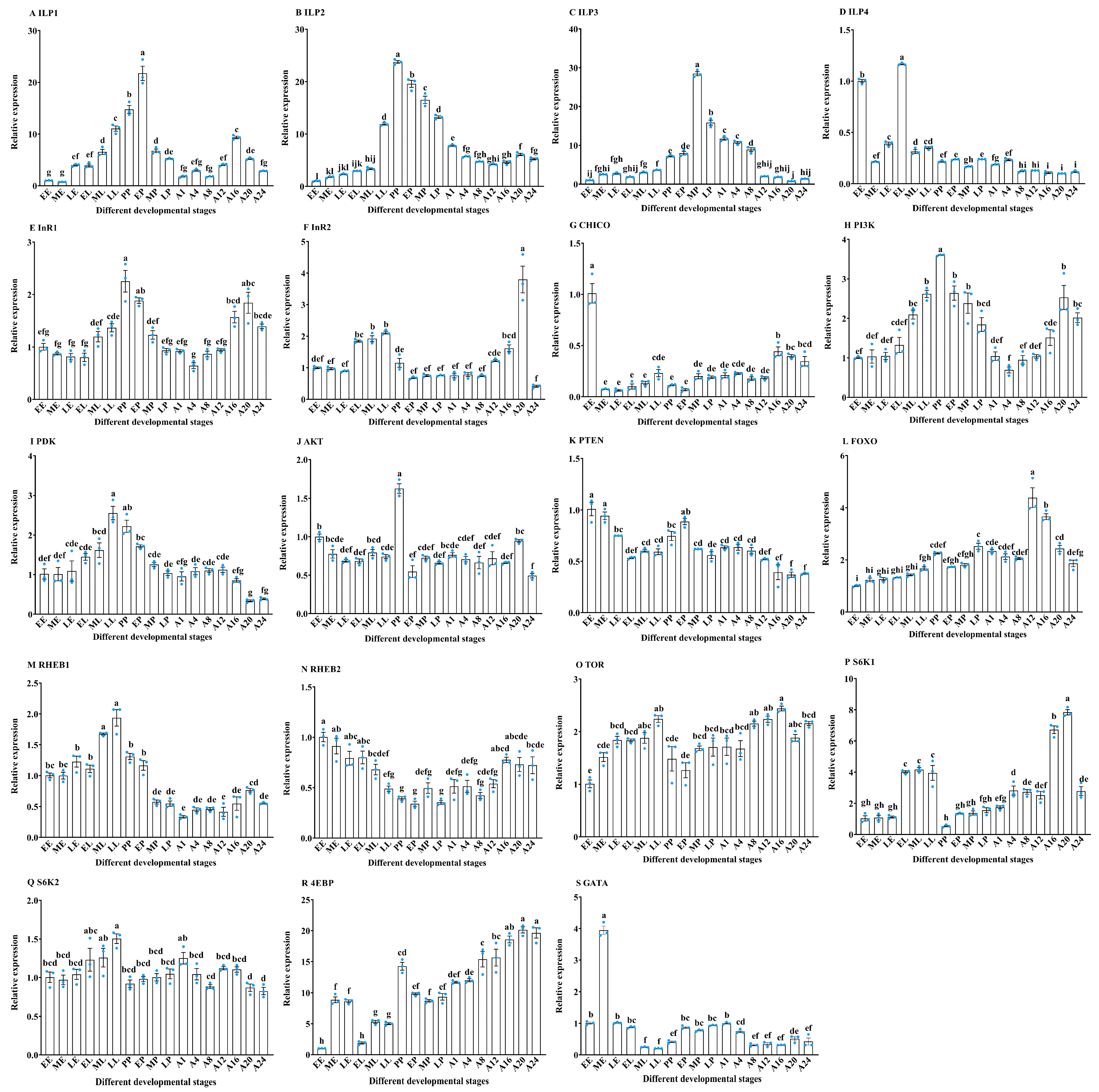
3.1. The Expression Pattern Analysis of IIS/TOR Signaling Pathway Related Genes in Different Developmental Stages of T. molitor

The IIS/TOR signaling pathway plays a critical role in regulating insect growth and development [10]. To investigate its involvement throughout the developmental stages of T. molitor, we assessed the expression of genes associated with this pathway across various developmental phases. RT-qPCR results indicated that ILP genes were expressed during different developmental stages of T. molitor, including three egg stages (EE, ME, LE), three larval stages (EL, ML, LL), one prepupal stage (PP), three pupal stages (EP, MP, LP), and seven adult stages (A1, A4, A8, A12, A16, A20, A24). The expression of ILP1 increased progressively from the EE to EP stage, peaked in the EP stage, and subsequently declined (F16,34 = 152.66, p < 0.001) (Figure 1A). In contrast, ILP2 expression was markedly elevated during the pupal stage compared to the other stages, with the highest levels detected in the PP stage (F16,34 = 574.45, p < 0.001) (Figure 1B). Furthermore, ILP2 expression exhibited an upward trend from EE to LL, declined from PP to LP, and remained stable from A1 to A24 (Figure 1B). Similarly, ILP3 expression was significantly higher during the pupal and early adult stages (A1–A8), peaking in the MP stage (F16,34 = 601.33, p < 0.001) (Figure 1C), while expression remained stable in the egg, larval, and late adult stages (A12–A24) (Figure 1C).
In contrast to the elevated expression of ILP1–3 during the pupal stage, ILP4 expression was significantly higher in the EL and EE stages compared to all other developmental stages (F16,34 = 1338.05, p < 0.001) (Figure 1D). The InR1 gene exhibited a gradual increase from the EE to PP stage, followed by a decline to A4, with a second peak at A20 (F16,34 = 23.88, p < 0.001). Meanwhile, InR2 showed relatively high expression during the larval stage, peaking at A20 (F16,34 = 47.32, p < 0.001) (Figure 1E,F). The CHICO gene had the highest expression at EE, gradually increasing from ME to LL, and remaining stable from MP to A12 (F16,34 = 48.84, p < 0.001) (Figure 1G). Reflecting the pattern of InR1, PI3K expression increased from EE to PP, peaked in the PP stage, decreased to A4, and then increased again from A4 to A20 (F16,34 = 28.43, p < 0.001) (Figure 1H). The PDK gene exhibited a gradual increase from EE to LL, with no significant difference between LL and PP stages, followed by a decline (F16,34 = 22.46, p < 0.001) (Figure 1I). The AKT gene expression peaked in the PP stage (F16,34 = 29.58, p < 0.001) (Figure 1J). The PTEN gene was highly expressed during the egg stage and increased in the PP stage, though it remained lower than in the egg stage (F16,34 = 30.96, p < 0.001) (Figure 1K). The FOXO gene demonstrated continuous upregulation after the EE stage, with heightened expression in the A12 and A16 stages (F16,34 = 56.14, p < 0.001) (Figure 1L). The RHEB1 gene was highly expressed in the LL stage (F16,34 = 57.38, p < 0.001), while RHEB2 peaked in the EE stage (F16,34 = 14.58, p < 0.001), and TOR expression was relatively high in the LL, A12, and A16 stages (F16,34 = 11.56, p < 0.001) (Figure 1M–O). The S6K gene expression in T. molitor developmental stages was responsive to TOR signal transduction. Specifically, S6K1 was more highly expressed during the larval and adult stages, peaking at A20 (F16,34 = 102.68, p < 0.001) (Figure 1P), while S6K2 was highly expressed in the LL stage (F16,34 = 6.31, p < 0.001) (Figure 1Q). The 4EBP gene exhibited a gradual increase from the LP to A24 stage, with high expression in the PP stage (F16,34 = 97.19, p < 0.001), whereas GATA expression peaked in the ME stage (F16,34 = 305.45, p < 0.001) (Figure 1R,S).

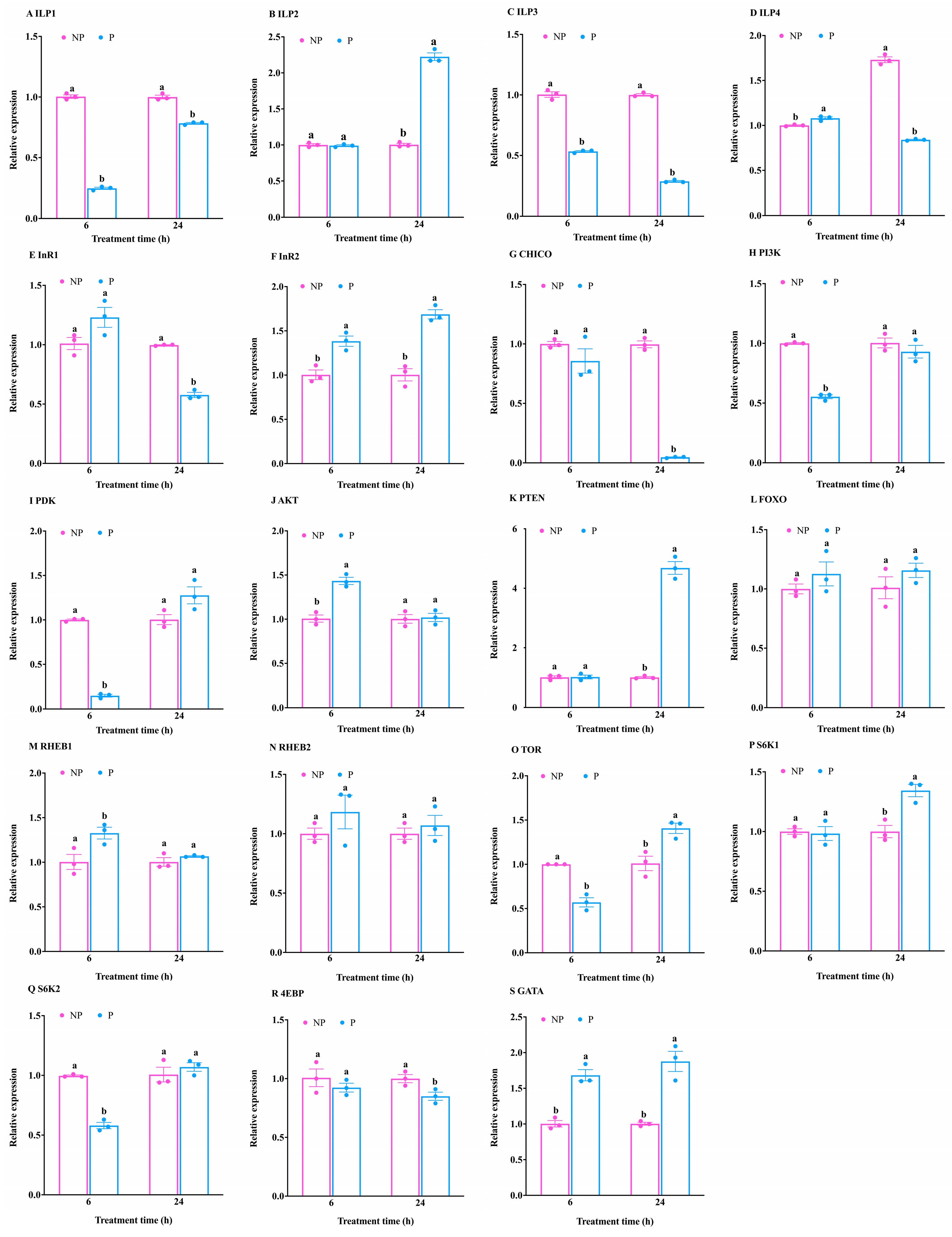
3.2. Expression Pattern Analysis of Genes Related to the IIS/TOR Signaling Pathway of T. molitor in Response to S. guani Parasitism

The above studies have shown that the IIS/TOR signaling pathway plays a role in various stages of T. molitor development. Given that parasitic wasps can influence the growth and development of their hosts, this study aimed to assess the impact of parasitism on the IIS/TOR signaling pathway in T. molitor.
At 6 h post-parasitism, significant reductions in the expression levels of the ILP1 and ILP3 genes were observed in T. molitor compared to the control group (ILP1: t = −44.52, df = 4, p < 0.001; ILP3: t = −19.37, df = 4, p < 0.001) (Figure 2A,C). Conversely, ILP4 expression was significantly elevated in the parasitized group (t = 4.90, df = 4, p < 0.01) (Figure 2D). Additionally, the expression of the InR2 gene was upregulated following parasitism (t = −4.79, df = 4, p < 0.001) (Figure 2F).
Moreover, parasitism was found to suppress the expression of the PI3K and PDK genes (PI3K: t = 25.94, df = 4, p < 0.001; PDK: t = 52.23, df = 4, p < 0.001), while the AKT gene exhibited upregulation (t = −7.56, df = 4, p < 0.01) (Figure 2H–J). The RHEB1 gene within the TOR signaling pathway was transcriptionally induced in response to the elevated AKT expression (t = −2.98, df = 4, p < 0.05) (Figure 2M). However, the expression of the TOR gene and S6K2 gene was significantly suppressed (TOR: t = 8.39, df = 4, p < 0.01; S6K2: t = 15.65, df = 4, p < 0.001) (Figure 2O,Q), while GATA expression was markedly increased (t = −7.40, df = 4, p < 0.01) (Figure 2S).
Importantly, there was no observed effect on the transcription levels of ILP2, InR1, CHICO, PTEN, FOXO, RHEB2, S6K1, and 4EBP genes (ILP2: t = −0.48, df = 4, p > 0.05; InR1: t = −2.23, df = 4, p > 0.05; CHICO: t = 1.35, df = 2.19, p > 0.05; PTEN: t = −0.14, df = 4, p > 0.05; FOXO: t = −1.16, df = 4, p > 0.05; RHEB2: t = −1.22, df = 4, p > 0.05; S6K1: t = 0.28, df = 4, p > 0.05; 4EBP: t = 0.96, df = 4, p > 0.05) (Figure 2B,E,G,K,L,N,P,R). These results suggest that parasitism for 6 h minimally affects the IIS signaling pathway, while exerting a significant inhibitory influence on the TOR pathway.
At 24 h after parasitism, the expression levels of ILP1, ILP3, and ILP4 genes in T. molitor were significantly lower than those in the control group (ILP1: t = −13.00, df = 4, p < 0.001; ILP3: t = −59.35, df = 4, p < 0.001; ILP4: t = −27.25, df = 4, p < 0.001); the expression level of ILP2 gene was significantly higher than that of the control group (t = 21.60, df = 4, p < 0.001) (Figure 2A–D). In addition, the expression of InR1 gene was inhibited (t = 17.69, df = 4, p < 0.001) and the expression of InR2 gene was induced (t = −8.03, df = 4, p < 0.01) after 24 h of parasitism (Figure 2E,F).
At the same time, the expression of CHICO was inhibited (t = 32.98, df = 4, p < 0.001) (Figure 2G), and the expression of PTEN gene was upregulated (t = −16.95, df = 4, p < 0.001) (Figure 2K), which affected the InR1/CHICO signal transduction, but the expression of FOXO, PI3K, PDK, and AKT genes was not affected (FOXO: t = −1.33, df = 4, p > 0.05; PI3K: t = 1.02, df = 4, p > 0.05; PDK: t = −2.45, df = 4, p > 0.05; AKT: t = −0.22, df = 4, p > 0.05) (Figure 2H–J,L). The expression of TOR gene was upregulated (t = −3.97, df = 4, p < 0.05) (Figure 2O), and the downstream signal transduction was activated, so that the expression of S6K1 and GATA genes was significantly upregulated (S6K1: t = −4.68, df = 4, p < 0.01; GATA: t = −6.05, df = 4, p < 0.01) (Figure 2P,S). The expression of 4EBP gene was significantly inhibited (t = 3.16, df = 4, p < 0.05) (Figure 2R). However, the expression of RHEB1, RHEB2, and S6K2 was not affected (RHEB1: t = −1.33, df = 2.09, p > 0.05; RHEB2: t = −0.70, df = 4, p > 0.05; S6K2: t = −0.93, df = 4, p > 0.05) (Figure 2M,N,Q). The above results showed that 24 h of parasitism affected the signal transduction of IIS pathway, but the effect was not significant and did not affect TOR signal transduction.

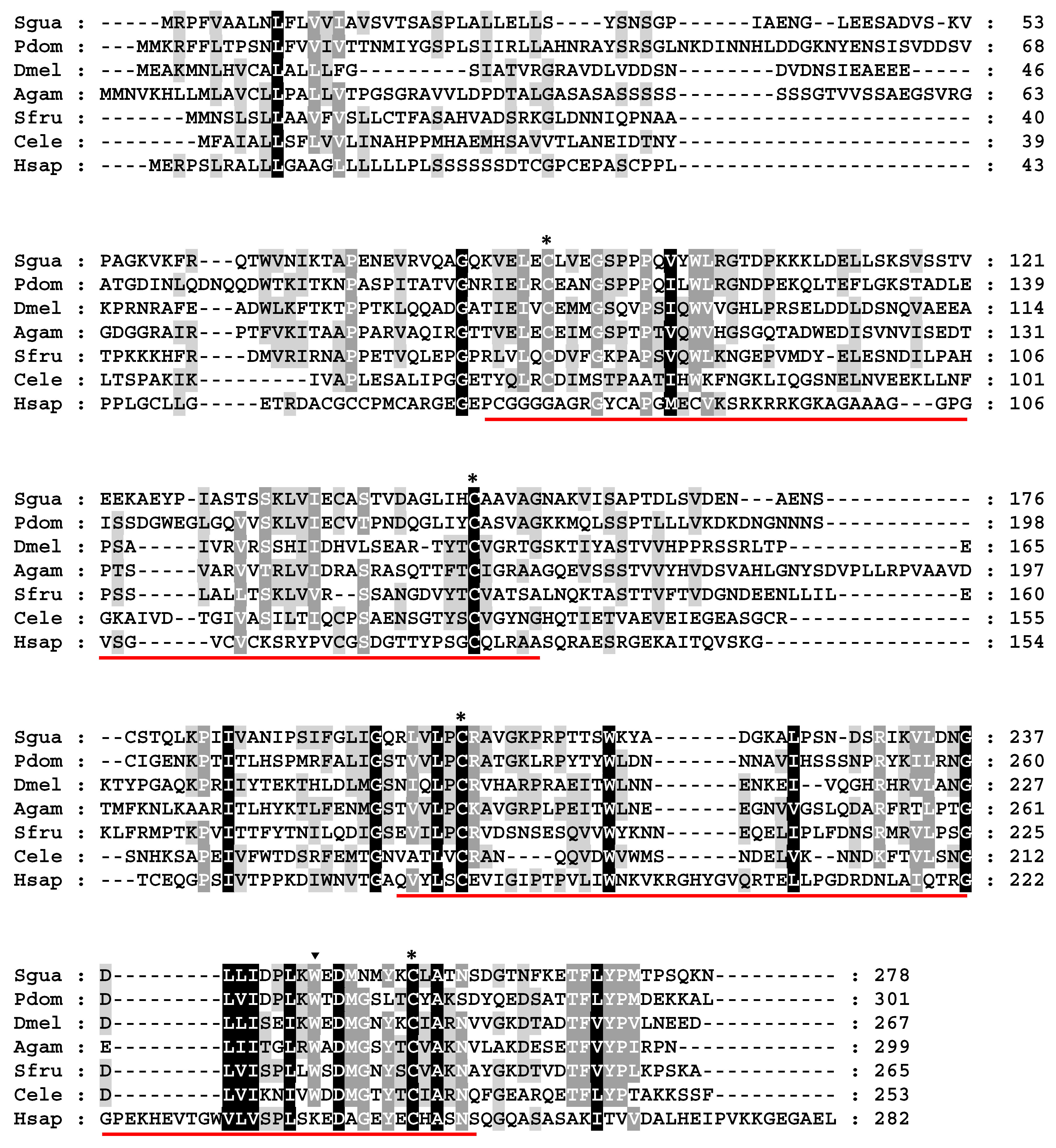
3.3. Cloning and Expression Analysis of IMP-L2 Gene from S. guani Venom

The aforementioned analyses demonstrated that the parasitism of S. guani significantly impacts the IIS/TOR signaling pathway, which is crucial for the growth and development of T. molitor. Parasitic wasps are known to impair host growth and development by introducing parasitic factors, including venom. Our previous research identified that the venom of S. guani is abundant in the IMP-L2 protein. Consequently, this study focused on cloning and analyzing the IMP-L2 gene, assessing its expression across various tissues under parasitic and non-parasitic conditions, as well as during different developmental stages. Additionally, we successfully obtained the recombinant protein.
We elucidated the bioinformatics characteristics of IMP-L2, particularly its expression in response to S. guani parasitism and nutritional status, providing a comprehensive foundation for subsequent functional validations of the IMP-L2 protein. The cloned IMP-L2 gene comprised a coding region of 837 bp, which encodes a protein of 278 amino acids (Figure 3). The identified signal peptide is MRPFVAALNLFLVVIAVSVTSA, with a theoretical molecular weight of 30.01 kDa and an isoelectric point of 5.63 (Figure 3). Notably, an N-glycosylation site (NDS at positions 225–227) was also identified (Figure 3).
Homologous alignment analyses revealed amino acid identities of 38%, 21%, 25%, 25%, 18%, and 13% when compared to the corresponding IMP-L2 proteins from Polistes dominula, Drosophila melanogaster, Anopheles gambiae, Spodoptera frugiperda, Caenorhabditis elegans, and humans, respectively (Figure 3). A phylogenetic tree constructed using the IMP-L2 proteins from various Hymenoptera species, with D. melanogaster as an outgroup, indicated the presence of a single IMP-L2 gene in Ichneumonoidea, Chalcidoidea, and Bethyloidea, while gene expansion was observed in the Vespoidea, Formicidae, and Apoidea lineages (Figure S1).
Simultaneously, RT-qPCR analysis revealed that the expression of the IMP-L2 gene in the venom apparatus of S. guani was significantly elevated compared to that in abdominal remnants (t = −8.86, df = 4, p < 0.01) (Figure 4A). The IMP-L2 gene was expressed under different developmental stages (Figure 4B). Notably, the IMP-L2 gene was highly expressed in adults (especially 1–5 days after eclosion) (F14,30 = 37.76, p < 0.001), which may be due to the fact that the venom organs of S.guani were not formed or not well developed during the egg, larva, and pupa stages, and the development was perfect after eclosion. Therefore, the IMP-L2 gene was highly expressed in adults (Figure 4B). Furthermore, significant upregulation of the IMP-L2 gene was observed following the parasitism of T. molitor pupae by S. guani (t = 5.04, df = 4, p < 0.01) (Figure 4C), suggesting that the IMP-L2 gene may play a role in the process of S. guani parasitizing T. molitor pupae.
The recombinant protein IMP-L2 was successfully produced through eukaryotic expression and subsequent purification. SDS-PAGE analysis revealed that the molecular weight of the recombinant IMP-L2 was approximately 30 kDa, aligning closely with the predicted theoretical molecular weight (Figure 5). Additionally, Western blot results confirmed that the purified protein corresponded to the recombinant IMP-L2 (Figure 5). This recombinant protein is suitable for use in subsequent injection experiments.

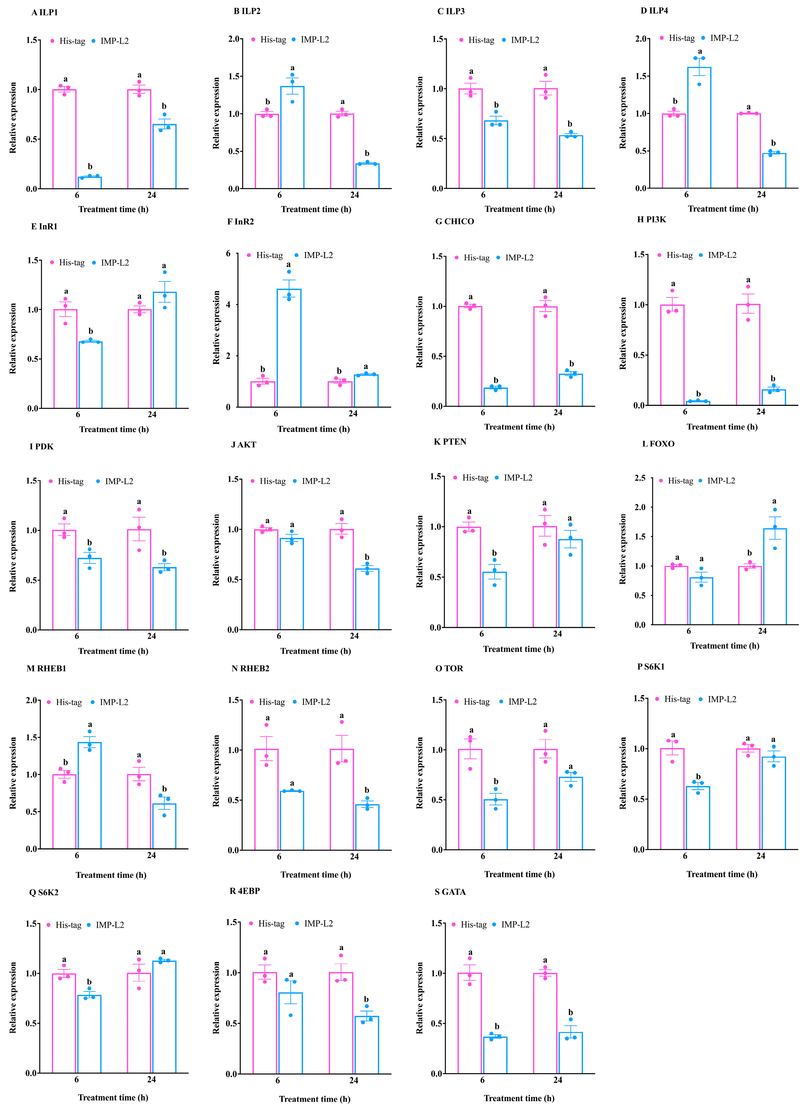
3.4. Analysis of the Expression Pattern of Genes Related to the IIS/TOR Signaling Pathway in T. molitor After Injection of Recombinant IMP-L2 Protein

The results from the aforementioned experiments indicate that the IMP-L2 gene is specifically and highly expressed in the venom apparatus and may play a role in parasitism. Previous studies have shown that IMP-L2 from non-venom sources can bind to insulin, inhibiting insulin signal transduction and consequently impeding growth and development [43,45]. To assess whether venom-derived IMP-L2 similarly affects the host, we conducted an injection experiment using recombinant IMP-L2 to evaluate its impact on gene expression within the growth and development-related IIS/TOR pathway.
Six hours post-injection of the IMP-L2 venom protein, the expression levels of the ILP1 and ILP3 genes were significantly reduced compared to the control group (ILP1: t = −33.43, df = 4, p < 0.001; ILP3: t = −4.64, df = 4, p < 0.05) (Figure 6A,C). Conversely, the expression levels of ILP2 and ILP4 genes in the treatment group were significantly increased relative to the control group (ILP2: t = 3.35, df = 4, p < 0.05; ILP4: t = 5.07, df = 2.30, p < 0.05) (Figure 6B,D). Additionally, the expression of the InR2 gene was induced (t = −10.25, df = 4, p < 0.01) (Figure 6F), while the expression of host genes InR1, CHICO, PI3K, PDK, and PTEN was inhibited (InR1: t = 4.30, df = 4, p < 0.05; CHICO: t = 36.94, df = 4, p < 0.001; PI3K: t = 14.02, df = 2.01, p < 0.01; PDK: t = 3.54, df = 4, p < 0.05; PTEN: t = 5.22, df = 4, p < 0.01) (Figure 6E,G–I,K).
These findings demonstrate that IMP-L2 inhibits the signal transduction of the InR1/PDK pathway; however, downstream AKT expression remained unaffected (t = 2.21, df = 4, p > 0.05), as did the transcription of FOXO (t = 2.23, df = 4, p > 0.05) (Figure 6J,L). Furthermore, the expression of the RHEB1 gene in the TOR signaling pathway was significantly elevated compared to the control group, while RHEB2 expression was unchanged (RHEB1: t = −4.72, df = 4, p < 0.01; RHEB2: t = 3.47, df = 2.00, p > 0.05) (Figure 6M,N).
In addition, downregulation of TOR expression inhibited the transcription of S6K1, S6K2, and GATA genes without affecting 4EBP expression (TOR: t = 4.34, df = 4, p < 0.05; S6K1: t = 4.90, df = 4, p < 0.01; S6K2: t = 4.15, df = 4, p < 0.05; GATA: t = 8.14, df = 4, p < 0.01; 4EBP: t = 1.51, df = 4, p > 0.05) (Figure 6O–S). Collectively, these results indicate that the injection of IMP-L2 has a minimal impact on the IIS signaling pathway while significantly inhibiting TOR pathway signal transduction.
The expression of ILP1-ILP4 gene was significantly lower than that in the control group at 24 h after injection of IMP-L2 venom protein (ILP1: t = −5.58, df = 4, p < 0.01; ILP2: t = −22.34, df = 4, p < 0.001; ILP3: t = −6.52, df = 4, p < 0.01; ILP4: t = −32.18, df = 4, p < 0.001) (Figure 6A–D). The expression levels of InR2 and FOXO genes were significantly increased (InR2: t = −3.12, df = 4, p < 0.05; FOXO: t = −3.30, df = 4, p < 0.05) (Figure 6F,L), while the expression of CHICO, PI3K, PDK, and AKT was significantly inhibited (CHICO: t = 11.47, df = 4, p < 0.001; PI3K: t = 8.71, df = 4, p < 0.01; PDK: t = 3.09, df = 4, p < 0.05; AKT: t = 6.58, df = 4, p < 0.01) (Figure 6G–J). The results showed that the venom IMP-L2 affected the IIS signal transduction and activated the FOXO signal by inhibiting the host CHICO/AKT signaling cascade, but the expression of InR1 and PTEN was not affected (InR1: t = −1.58, df = 4, p > 0.05; PTEN: t = 0.97, df = 4, p > 0.05) (Figure 6E,K). At the same time, the expression of RHEB1 and RHEB2 genes was inhibited (RHEB1: t = 3.19, df = 4, p < 0.05; RHEB2: t = 4.03, df = 2.23, p < 0.05) (Figure 6M,N), and then inhibited the expression of GATA gene downstream of TOR signaling pathway (t = 8.45, df = 4, p < 0.01) (Figure 6S), and inhibited the expression of 4EBP (t = 4.55, df = 4, p < 0.05) (Figure 6R), but did not affect the expression of TOR, S6K1 and S6K2 genes (TOR: t = 2.71, df = 4, p > 0.05; S6K1: t = 1.21, df = 4, p > 0.05; S6K2: t = −1.45 df = 4, p > 0.05) (Figure 6O–Q). These results suggest that injection of IMP-L2 after 24 h inhibits IIS/TOR pathway signal transduction.

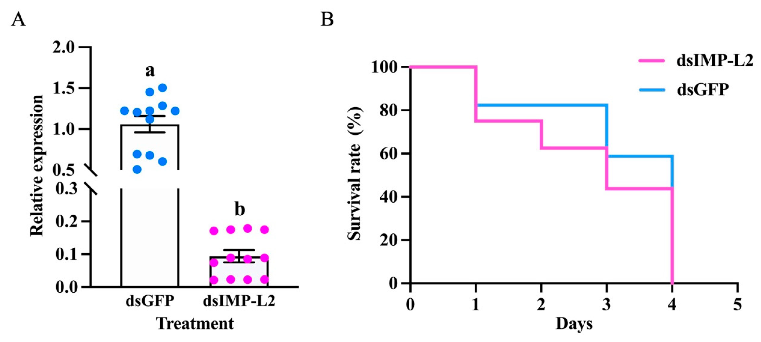
3.5. S. guani IMP-L2 Affects the Longevity of T. molitor

At 24 h after RNAi, compared with dsGFP injection, the relative expression of the IMP-L2 gene decreased significantly. The expression level of IMP-L2 gene decreased by 85.47% (Figure 7A). When S. guani IMP-L2 was silenced and parasitized T. molitor pupae, the death rate of T. molitor pupae was faster than that of T. molitor pupae parasitized by S. guani treated with dsGFP (Figure 7B).

4. Discussion

The IIS/TOR signaling pathway regulates the developmental stages of T. molitor. Our findings indicate that CHICO, PTEN, RHEB2, and GATA are primarily involved in embryonic development. Previous studies have linked CHICO to embryonic development in Drosophila, where CHICO mutants extend egg diapause [56]. Similarly, PTEN is implicated in the embryonic development of Aedes aegypti and Bombyx mori, exhibiting high expression during the egg stage [57]. In contrast to our results showing RHEB2’s role in T. molitor embryogenesis, it appears that RHEB2 does not influence yolk formation or embryonic development in Tribolium castaneum [58]. Furthermore, Pannier (pnr), a GATA family member in Bactrocera dorsalis, is essential for both embryonic and post-embryonic development, with knockout leading to significant mortality during these stages [59].
High expression levels of ILP4, PDK, RHEB1, TOR, and S6K2 during larval development suggest their functional roles at this stage. ILP4 similarly influences larval development in Gnatocerus cornutus, where GcorILP4 is highly expressed in the larval stage [60]. PDK is predominantly expressed during late larval stages, potentially aiding prepupal development [61]. Notably, RHEB1’s involvement extends beyond yolk development to include oviposition, cell volume regulation, neuronal function, and lifespan [58,62,63]. Our findings align with previous research indicating that TOR is crucial for Drosophila larval development, with reduced activity resulting in larval mortality [14]. Moreover, S6K2’s role in larval growth and development highlights its additional functions beyond egg development [64,65].
We observed that IIS pathway-related genes are critically important during the prepupal and pupal stages, with seven genes (ILP1, ILP2, ILP3, InR1, PI3K, PDK, AKT) identified. GCORILP3 in G. cornutus and Mv-ILP1 in Maruca vitrata also exhibit high expression during these stages [60,66]. Interestingly, ILP2 showed peak expression during the prepupal stage, contrasting with its predominance in insect larvae and adults [67]. Consistent with our findings, InR, PI3K, PDK, and AKT are also highly expressed during the prepupal stage in Bombyx [61]. These results suggest that the prepupal stage of T. molitor primarily relies on the IIS pathway for growth and development.
InR and FOXO affect ovarian development and oviposition in adults, and TOR, S6K1, and 4EBP are involved in vitellogenesis in adults [68,69,70]. The significant upregulation of these genes in T.molitor may be involved in the regulation of these physiological processes.
At early stage (6 h post-parasitism), the expressions of ILP1 and ILP3 were suppressed, while ILP4 expression appeared compensatory, leading to an increase in ILP1 and ILP3 levels, consistent with observations in Drosophila [71]. The upregulation of InR2 did not inhibit InR1 expression, resulting in unaffected downstream CHICO expression. Although PI3K/PDK signaling was inhibited, AKT expression was ultimately upregulated, indicating that IIS signals were still transmitted to the TOR pathway. This upregulation may be attributed to the activation of the ROS/AKT/S6K/CREB/HIF1 pathway, which regulates pupal diapause during parasitic stages, thereby compensating for the inhibitory effects on AKT expression stemming from PI3K/PDK/AKT signaling inhibition [72].
Concurrently, RHEB1 of the TOR pathway was upregulated in response to increased AKT levels. However, TOR complex expression was suppressed at 6 h post-parasitism, which inhibited TOR signaling and consequently delayed the growth and development of T. molitor. This inhibition of the TOR pathway may induce a state akin to “starvation restriction,” which is known to prolong lifespan and reduce fecundity in various organisms, similar to effects observed in Drosophila [73,74].
At 24 h after parasitism, ILP1, ILP3, and ILP4 expressions remained suppressed, while ILP2 was upregulated, potentially in response to IMP-L2 induction, analogous to the upregulation of DILP2 in Drosophila following IMP-L2 overexpression [43]. Despite significant InR2 expression inhibiting InR1/CHICO signaling, PTEN was also upregulated, yet it did not impact PI3K/PDK/AKT signaling, leaving TOR signaling unaffected. This suggests that the inhibitory effects of parasitic factors on the IIS/TOR pathway may diminish following host consumption and metabolism, leading to a gradual restoration of IIS/TOR signaling.
The insulin-binding protein IMP-L2 is crucial in Hymenoptera species, exhibiting high expression levels specifically within the venom apparatus of S. guani. The cloned IMP-L2 from S. guani possesses an immunoglobulin domain that shows significant homology with IMP-L2 from other organisms, suggesting a common ancestral origin as an insulin-binding protein [38,39]. We found that gene duplication may occur in the common ancestor of the post-differentiated species, and the phenomenon of gene number amplification occurs in the post-differentiated species.
Additionally, the elevated expression of IMP-L2 in the venom apparatus corroborates our earlier findings that identified this protein as prevalent in the venom of S. guani [50]. Both IMP-L2 and its homolog EpIBP have been detected in the venoms of N. vitripennis and E. pomiformis. In E. pomiformis, EpIBP serves as a primary venom component that interacts with the apolipophorin III (apoLp III) protein of Lepidoptera, influencing host metabolism through this interaction [51,52]. It is plausible that IMP-L2 in S. guani may exhibit analogous functions.
The venom-derived IMP-L2 from S. guani is induced during parasitism, potentially facilitating the delay of host development and lifespan extension by inhibiting IIS and the TOR pathway during the parasitism of T. molitor. The IMP-L2 gene is highly expressed in parasitized S. guani, likely due to transcription activation coinciding with venom secretion via the sting, suggesting its functional role in the parasitic process.
Following the injection of recombinant IMP-L2, inhibition of host IIS/TOR signal transduction was observed at both 6 h and 24 h post-injection. Consistent with the parasitism results, the expression of ILP1 and ILP3 was suppressed at 6 h, while ILP2 showed compensatory expression alongside ILP4. Notably, increased InR2 expression inhibited the entire InR1/Chico/PI3K/PDK pathway, although AKT levels remained comparable to controls. This suggests that compensatory AKT activation within the pupal diapause-related pathway offsets the reduction in AKT associated with the IIS signaling pathway.
In contrast to parasitism, IMP-L2’s inhibition of the TOR complex resulted in a broader impact on downstream gene expression. The venom composition of parasitic wasps is complex, and the concentration of free amino acids in the hemolymph of the host will increase after the venom is injected into the host [75,76]. High concentrations of amino acids can stimulate the TOR signaling cascade [30]. Therefore, the reason why the inhibitory effect of TOR signaling pathway caused by parasitism is not as good as that of pure IMP-L2 may be that high concentration of amino acids stimulation TOR signaling cascade offsets the inhibitory effect of some IMP-L2. At 24 h post-injection, ILP1-4 expression was down-regulated, mirroring results observed during parasitism for ILP1, ILP3, and ILP4. The upregulation of InR2 was linked to the suppression of signaling throughout the InR1/Chico/PI3K/PDK/AKT pathway, while significant downregulation of AKT activated FOXO transcription, potentially triggering diapause in pupae.
The lack of compensation for the downregulation of ILP2 and AKT at 24 h indicates a robust inhibition of IIS signaling. In Drosophila, IIS pathway suppression results in reduced food intake, extended lifespan, and decreased fecundity, suggesting similar effects may occur in T. molitor due to IIS pathway inhibition. Additionally, at 24 h post-injection, IMP-L2 inhibited the TOR pathway in T. molitor, primarily affecting the expression of RHEB1/2 and GATA. Collectively, these results imply that S. guani predominantly targets the host IIS pathway through venom-derived IMP-L2 at the 24 h mark, with high concentrations of IMP-L2 exerting potent inhibitory effects on both the IIS and TOR signaling pathways.
In addition, we found that after IMP-L2 knockdown, the pupae of T. molitor parasitized by S. guani accelerated death and the death time was shortened. This result indicates that IMP-L2 affects the lifespan of T. molitor. It may be that IMP-L2 knockdown reduces the binding of ILP, restores the signal transduction of IIS/TOR pathway, accelerates nutrient consumption, and accelerates its death.

5. Conclusions

In conclusion, the IIS/TOR pathway is crucial for regulating the growth and development of all stages of T. molitor. In the initial phase (6 h) following parasitism, S. guani modulates the growth, development, and longevity of T. molitor by targeting the TOR signaling pathway. By the later stage (24 h) post-parasitism, host IIS/TOR signal transduction begins to recover as parasitic factors diminish. Notably, the venom of S. guani is particularly enriched in the insulin-binding protein IMP-L2, which is vital for Hymenoptera insects, and responds to parasitic stimuli.
In the early phase post-injection, the strategy mirrors that of early parasitism, wherein IMP-L2 inhibits the host TOR signaling pathway, albeit with a more pronounced effect at higher concentrations. This inhibition extends into the late stage of injection, demonstrating that S. guani primarily employs venom-derived IMP-L2 to impede the IIS/TOR signaling pathway, thereby delaying host growth and development. In addition, the absence of IMP-L2 accelerates the death of the host. This mechanism ultimately prolongs the host’s lifespan, facilitating the successful development of parasitic wasp eggs and larvae.

Supplementary Materials

The following supporting information can be downloaded at: https://www.mdpi.com/article/10.3390/insects16080763/s1, Figure S1: Phylogenetic tree of IMP-L2 sequences from venom of Scleroderma guani and other species; Figure S2: Original Western blot images for Figure 5; Table S1: The primer for RT-PCR or RT-qPCR of related genes of IMP-L2 and IIS/TOR pathway from Scleroderma guani or Tenebrio molitor respectally.

Author Contributions

W.W.: writing—original draft, methodology, validation, visualization. Z.Z., X.R.: methodology, visualization. C.W.: visualization. J.Z.: writing—review and editing, funding acquisition, project administration. All authors have read and agreed to the published version of the manuscript.

Funding

This work was supported by the, the National Natural Science Foundation of China (32060126, 31660629), the Xing Dian Talents Support Program of Yunnan Province to Jiaying Zhu, the Science and Technology Innovation Team of Yunnan Provincial Department of Education, and the Funding for the Construction of First-Class Discipline of Forestry in Yunnan Province.

Data Availability Statement

The data presented in this study are openly available in NCBI at (https://www.ncbi.nlm.nih.gov/genbank/, accessed on 5 October 2024). The accession numbers for each dataset are provided in Table S1.

Acknowledgments

We thank the data platform and the researchers who uploaded the data, who helped the research go smoothly.

Conflicts of Interest

All the authors declare that they have no conflicts of interest.

References

  1. Becchimanzi, A.; Avolio, M.; Bostan, H.; Colantuono, C.; Cozzolino, F.; Mancini, D.; Chiusano, M.L.; Pucci, P.; Caccia, S.; Pennacchio, F. Venomics of the ectoparasitoid wasp Bracon nigricans. BMC Genom. 2020, 21, 34. [Google Scholar] [CrossRef] [PubMed]
  2. Samková, A.; Hadrava, J.; Skuhrovec, J.; Janšta, P. Host specificity of the parasitic wasp Anaphes flavipes (Hymenoptera: Mymaridae) and a new defence in its hosts (Coleoptera: Chrysomelidae: Oulema spp.). Insects 2020, 11, 175. [Google Scholar] [CrossRef] [PubMed]
  3. Wang, C.; Jin, F.; De Mandal, S.; Zeng, L.; Zhang, Y.; Hua, Y.; Hong, Y.; Zhao, C.; Li, J.; Li, D.; et al. Insights into the venom protein components of the egg parasitoid Anastatus japonicus (Hymenoptera: Eupelmidae). Pest Manag. Sci. 2020, 76, 2113–2126. [Google Scholar] [CrossRef] [PubMed]
  4. Burke, G.R. Common themes in three independently derived endogenous nudivirus elements in parasitoid wasps. Curr. Opin. Insect Sci. 2019, 32, 28–35. [Google Scholar] [CrossRef] [PubMed]
  5. Pennacchio, F.; Strand, M.R. Evolution of developmental strategies in parasitic hymenoptera. Annu. Rev. Entomol. 2006, 51, 233–258. [Google Scholar] [CrossRef] [PubMed]
  6. Moreau, S.J.; Asgari, S. Venom proteins from parasitoid wasps and their biological functions. Toxins 2015, 7, 2385–2412. [Google Scholar] [CrossRef] [PubMed]
  7. Wan, B.; Yang, L.; Zhang, J.; Qiu, L.; Fang, Q.; Yao, H.; Poirié, M.; Gatti, J.L.; Ye, G. The venom of the ectoparasitoid wasp Pachycrepoideus vindemiae (Hymenoptera: Pteromalidae) induces apoptosis of Drosophila melanogaster Hemocytes. Insects 2020, 11, 363. [Google Scholar] [CrossRef] [PubMed]
  8. De Loof, A. Ecdysteroids, juvenile hormone and insect neuropeptides: Recent successes and remaining major challenges. Gen. Comp. Endocrinol. 2008, 155, 3–13. [Google Scholar] [CrossRef] [PubMed]
  9. Renehan, A.G.; Frystyk, J.; Flyvbjerg, A. Obesity and cancer risk: The role of the insulin–IGF axis. Trends Endocrinol. Metab. 2006, 17, 328–336. [Google Scholar] [CrossRef] [PubMed]
  10. Saltiel, A.R.; Kahn, C.R. Insulin signalling and the regulation of glucose and lipid metabolism. Nature 2001, 414, 799–806. [Google Scholar] [CrossRef] [PubMed]
  11. Corona, M.; Velarde, R.A.; Remolina, S.; Moran-Lauter, A.; Wang, Y.; Hughes, K.A.; Robinson, G.E. Vitellogenin, juvenile hormone, insulin signaling, and queen honey bee longevity. Proc. Natl. Acad. Sci. USA 2007, 104, 7128–7133. [Google Scholar] [CrossRef] [PubMed]
  12. de Azevedo, S.V.; Hartfelder, K. The insulin signaling pathway in honey bee (Apis mellifera) caste development—differential expression of insulin-like peptides and insulin receptors in queen and worker larvae. J. Insect Physiol. 2008, 54, 1064–1071. [Google Scholar] [CrossRef] [PubMed]
  13. Das, D.; Arur, S. Conserved insulin signaling in the regulation of oocyte growth, development, and maturation. Mol. Reprod. Dev. 2017, 84, 444–459. [Google Scholar] [CrossRef] [PubMed]
  14. Lin, X.; Smagghe, G. Roles of the insulin signaling pathway in insect development and organ growth. Peptides 2019, 122, 169923. [Google Scholar] [CrossRef] [PubMed]
  15. Marquez, A.G.; Pietri, J.E.; Smithers, H.M.; Nuss, A.; Antonova, Y.; Drexler, A.L.; Riehle, M.A.; Brown, M.R.; Luckhart, S. Insulin-like peptides in the mosquito Anopheles stephensi: Identification and expression in response to diet and infection with Plasmodium falciparum. Gen. Comp. Endocrinol. 2011, 173, 303–312. [Google Scholar] [CrossRef] [PubMed]
  16. Nässel, D.R.; Kubrak, O.I.; Liu, Y.; Luo, J.; Lushchak, O.V. Factors that regulate insulin producing cells and their output in Drosophila. Front. Physiol. 2013, 4, 252. [Google Scholar] [CrossRef] [PubMed]
  17. Riehle, M.A.; Fan, Y.; Cao, C.; Brown, M.R. Molecular characterization of insulin-like peptides in the yellow fever mosquito, Aedes aegypti: Expression, cellular localization, and phylogeny. Peptides 2006, 27, 2547–2560. [Google Scholar] [CrossRef] [PubMed]
  18. Hun, L.V.; Cheung, K.W.; Brooks, E.; Zudekoff, R.; Luckhart, S.; Riehle, M.A. Increased insulin signaling in the Anopheles stephensi fat body regulates metabolism and enhances the host response to both bacterial challenge and Plasmodium falciparum infection. Insect Biochem. Mol. Biol. 2021, 139, 103669. [Google Scholar] [CrossRef] [PubMed]
  19. Rulifson, E.J.; Kim, S.K.; Nusse, R. Ablation of insulin-producing neurons in flies: Growth and diabetic phenotypes. Science 2002, 296, 1118–1120. [Google Scholar] [CrossRef] [PubMed]
  20. Bai, H.; Kang, P.; Tatar, M. Drosophila insulin-like peptide-6 (dilp6) expression from fat body extends lifespan and represses secretion of Drosophila insulin-like peptide-2 from the brain. Aging Cell 2012, 11, 978–985. [Google Scholar] [CrossRef] [PubMed]
  21. Arrese, E.L.; Soulages, J.L. Insect fat body: Energy, metabolism, and regulation. Annu. Rev. Entomol. 2010, 55, 207–225. [Google Scholar] [CrossRef] [PubMed]
  22. Koyama, T.; Mendes, C.C.; Mirth, C.K. Mechanisms regulating nutrition-dependent developmental plasticity through organ-specific effects in insects. Front. Physiol. 2013, 4, 263. [Google Scholar] [CrossRef] [PubMed]
  23. Kremer, L.P.M.; Korb, J.; Bornberg-Bauer, E. Reconstructed evolution of insulin receptors in insects reveals duplications in early insects and cockroaches. J. Exp. Zool. Part B Mol. Dev. Evol. 2018, 330, 305–311. [Google Scholar] [CrossRef] [PubMed]
  24. Xu, H.J.; Xue, J.; Lu, B.; Zhang, X.C.; Zhuo, J.C.; He, S.F.; Ma, X.F.; Jiang, Y.Q.; Fan, H.W.; Xu, J.Y.; et al. Two insulin receptors determine alternative wing morphs in planthoppers. Nature 2015, 519, 464–467. [Google Scholar] [CrossRef] [PubMed]
  25. Gao, X.; Neufeld, T.P.; Pan, D. Drosophila PTEN regulates cell growth and proliferation through PI3K-dependent and -independent pathways. Dev. Biol. 2000, 221, 404–418. [Google Scholar] [CrossRef] [PubMed]
  26. Goberdhan, D.C.; Paricio, N.; Goodman, E.C.; Mlodzik, M.; Wilson, C. Drosophila tumor suppressor PTEN controls cell size and number by antagonizing the Chico/PI3-kinase signaling pathway. Genes Dev. 1999, 13, 3244–3258. [Google Scholar] [CrossRef] [PubMed]
  27. Kramer, J.M.; Davidge, J.T.; Lockyer, J.M.; Staveley, B.E. Expression of Drosophila FOXO regulates growth and can phenocopy starvation. BMC Dev. Biol. 2003, 3, 5. [Google Scholar] [CrossRef] [PubMed]
  28. Jünger, M.A.; Rintelen, F.; Stocker, H.; Wasserman, J.D.; Végh, M.; Radimerski, T.; Greenberg, M.E.; Hafen, E. The Drosophila forkhead transcription factor FOXO mediates the reduction in cell number associated with reduced insulin signaling. J. Biol. 2003, 2, 20. [Google Scholar] [CrossRef] [PubMed]
  29. Slack, C.; Giannakou, M.E.; Foley, A.; Goss, M.; Partridge, L. dFOXO-independent effects of reduced insulin-like signaling in Drosophila. Aging Cell 2011, 10, 735–748. [Google Scholar] [CrossRef] [PubMed]
  30. Gao, X.; Zhang, Y.; Arrazola, P.; Hino, O.; Kobayashi, T.; Yeung, R.S.; Ru, B.; Pan, D. Tsc tumour suppressor proteins antagonize amino-acid-TOR signalling. Nat. Cell Biol. 2002, 4, 699–704. [Google Scholar] [CrossRef] [PubMed]
  31. Gao, X.; Pan, D. TSC1 and TSC2 tumor suppressors antagonize insulin signaling in cell growth. Genes Dev. 2001, 15, 1383–1392. [Google Scholar] [CrossRef] [PubMed]
  32. Garami, A.; Zwartkruis, F.J.; Nobukuni, T.; Joaquin, M.; Roccio, M.; Stocker, H.; Kozma, S.C.; Hafen, E.; Bos, J.L.; Thomas, G. Insulin activation of Rheb, a mediator of mTOR/S6K/4E-BP signaling, is inhibited by TSC1 and 2. Mol. Cell 2003, 11, 1457–1466. [Google Scholar] [CrossRef] [PubMed]
  33. Bennett-Keki, S.; Fowler, E.K.; Folkes, L.; Moxon, S.; Chapman, T. Sex-biased gene expression in nutrient-sensing pathways. Proc. R. Soc. B Biol. Sci. 2023, 290, 20222086. [Google Scholar] [CrossRef] [PubMed]
  34. Ling, L.; Raikhel, A.S. Amino acid-dependent regulation of insulin-like peptide signaling is mediated by TOR and GATA factors in the disease vector mosquito Aedes aegypti. Proc. Natl. Acad. Sci. USA 2023, 120, e2303234120. [Google Scholar] [CrossRef] [PubMed]
  35. Jiang, Q.; Zhang, X.; Dai, X.; Han, S.; Wu, X.; Wang, L.; Wei, W.; Zhang, N.; Xie, W.; Guo, J. S6K1-mediated phosphorylation of PDK1 impairs AKT kinase activity and oncogenic functions. Nat. Commun. 2022, 13, 1548. [Google Scholar] [CrossRef] [PubMed]
  36. Teleman, A.A. Molecular mechanisms of metabolic regulation by insulin in Drosophila. Biochem. J. 2009, 425, 13–26. [Google Scholar] [CrossRef] [PubMed]
  37. Hietakangas, V.; Cohen, S.M. Re-evaluating AKT regulation: Role of TOR complex 2 in tissue growth. Genes Dev. 2007, 21, 632–637. [Google Scholar] [CrossRef] [PubMed]
  38. Garbe, J.C.; Yang, E.; Fristrom, J.W. IMP-L2: An essential secreted immunoglobulin family member implicated in neural and ectodermal development in Drosophila. Development 1993, 119, 1237–1250. [Google Scholar] [CrossRef] [PubMed]
  39. Sloth Andersen, A.; Hertz Hansen, P.; Schaffer, L.; Kristensen, C. A new secreted insect protein belonging to the immunoglobulin superfamily binds insulin and related peptides and inhibits their activities. J. Biol. Chem. 2000, 275, 16948–16953. [Google Scholar] [CrossRef] [PubMed]
  40. Yamanaka, Y.; Wilson, E.M.; Rosenfeld, R.G.; Oh, Y. Inhibition of insulin receptor activation by insulin-like growth factor binding proteins. J. Biol. Chem. 1997, 272, 30729–30734. [Google Scholar] [CrossRef] [PubMed]
  41. Oh, Y.; Nagalla, S.R.; Yamanaka, Y.; Kim, H.S.; Wilson, E.; Rosenfeld, R.G. Synthesis and characterization of insulin-like growth factor-binding protein (IGFBP)-7. Recombinant human mac25 protein specifically binds IGF-I and -II. J. Biol. Chem. 1996, 271, 30322–30325. [Google Scholar] [CrossRef] [PubMed]
  42. Roed, N.K.; Viola, C.M.; Kristensen, O.; Schluckebier, G.; Norrman, M.; Sajid, W.; Wade, J.D.; Andersen, A.S.; Kristensen, C.; Ganderton, T.R.; et al. Structures of insect Imp-L2 suggest an alternative strategy for regulating the bioavailability of insulin-like hormones. Nat. Commun. 2018, 9, 3860. [Google Scholar] [CrossRef] [PubMed]
  43. Alic, N.; Hoddinott, M.P.; Vinti, G.; Partridge, L. Lifespan extension by increased expression of the Drosophila homologue of the IGFBP7 tumour suppressor. Aging Cell 2011, 10, 137–147. [Google Scholar] [CrossRef] [PubMed]
  44. Owusu-Ansah, E.; Song, W.; Perrimon, N. Muscle mitohormesis promotes longevity via systemic repression of insulin signaling. Cell 2013, 155, 699–712. [Google Scholar] [CrossRef] [PubMed]
  45. Honegger, B.; Galic, M.; Köhler, K.; Wittwer, F.; Brogiolo, W.; Hafen, E.; Stocker, H. Imp-L2, a putative homolog of vertebrate IGF-binding protein 7, counteracts insulin signaling in Drosophila and is essential for starvation resistance. J. Biol. 2008, 7, 10. [Google Scholar] [CrossRef] [PubMed]
  46. Lee, G.J.; Han, G.; Yun, H.M.; Lim, J.J.; Noh, S.; Lee, J.; Hyun, S. Steroid signaling mediates nutritional regulation of juvenile body growth via IGF-binding protein in Drosophila. Proc. Natl. Acad. Sci. USA 2018, 115, 5992–5997. [Google Scholar] [CrossRef] [PubMed]
  47. Taghert, P.H.; Hewes, R.S.; Park, J.H.; O’Brien, M.A.; Han, M.; Peck, M.E. Multiple amidated neuropeptides are required for normal circadian locomotor rhythms in Drosophila. J. Neurosci. Off. J. Soc. Neurosci. 2001, 21, 6673–6686. [Google Scholar] [CrossRef] [PubMed]
  48. Bader, R.; Sarraf-Zadeh, L.; Peters, M.; Moderau, N.; Stocker, H.; Köhler, K.; Pankratz, M.J.; Hafen, E. The IGFBP7 homolog Imp-L2 promotes insulin signaling in distinct neurons of the Drosophila brain. J. Cell Sci. 2013, 126, 2571–2576. [Google Scholar] [CrossRef] [PubMed]
  49. Park, D.; Veenstra, J.A.; Park, J.H.; Taghert, P.H. Mapping peptidergic cells in Drosophila: Where DIMM fits in. PLoS ONE 2008, 3, e1896. [Google Scholar] [CrossRef] [PubMed]
  50. Zhu, J.-Y. Deciphering the main venom components of the ectoparasitic ant-like bethylid wasp, Scleroderma guani. Toxicon 2016, 113, 32–40. [Google Scholar] [CrossRef] [PubMed]
  51. Baek, J.H.; Lee, S.H.; Kim, W.Y.; Kim, M.G. An insulin-binding protein from the venom of a solitary wasp Eumenes pomiformis binds to apolipophorin III in lepidopteran hemolymph. Toxicon 2016, 111, 62–64. [Google Scholar] [CrossRef] [PubMed]
  52. de Graaf, D.C.; Aerts, M.; Brunain, M.; Desjardins, C.A.; Jacobs, F.J.; Werren, J.H.; Devreese, B. Insights into the venom composition of the ectoparasitoid wasp Nasonia vitripennis from bioinformatic and proteomic studies. Insect Mol. Biol. 2010, 19 (Suppl. S1), 11–26. [Google Scholar] [CrossRef] [PubMed]
  53. Zhu, J.Y.; Yang, P.; Zhang, Z.; Wu, G.X.; Yang, B. Transcriptomic immune response of Tenebrio molitor pupae to parasitization by Scleroderma guani. PLoS ONE 2013, 8, e54411. [Google Scholar] [CrossRef] [PubMed]
  54. Wang, W.; Chen, F. Role of the subtilisin-like serine protease CJPRB from Cordyceps javanica in eliciting an immune response in Hyphantria cunea. Int. J. Mol. Sci. 2023, 24, 4170. [Google Scholar] [CrossRef] [PubMed]
  55. Livak, K.J.; Schmittgen, T.D. Analysis of relative gene expression data using real-time quantitative PCR and the 2(-Delta Delta C(T)) method. Methods 2001, 25, 402–408. [Google Scholar] [CrossRef] [PubMed]
  56. Schiesari, L.; Andreatta, G.; Kyriacou, C.P.; O’Connor, M.B.; Costa, R. The insulin-like proteins dILPs-2/5 determine diapause inducibility in Drosophila. PLoS ONE 2016, 11, e0163680. [Google Scholar] [CrossRef] [PubMed]
  57. Riehle, M.A.; Brown, J.M. Characterization of phosphatase and tensin homolog expression in the mosquito Aedes aegypti: Six splice variants with developmental and tissue specificity. Insect Mol. Biol. 2007, 16, 277–286. [Google Scholar] [CrossRef] [PubMed]
  58. Parthasarathy, R.; Palli, S.R. Molecular analysis of nutritional and hormonal regulation of female reproduction in the red flour beetle, Tribolium castaneum. Insect Biochem. Mol. Biol. 2011, 41, 294–305. [Google Scholar] [CrossRef] [PubMed]
  59. Zhang, Q.; Dou, W.; He, L.Q.; Yu, S.S.; Chen, J.Q.; Zheng, L.Y.; Wang, L.; Smagghe, G.; Wang, J.J. Pannier is a key regulator of embryogenesis, pupal development and female reproduction in the insect pest Bactrocera dorsalis. Pest Manag. Sci. 2023, 79, 1352–1361. [Google Scholar] [CrossRef] [PubMed]
  60. Okada, Y.; Katsuki, M.; Okamoto, N.; Fujioka, H.; Okada, K. A specific type of insulin-like peptide regulates the conditional growth of a beetle weapon. PLoS Biol. 2019, 17, e3000541. [Google Scholar] [CrossRef] [PubMed]
  61. Liu, Y.; Zhou, S.; Ma, L.; Tian, L.; Wang, S.; Sheng, Z.; Jiang, R.J.; Bendena, W.G.; Li, S. Transcriptional regulation of the insulin signaling pathway genes by starvation and 20-hydroxyecdysone in the Bombyx fat body. J. Insect Physiol. 2010, 56, 1436–1444. [Google Scholar] [CrossRef] [PubMed]
  62. Honjoh, S.; Yamamoto, T.; Uno, M.; Nishida, E. Signalling through RHEB-1 mediates intermittent fasting-induced longevity in C. elegans. Nature 2009, 457, 726–730. [Google Scholar] [CrossRef] [PubMed]
  63. Brown, H.L.; Kaun, K.R.; Edgar, B.A. The small GTPase Rheb affects central brain neuronal morphology and memory formation in Drosophila. PLoS ONE 2012, 7, e44888. [Google Scholar] [CrossRef] [PubMed]
  64. Lenaerts, C.; Monjon, E.; Van Lommel, J.; Verbakel, L.; Vanden Broeck, J. Peptides in insect oogenesis. Curr. Opin. Insect Sci. 2019, 31, 58–64. [Google Scholar] [CrossRef] [PubMed]
  65. Santos, C.G.; Humann, F.C.; Hartfelder, K. Juvenile hormone signaling in insect oogenesis. Curr. Opin. Insect Sci. 2019, 31, 43–48. [Google Scholar] [CrossRef] [PubMed]
  66. Al Baki, M.A.; Lee, D.W.; Jung, J.K.; Kim, Y. Insulin-like peptides of the legume pod borer, Maruca vitrata, and their mediation effects on hemolymph trehalose level, larval development, and adult reproduction. Arch. Insect Biochem. Physiol. 2019, 100, e21524. [Google Scholar] [CrossRef] [PubMed]
  67. Chowański, S.; Walkowiak-Nowicka, K.; Winkiel, M.; Marciniak, P.; Urbański, A.; Pacholska-Bogalska, J. Insulin-like peptides and cross-talk with other factors in the regulation of insect metabolism. Front. Physiol. 2021, 12, 701203. [Google Scholar] [CrossRef] [PubMed]
  68. Dong, Y.; Chen, W.; Kang, K.; Pang, R.; Dong, Y.; Liu, K.; Zhang, W. FoxO directly regulates the expression of TOR/S6K and vitellogenin to modulate the fecundity of the brown planthopper. Sci. China Life Sci. 2021, 64, 133–143. [Google Scholar] [CrossRef] [PubMed]
  69. Hansen, I.A.; Sieglaff, D.H.; Munro, J.B.; Shiao, S.H.; Cruz, J.; Lee, I.W.; Heraty, J.M.; Raikhel, A.S. Forkhead transcription factors regulate mosquito reproduction. Insect Biochem. Mol. Biol. 2007, 37, 985–997. [Google Scholar] [CrossRef] [PubMed]
  70. Liu, Y.-K.; Luo, Y.-J.; Deng, Y.-M.; Li, Y.; Pang, X.-Q.; Xu, C.-D.; Wang, S.-G.; Tang, B. Insulin receptors regulate the fecundity of Nilaparvata lugens (Stål) (Hemiptera: Delphacidae). J. Asia-Pac. Entomol. 2020, 23, 1151–1159. [Google Scholar] [CrossRef]
  71. Grönke, S.; Clarke, D.F.; Broughton, S.; Andrews, T.D.; Partridge, L. Molecular evolution and functional characterization of Drosophila insulin-like peptides. PLoS Genet. 2010, 6, e1000857. [Google Scholar] [CrossRef] [PubMed]
  72. Wang, X.-X.; Geng, S.-L.; Zhang, X.-S.; Xu, W.-H. P–S6K is associated with insect diapause via the ROS/AKT/ S6K/CREB/HIF-1 pathway in the cotton bollworm, Helicoverpa armigera. Insect Biochem. Mol. Biol. 2020, 120, 103262. [Google Scholar] [CrossRef] [PubMed]
  73. Kapahi, P.; Zid, B.M.; Harper, T.; Koslover, D.; Sapin, V.; Benzer, S. Regulation of lifespan in Drosophila by modulation of genes in the TOR signaling pathway. Curr. Biol. 2004, 14, 885–890. [Google Scholar] [CrossRef] [PubMed]
  74. Partridge, L.; Alic, N.; Bjedov, I.; Piper, M.D.W. Ageing in Drosophila: The role of the insulin/Igf and TOR signalling network. Exp. Gerontol. 2011, 46, 376–381. [Google Scholar] [CrossRef] [PubMed]
  75. Rahbe, Y.; Digilio, M.C.; Febvay, G.; Guillaud, J.; Fanti, P.; Pennacchio, F. Metabolic and symbiotic interactions in amino acid pools of the pea aphid, Acyrthosiphon pisum, parasitized by the braconid Aphidius ervi. J. Insect Physiol. 2002, 48, 507–516. [Google Scholar] [CrossRef] [PubMed]
  76. Mrinalini; Siebert, A.L.; Wright, J.; Martinson, E.; Wheeler, D.; Werren, J.H. Parasitoid venom induces metabolic cascades in flyhosts. Metabolomics 2014, 11, 1–17. [Google Scholar] [CrossRef]
Figure 1. The expression of genes related to IIS/TOR signaling pathway in different developmental stages of T. molitor. (AD) Expression of insulin-like peptide (ILP)-1/2/3/4; (E,F) expression of insulin receptor (InR)-1/2; (G) expression of insulin receptor substrate (CHICO); (H) expression of phosphatidylinositol 3-kinase (PI3K); (I) expression of phosphoinositide-dependent protein kinase (PDK); (J) expression of protein kinase B (AKT); (K) expression of phosphatase and tensin homolog (PTEN); (L) expression of transcription factor forkhead box class O (FOXO); (M,N) expression of ras homolog enhanced in brain (RHEB)-1/2; (O) expression of target of rapamycin (TOR); (P,Q) expression of stimulating ribosomal protein S6 kinase (S6K)-1/2; (R) expression of translational repressor 4e-binding protein (4EBP); (S) expression of GATA transcription factor (GATA) in different developmental stages. EE, early egg; ME, middle egg; LE, later egg; EL, early larvae; ML, middle larvae; LL, later larvae; PP, prepupae; EP, early pupae; MP, middle pupae; LP, later pupae; A1, adult after 1 day of emergence; A4, adult after 4 days of emergence; A8, adult after 8 days of emergence; A12, adult after 12 days of emergence; A16, adult after 16 days of emergence; A20, adult after 20 days of emergence; A24, adult after 24 days of emergence. Means ± standard error (means ± SE) corresponding to different letters within a same gene was significantly different (p < 0.05, Tukey’s multiple comparison test), while solid circles indicate values for individual biological replicates.
Insects 16 00763 g001
Figure 2. Effect of parasitization by S. guani on the expression of IIS/TOR signaling related genes of T. molitor. (AD) Expression of insulin-like peptide (ILP)-1/2/3/4; (E,F) expression of insulin receptor (InR)-1/2; (G) expression of insulin receptor substrate protein (CHICO); (H) expression of phosphatidylinositol 3-kinase (PI3K); (I) expression of phosphoinositide-dependent protein kinase (PDK); (J) expression of protein kinase B (AKT); (K) expression of phosphatase and tensin homolog (PTEN); (L) expression of transcription factor forkhead box class O (FOXO); (M,N) expression of ras homolog enhanced in brain (RHEB)-1/2; (O) expression of target of rapamycin (TOR); (P,Q) expression of stimulating ribosomal protein S6 kinase (S6K)-1/2; (R) expression of translational repressor 4e-binding protein (4EBP); (S) expression of GATA transcription factor (GATA) at different time under parasitized and non-parasitized conditions. P, parasitization; NP, non-parasitization. Means ± standard error (means ± SE) corresponding to different letters within a same gene at same time was significantly different (p < 0.05, Student’s t test) while solid circles indicate values for individual biological replicates.
Insects 16 00763 g002
Figure 3. Analysis of multiple alignment of IMP-L2 sequence from S. guani venom and other species. Multiple sequence alignment of IMP-L2 in invertebrates and humans. The black shadow represents the identical of residual site, and the gray shadow represents the similar of residual site. The triangle marker IMP-L2 premature termination codon, the asterisk marker is the formation of two disulfide bonds of cysteine, and the red line refers to the IG domain. Sgua: Sclernderma guani; Pdom: Polistes dominula (XP_015175065.1); Dmel: Drosophila melanogaster (Q09024.4); Agam: Anopheles gambiae (XP_312831.5); Sfru: Spodoptera frugiperda (AAF61949.1); Cele: Caenorhabditis elegans (CCD63842.1); Hsap: Homo sapiens, (Q16270.1).
Insects 16 00763 g003
Figure 4. Analysis of venom IMP-L2 gene expression pattern in S. guani. A Expression of IMP-L2 gene in different tissues. (A), abdominal remnants; V, venom apparatus. (B) The expression of IMP-L2 gene at different developmental stages. E, egg; EL, early larvae; LL, late larvae; ML, mature larvae; SL, spinning mature larvae; WP, pupae in white cocoon; YP, pupae in yellow cocoon; BP, pupae in black cocoon; A1, adult after 1 day of emergence; A3, adult after 3 days of emergence; A5, adult after 5 days of emergence; A10, adult after 10 days of emergence; A15, adult after 15 days of emergence; A20, adult after 20 days of emergence; A25, adult after 25 days of emergence. (C) Effect of parasitization on expression of IMP-L2 gene. P, parasitization; NP, non-parasitization. (A,C) means ± SE within different tissues and different treatments followed by the different lower-case letters are significantly different (p < 0.05, Student’s t test), while solid circles indicate values for individual biological replicates. (B) means ± SE within different tissues and different treatments followed by the different lower-case letter are significantly different (p < 0.05, Tukey’s multiple comparison test) while solid circles indicate values for individual biological replicates.
Insects 16 00763 g004
Figure 5. Eukaryotic expression of venom IMP-L2 protein of S. guani. M, standard of protein molecular weight; 1, the purified IMP-L2 protein was detected by SDS-PAGE; 2, the purified IMP-L2 protein was detected by Western blot.
Insects 16 00763 g005
Figure 6. Effect of venom IMP-L2 of S. guani on the expression of IIS/TOR related genes of T. molitor. (AD) Expression of insulin-like peptide (ILP)-1/2/3/4; (E,F) expression of insulin receptor (InR)-1/2; (G) expression of insulin receptor substrate protein (CHICO); (H) expression of phosphatidylinositol 3-kinase (PI3K); (I) expression of phosphoinositide-dependent protein kinase (PDK); (J) expression of protein kinase B (AKT); (K) expression of phosphatase and tensin homolog (PTEN); (L) expression of transcription factor forkhead box class O (FOXO); (M,N) expression of ras homolog enhanced in brain (RHEB)-1/2; (O) expression of target of rapamycin (TOR); (P,Q) expression of stimulating ribosomal protein S6 kinase (S6K)-1/2; (R) expression of translational repressor 4e-binding protein (4EBP); (S) expression of GATA transcription factor (GATA) at different time after His-tag injection and IMP-L2 injection. Means ± standard error (means ± SE) corresponding to different letters within a same gene at same time was significantly different (p < 0.05, Student’s t test) while solid circles indicate values for individual biological replicates.
Insects 16 00763 g006
Figure 7. The interference efficiency of S.guani IMP-L2 (after 24 h) and its effect on the survival of T. molitor pupae after silencing. (A), the interference efficiency of IMP-L2 after 24 h of RNAi. (B), surviva curve of pupae of T. molitor after RNAi. Different lowercase letters indicate significant differences (p < 0.05, Student’s t test), while solid circles indicate values for individual replicates.
Insects 16 00763 g007
Disclaimer/Publisher’s Note: The statements, opinions and data contained in all publications are solely those of the individual author(s) and contributor(s) and not of MDPI and/or the editor(s). MDPI and/or the editor(s) disclaim responsibility for any injury to people or property resulting from any ideas, methods, instructions or products referred to in the content.

