Article

Adipokinetic Hormones and Their Receptor Regulate the Locomotor Behavior in Tribolium castaneum

1
Guizhou Provincial Key Laboratory for Agricultural Pest Management of the Mountainous Region, Institute of Entomology, College of Agriculture, Guizhou University, Guiyang 550025, China
2
Institute of Plant Health and Medicine, Guizhou University, Guiyang 550025, China
*
Author to whom correspondence should be addressed.
Insects 2025, 16(4), 407; https://doi.org/10.3390/insects16040407
Submission received: 15 March 2025 / Revised: 8 April 2025 / Accepted: 10 April 2025 / Published: 12 April 2025
(This article belongs to the Special Issue Research on Insect Molecular Biology)

Simple Summary

The insect nervous system regulates locomotor behavior through the use of specialized signaling molecules known as neuropeptides. One such neuropeptide, adipokinetic hormone (AKH), is known to help insects use energy from fats. In this study, we explored how AKH and its receptor (AKHR) affect locomotion in the red flour beetle, Tribolium castaneum, a pest that damages stored grains. We found that AKH strongly activates its receptor, triggering responses at very low concentrations. The genes for AKH were active in the brain, while the receptor was found in the fat-storing tissue. When we reduced AKH or AKHR levels using RNA interference techniques, the beetles moved much less, showing that this signaling pathway is crucial for locomotion. These findings help us understand how insects control their activity, which could lead to better ways to manage pest insects in agriculture and food storage.

Abstract

The regulation of locomotor behavior is essential for insects to perform their life activities. The central nervous system plays a pivotal role in modulating physiological behaviors, particularly movement, with neuropeptides serving as key modulators of these processes. Among these, adipokinetic hormone (AKH) was originally identified in insects as a neurohormone involved in lipid mobilization. This study investigates the functional role of AKHs (AKH1 and AKH2) and their receptor (AKHR) in regulating locomotion in the red flour beetle, Tribolium castaneum. Using functional calcium reporter assays, we demonstrated that AKHR is activated by two mature AKH peptides from T. castaneum, with half-maximal effective concentrations (EC50) falling within the nanomolar range. Gene expression analysis confirmed the presence of AKH1 and AKH2 transcripts in the brain, while AKHR expression was localized to the fat body and carcass. The silencing of AKHs or AKHR through RNA interference resulted in a significant reduction in both movement distance and duration. Collectively, these findings highlight the regulatory influence of AKH/AKHR signaling in locomotor activity in T. castaneum, thereby advancing our understanding of the molecular mechanisms underlying locomotor control in this economically important insect species.

1. Introduction

The locomotor behavior of insects is fundamental to their survival, impacting essential behaviors such as searching for food, finding mates, escaping predators, and adapting to changing environments. Coordinated movement requires the seamless integration of internal physiological states and external sensory inputs in insects [1,2]. Insect neuropeptides, as small intercellular signaling molecules primarily secreted by the central nervous system, play an integral role in regulating various physiological functions, including locomotion, by interacting with their specific receptors [3,4]. Previous studies have identified neuropeptide–receptor systems being involved in controlling the locomotor behavior in insects. For instance, neuropeptide F had suppressive effects on phase-related locomotor activity in Locusta migratoria [5]. Silencing the diuretic hormone receptor resulted in reduced mean velocity in Drosophila melanogaster [6]. Moreover, the tachykinin and short neuropeptide F pathways in the central complex have specific roles in the fine-tuning of locomotor activity in adult D. melanogaster [7]. Despite these findings, limited attention has been given to the regulatory impact of neuropeptide–receptor systems on locomotor behavior in only a small number of insect species.
Adipokinetic hormone (AKH), a neuropeptide initially isolated from locusts as a neurohormone that facilitates lipid mobilization during flight, is now known to be widespread across insect species [8]. It is documented that AKH plays a vital role in regulating energy mobilization, impacting various physiological processes such as starvation responses, stress adaptation, development, and reproductive capacity [9,10,11,12,13]. These functions are mediated through adipokinetic hormone receptors (AKHRs) that share similarities with vertebrate gonadotropin-releasing hormone receptors [14].
Tribolium castaneum, commonly known as the red flour beetle, is a widespread pest of stored grains, flour, cereals, and other dried food products, causing significant economic damage through contamination. Both the larvae and adults feed on these materials, thriving in warm, dry environments typical of storage facilities. Beyond its role as a pest, T. castaneum is a valuable model organism in scientific research. Its short life cycle, ease of lab rearing, and fully sequenced genome make it ideal for studying development, genetics, evolution, and pest control strategies. Notably, it is also used to explore questions in insect biology that complement research on other insects like D. melanogaster [15].
In this study, we researched the regulation of the locomotor behavior in T. castaneum by AKHs and their receptor. Using molecular profiling and calcium mobilization assays, we comprehensively characterized the T. castaneum AKHs (Tc-AKHs) and their receptor (Tc-AKHR), thereby confirming the receptor functional activity of the receptor. Through RNA interference (RNAi), we further demonstrated that silencing the expression of Tc-AKHs or their receptor significantly altered locomotor patterns in adult beetles. Collectively, these findings provide valuable insights into the neuromodulatory mechanism underlying insect locomotion.

2. Materials and Methods

2.1. Insect Rearing and Cell Line Maintenance

The T. castaneum colony was reared in a controlled growth chamber under specific environmental conditions of 30 ± 1 °C temperature, 70 ± 5% relative humidity, and continuous darkness [16,17]. The insects were provided with a diet consisting of whole wheat flour and brewer’s yeast in a 19:1 ratio. Concurrently, the CHO-WTA11 cell line (generously provided by Professor Jiang Hongbo from Southwest University, Chongqing, China) was cultured in a high-glucose Dulbecco’s Modified Eagle Medium (DMEM; Sigma, Taufkirchen, Germany) supplemented with 10% fetal bovine serum (FBS; Gibco, Jenks, OK, USA). The cells were maintained in a 5% CO2 incubator to ensure optimal growth conditions.

2.2. Sequence Analysis of Tc-AKHR and Its Ligands in T. castaneum

Previous studies enabled the identification of the pre-mRNA sequences of Tc-AKH1 (NP_001107797.1) and Tc-AKH2 (NP_001107818.1), as well as the Tc-AKHR sequence (NM_001083340.1), through the NCBI database. The transmembrane domains of Tc-AKHR were predicted using the TMHMM version 2.0. The corresponding amino acid sequences were derived via ExPASy and subsequently aligned with homologous sequences using NCBI’s blastp tool. Phylogenetic analysis was performed using MEGA 5.1, while sequence alignment was conducted with Jalview 2.11. Signal peptides within the Tc-AKH precursor were predicted using the SignalP server, and C-terminal motifs were visualized using WebLogo.

2.3. Structural Modeling and Molecular Docking

The amino acid structures of Tc-AKHs were drawn using ChenDraw (v21.0), while homology modeling of Tc-AHKR was conducted via the Alphafold3 tool to generate a three-dimensional structural model. Molecular docking simulations were performed using the CDDOCKER module in Discovery Studio 21.0. The interaction patterns between Tc-AKHs and Tc-AKHR were visualized and analyzed using PyMOL v.3.11.

2.4. Functional Calcium Reporter Assays

The open reading frames (ORFs) of Tc-AKHR were cloned into the pcDNA 3.1 (+) expression vector (Tsingke, Beijing, China) for heterologous expression in CHO-WTA11 cells, which served as the reporter system for Tc-AKH-induced calcium mobilization. Tc-AKH peptides were chemically synthesized based on high-performance liquid chromatography (HPLC) and mass spectrometry (MS) analyses (Zoonbio, Nanjing, China). For GPCR assays, a mixture of expression vectors, containing Tc-AKHR (2.5 μg) and apoaequorin in a 1:1 ratio, was transfected into CHO cells using TransIT®-LT1 (Mirus, Madison, WI, USA). The transfected cells were harvested, resuspended in a serum-free high-glucose medium, and incubated with coelenterazine h (Thermo Fisher Scientific, Waltham, MA, USA) under dark conditions. Synthetic Tc-AKH peptides, prepared in 10-fold serial dilutions ranging from 2 pM to 20 μM, were added to an opaque 96-well plate. Luminescence was quantified using a SpectraMax L chemiluminescence microplate reader (Molecular Devices, San Jose, CA, USA) [18]. All experiments were conducted in three biological replicates.

2.5. Gene Expression in Different Tissues

Tissue-specific gene expression profiles of Tc-AKHR and Tc-AKHs were obtained from the Beetle Atlas database (https://motif.mvls.gla.ac.uk/BeetleAtlas/). Origin 2024 was employed to analyze and visualize the differential expression patterns across various tissues.

2.6. Total RNA Isolation and cDNA Synthesis

For total RNA isolation, three adult beetles were placed in RNase-free centrifuge tubes, flash-frozen in liquid nitrogen, and homogenized using a disposable tissue grinding rod under continuous liquid nitrogen cooling. Total RNA was extracted from whole-body samples using the Eastep® Super Total RNA Extraction Kit (Promega, Madison, WI, USA). RNA integrity was assessed on 1% agarose gels, and purity was determined using a Nanodrop 2000 spectrophotometer (Thermo Fisher Scientific, USA). Subsequently, 1 µg of total RNA was reverse-transcribed into cDNA using the StarScript Pro All-in-one RT Mix with gDNA Remover (GenStar, Beijing, China), following the manufacturer’s protocol.

2.7. Synthesis of Double-Stranded RNA

The synthesis of double-stranded RNA (dsRNA) targeting specific genes (dsEGFP, dsAKHR, dsAKH1, and dsAKH2) was initiated by generating templates through PCR amplification. Gene-specific primers, incorporating the T7 polymerase promoter sequence at their 5′ ends (Table S1), were used to amplify the target gene fragments. The resulting PCR products were purified and subsequently employed as templates for dsRNA synthesis using the TranscriptAid™ T7 High Yield Transcription Kit (Thermo Fisher Scientific, USA), following the manufacturer’s instructions. Post-synthesis, the dsRNA was purified via phenol/chloroform extraction and ethanol precipitation and then resuspended in DEPC-treated water. The concentration of the synthesized dsRNA was quantified using a Nanodrop 2000 spectrophotometer (Thermo Fisher Scientific, USA) at 260 nm, and its integrity was verified via electrophoresis on 1% agarose gels.

2.8. Quantitative Reverse Transcription PCR

Quantitative real-time PCR (qRT-PCR) was conducted using the CFX Opus Real-Time PCR System (Bio-Rad, Hercules, CA, USA). Each reaction mixture consisted of 2 µL of cDNA (200 ng/µL), 1 µL each of forward and reverse gene-specific primers (10 µM), 6 µL of DEPC-treated water, and 10 µL of 2×RealStar Fast SYBR qPCR Mix (GenStar, China). The thermal cycling protocol included an initial denaturation step at 95 °C for 2 min, followed by 39 cycles of 95 °C for 15 s and 60 °C for 30 s. Gene expression levels were quantified in duplicate and normalized to the internal control gene, T. castaneum ribosomal protein S3 (Tcrp3) [19]. Primer sequences for the target genes are provided in Table S1. Relative gene expression was calculated using the 2−∆∆CT method [20].

2.9. RNA Interference and Movement Analysis

Three-day-old adult T. castaneum beetles were anesthetized with CO2 and immobilized on double-sided tape for microinjection. Approximately 300 ng of dsRNA was injected into the ventral side of the first abdominal segment using a microinjection system (WPI-PV830, World Precision Instruments, Sarasota, FL, USA) equipped with a glass capillary needle [21]. Beetles injected with dsEGFP served as the control group. Following injection, all adults were maintained under identical conditions for 48 h prior to analysis.
For movement assessments, all tests were conducted at room temperature. Groups consisting of three adults each from the treatment (dsAKHR, dsAKH1, and dsAKH2) and control (dsEGFP) groups were transferred to Petri dishes with a diameter of 125 mm. Their motion was recorded for 1 min using a camera (Sony FDR-AX60, Tokyo, Japan) under controlled conditions. The recorded videos were analyzed using the EthoVision XT 17.5 (Noldus, Wageningen, The Netherlands) to quantify movement parameters and generate trajectories. Subject detection was achieved with gray scaling. Thresholds for “moving” and “resting” states were defined as 5 mm/s and 1 mm/s, respectively, based on slight modifications of previously established criteria [17]. For the determination of the active time, the tracking target was divided into a certain number of pixels according to the automatic setting of the software. When 5–60% of the pixels changed, it was considered to be in an active state.

2.10. Statistical Analysis

Statistical analysis began with the Shapiro–Wilk test to evaluate the normality of the datasets. For data conforming to a normal distribution (Shapiro–Wilk test: p > 0.05), an independent Student’s t-test was used to compare mean values of gene expression levels and other parameters between the RNAi-treated and control groups. In cases where data deviated from normality (Shapiro–Wilk test: p ≤ 0.05), the nonparametric Mann–Whitney U test was applied. All results are presented as the mean ± SEM from five or more independent experiments. A p-value of <0.05 was considered statistically significant.

3. Results

3.1. Sequence Analysis and Molecular Characterization of AKHs and AKHR in T. castaneum

The full-length pre-mRNA sequences of Tc-AKH1 and Tc-AKH2 each consist of two exons. The exons in Tc-AKH1 are 69 bp and 171 bp in length and encode a protein of 73 amino acids, while those in Tc-AKH2 are 162 bp and 45 bp in length and encode a 68-amino acid protein, respectively (Figure 1A,C). Notably, the first 19 amino acid residues in both Tc-AKH1 and Tc-AKH2 are predicted to function as secretory signal peptides, followed by a conserved mature peptide that ends with a predicted amidation site and a dibasic cleavage signal (GKR) (Figure 1C). When compared to the mature AKH peptide sequences of T. castaneum, D. melanogaster, Bombyx mori, L. migratoria, Acyrthosiphon pisum, Spodoptera frugiperda, Apis mellifera, and Leptinotarsa decemlineata, it was observed that the AKH peptides are highly conserved and displayed the following characteristic motif: QxxxxxxWamide (Figure 1B).
The Tc-AKHR mRNA features a putative ORF of 1137 nucleotides that encodes a protein consisting of 378 amino acids. Tc-AKHR is a membrane-bound protein with seven transmembrane domains, consistent with its classification as a G protein-coupled receptor (GPCR). Comparative analysis with AKHR sequences from D. melanogaster and B. mori revealed a high degree of conservation. The Tc-AKHR sequence shares 52.77% similarity with D. melanogaster AKHR and 59.73% similarity with B. mori AKHR, particularly in the amino acid sequence of the transmembrane domains (Figure 2B).
Phylogenetic analysis was further conducted using MEGA5.1 to clarify the evolutionary relationship of Tc-AKHR within the arthropod phylum. Using the neighbor-joining method, a gene cluster analysis was performed, which showed that the Tc-AKHR from T. castaneum (order: Coleoptera) is most closely related to the AKHR from Zophobas morio (order: Coleoptera), followed by that from A. mellifera (order: Hymenoptera) (Figure 2A).

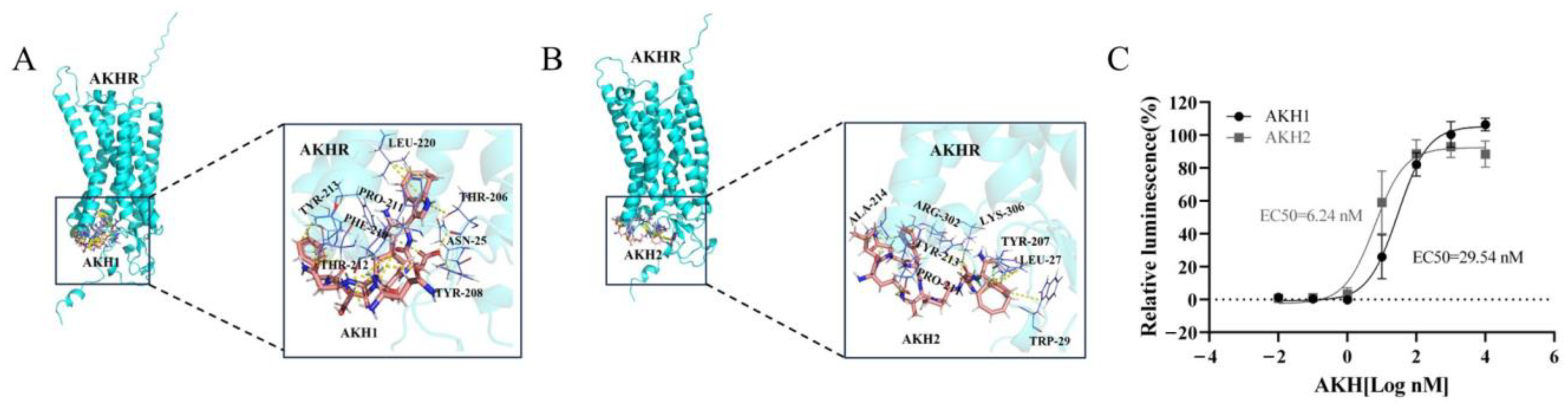
3.2. Activity Determination of Tc-AKHs on Tc-AKHR

Docking simulations revealed that both Tc-AKH1 and Tc-AKH2 bind to specific docking sites on Tc-AKHR, with each ligand interacting with a distinct set of eight amino acids on the receptor (Figure 3A,B). These sites are crucial for the binding of Tc-AKHs to Tc-AKHR.
To evaluate the interaction between Tc-AKHs and Tc-AKHR, heterologous expression of Tc-AKHR was carried out in CHO-WTA11 cells, followed by calcium reporter assays. The results indicated that the CHO cells expressing Tc-AKHR were successfully activated by chemically synthesized Tc-AKHs. Among the two ligands, Tc-AKH1 demonstrated a higher sensitivity, with an effective concentration (EC50) of 6.24 nM, compared to Tc-AKH2, which exhibited an EC50 of 29.54 nM (Figure 3C).

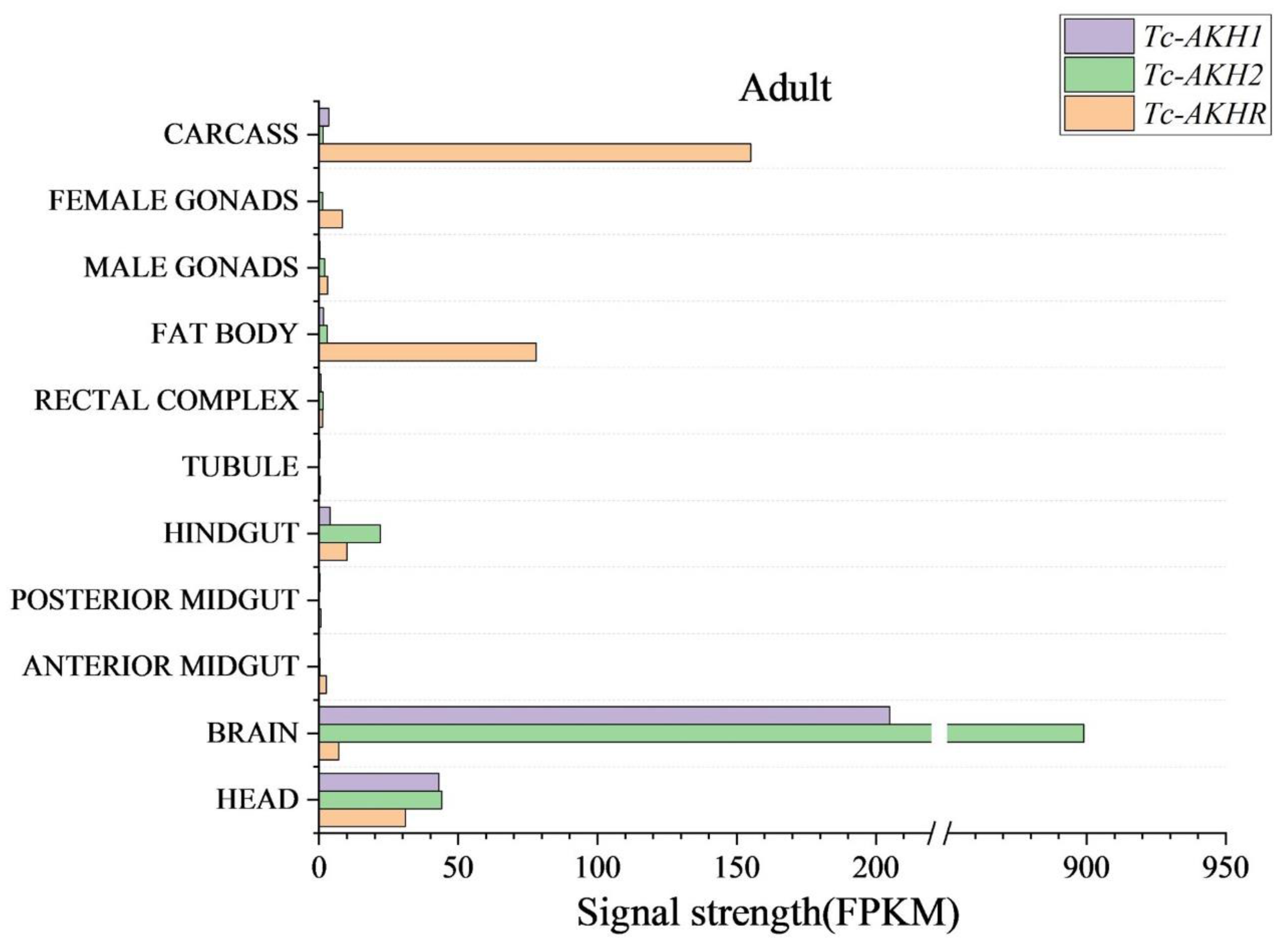
3.3. Expression of Tc-AKHs and Tc-AKHR in Different Tissues of T. castaneum

To investigate the tissue-specific expression patterns of Tc-AKHR and Tc-AKHs, transcript levels were assessed across various tissues in adult T. castaneum according to data from the Beetle Atlas database. The expression of Tc-AKHR was found to be higher in the fat body and carcass. The two Tc-AKHs exhibited a very high expression in the brain (Figure 4).

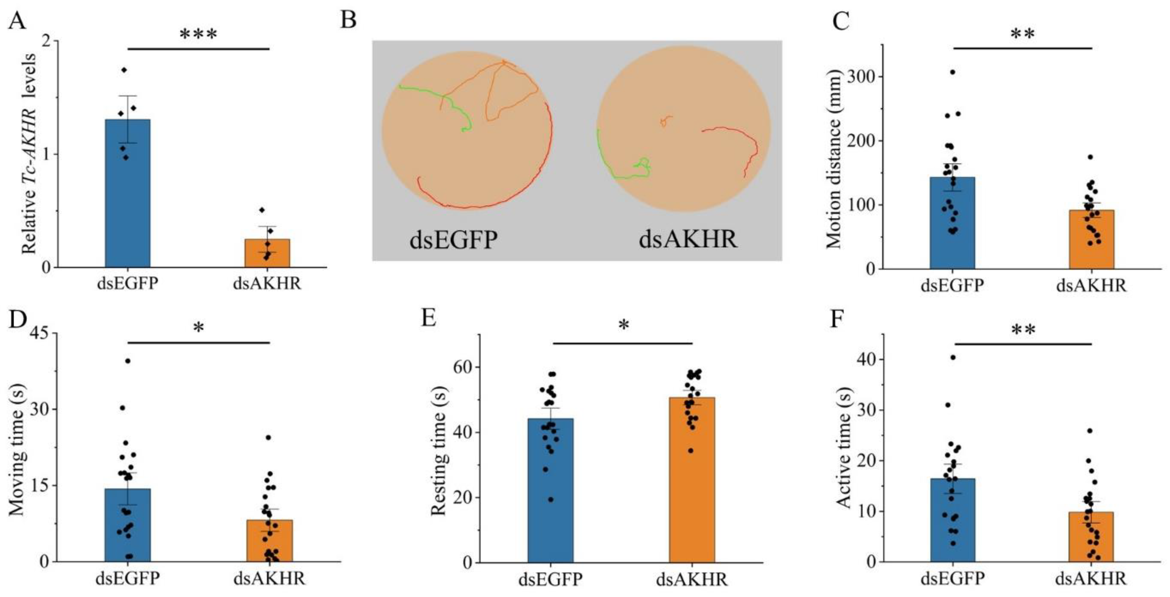
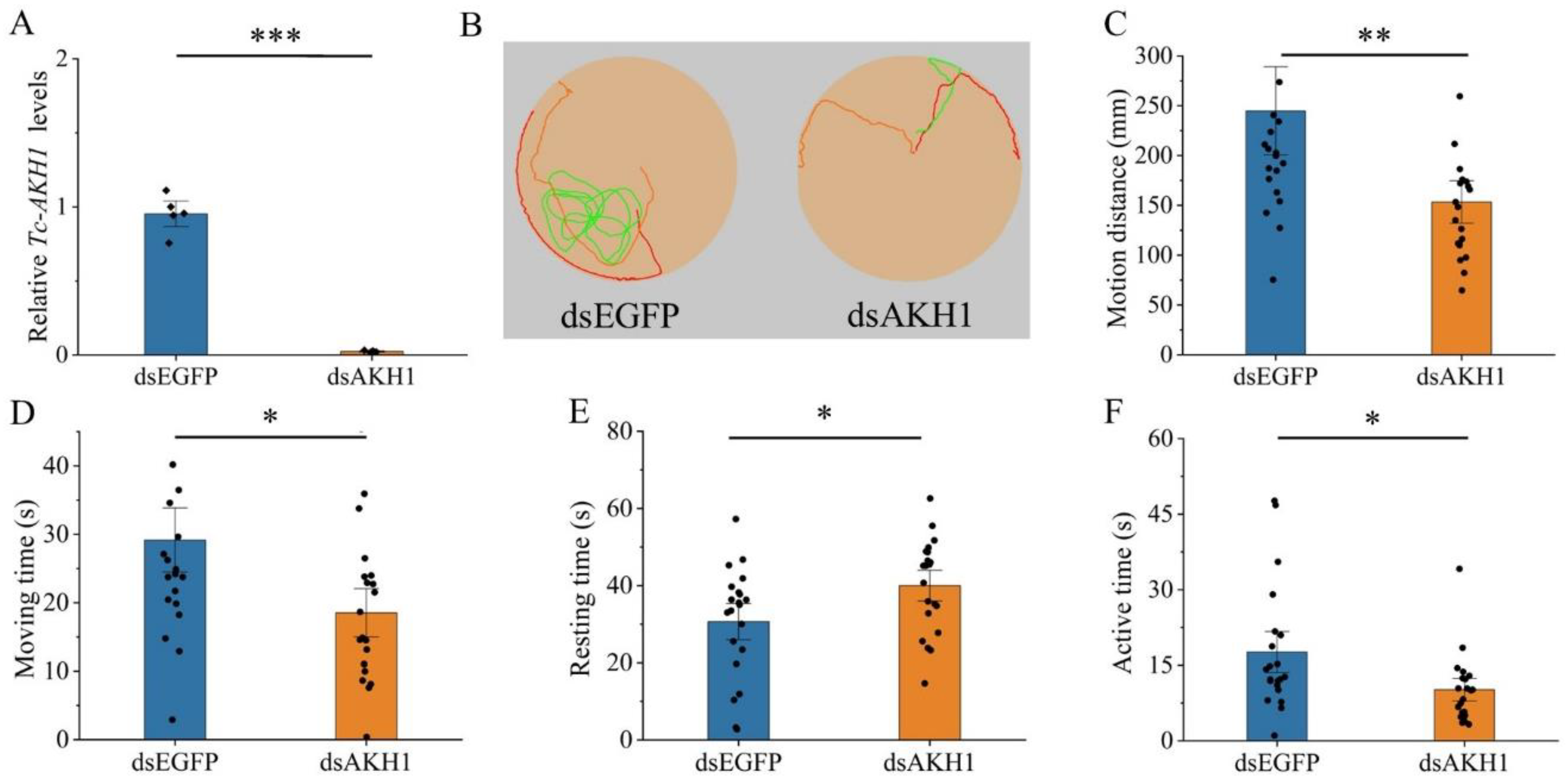
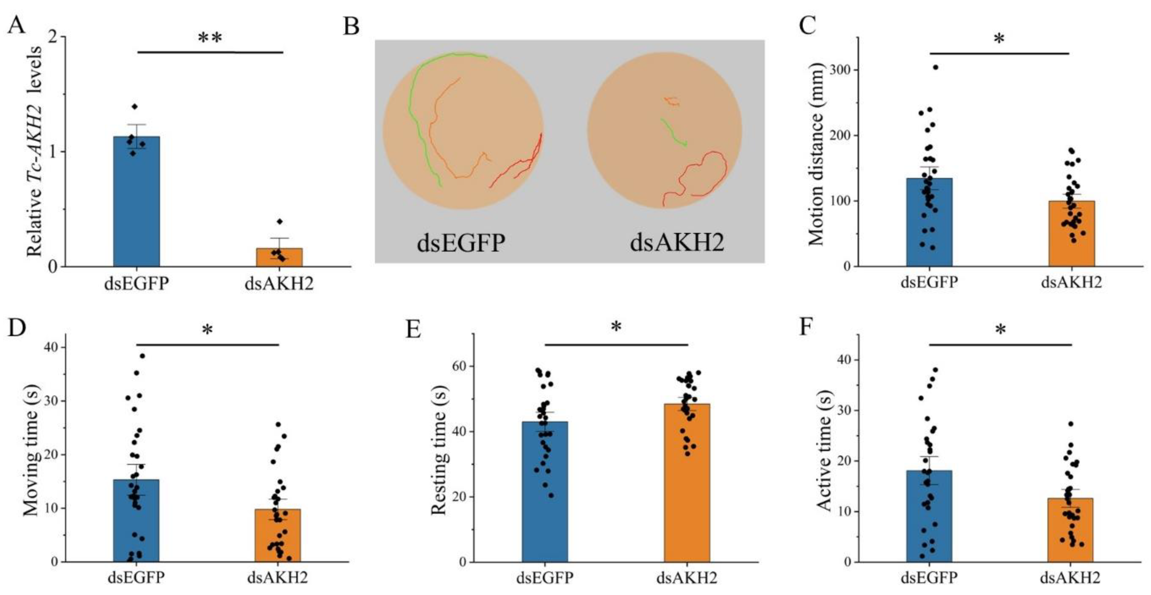
3.4. Effects of Tc-AKH and Tc-AKHR Knockdown on Locomotion

To investigate the role of Tc-AKHs and Tc-AKHR in locomotor behavior, RNAi was employed to suppress their expression, followed by behavioral analysis in adult beetles. The knockdown efficiency was confirmed through qRT-PCR, revealing significant reductions in mRNA levels: Tc-AKHR by 81%, Tc-AKH1 by 97%, and Tc-AKH2 by 86% (Figure 5A, Figure 6A and Figure 7A). No cross-regulatory effects were observed between dsAKH1 and dsAKH2 (Figure S1).
The subsequent tracking of adult movement behavior demonstrated that the locomotor trajectories of RNAi-treated groups (dsAKHR and dsAKHs) were significantly shorter than those of the control group (dsEGFP) (Figure 5B, Figure 6B and Figure 7B). Further quantitative analysis indicated a marked change in key locomotor parameters, including total motion distance, movement duration, resting time, and active time, across all RNAi groups compared to controls (p < 0.05). Specifically, motion distance was significantly reduced with dsAKHR (91.73 mm, p < 0.01), dsAKH1 (153.28 mm, p < 0.01), and dsAKH2 (99.73 mm, p < 0.05) relative to dsEGFP controls (140.62 mm, 244.86 mm, and 134.57 mm, respectively) (Figure 5C, Figure 6C and Figure 7C). Similarly, movement duration was significantly decreased with dsAKHR (8.17 s, p < 0.05), dsAKH1 (18.53 s, p < 0.05), and dsAKH2 (9.80 s, p < 0.05) compared to the control groups (14.33 s, 29.17 s, and 15.30 s, respectively) (Figure 5D, Figure 6D and Figure 7D).
Furthermore, RNAi treatment significantly altered resting and active times. Resting times with dsAKHR (50.66 s, p < 0.05), dsAKH1 (39.99 s, p < 0.05), and dsAKH2 (48.45 s, p < 0.05) were significantly longer than those in controls (44.17 s, 30.64 s, and 42.98 s, respectively) (Figure 5E, Figure 6E and Figure 7E). Active time also exhibited significant reductions in dsAKHR (9.82 s, p < 0.01), dsAKH1 (10.16 s, p < 0.05), and dsAKH2 (12.60 s, p < 0.05) relative to control values (16.43 s, 17.62 s, and 18.10 s, respectively) (Figure 5F, Figure 6F and Figure 7F).

4. Discussion

In this study, two AKH genes and a single AKHR gene were identified in T. castaneum. In most insect species, one or two genes that encode AKH precursor genes have been identified, whereas, for instance, in L. migratoria, four AKH-encoding genes have been identified. So far, the evolutionary dynamics and functional divergence of AKH genes in regulating species-specific behavioral adaptations remain unknown. Also, it is unclear whether gene duplication leads to functional differentiation in T. castaneum. Tc-AKHR shares high similarity with AKHRs from other insect orders, including Lepidoptera and Diptera. Phylogenetic analysis confirmed that T. castaneum AKHR is orthologous to AKHRs in other insects. Functional calcium reporter assays showed that Tc-AKHR-transfected CHO cells responded to two AKH peptides in a concentration-dependent manner, consistent with previous findings in D. melanogaster, which confirmed the specificity of receptors for their corresponding ligands [22]. However, the use of CHO cells as a heterologous expression system raises inherent questions regarding the reconstitution of the endogenous cellular milieu of T. castaneum, which may bias receptor signal kinetics. Further research could use compatible T. castaneum cells. The Tc-AKH1, Tc-AKH2, and Tc-AKHR expressions were assessed in different tissues of adults according to data from the Beetle Atlas database. We found very high levels of Tc-AKHs only in the brain, which is not strange for a neuropeptide gene. For Tc-AKHR, high levels of expression were found in the fat body, which is the principal insect organ that stores both fat and sugar reserves, consistent with findings in other species [5,23,24,25]. The fat body is a key organ for lipid storage and energy regulation, and AKHR is known to mobilize lipids from this organ [26]. Tc-AKHR was also highly expressed in the carcass, suggesting its involvement in other physiological functions, such as reproduction, muscle tonus, and heart rate [27,28,29].
RNAi techniques were applied to suppress the expression of Tc-AKH1, Tc-AKH2, and Tc-AKHR in T. castaneum, resulting in decreases in locomotor activity. The silencing of Tc-AKH1 or Tc-AKH2 produced the same results in locomotion; therefore, we did not find evidence to support the notion that the gene duplication of AKH led to functional differentiation. It is hypothesized that the duplication of the AKH gene resulted in a compensatory effect on locomotion, potentially enhancing its adaptive significance. The influence of AKH on T. castaneum’s locomotion is likely attributed to its role in energy regulation. AKH specifically binds to its receptor, triggering enzymatic activation, including glycogen phosphorylase and triglyceride lipases, which facilitate energy mobilization [30,31]. The primary function of AKH is to induce lipid and carbohydrate release from the fat body. The lipid mobilization mechanism is mediated by AKH through triglyceride lipase [32,33]. In parallel, an alternative lipolytic pathway involves Brummer lipase, an enzyme homologous to mammalian adipose triglyceride lipase. Drosophila flies deficient in Brummer exhibit impaired lipid mobilization and excessive fat accumulation, while AKH responds to acute lipid demands. Brummer maintains basal lipid homeostasis and contributes to energy supply during metabolic exigencies [34,35]. AKH-mediated lipid mobilization is facilitated by intracellular signaling cascades [36]. One pathway involves calcium release from the endoplasmic reticulum, which acts as a crucial lipid mobilization signal. Another pathway entails AKH binding to its receptor on fat body cells, activating adenylate cyclase and increasing cyclic adenosine monophosphate (cAMP) levels. This cascade activates cAMP-dependent protein kinase A, which subsequently phosphorylates lipid droplets, triglyceride lipase, and perilipin 1/lipid storage droplet-1 protein [37]. Collectively, these findings indicate that AKH regulates peripheral energy reserves and locomotor activity via an intricate signaling network. Further research is necessary to unravel the molecular mechanisms behind this network.
In conclusion, the molecular characteristics of AKHs and AKHR in T. castaneum, along with their pharmacological properties, have been described. Through RNAi-mediated suppression of AKHs or AKHR, the pivotal role of AKH/AKHR signaling in locomotor activity has been confirmed. These findings not only strengthen the established function of AKH in locomotor control but also offer mechanistic insights into the neuromodulatory regulation of insect movement. This contributes to a more comprehensive understanding of neurohormonal pathways related to locomotion.

Supplementary Materials

The following supporting information can be downloaded at: https://www.mdpi.com/article/10.3390/insects16040407/s1. Figure S1. (A) Effect of dsAKH1 injected into three-day-old adults on the gene transcript levels of Tc-AKH2. (B) Effect of dsAKH2 injected into three-day-old adults on the gene transcript levels of Tc-AKH1; Video S1. Motion video after injection with dsAKHR; Video S2. Motion video after injection with dsAKH1; Video S3. Motion video after injection with dsAKH2. Table S1. Primers of this study.

Author Contributions

R.-H.L., X.-D.P., S.-H.G., G.S. and T.-X.L. conceived the study and participated in its design. R.-H.L., X.-D.P. and S.-Q.W. performed the experiments. R.-H.L., X.-D.P. and S.-H.G. analyzed the data. R.-H.L., X.-D.P., S.-H.G., G.S. and T.-X.L. wrote the paper. All authors have read and agreed to the published version of the manuscript.

Funding

This research was funded by Guizhou Provincial Science and Technology Plan Project, grant number Qiankehe Platform Talents-BQW [2024] 008, and Guizhou University Students’ Innovation and Entrepreneurship Training Program, grant number gzugc2024034.

Data Availability Statement

The original contributions presented in this study are included in the article and supplementary material. Further inquiries can be directed to the corresponding author.

Conflicts of Interest

The authors declare that the research was conducted in the absence of any commercial or financial relationships that could be construed as potential conflicts of interest.

References

  1. Huston, S.J.; Jayaraman, V. Studying sensorimotor integration in insects. Curr. Opin. Neurobiol. 2011, 21, 527–534. [Google Scholar] [CrossRef] [PubMed]
  2. Dickinson, M.H.; Farley, C.T.; Full, R.J.; Koehl, M.A.; Kram, R.; Lehman, S. How animals move: An integrative view. Science 2000, 288, 100–106. [Google Scholar] [CrossRef] [PubMed]
  3. Chen, X.; Peterson, J.; Nachman, R.J.; Ganetzky, B. Drosulfakinin activates CCKLR-17D1 and promotes larval locomotion and escape response in Drosophila. Fly 2012, 6, 290–297. [Google Scholar] [CrossRef] [PubMed]
  4. Nässel, D.R.; Zandawala, M. Recent advances in neuropeptide signaling in Drosophila, from genes to physiology and behavior. Prog. Neurobiol. 2019, 179, 101607. [Google Scholar] [CrossRef]
  5. Hou, L.; Yang, P.; Jiang, F.; Liu, Q.; Wang, X.; Kang, L. The neuropeptide F/nitric oxide pathway is essential for shaping locomotor plasticity underlying locust phase transition. eLife 2017, 6, e22526. [Google Scholar] [CrossRef]
  6. Wang, S.; Li, M.; Wang, N.; Song, Y.; Peng, X.; Chen, M. Functional characterization of two DH44R genes associated with starvation and desiccation in Rhopalosiphum padi (Hemiptera: Aphididae). Pestic. Biochem. Physiol. 2024, 201, 105902. [Google Scholar] [CrossRef]
  7. Kahsai, L.; Martin, J.R.; Winther, A.M. Neuropeptides in the Drosophila central complex in modulation of locomotor behavior. J. Exp. Biol. 2010, 213, 2256–2265. [Google Scholar] [CrossRef]
  8. Stone, J.V.; Mordue, W.; Batley, K.E.; Morris, H.R. Structure of locust adipokinetic hormone, a neurohormone that regulates lipid utilisation during flight. Nature 1976, 263, 207–211. [Google Scholar] [CrossRef]
  9. Marco, H.G.; Gäde, G. Structure and function of adipokinetic hormones of the large white butterfly Pieris brassicae. Physiol. Entomol. 2017, 42, 103–112. [Google Scholar] [CrossRef]
  10. Kong, X.; Xie, Y.; Cao, Y.; Li, Y.; Zhang, Y.; Zou, Z.; Xia, B.; Xin, T. Expression and functional analysis of adipokinetic hormone reveal its different roles in larval development and female fecundity in Panonychus citri (McGregor) (Acari: Tetranychidae). Insect Mol. Biol. 2024, online ahead of print. [Google Scholar] [CrossRef]
  11. Hou, Q.L.; Chen, E.H.; Jiang, H.B.; Wei, D.D.; Gui, S.H.; Wang, J.J.; Smagghe, G. Adipokinetic hormone receptor gene identification and its role in triacylglycerol mobilization and sexual behavior in the oriental fruit fly (Bactrocera dorsalis). Insect Biochem. Mol. Biol. 2017, 90, 1–13. [Google Scholar] [CrossRef] [PubMed]
  12. Sajwan, S.; Sidorov, R.; Stašková, T.; Žaloudíková, A.; Takasu, Y.; Kodrík, D.; Zurovec, M. Targeted mutagenesis and functional analysis of adipokinetic hormone-encoding gene in Drosophila. Insect Biochem. Mol. Biol. 2015, 61, 79–86. [Google Scholar] [CrossRef] [PubMed]
  13. Tang, B.; Cheng, Y.; Li, Y.; Li, W.; Ma, Y.; Zhou, Q.; Lu, K. Adipokinetic hormone regulates cytochrome P450-mediated imidacloprid resistance in the brown planthopper, Nilaparvata lugens. Chemosphere 2020, 259, 127490. [Google Scholar] [CrossRef] [PubMed]
  14. Jiang, S.; Marco, H.G.; Scheich, N.; He, S.; Wang, Z.; Gäde, G.; McMahon, D.P. Comparative analysis of adipokinetic hormones and their receptors in Blattodea reveals novel patterns of gene evolution. Insect Mol. Biol. 2023, 32, 615–633. [Google Scholar] [CrossRef]
  15. Richards, S.; Gibbs, R.A.; Weinstock, G.M.; Brown, S.J.; Denell, R.; Beeman, R.W.; Gibbs, R.; Bucher, G.; Friedrich, M.; Grimmelikhuijzen, C.J.; et al. The genome of the model beetle and pest Tribolium castaneum. Nature 2008, 452, 949–955. [Google Scholar] [CrossRef]
  16. Walski, T.; Van Damme, E.J.M.; Smargiasso, N.; Christiaens, O.; De Pauw, E.; Smagghe, G. Protein N-glycosylation and N-glycan trimming are required for postembryonic development of the pest beetle Tribolium castaneum. Sci. Rep. 2016, 6, 35151. [Google Scholar] [CrossRef]
  17. Pang, X.D.; Li, Y.S.; Lu, R.H.; Smagghe, G.; Liu, T.X.; Gui, S.H. miR-7977 regulates the locomotor behavior by targeting diuretic hormone and SIFamide receptors in Tribolium castaneum. Int. J. Biol. Macromol. 2025, online ahead of print. [Google Scholar] [CrossRef]
  18. Gui, S.H.; Jiang, H.B.; Liu, X.Q.; Xu, L.; Wang, J.J. Molecular characterizations of natalisin and its roles in modulating mating in the oriental fruit fly, Bactrocera dorsalis (Hendel). Insect Mol. Biol. 2017, 26, 103–112. [Google Scholar] [CrossRef]
  19. Lord, J.C.; Hartzer, K.; Toutges, M.; Oppert, B. Evaluation of quantitative PCR reference genes for gene expression studies in Tribolium castaneum after fungal challenge. J. Microbiol. Methods 2010, 80, 219–221. [Google Scholar] [CrossRef]
  20. Livak, K.J.; Schmittgen, T.D. Analysis of relative gene expression data using real-time quantitative PCR and the 2(-delta delta C(T)) method. Methods 2001, 25, 402–408. [Google Scholar] [CrossRef]
  21. Noh, M.Y.; Beeman, R.W.; Arakane, Y. RNAi-based functional genomics in Tribolium castaneum and possible application for controlling insect pests. Entomol. Res. 2012, 42, 1–10. [Google Scholar] [CrossRef]
  22. Park, Y.; Kim, Y.J.; Adams, M.E. Identification of G protein-coupled receptors for Drosophila PRXamide peptides, CCAP, corazonin, and AKH supports a theory of ligand-receptor coevolution. Proc. Natl. Acad. Sci. USA 2002, 99, 11423–11428. [Google Scholar] [CrossRef] [PubMed]
  23. Staubli, F.; Jorgensen, T.J.; Cazzamali, G.; Williamson, M.; Lenz, C.; Sondergaard, L.; Roepstorff, P.; Grimmelikhuijzen, C.J. Molecular identification of the insect adipokinetic hormone receptors. Proc. Natl. Acad. Sci. USA 2002, 99, 3446–3451. [Google Scholar] [CrossRef] [PubMed]
  24. Ziegler, R.; Isoe, J.; Moore, W.; Riehle, M.A.; Wells, M.A. The putative AKH receptor of the tobacco hornworm, Manduca sexta, and its expression. J. Insect Sci. 2011, 11, 40. [Google Scholar] [CrossRef]
  25. Konuma, T.; Morooka, N.; Nagasawa, H.; Nagata, S. Knockdown of the adipokinetic hormone receptor increases feeding frequency in the two-spotted cricket Gryllus bimaculatus. Endocrinology 2012, 153, 3111–3122. [Google Scholar] [CrossRef]
  26. Alves-Bezerra, M.; De Paula, I.F.; Medina, J.M.; Silva-Oliveira, G.; Medeiros, J.S.; Gäde, G.; Gondim, K.C. Adipokinetic hormone receptor gene identification and its role in triacylglycerol metabolism in the blood-sucking insect Rhodnius prolixus. Insect Biochem. Mol. Biol. 2016, 69, 51–60. [Google Scholar] [CrossRef]
  27. Lorenz, M.W. Adipokinetic hormone inhibits the formation of energy stores and egg production in the cricket Gryllus bimaculatus. Comp. Biochem. Physiol. B Biochem. Mol. Biol. 2003, 136, 197–206. [Google Scholar] [CrossRef]
  28. O’Shea, M.; Witten, J.; Schaffer, M. Isolation and characterization of two myoactive neuropeptides: Further evidence of an invertebrate peptide family. J. Neurosci. 1984, 4, 521–529. [Google Scholar] [CrossRef]
  29. Scarborough, R.M.; Jamieson, G.C.; Kalish, F.; Kramer, S.J.; McEnroe, G.A.; Miller, C.A.; Schooley, D.A. Isolation and primary structure of two peptides with cardioacceleratory and hyperglycemic activity from the corpora cardiaca of Periplaneta americana. Proc. Natl. Acad. Sci. USA 1984, 81, 5575–5579. [Google Scholar] [CrossRef]
  30. Van der Horst, D.J. Insect adipokinetic hormones: Release and integration of flight energy metabolism. Comp. Biochem. Physiol. B Biochem. Mol. Biol. 2003, 136, 217–226. [Google Scholar] [CrossRef]
  31. Toprak, U. The role of peptide hormones in insect lipid metabolism. Front. Physiol. 2020, 11, 434. [Google Scholar] [CrossRef] [PubMed]
  32. Arrese, E.L.; Patel, R.T.; Soulages, J.L. The main triglyceride-lipase from the insect fat body is an active phospholipase A (1): Identification and characterization. J. Lipid Res. 2006, 47, 2656–2667. [Google Scholar] [CrossRef] [PubMed]
  33. Arrese, E.L.; Soulages, J.L. Insect fat body: Energy, metabolism, and regulation. Annu. Rev. Entomol. 2010, 55, 207–225. [Google Scholar] [CrossRef]
  34. Grönke, S.; Mildner, A.; Fellert, S.; Tennagels, N.; Petry, S.; Müller, G.; Jäckle, H.; Kühnlein, R.P. Brummer lipase is an evolutionary conserved fat storage regulator in Drosophila. Cell Metab. 2005, 1, 323–330. [Google Scholar] [CrossRef]
  35. Grönke, S.; Müller, G.; Hirsch, J.; Fellert, S.; Andreou, A.; Haase, T.; Jäckle, H.; Kühnlein, R.P. Dual lipolytic control of body fat storage and mobilization in Drosophila. PLoS Biol. 2007, 5, e137. [Google Scholar] [CrossRef]
  36. Canavoso, L.E.; Wells, M.A. Role of lipid transfer particle in delivery of diacylglycerol from midgut to lipophorin in larval Manduca sexta. Insect Biochem. Mol. Biol. 2001, 31, 783–790. [Google Scholar] [CrossRef]
  37. Arrese, E.L.; Mirza, S.; Rivera, L.; Howard, A.D.; Chetty, P.S.; Soulages, J.L. Expression of lipid storage droplet protein-1 may define the role of AKH as a lipid mobilizing hormone in Manduca sexta. Insect Biochem. Mol. Biol. 2008, 38, 993–1000. [Google Scholar] [CrossRef]
Figure 1. Gene structures and deduced amino acid sequences of AKHs in T. castaneum. (A) Gene structures of Tc-AKH1 and Tc-AKH2. Exons are represented by boxes and introns by lines. (B) Sequence alignment of mature AKH peptides from T. castaneum, D. melanogaster, B. mori, L. migratoria, A. pisum, S. frugiperda, A. mellifera and L. decemlineata. The calculated sequence logo is shown at the bottom. (C) Deduced amino acid sequences of Tc-AKHs. Putative signal peptides are underlined; mature peptides are shaded in gray, and predicted amidation signals with dibasic cleavage sites (KR) are shown in bold.
Insects 16 00407 g001
Figure 2. Amino acid sequence alignment and cluster analysis of AKHR in T. castaneum. (A) Evolutionary tree analysis of Tc-AKHR (neighbor-joining). The insect AKHR protein sequences of T. castaneum, Z. morio, A. mellifera, A. pisum, N. vitripennis, P. americana, D. pulex, A. aegypti, A. gambiae, B. dorsalis, D. melanogaster, G. morsitans, and B. mori were selected to construct the evolutionary tree. Tc-AKHR was assigned with “▲”. (B) Comparative amino acid sequence alignment of T. castaneum, D. melanogaster, and B. mori AKHR sequences.
Insects 16 00407 g002
Figure 3. (A,B) Tc-AKH1 and Tc-AKH2 docked to Tc-AKHR. The hydrogen bond is represented by a yellow dotted line; the three letters are the abbreviation of the amino acid. and the number is the amino acid number of the receptor. (C) Dose–response curves and EC50 values of Tc-AKH1 and Tc-AKH2, tested on Tc-AKHR expressed in CHO-WTA11 cells.
Insects 16 00407 g003
Figure 4. The relative expression of Tc-AKHR, Tc-AKH1, and Tc-AKH2 in different adult tissues of T. castaneum. Data were obtained from the Beetle Atlas database.
Insects 16 00407 g004
Figure 5. Effect of dsAKHR injected into three-day-old adults on the gene transcript levels and motion situation of Tc-AKHR. The data were analyzed using Student’s t-test or the Mann–Whitney U test. Date are means ± SE. * p < 0.05, ** p < 0.01, and *** p < 0.001. (A) Relative expression levels of Tc-AKHR at 48 h after injection with dsAKHR; dsEGFP was used as a control. (B) Movement trajectory after injection with dsAKHR. (C) Changes in motion distance. (D) Changes in moving time. (E) Changes in resting time. (F) Changes in active time.
Insects 16 00407 g005
Figure 6. Effect of dsAKH1 injected into three-day-old adults on the gene transcript levels and motion situation of Tc-AKH1. Date are means ± SE. * p < 0.05, ** p < 0.01, and *** p < 0.001. (A) Relative expression levels of Tc-AKH1 at 48 h after injection with dsAKH1; dsEGFP was used as a control (n = 5). (B) Movement trajectory after injection with dsAKH1. (C) Changes in motion distance. (D) Changes in moving time. (E) Changes in resting time. (F) Changes in active time. The data in (A,D,E) were analyzed using Student’s t-test. The data in (C,F) were analyzed using the Mann–Whitney U test.
Insects 16 00407 g006
Figure 7. Effect of dsAKH2 injected into three-day-old adults on the gene transcript levels and motion situation of Tc-AKH2. Date are means ± SE. * p < 0.05, and ** p < 0.01. (A) Relative expression levels of Tc-AKH2 at 48 h after injection with dsAKH2; dsEGFP was used as a control (n = 5). The data were analyzed using the Mann–Whitney U test. (B) Movement trajectory after injection with dsAKH2. (C) Changes in motion distance. (D) Changes in moving time. (E) Changes in resting time. (F) Changes in active time. The data in (CF) were analyzed using Student’s t-test.
Insects 16 00407 g007
Disclaimer/Publisher’s Note: The statements, opinions and data contained in all publications are solely those of the individual author(s) and contributor(s) and not of MDPI and/or the editor(s). MDPI and/or the editor(s) disclaim responsibility for any injury to people or property resulting from any ideas, methods, instructions or products referred to in the content.

