Article

Implication of Two Small Heat Shock Proteins in the Thermotolerance of Bradysia odoriphaga (Diptera: Sciaridae) Yang et Zhang

1
Plant Protection Institute, Hebei Academy of Agriculture and Forestry Sciences, Baoding 071000, China
2
Key Laboratory of Integrated Pest Management on Crops in Northern Region of North China, Ministry of Agriculture and Rural Affairs, Baoding 071000, China
3
IPM Innovation Center of Hebei Province, Baoding 071000, China
4
International Science and Technology Joint Research Center on IPM of Hebei Province, Baoding 071000, China
5
Key Laboratory of Green Control of Crop Pests in Hunan Higher Education, Hunan University of Humanities, Science and Technology, Loudi 417000, China
6
State Key Laboratory of Vegetable Biobreeding, Institute of Vegetables and Flowers, Chinese Academy of Agricultural Sciences, Beijing 100081, China
*
Authors to whom correspondence should be addressed.
Insects 2025, 16(11), 1107; https://doi.org/10.3390/insects16111107
Submission received: 27 August 2025 / Revised: 19 October 2025 / Accepted: 28 October 2025 / Published: 30 October 2025
(This article belongs to the Section Insect Molecular Biology and Genomics)

Simple Summary

Bradysia odoriphaga is an important underground pest that can damage more than 30 plant species. It has been proven that B. odoriphaga can be killed when the temperature exceeds 40 °C for 4 h. This study identified two small heat shock protein genes, BoHsp21.9 and BoHsp22.3, as essential for heat stress. The results showed that BoHsp21.9 and BoHsp22.3 are expressed in all developmental stages and body segments, especially expressed when induced by heat stress. RNAi-mediated silencing of BoHsp21.9 and BoHsp22.3 significantly decreased survival rate of fourth-instar larvae when exposed to 38 °C. This is the first study on small heat shock proteins in B. odoriphaga.

Abstract

Bradysia odoriphaga Yang et Zhang damages roots of 30 plant species, resulting in >50% yield loss. Heat stress can not only affect the survival but also affect the expression of heat shock proteins of B. odoriphaga. In this study, two small heat shock protein genes, Hsp21.9 and Hsp22.3, were cloned from B. odoriphaga. The full-length cDNA sequences of BoHsp21.9 and BoHsp22.3 were 749 and 941 bp in length and contained a 588 and 594 bp open reading frame (ORF), encoding a protein of 196 and 198 amino acids with a calculated molecular weight of 21.9 and 22.3 kDa and an isoelectric point of 6.84 and 6.91. Phylogenetic tree analysis showed that BoHsp21.9 and BoHsp22.3 clustered into one branch with flies. qRT-PCR analyses indicated that BoHsp21.9 and BoHsp22.3 were expressed in all tested developmental stages and body segments, especially induced by heat stress. RNAi-mediated silencing of BoHsp21.9 and BoHsp22.3 significantly decreased the survival rate of fourth-instar larvae when exposed to 38 °C. This is the first study on small heat shock proteins in B. odoriphaga, and BoHsp21.9, and BoHsp22.3 play important roles in the molecular mechanism of B. odoriphaga to theromotolerance.

1. Introduction

Bradysia odoriphaga Yang et Zhang, a kind of root maggot that seriously devastates a variety of vegetables such as Chinese chive and onion, can survive in both open fields and protected cultivation [1,2,3]. The larvae cluster and destruct roots and bulb tissues, resulting in moisture loss and even death [2,3,4,5]. Among the numerous control methods of B. odoriphaga, a new, convenient, efficient, and environment-friendly physical method, soil solarization, has achieved great success, which is to kill B. odoriphaga by using plastic film to make the ground temperature reach 40 °C.
B. odoriphaga has two opposite characteristics of cold resistance and heat sensitivity. We found that B. odoriphaga is mainly distributed north of 30° N in China, where it occurs in multiple generations (Figure 1). Field dynamic monitoring showed that B. odoriphaga populations usually decreased in summer and winter [6]. A series of biological experiments indicated that the optimal growth temperature of B. odoriphaga is 20 °C to 25 °C [5]. The developmental minimum temperature threshold of B. odoriphaga is 7.8 °C [7] and B. odoriphaga could overwinter with larvae in Beijing [8]. However, the survival of B. odoriphaga was affected by high temperature. When temperature exceeded 30 °C, B. odoriphaga shortened its development duration [5]. When the temperature exceeded 37 °C for 2 h, the eggs laid by the adults of B. odoriphaga did not hatch [6]. When the temperature exceeded 40 °C for 4 h, all stages of B. odoriphaga could be killed [9]. Among all developmental stages, larvae had a lower sensitivity than other stages [9]. In addition, the transcriptome data of larvae exposed to heat stress showed that small heat shock proteins were the highest up-regulated [10].
Small heat shock proteins (sHsps) represent a diverse group of proteins with molecular weights ranging from 12 kDa to 42 kDa [11,12,13,14,15]. These proteins are ubiquitous across nearly all organisms and exhibit significant variation in their sequence, structure, size, and function [16,17]. The sHsps have a conserved α-crystallin domain which comprise about 100 amino acid residues, and variable N- and C-terminal extensions [16,18,19,20]. The α-crystallin domain forms a conserved β-sheet sandwich in sHsp secondary structure, which helps sHsps assemble into oligomeric complexes that prevent irreversible protein aggregation under extreme temperature conditions [21,22,23,24]. The expression of Hsp27 in Drosophila was heat-induced in a wild temperature range of 30–37 °C, with a maximum level at 35 °C [25]. The expression of Hsp22.6 in Apis cerana cerana was significantly up-regulated by temperature variations at 4 °C, 16 °C, and 42 °C [26]. In Aedes aegypi, Hsp26 was up-regulated under thermal stress to protect the larvae and pupae against stressful conditions [27]. In Bombyx mori, the expression of Hsp19.9, Hsp21.4, Hsp23.7, Hsp25.4, and Hsp27.4 could be induced by exposure to high temperature [22,28,29,30].
Based on field control results, field investigation results, biological experiment results, and transcriptomics results, we would like to know whether sHsps are involved in the thermotolerance of B. odoriphaga. Thus, in this study, we used B. odoriphaga larvae as materials and studied their sequence cloning, evolutionary analysis, spatiotemporal expression, and response expression to different high temperatures; and the functional study of two sHsps laid the foundation for further study on the response mechanism of B. odoriphaga to thermotolerance.

2. Materials and Methods

2.1. Insects

B. odoriphaga used in this study were the laboratory populations established in 2016 [6].

2.2. RNA Extraction and cDNA Synthesis

We referred to Cheng et al. for the methods used [10].

2.3. Cloning and Confirmation of BoHsp21.9 and BoHsp22.3

Two small heat shock proteins, one of which is partial, were identified from previous transcriptome data of B. odoriphaga [10]. A 3/5-RACE Kit (Clontech, Dalian, China) was used to obtain the full-length cDNA of BoHsp21.9. For 5′-RACE, the PCR procedures for the first round were 5 cycles of 94 °C for 30 s, 64 °C for 30 s, and 72 °C for 3 min, followed by 5 cycles of 94 °C for 30 s, 60 °C for 30 s, and 72 °C for 3 min, and followed by 25 cycles of 94 °C for 30 s, 56 °C for 30 s, and 72 °C for 3 min. The PCR conditions for the second round were 20 cycles of 94 °C for 30 s, 56 °C for 30 s, and 72 °C for 3 min.
Based on the obtained sequence of the 5′-RACE, the putative full-length of BoHsp21.9 and BoHsp22.3 genes were amplified from the transcriptome data using specific primers Hsp21.9-F, Hsp21.9-R, Hsp22.3-F, and Hsp22.3-R. The PCR was performed at 94 °C for 5 min, followed by 35 cycles of 94 °C for 45 s, 58 °C for 60 s, and 72 °C for 30 s, with a final extension at 72 °C for 10 min. All primers used are shown in Table 1.

2.4. Bioinformatic Analysis

Two full-length sHsp cDNAs were utilized to blast for homologs at the NCBI website (https://blast.ncbi.nlm.nih.gov/Blast.cgi) (accessed on 19 October 2025). Open Reading Frames (ORFs) were identified with the aid of the online ORF Finder software (http://www.ncbi.nlm.nih.gov/orffinder/) (accessed on 19 October 2025). The isoelectric point (pI) and molecular weights (KDa) of the predicted proteins were calculated by the SWISS-PROT (ExPASy server) program “Compute pI/Mw” (http://web.expasy.org/compute_pi/) (accessed on 19 October 2025). For phylogenetic analysis, twenty-one sHsps protein sequences from 10 insect species including Diptera, Lepidoptera, Hymenoptera, and Coleoptera were downloaded from GenBank. A phylogenetic tree was constructed using the MEGA 6.0 software, employing the Maximum Likelihood (ML) method with LG + G model and supported by 1000 bootstrap replicates.

2.5. Sampling of BoHsp21.9 and BoHsp22.3 in Different Developmental Stages and Body Segments

To create age-synchronized cohorts, approximately 10 pairs of adult B. odoriphaga were transferred into new Petri dishes. Fresh Chinese chive rhizomes were placed in the dishes, allowing the gnats to mate and lay eggs for 24 h. For different developmental stages expression analysis, ten developmental stages were defined as 200 eggs of two periods, E1 (24 h after oviposition), E2 (black eye), 20 entire bodies of four instars larvae (L1, L2, L3, L4), 20 pupae of two periods, P1 (white eyes), P2 (black eyes), and 20 newly emerged (<24 h) female and male adults. For the analysis of body segments expression, three body segments including head, thorax, and abdomen from 200 newly emerged female and male adults were dissected on ice. Each treatment was conducted in triplicate.

2.6. Sampling of BoHsp21.9 and BoHsp22.3 in Response to Heat Stress

To evaluate the expression of two sHsps in response to temperature, the fourth-instar larvae of B. odoriphaga were subjected to different thermal regimes. Temperature treatments were conducted in an environmental chamber (MLR-351H, Sanyo Electric Co., Ltd., Osaka, Japan), in which larvae were held in Petri dishes covered by plastic cups with holes. For this study, 30 fourth-instar larvae for each treatment were exposed to a particular temperature (30, 32, 34, 36, or 38 °C, respectively) for varying durations (1, 2, 4, 6, 8, 10, or 12 h, respectively). Temperature was controlled such that it fluctuated no more than ± 0.5 °C. After exposure, five live larvae were quickly frozen in liquid nitrogen and stored at −80 °C. And other live larvae in Petri dishes were transferred to another environmental chamber at 25 °C to allow for recovery for 1 h and 2 h. After recovery, five survival larvae were stored at −80 °C until analysis. Larvae maintained at 25 °C were used as reference control. Each treatment was conducted in triplicate.

2.7. Real-Time Quantitative PCR

qRT-PCR was performed on an ABI QuantStudio 3 real-time PCR system (Applied Biosystems Inc., Foster City, CA, USA) using FastFire qPCR PreMix (SYBR Green) (Tiangen, Beijing, China) with the conditions of 95 °C for 1 min, followed by 40 cycles of 95 °C for 5 s and 60 °C for 15 s. Specific primers Hsp21.9-FP, Hsp21.9-RP and Hsp22.3-FP, Hsp22.3-RP of BoHsp21.9 and BoHsp22.3 are shown in Table 1. The amplification efficiency (E) was determined by constructing a standard curve with a 3-fold cDNA serial dilution. The genes RPS15 and RPL28 were used as internal reference genes to normalize the expression levels of BoHsp21.9 and BoHsp22.3 among the samples, of which stability analysis under heat stress was provided by RefFiner online (http://blooge.cn/RefFinder/) (accessed on 19 October 2025) (Table S1) [31]. Three technical replications were performed for each biological replication. 2−ΔΔCT methods were used to calculate the relative expression of BoHsp21.9 and BoHsp22.3 [32].

2.8. Double-Stranded (dsRNA) Synthesis

BoHsp21.9 and BoHsp22.3 dsRNA were synthesized using the AmpliScribeTM T7-FlashTM Transcription Kit (Lucigen Simplifying Genomics, Middleton, WI, USA). Specific dsRNA primers of BoHsp21.9 and BoHsp22.3 are listed in Table 1. PCR was performed with the procedures of 94 °C for 5 min, followed by 35 cycles of 94 °C for 30 s, 57 °C for 30 s, and 72 °C for 30 s, with a final extension at 72 °C for 10 min. Ethanol precipitation and nuclease-free water were utilized to purify and elute dsRNA. Double strands of green fluorescent protein (dsGFP) was used as the non-target negative control. Nuclease-free water was used as the negative control.

2.9. dsRNA Feeding

The established method by Chen et al. [33] was used to perform RNAi by feeding fourth-instar larvae dsRNA (30 µg/g artificial diet). Larvae fed with the same amount of artificial diet containing the same amount of dsGFP and only fed with the artificial diet were used as control, respectively. RNAi efficiency on transcript expression was analyzed using qRT-PCR at 12, 24, 48 h. The relative expression level of BoHsp21.9 and BoHsp22.3 were normalized with those only from the group fed with the artificial diet.

2.10. Heat Stress After RNA Interference

For heat stress treatments after dsRNA uptake, the exposure time (2 h and 3 h) of 38 °C was used for the survival assessment. At the time point showing the highest RNAi efficiency (48 h), each group of 30 fourth-instar larvae of B. odoriphaga were selected for survival estimates. The larvae were counted after a recovery period of 3 h at 25 °C to exclude those individuals that were in suspended animation. Each treatment was repeated four times.

2.11. Statistical Analysis

Results were shown as mean ± standard error (SE), and statistical analyses were carried out using SPSS software 19.0 (SPSS, Chicago, IL, USA). For qRT-PCR results, one-way analysis of variance (ANOVA) followed by Tukey’s multiple comparison tests was applied, and some data were transformed with log10 to avoid heterogeneity of variance. For survival assessment results, Kaplan–Meier followed by log-rank test was applied. Statistical differences were considered significant at p < 0.05.

3. Results

3.1. Cloning and Sequence Analysis of BoHsp21.9 and BoHsp22.3

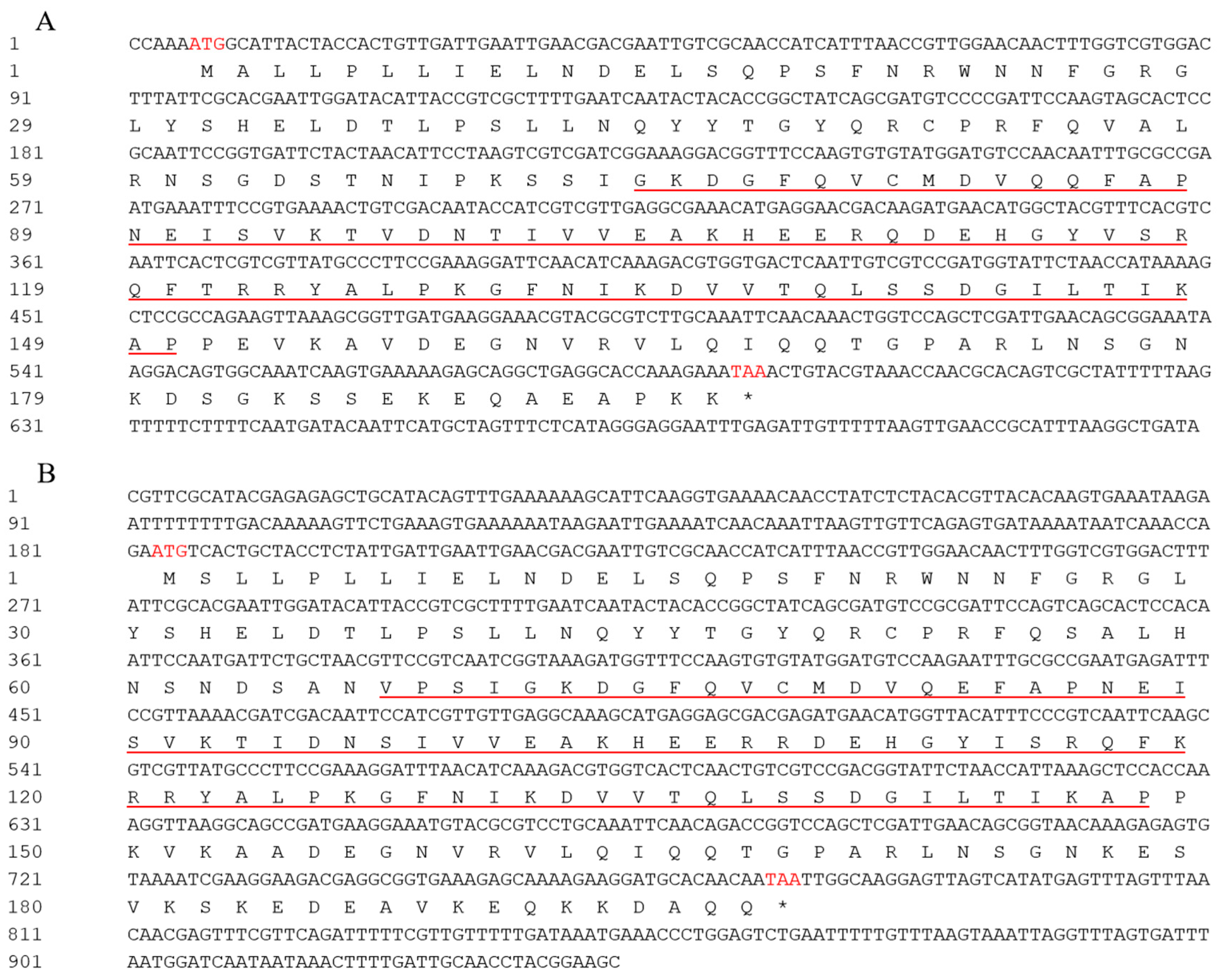
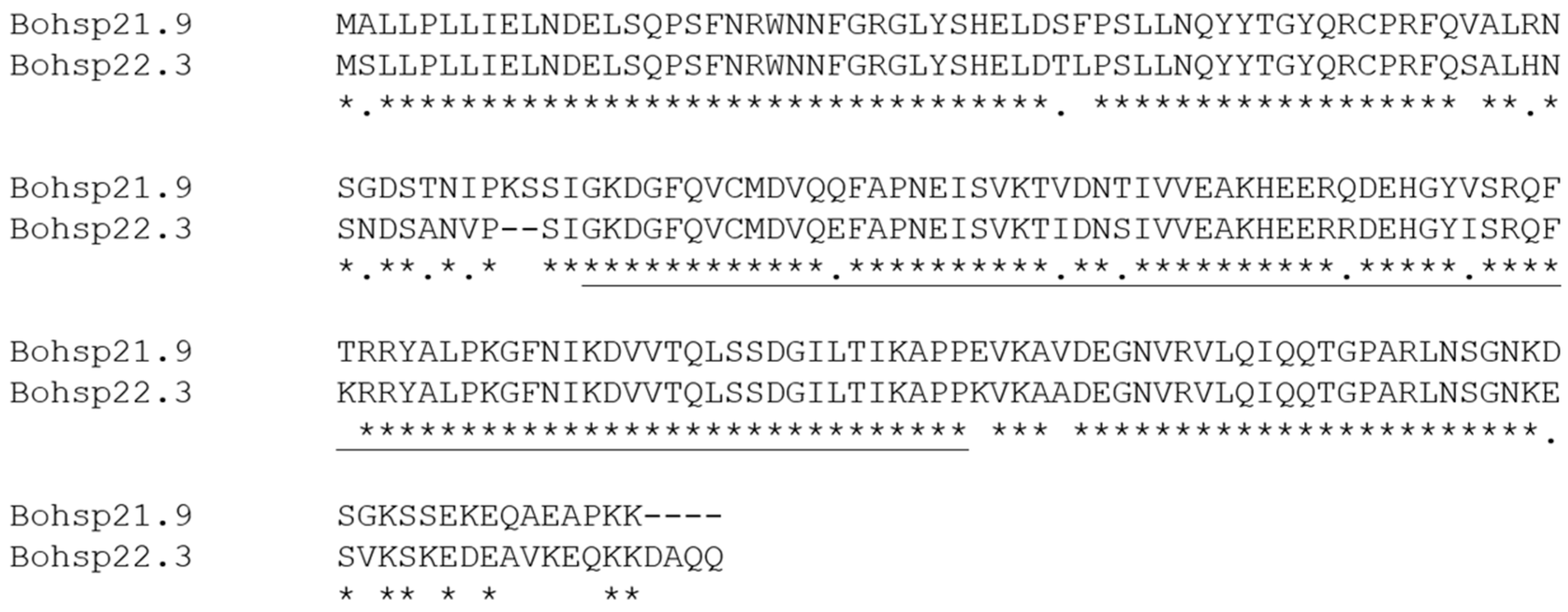
Based on previous B. odoriphaga transcriptome data, we identified and cloned BoHsp21.9 (GenBase accession number: C_AA121213.1) and BoHsp22.3 (GenBase accession number: C_AA121212.1) which might be involved in short term heat stress. BoHsp21.9 and BoHsp22.3 are 749 and 941 bp in length, with an open reading frame (ORF) of 588 and 594 bp that encodes 196 and 198 amino acids, respectively. The predicted isoelectric points are 6.84 and 6.91, and the calculated molecular masses of Bohsp21.9 and Bohsp22.3 proteins are 21.9 and 22.3 kDa, respectively. We named the genes based on their predicted molecular weights. Their deduced amino acid sequences contain the typical α-crystallin domain suggesting that they are small heat shock protein genes (Figure 2). And the amino acid sequence of BoHsp21.9 showed 86.15% identification with BoHsp22.3 (Figure 3).

3.2. Phylogenetic Analysis

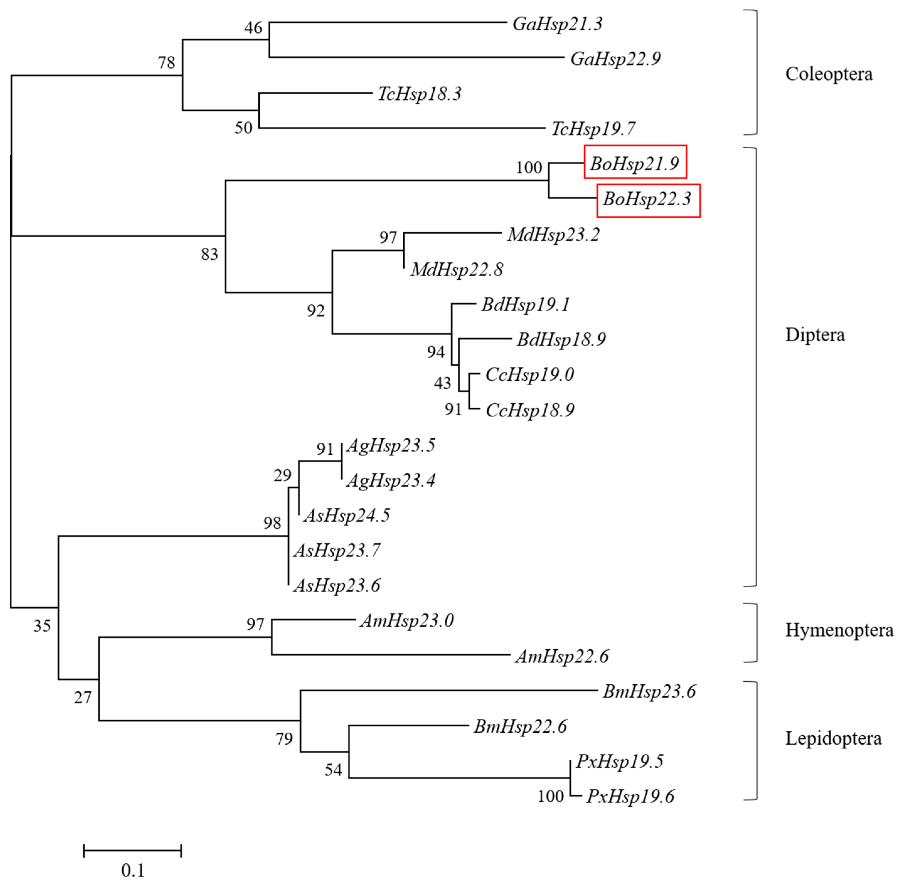
To study the evolutionary relationships of BoHsp21.9 and BoHsp22.3, twenty-one full-length sHsp amino acid sequences from Coleoptera, Hymenoptera, Diptera, and Lepidoptera were employed for phylogenetic analysis, which name and accession numbers are listed in the figure captions. As shown in Figure 4, BoHsp21.9 and BoHsp22.3 clustered together with other Diptera insects, and clustered with flies more nearly than midges.

3.3. Developmental Stage Expression Patterns of BoHsp21.9 and BoHsp22.3

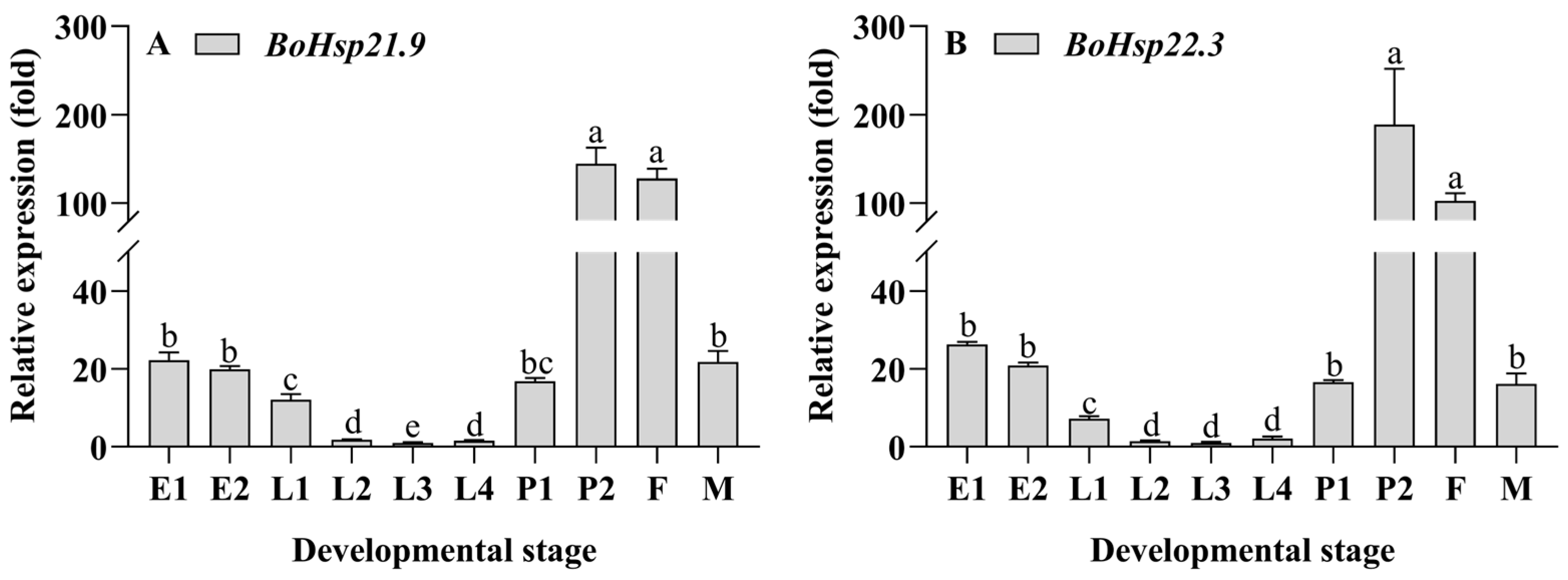
Developmental stage expression patterns of BoHsp21.9 and BoHsp22.3 were measured by qRT-PCR. As shown in Figure 5 and Table S2, the expression pattern of BoHsp21.9 and BoHsp22.3 showed a gradual decrease from E1 stage (eggs of 24 h after oviposition) to E2 stage (eggs of black eye). During the larval stage, the expression pattern firstly decreased from L1 stage (1st-instar larvae) to L3 stage (3rd-instar larvae), followed by an increase at L4 stage (4th-instar larvae). However, the expression pattern was opposite from egg and larvae stage, with a rapid increase from P1 stage (pupae with white eyes) to P2 stage (pupae with black eyes). In addition, the expression pattern of female was much higher than that of male.

3.4. Body Segment Expression Patterns of BoHsp21.9 and BoHsp22.3

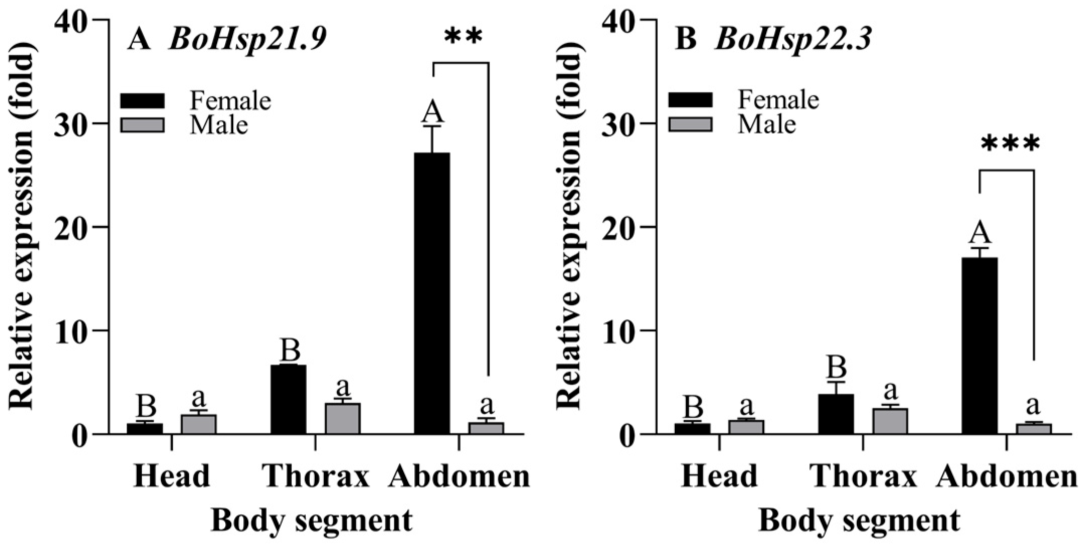
Body segment expression patterns of BoHsp21.9 and BoHsp22.3 were measured by qRT-PCR. As shown in Figure 6 and Table S3, the expressions of BoHsp21.9 and BoHsp22.3 were higher in the abdomen of female adults than in males, with 20-fold higher for BoHsp21.9 (F1,4 = 95.89, p = 0.001) and 22-fold higher for BoHsp22.3 (F1,4 = 317.081, p = 0.000), respectively. In addition, both genes were expressed in all parts of female and male adults, and the expression levels were lowest in the head of female adult and lowest in the abdomen of male adult.

3.5. Expression Patterns of BoHsp21.9 and BoHsp22.3 After Heat Stress

We used heat stress to further investigate the expression of BoHsp21.9 and BoHsp22.3. As shown in Figure 7, and Tables S4 and S5, BoHsp21.9 and BoHsp22.3 exhibited a significant upregulation in response to heat stress. The expression of BoHsp21.9 and BoHsp22.3 reached the maximum value at 30 °C, 32 °C, 34 °C for 1 h or 2 h. With the increase in temperature of heat stress, the maximum value of expression increased from 23-fold of 30 °C (F7,16 = 399.650, p = 0.000) to 257-fold of 32 °C (F7,16 = 303.093, p = 0.000) and 1211-fold of 34 °C (F7,16 = 307.010, p = 0.000) of BoHsp21.9 (Figure 7A–C); 26-fold of 30 °C (F7,16 = 537.170, p = 0.000) to 268-fold of 32 °C (F7,16 = 335.605, p = 0.000) and 890-fold of 34 °C (F7,16 = 1186.764, p = 0.000) of BoHsp22.3 (Figure 7F–H), respectively. At 30 °C, 32 °C, and 34 °C, the expression of BoHsp21.9 and BoHsp22.3 decreased with the increase in exposure time, and finally tended to be gentle, reaching 2-, 6-, 5-fold of BoHsp21.9 and 1-, 5-, 7-fold of BoHsp22.3 at normal temperature, respectively. However, the expression patterns of BoHsp21.9 and BoHsp22.3 under heat stress at 36 °C were different from those at 30 °C, 32 °C, and 34 °C. Specifically, the expression levels of BoHsp21.9 (F7,16 = 58.495, p = 0.000) and BoHsp22.3 (F7,16 = 59.574, p = 0.000) remained at a high and almost stable expression under 1–12 h exposure time (Figure 7D,I). In addition, when the heat stress temperature reached 38 °C, and the exposure time was 4 h, the larvae of B. odoriphaga died (Figure 7E,J).
The expression patterns of BoHsp21.9 and BoHsp22.3 during 1 h and 2 h recovery at 25 °C from 1 to 12 h under 30–36 °C heat stress were also examined (Figure 8, Tables S6 and S7). The results showed that the expression of both BoHsp21.9 and BoHsp22.3 decreased significantly after 1 h or 2 h of recovery at 25 °C from 30 °C, 32 °C, 34 °C of heat stress (Figure 8A–C,E–G). The longer the exposure time, the lower the expression after recovery at 25 °C, and finally almost the same as 25 °C. However, the expression of BoHsp21.9 and BoHsp22.3 were significantly higher after recovery at 25 °C from 36 °C (Figure 8D,H).

3.6. Functional Analysis of BoHsp21.9 and BoHsp22.3 by RNAi

qRT-PCR analysis showed that transcription levels of BoHsp21.9 and BoHsp22.3 of B. odoriphaga were reduced after dsRNA uptake at 12, 24, and 48 h compared with the dsGFP group (Figure 9A,B). After treatment with dsBoHsp21.9 and dsBoHsp22.3 at 48 h, BoHsp21.9 (F2,6 = 4.242, p = 0.071) and BoHsp22.3 (F2,6 = 4.749, p = 0.058) expression were reduced by 60% in larvae. Thus, at 48 h after the uptake of dsBoHsp21.9 and dsBoHsp22.3, we evaluated the survival of dsRNA-fed B. odoriphaga larvae at 38 °C. Compared with the dsGFP group, RNAi-mediated silencing of BoHsp21.9 obviously decreased survival rate by 5.8% (p = 0.0740) and 10.8% (p = 0.0261) when the larvae were exposed to 38 °C for 2 h and 3 h, respectively. RNAi-mediated silencing of BoHsp22.3 obviously decreased survival rate by 4.2% (p = 0.0241) and 10.0% (p = 0.0381) when the larvae were exposed to 38 °C for 2 h and 3 h, respectively. RNAi-mediated silencing of BoHsp21.9 and BoHsp22.3 obviously decreased survival rate by 9.2% (p = 0.0007) and 19.1% (p = 0.0003) when the larvae were exposed to 38 °C for 2 h and 3 h, respectively (Figure 9C).

4. Discussion

Small heat shock proteins (sHsps) exhibit chaperone activity and reflect the response mechanism of insects to environmental extreme stress [22], which have been studied in Drosophila melanogaster [34], Plutella xylostella [35], Mamestra brassicae [36], Ceratitis capitate [37], Sesamia nonagrioides [38], Liriomyza sativa [39], and Macrocentrus cingulum [40]. However, the study of sHsps in B. odoriphaga has not been conducted. In this study, BoHsp21.9 and BoHsp22.3 were identified by PCR, which contained 588 and 594 bp ORFs encoding 196 and 198 amino acids, respectively. The predicted BoHsps21.9 and BoHsp22.3 shared high amino acid sequence similarity and grouped in same clusters with flies when analyzed by the maximum likelihood method. The significant sequence homology of sHsp indicated their evolutionary conserved and functional response to heat protection [41]. Therefore, we inferred that the function of sHsp of B. odoriphaga is similar to that of flies.
sHsps may play an important role in regulating development stages [42] and maintaining the normal functioning of tissues [43]. In this study, BoHsp21.9 and BoHsp22.3 displayed similar developmental expression patterns, with the lowest expression in larvae among all developmental stages. The phenomenon of low sHsp expression in larvae was also found in Hsp20.5, Hsp20.6, and Hsp20.7 in locust [44] and Hsp19.7 and Hsp20.7 in Spodoptera litura [45]. In B. odoriphaga, the expression patterns of larvae developmental stages decreased from L1 to L3, followed by an increase in L4. As gregarious insects, small body size in stages L1 to L3 occupies a limited amount of space to reduce individual movement and energy consumption [46]. However, the increased expression in L4 may be due to the increasing demand for energy (i.e., fold) by maturing insects [47]. In addition, BoHsp21.9 and BoHsp22.3 remarkably up-regulated after the larval–pupal transformation, which is highly expressed in pupal stages. The phenomenon of up-regulation after the larval–pupal transformation can be found in Hsp20 in S. litura [45]. High expression of sHsps in pupal was also found in Hsp19.5 in P. xylostella [35] and Hsp19.5, Hsp20.8, and Hsp21.7 in L. sativa [39]. High expression of BoHsp21.9 and BoHsp22.3 in pupal stages of B. odoriphaga suggests that they may be involved in metamorphosis. Insect metamorphosis is a process of degradation and reconstruction of tissues and organs, which may greatly induce the expression of heat shock protein genes [39,48]. On the other hand, it has been reported that the more abundant the expression level of ApsHsp20.8 tissue, the more sensitive it is to stress [49]. The low expression level of larvae indicated that compared with other developmental states, it was the most difficult to change its survival state in the face of high temperature stress. Therefore, as long as it is lethal to larvae, it is also lethal to other development states. This is consistent with the biological experiment results of Shi et al. [9] which show that more time is needed to kill the larvae. The expression levels in female adults were found to be higher than those in male adults, indicating that female adults exhibit greater sensitivity to high temperature stress compared to male adults. Therefore, when exposed to high temperature, male adults are more likely to show heat tolerance than female adults. This is consistent with the biological experiment results of Cheng et al. [6], whereby the survival rate of male adults is higher than female adults when faced high temperatures.
The expression of BoHsp21.9 and BoHsp22.3 was different between male and female adults; therefore, we dissected and verified the expression in the body segments of male and female adults. There were no differences between male and female adults in head and thorax, but there were significantly differences in the abdomen. We speculate that the difference in abdominal expression between male and female adults is caused by the difference in reproductive system. It has been verified that sHsps were highly expressed in the ovary of Tribolium castaneum; moreover, sHsps expressed in the ovary without stress played a crucial role in maintaining normal cell development [48].
All species respond to heat shock responses by synthesizing Hsps, which is a conservative defense mechanism in acute extreme environments [50,51]. Typically, Hsps are rapidly up-regulated at the onset of stress and downregulated when favorable conditions return [52]. In addition, Hsp expression patterns may differ depending on the specific type of high temperature stress exposure [53]. In our study, we exposed the larvae to 30–38 °C for 1–12 h. The expression of BoHsp21.9 and BoHsp22.3 reached the highest level at 30 °C, 32 °C, and 34 °C when the larvae were exposed for 1 h or 2 h, and decreased with the increase in exposure duration. The expression of BoHsp21.9 and BoHsp22.3 was stable and high in different exposure times under 36 °C of heat shock. The larvae died when exposed at 38 °C for 4 h. In addition, the expression of BoHsp21.9 and BoHsp22.3 increased with the rise in exposure temperature. Our findings indicate that sHsp expression can be induced to varying levels depending on the degree of heat shock exposure. In sub-lethal temperature tolerance, sHsps may prevent stress-induced cytoskeletal destruction by interacting with microfilaments or stabilizing actin polymers [54,55,56]. When stress intensity exceeds the regulation capacity of the organism, denaturation of proteins can occur in the cells [57]. The expression of BoHsp21.9 and BoHsp22.3 recovered at 25 °C after heat shock shows that BoHsp21.9 and BoHsp22.3 could be accumulated. With the increase in heat shock temperature, the greater the degree of accumulation obtained, and correspondingly, more time is needed to recover to the normal level. Therefore, when the lethal temperature could not be reached, the larvae of B. odoriphaga could survive in large numbers and may have the ability to withstand high temperature again.
RNA interference (RNAi)-mediated gene silencing is a powerful tool for exploring gene function [58]. RNAi has been effectively employed to investigate gene function in Diptera [59,60,61]. By using oral delivery RNAi technology, we found that silencing of BoHsp21.9 and BoHsp22.3 significantly decreased the expression of BoHsp21.9 and BoHsp22.3 by 60% at 48 h after dsRNA feeding compared with the dsGFP group. We subsequently applied gene silencing followed by a survival assay to evaluate the role of BoHsp21.9 and BoHsp22.3 in temperature tolerance. The results showed the decreased survival rate of B. odoriphaga when exposed to 38 °C for 2 h and 3 h. Compared with the dsGFP group, the reduced expression level after interference made it easier for B. odoriphaga to change survival state under high temperature stress. Therefore, we speculate that BoHsp21.9 and BoHsp22.3 may be important genes involved in thermotolerance in B. odoriphaga.

5. Conclusions

In conclusion, on the basis of successful cloning of the full-length of Hsp21.9 and Hsp22.3 of B. odoriphaga, we analyzed the deduced protein sequence characteristic motifs, which indicated highly conserved structures as compared with Diptera insects of fly. Developmental stages and body segments expression patterns suggested that BoHsp21.9 and BoHsp22.3 might be involved in regulating development and maintaining the normal function of B. odoriphaga. Moreover, the expression of BoHsp21.9 and BoHsp22.3 can be up-regulated by heat stress, and oral delivery-mediated RNAi of BoHsp21.9 and BoHsp22.3 can effectively suppress the gene expression, consequently resulting in the low survival rate of B. odoriphaga under heat stress. These results suggest that BoHsp21.9 and BoHsp22.3 play an important role in thermotolerance. The results of this study have preliminarily elucidated the close relationship between small heat shock proteins and the temperature adaptability of B. odoriphaga, providing a theoretical basis for further exploring the temperature tolerance mechanism of this insect.

Supplementary Materials

The following supporting information can be downloaded at https://www.mdpi.com/article/10.3390/insects16111107/s1.

Author Contributions

Conceptualization, J.C., Q.W. and J.S.; methodology, J.C., Q.W. and J.S.; software, H.Z.; validation, J.C., S.F. and W.C.; resources, Q.W. and J.S.; writing—original draft preparation, J.C.; writing—review and editing, all authors; visualization, all authors; supervision, Q.W. and J.S.; project administration, Q.W. and J.S. All authors have read and agreed to the published version of the manuscript.

Funding

This research was funded by the National Natural Science Foundation of China (32202309), the China Agriculture Research System of MOF and MARA (CARS-24-C-02), HAAFS Agriculture Science and Technology Innovation Project (2022KJCXZX-ZBS-6).

Data Availability Statement

The original contributions presented in this study are included in the article/Supplementary Material. Further inquiries can be directed to the corresponding authors.

Acknowledgments

The authors are grateful to the Institute of Vegetables and Flowers, Chinese Academy of Agricultural Sciences for providing the GFP material.

Conflicts of Interest

The authors declare no conflicts of interest.

References

  1. Yang, J.K.; Zhang, X.M. Notes on the fragrant onion gnats with descriptions of two new species of Bradysia (Sciaridae: Diptera). Acta Agric. Univ. Pekin. 1985, 11, 153–156. [Google Scholar]
  2. Xue, M.; Pang, Y.H.; Wang, C.X.; Li, Q. Biological effect of liliaceous host plants on Bradysia odoriphaga Yang et Zhang (Diptera: Sciaridae). Acta Entomol. Sin. 2005, 48, 914–921. [Google Scholar]
  3. Zhu, G.D.; Xue, M.; Luo, Y.; Ji, G.X.; Liu, F.; Zhao, H.P.; Sun, X. Effects of short-term heat shock and physiological responses to heat stress in two Bradysia adults, Bradysia odoriphaga and Bradysia difformis. Sci. Rep. 2017, 7, 13381. [Google Scholar] [CrossRef]
  4. Zhang, P.; Liu, F.; Mu, W.; Wang, Q.H.; Li, H. Comparison of Bradysia odoriphaga Yang and Zhang reared on artificial diet and different host plants based on an age-stage, two-sex life table. Phytoparasitica 2015, 43, 107–120. [Google Scholar] [CrossRef]
  5. Li, W.X.; Yang, Y.T.; Xie, W.; Wu, Q.J.; Xu, B.Y.; Wang, S.L.; Zhu, X.; Wang, S.J.; Zhang, Y.J. Effects of Temperature on the Age-Stage, Two-Sex Life Table of Bradysia odoriphaga (Diptera: Sciaridae). J. Econ. Entomol. 2015, 108, 126–134. [Google Scholar] [CrossRef]
  6. Cheng, J.X.; Su, Q.; Jiao, X.G.; Shi, C.H.; Yang, Y.T.; Han, H.L.; Xie, W.; Guo, Z.J.; Wu, Q.J.; Xu, B.Y.; et al. Effects of Heat Shock on the Bradysia odoriphaga (Diptera: Sciaridae). J. Econ. Entomol. 2017, 110, 1630–1638. [Google Scholar] [CrossRef] [PubMed]
  7. Mei, Z.X. Studies on Biological Characteristics and Cold Hardness of Bradysia odoriphaga. Master’s Thesis, Northwest Sci-Tech University of Agriculture and Forestry, Yangling, China, 2003. [Google Scholar]
  8. Shi, C.H.; Yang, Y.T.; Han, H.L.; Cheng, J.X.; Wu, Q.J.; Xu, B.Y.; Zhang, Y.J. Population dynamics and summer and winter habitats of Bradysia odoriphaga in the Beijing area. Chin. J. Appl. Entomol. 2016, 53, 1174–1183. [Google Scholar]
  9. Shi, C.H.; Hu, J.R.; Wei, Q.W.; Yang, Y.T.; Cheng, J.X.; Han, H.L.; Wu, Q.J.; Wang, S.L.; Xu, B.Y.; Su, Q.; et al. Control of Bradysia odoriphaga (Diptera: Sciaridae) by soil solarization. Crop Prot. 2018, 114, 76–82. [Google Scholar] [CrossRef]
  10. Cheng, J.X.; Su, Q.; Xia, J.X.; Yang, Z.Z.; Shi, C.H.; Wang, S.L.; Wu, Q.J.; Li, C.R.; Zhang, Y.J. Comparative transcriptome analysis of differentially expressed genes in Bradysia odoriphaga Yang et Zhang (Diptera: Sciaridae) at different acute stress temperatures. Genomics 2020, 112, 3739–3750. [Google Scholar] [CrossRef] [PubMed]
  11. Kim, K.K.; Kim, R.; Kim, S.H. Crystal structure of a small heat-shock protein. Nature 1998, 394, 595–599. [Google Scholar] [CrossRef] [PubMed]
  12. Waters, E.R.; Rioflorido, I. Evolutionary analysis of the small heat shock proteins in five complete algal genomes. J. Mol. Evol. 2007, 65, 162–174. [Google Scholar] [CrossRef]
  13. Waters, E.R.; Aevermann, B.D.; Sanders-Reed, Z. Comparative analysis of the small heat shock proteins in three angiosperm genomes identifies new subfamilies and reveals diverse evolutionary patterns. Cell Stress Chaperon. 2008, 13, 127–142. [Google Scholar]
  14. Aevermann, B.D.; Waters, E.R. A comparative genomic analysis of the small heat shock proteins in Caenorhabditis elegans and briggsae. Genetica 2008, 133, 307–319. [Google Scholar]
  15. Singh, M.K.; Sharma, B.; Tiwari, P.K. The small heat shock protein Hsp27: Present understanding and future prospects. J. Therm. Biol. 2017, 69, 149–154. [Google Scholar] [CrossRef]
  16. de Jong, W.W.; Caspers, G.J.; Leunissen, J.A.M. Genealogy of the α-crystallin-small heat-shock protein superfamily. Int. J. Biol. Macromol. 1998, 22, 151–162. [Google Scholar] [CrossRef] [PubMed]
  17. Franck, E.; Madsen, O.; Rheede, T.V.; Ricard, G.; Huynen, M.A.; de Jong, W.W. Evolutionary Diversity of Vertebrate Small Heat Shock Proteins. J. Mol. Evol. 2004, 59, 792–805. [Google Scholar] [CrossRef] [PubMed]
  18. Caspers, G.J.; Leunissen, J.A.M.; de Jong, W.W. The expanding small heat-shock protein family, and structure predictions of the conserved “α-crystallin domain”. J. Mol. Evol. 1995, 40, 238–248. [Google Scholar] [PubMed]
  19. Fu, X.M.; Jiao, W.W.; Chang, Z.Y. Phylogenetic and Biochemical Studies Reveal a Potential Evolutionary Origin of Small Heat Shock Proteins of Animals from Bacterial Class A. J. Mol. Evol. 2006, 62, 257–266. [Google Scholar] [CrossRef]
  20. Stromer, T.; Fischer, E.; Richter, K.; Haslbeck, M.; Buchner, J. Analysis of the Regulation of the Molecular Chaperone Hsp26 by Temperature-induced Dissociation: The N-terminal domain is important for oligomer assembly and the binding of unfolding proteins. J. Biol. Chem. 2004, 279, 11222–11228. [Google Scholar]
  21. Pérez-Morales, D.; Ostoa-Saloma, P.; Espinoza, B. Trypanosoma cruzi SHSP16: Characterization of an a-crystallin small heat shock protein. Exp. Parasitol. 2009, 123, 182–189. [Google Scholar]
  22. Li, Z.W.; Li, X.; Yu, Q.Y.; Xiang, Z.H.; Kishino, H.; Zhang, Z. The small heat shock protein (sHSP) genes in the silkworm, Bombyx mori, and comparative analysis with other insect sHSP genes. BMC Evol. Biol. 2009, 9, 215. [Google Scholar] [CrossRef]
  23. Reineke, A. Identification and expression of a small heat shock protein in two lines of the endoparasitic wasp Venturia canescens. Comp. Biochem. Phys. A 2005, 141, 60–69. [Google Scholar] [CrossRef] [PubMed]
  24. Liu, Z.H.; Xi, D.M.; Kang, M.J.; Guo, X.Q.; Xu, B.H. Molecular cloning and characterization of Hsp27.6: The first reported small heat shock protein from Apis cerana cerana. Cell Stress Chaperon. 2012, 17, 539–551. [Google Scholar] [CrossRef] [PubMed]
  25. Vazquez, J.; Pauli, D.; Tissières, A. Transcriptional regulation in Drosophila during heat shock: A nuclear run-on analysis. Chromosoma 1993, 102, 233–248. [Google Scholar] [CrossRef]
  26. Zhang, Y.Y.; Liu, Y.L.; Guo, X.L.; Li, Y.L.; Gao, H.R.; Guo, X.Q.; Xu, B.H. sHsp22.6, an intronless small heat shock protein gene, is involved in stress defence and development in Apis cerana cerana. Insect Biochem. Mol. Biol. 2014, 53, 1–12. [Google Scholar] [CrossRef]
  27. Zhao, L.M.; Becnel, J.J.; Clark, G.G.; Linthicum, K.J. Expression of AeaHsp26 and AeaHsp83 in Aedes aegypti (Diptera: Culicidae) larvae and Pupae in Response to Heat Shock Stress. J. Med. Entomol. 2010, 47, 367–375. [Google Scholar]
  28. Sakano, D.; Lin, B.; Xia, Q.Y.; Yamamoto, K.; Aso, Y. Genes encoding small heat shock proteins of the silkworm, Bombyx mori. Biosci. Biotechnol. Biochem. 2006, 70, 2443–2450. [Google Scholar] [CrossRef]
  29. Sheng, Q.; Xia, J.Y.; Nie, Z.M.; Zhang, Y.Z. Cloning, Expression, and Cell Localization of a Novel Small Heat Shock Protein Gene: BmHSP25.4. Appl. Biochem. Biotechnol. 2010, 162, 1297–1305. [Google Scholar] [CrossRef]
  30. Wang, H.; Fang, Y.; Bao, Z.Z.; Jin, X.; Zhu, W.J.; Wang, L.P.; Liu, T.; Ji, H.P.; Wang, H.Y.; Xu, S.Q.; et al. Identification of a Bombyx mori gene encoding small heat shock protein BmHsp27.4 expressed in response to high-temperature stress. Gene 2014, 538, 56–62. [Google Scholar]
  31. Shi, C.H.; Yang, F.S.; Zhu, X.; Du, E.; Yang, Y.T.; Wang, S.L.; Wu, Q.J.; Zhang, Y.J. Evalution of Housekeeing Genes for Quantitative Real-Time PCR Analysis of Bradysia odoriphaga (Diptera: Sciaridae). Int. J. Mol. Sci. 2016, 17, 1034. [Google Scholar] [CrossRef]
  32. Livak, K.J.; Schmittgen, T.D. Analysis of Relative Gene Expression Data Using Real-Time Quantitative PCR and the 2−ΔΔCT Method. Methods 2001, 25, 402–408. [Google Scholar] [CrossRef]
  33. Chen, C.Y.; Shan, T.S.; Liu, Y.; Shi, X.Y.; Gao, X.W. Identification of a novel cytochrome P450 CYP3356A1 linked with insecticide detoxification in Bradysia odoriphaga. Pest Manag. Sci. 2019, 75, 1006–1013. [Google Scholar] [CrossRef]
  34. Michaud, S.; Morrow, G.; Marchand, J.; Tanguay, R.M. Drosophila small heat shock proteins: Cell and organelle-specific chaperones? Prog. Mol. Subcell. Biol. 2002, 28, 79–101. [Google Scholar]
  35. Sonoda, S.; Ashfaq, M.; Tsumuki, H. Cloning and nucleotide sequencing of three heat shock protein genes (hsp90, hsc70, and hsp19.5) from the diamondback moth, Plutella xylostella (L.) and their expression in relation to developmental stage and temperature. Arch. Insect Biochem. Physiol. 2006, 62, 80–90. [Google Scholar] [CrossRef]
  36. Sonoda, S.; Ashfaq, M.; Tsumuki, H. A comparison of heat shock protein genes from cultured cells of the Cabbage armyworm, Mamestra brassicae, in response to heavy metals. Arch. Insect Biochem. Physiol. 2007, 65, 210–222. [Google Scholar] [CrossRef]
  37. Kokolakis, G.; Tatari, M.; Zacharopoulou, A.; Mintzas, A.C. The hsp27 gene of the Mediterranean fruit fly, Ceratitis capitata: Structural characterization, regulation and developmental expression. Insect Mol. Biol. 2008, 17, 699–710. [Google Scholar] [CrossRef] [PubMed]
  38. Gkouvitsas, T.; Kontogiannatos, D.; Kourti, A. Differential expression of two small Hsps during diapause in the corn stalk borer Sesamia nonagrioides (Lef.). J. Insect Physiol. 2008, 54, 1503–1510. [Google Scholar] [CrossRef]
  39. Huang, L.H.; Wang, C.Z.; Kang, L. Cloning and expression of five heat shock protein genes in relation to cold hardening and development in the leafminer, Liriomyza sativa. J. Insect Physiol. 2009, 55, 279–285. [Google Scholar] [CrossRef] [PubMed]
  40. Xu, P.J.; Xiao, J.H.; Liu, L.; Li, T.; Huang, D.W. Molecular cloning and characterization of four heat shock protein genes from Macrocentrus cingulum (Hymenoptera: Braconidae). Mol. Biol. Rep. 2010, 37, 2265–2272. [Google Scholar] [CrossRef] [PubMed]
  41. Singh, M.K.; Tiwari, P.K. Cloning & sequence identification of Hsp27 gene and expression analysis of the protein on thermal stress in Lucilia cuprina. Insect Sci. 2016, 23, 555–568. [Google Scholar]
  42. Michaud, S.; Marin, R.; Tanguay, R.M. Regulation of heat shock gene induction and expression during Drosophila development. Cell Mol. Life Sci. 1997, 53, 104–113. [Google Scholar] [CrossRef]
  43. Gu, J.; Huang, L.X.; Shen, Y.; Huang, L.H.; Feng, Q.L. Hsp70 and small Hsps are the major heat shock protein members involved in midgut metamorphosis in the common cutworm, Spodoptera litura. Insect Mol. Bio. 2012, 5, 535–543. [Google Scholar] [CrossRef]
  44. Wang, H.S.; Wang, X.H.; Zhou, C.S.; Huang, L.H.; Zhang, S.F.; Guo, W.; Kang, L. cDNA cloning of heat shock proteins and their expression in the two phases of the migratory locust. Insect Mol. Biol. 2007, 16, 207–219. [Google Scholar] [CrossRef]
  45. Shen, Y.; Gu, J.; Huang, L.H.; Zheng, S.C.; Liu, L.; Xu, W.H.; Feng, Q.L.; Kang, L. Cloning and expression analysis of six small heat shock protein genes in the common cutworm, Spodoptera litura. J. Insect Physiol. 2011, 57, 908–914. [Google Scholar] [CrossRef]
  46. Collett, M.; Despland, E.; Simpson, S.J.; Krakauer, D. Spatial scales of desert locust gregarization. Proc. Natl. Acad. Sci. USA 1998, 95, 13052–13055. [Google Scholar]
  47. Pener, M.P. Locust Phase Polymorphism and its Endocrine Relations. Adv. Insect Physiol. 1991, 23, 1–79. [Google Scholar]
  48. Xie, J.; Hu, X.X.; Zhai, M.F.; Yu, X.J.; Song, X.W.; Gao, S.S.; Wu, W.; Li, B. Characterization and functional analysis of hsp18.3 gene in the red flour beetle, Tribolium castaneum. Insect Sci. 2019, 26, 263–273. [Google Scholar] [CrossRef]
  49. Zhang, C.F.; Dai, L.S.; Wang, L.; Qian, C.; Wei, G.Q.; Li, J.; Zhu, B.J.; Liu, C.L. Eicosanoids mediate sHSP 20.8 gene response to biotic stress in larvae of the Chinese oak silkworm Antheraea pernyi. Gene 2015, 562, 32–39. [Google Scholar] [CrossRef] [PubMed]
  50. Lü, Z.C.; Wan, F.H. Using double-stranded RNA to explore the role of heat shock protein genes in heat tolerance in Bemisia tabaci (Gennadius). J. Exp. Biol. 2011, 214, 764–769. [Google Scholar] [CrossRef] [PubMed]
  51. Lindquist, S. The Heat-Shock Response. Ann. Rev. Biochem. 1986, 55, 1151–1191. [Google Scholar] [CrossRef]
  52. Feder, J.H.; Rossi, J.M.; Solomon, J.M.; Solomon, N.M.; Lindquist, S. The consequences of expressing hsp70 in Drosophila cells at normal temperatures. Gene Dev. 1992, 6, 1402–1413. [Google Scholar] [CrossRef]
  53. Krebs, R.A. A comparison of Hsp70 expression and thermotolerance in adults and larvae of three Drosophila species. Cell Stress Chaperon. 1999, 4, 243–249. [Google Scholar] [CrossRef]
  54. Mahadav, A.; Kontsedalov, S.; Czosnek, H.; Ghanim, M. Thermotolerance and gene expression following heat stress in the whitefly Bemisia tabaci B and Q biotypes. Insect Biochem. Mol. Biol. 2009, 39, 668–676. [Google Scholar] [CrossRef]
  55. Liang, P.; MacRae, T.H. Molecular chaperones and the cytoskeleton. J. Cell Sci. 1997, 110, 1431–1440. [Google Scholar] [CrossRef]
  56. Wang, K.Y.; Spector, A. α-crystallin stabilizes actin filaments and prevents cytochalasin-induced depolymerization in a phosphorylation-dependent manner. Eur. J. Biochem. 1996, 242, 56–66. [Google Scholar]
  57. Somero, G.N. Proteins and Temperature. Annu. Rev. Physiol. 1995, 57, 43–68. [Google Scholar] [CrossRef]
  58. Colinet, H.; Lee, S.F.; Hoffmann, A.A. Knocking down expression of Hsp22 and Hsp23 by RNA interference affects recovery from chill coma in Drosophila melanogaster. J. Exp. Biol. 2010, 213, 4146–4150. [Google Scholar] [CrossRef]
  59. Chapman, T.; Bangham, J.; Vinti, G.; Seifried, B.; Lung, O.; Wolfner, M.F.; Smith, H.K.; Partridge, L. The sex peptide of Drosophila melanogaster: Female post-mating responses analyzed by using RNA interference. Proc. Natl. Acad. Sci. USA 2003, 100, 9923–9928. [Google Scholar] [PubMed]
  60. Huvenne, H.; Smagghe, G. Mechanisms of dsRNA uptake in insects and potential of RNAi for pest control: A review. J. Insect Physiol. 2010, 56, 227–235. [Google Scholar] [CrossRef]
  61. Olson, K.E.; Blair, C.D. Arbovirus-mosquito interactions: RNAi pathway. Curr. Opin. Virol. 2015, 15, 119–126. [Google Scholar] [CrossRef] [PubMed]
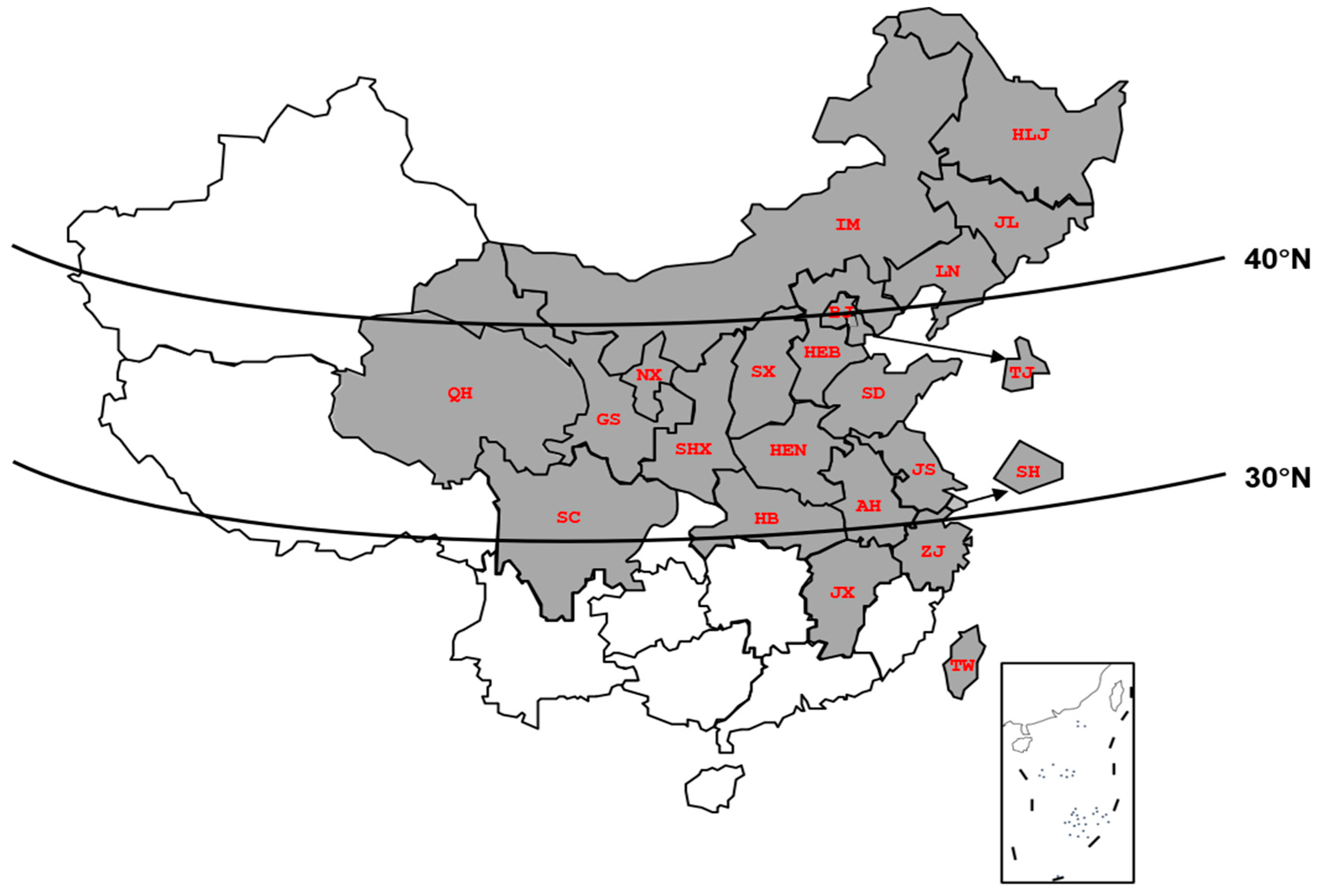
Figure 1. The occurrence of B. odoriphaga in China. The gray part indicates the presence of B. odoriphaga.
Insects 16 01107 g001
Figure 2. Complete cDNA sequences of B. odoriphaga Hsp21.9 and Hsp22.3. (A) BoHsp21.9. (B) BoHsp22.3. The α-crystallin domain is underlined. Red font of “ATG” denote initiation codon. The asterisk and red font of “TAA” denote termination codon.
Insects 16 01107 g002
Figure 3. Amino acid sequence comparison of BoHsp21.9 and BoHsp22.3. The asterisks and single dots denote fully and weakly conserved residues, respectively. The α-crystallin domain is underlined.
Insects 16 01107 g003
Figure 4. Phylogenetic analysis of BoHsp21.9 and BoHsp22.3. Ten insect species full names and gene accession numbers are designated by the following abbreviations: TcHsp19.7 (Tribolium castaneum, XP_973344.1), TcHsp18.3 (Tribolium castaneum, XP_974367.1), GaHsp22.9 (Gastrophysa atrocyanea, BAD91165.1), GaHsp21.3 (Gastrophysa atrocyanea, BAD91164.1), AmHsp23.0 (Apis mellifera, XP_001120194.1), AmHsp22.6 (Apis mellifera, XP_001119884.1), BmHsp23.6 (Bombyx mori, BAD74198.1), BmHsp22.6 (Bombyx mori, ACM24354.1), PxHsp19.5 (Plutella xylostella, BAE48744.1), PxHsp19.6 (Plutella xylostella, AHA36865.1), AgHsp23.5 (Anopheles gambiae, XP_315549.4), AgHsp23.4 (Anopheles gambiae, XP_315550.4), AsHsp24.5 (Anopheles sinensis, KFB40373.1), AsHsp23.7 (Anopheles sinensis, KFB40371.1), AsHsp23.6 (Anopheles sinensis, KFB40372.1), MdHsp22.8 (Musca domestica, XP_005190092.1), MdHsp23.2 (Musca domestica, XP_005190102.1), BdHsp18.9 (Bactrocera dorsalis, XP_011198114.1), BdHsp19.1 (Bactrocera dorsalis, XP_011198115.1), CcHsp18.9 (Ceratitis capitate, ACG58884.1), and CcHsp19.0 (Ceratitis capitate, XP_004523808.1).
Insects 16 01107 g004
Figure 5. Expression patterns of BoHsp21.9 and BoHsp22.3 in different developmental stages. E1: 200 eggs of 24 h after oviposition. E2: 200 eggs of black eyes. L1–L4: 1st–4th instar of 20 larvae. P1: 20 pupae of white eyes. P2: 20 pupae of black eyes. F/M: 20 newly emerged female and male adults. Data are represented by means ± standard errors (Table S2). Different letters represent statistically significant differences (p < 0.05, Tukey HSD in one-way ANOVA).
Insects 16 01107 g005
Figure 6. Expression patterns of BoHsp21.9 and BoHsp22.3 in different body segments. Head, thorax, abdomen dissected from 200 newly emerged female and male adults. Data are represented by means ± standard errors (Table S3). Different uppercase/lowercase letters represent statistically significant differences in female/male (p < 0.05, Tukey HSD in one-way ANOVA). “*” represents significant differences; ** p < 0.01, *** p < 0.001.
Insects 16 01107 g006
Figure 7. Expression patterns of BoHsp21.9 and BoHsp22.3 after heat stress. (AE) represent BoHsp21.9 at heat stress temperature of 30 °C, 32 °C, 34 °C, 36 °C, 38 °C. (FJ) represent BoHsp22.3 at heat stress temperature of 30 °C, 32 °C, 34 °C, 36 °C, 38 °C. CK represents 25 °C. Data are represented by means ± standard errors (Tables S4 and S5). Different letters represent statistically significant differences (p < 0.05, Tukey HSD in one-way ANOVA).
Insects 16 01107 g007
Figure 8. Heatmap of BoHsp21.9 and BoHsp22.3 expression patterns after recovery of 1 h and 2 h from heat stress. The color scale at the right ranges from the lowest (yellow) to the highest (red) relative expression fold (transformed with log10). CK represents 25 °C, and 0 h, 1 h, 2 h represent recovery time. Untransformed data are shown in Tables S6 and S7.
Insects 16 01107 g008
Figure 9. RNAi of BoHsp21.9 and BoHsp22.3. (A) Effect of RNAi treatment on the transcript level of BoHsp21.9 for different time points. (B) Effect of RNAi treatment on the transcript level of BoHsp22.3 for different time points. The control group was fed with dsGFP. The transcript levels of BoHsp21.9 and BoHsp22.3 were examined using qRT-PCR; RPS15 and RPL28 were selected as reference genes. Data are represented by means ± standard errors (Table S8). Different letters represent statistically significant differences (p < 0.05, Tukey HSD in one-way ANOVA). (C) Survival rate of B. odoriphaga under 38 °C for 2 h and 3 h after 48 h RNAi. Total sample size notations: 120 4th-instar larvae (groups of 30 larvae with four replicates). Data are represented by means. Different letters represent statistically significant differences (p < 0.05, log-rank test).
Insects 16 01107 g009
Table 1. Specific primers used in this study.
NamePrimer UsedPrimer Sequence (5′-3′)Efficiency (%)R2
GSP1-Hsp21.95′-RACECCTTCATCAACCGCTTTTACTTCTGGCG
GSP2-Hsp21.95′-RACETTTGGAAGGGCATAACGGCGAGTGA
10 × Universal Primer A Mix (UPM)RACETAATACGACTCACTATAGGGCAAGCAGTGGTATCAACGCAGAGT
10 × Universal Primer short (UPS)RACECTAATACGACTCACTATAGGGC
Hsp21.9-FPCRCCAAAATGGCATTACTACCAC
Hsp21.9-RPCRTATCAGCCTTAAATGCGGTTC
Hsp22.3-FPCRCGTTCGCATACGAGAGAGC
Hsp22.3-RPCRGCTTCCGTAGGTTGCAATC
Hsp21.9-FPqRT-PCRTCGTCCGATGGCATTCTAACC93.600.995
Hsp21.9-RPqRT-PCRTTCCGCTGTTCAATCGAGCT
Hsp22.3-FPqRT-PCRGTCGATCGGAAAGGACGGTT105.130.990
Hsp22.3-RPqRT-PCRTCTTGGCGTTCCTCATGCTT
RPS15-FPqRT-PCRATCGTGGCGTCGATTTGGAT101.03 *0.997 *
RPS15-RPqRT-PCRCTCATTTGGTGGGGCTTCCT
RPL28-FPqRT-PCRCGTGCCCGACATTTTCATCA105.18 *1.000 *
RPL28-RPqRT-PCRGACCAAGCCACTGTAACGGA
Hsp21.9-RNAi FPRNAiTAATACGACTCACTATAGGGTTGGAACAACTTTGGTCGTG
Hsp21.9-RNAi RPRNAiTAATACGACTCACTATAGGGGAAGGGCATAACGACGAGTG
Hsp22.3-RNAi FPRNAiTAATACGACTCACTATAGGGAATTCAAGCGTCGTTATGCC
Hsp22.3-RNAi RPRNAiTAATACGACTCACTATAGGGTTCTTTTGCTCTTTCACCGC
dsGFP-FPRNAiTAATACGACTCACTATAGGCAGTGCTTCAGCCGCTAC
dsGFP-RPRNAiTAATACGACTCACTATAGGGTTCACCTTGA
* represents data obtained from Shi et al. [31].
Disclaimer/Publisher’s Note: The statements, opinions and data contained in all publications are solely those of the individual author(s) and contributor(s) and not of MDPI and/or the editor(s). MDPI and/or the editor(s) disclaim responsibility for any injury to people or property resulting from any ideas, methods, instructions or products referred to in the content.