Article

Thought to Be Extinct, but Still Alive Today: The Miocene Genus Primascena Klimaszewsi, 1997 (Hemiptera: Psyllidae) in the Light of Two Extant Species from Brazil

by
Daniel Burckhardt
1,*,† and
Dalva L. Queiroz
2
1
Naturhistorisches Museum, Augustinergasse 2, 4052 Basel, Switzerland
2
Embrapa Florestas, Empresa Brasileira de Pesquisa Agropecuária, Km 111, Bairro Guaraituba, Colombo 83411-000, PR, Brazil
*
Author to whom correspondence should be addressed.
LSID: urn:lsid:zoobank.org:pub:96BB823E-9465-4734-83C9-5CFB221ADB15.
Insects 2024, 15(6), 382; https://doi.org/10.3390/insects15060382
Submission received: 26 April 2024 / Revised: 21 May 2024 / Accepted: 21 May 2024 / Published: 23 May 2024
(This article belongs to the Special Issue Phylogeny and Morphological Evolution of Hemiptera)

Abstract

:

Simple Summary

The psyllid genus Primascena was erected for a fossil species from Dominican amber, P. subita. As the single known specimen of this species is a partly damaged male, the description was quite incomplete. Recently, two undescribed species were discovered in Brazil (Mato Grosso do Sul) that are ascribed to this genus. The extant species allow the description of the previously unknown female and immature, and to complete information on the morphology of the male. A cladistic morphological analysis confirms the position of the genus in the subfamily Aphalaroidinae and supports the monophyly of the fossil and the two extant species. The extant Primascena species develop on plants of the Polygonaceae: Eriogonoideae, an unusual psyllid host, shared by only one other species of the same subfamily. Aphalaroidinae is most species-rich in the neotropics, with nine out of 13 genera being restricted to South America.

Abstract

Fossils can document the morphological diversification through time and date lineages, providing relevant characters are preserved. Primascena Klimaszewsi, 1997 was erected for P. subita Klimaszewsi, 1997 on the basis of a single, partly damaged male from Dominican amber. Originally assigned to Rhinocolidae: Paurocephalinae, the genus was subsequently transferred to Psyllidae: Aphalaroidinae. Recently, two undescribed species resembling the fossil species were discovered in Brazil (Mato Grosso do Sul), allowing a detailed morphological study of adults and immatures. Based on the morphological study, a revised diagnosis of the genus is provided, including the previously unknown female and fifth instar immatures. Primascena subita is redescribed and P. empsycha n. spec. and P. ruprechtiae n. spec. are formally described and illustrated. An identification key is provided for the species of Primascena. A cladistic morphological analysis supports the placement of the two new species in Primascena, and of this genus in the Aphalaroidinae. It is sister to all but Aphalaroida, though with little support. The two Brazilian species develop on Ruprechtia spp. (Polygonaceae: Eriogonoideae), an unusual psyllid host. Immatures of P. ruprechtiae are free-living on the lower leaf face and do not induce galls.

1. Introduction

Fossils illustrate the diversification of morphological structures through time and may help dating the age of lineages, providing the diagnostic characters are preserved. This is, however, often not the case, making fossils difficult to place in a phylogeny/classification of extant taxa. The jumping plant lice or psyllids (Hemiptera: Sternorrhyncha: Psylloidea) serve as an example. In representatives of this taxon, phylogenetically relevant characters are often difficult to observe (e.g., chaetotaxy of immatures), and morphological homoplasy is widespread (e.g., presence/absence of a genal processes) [1,2]. Psyllids constitute arguably the least known superfamily of Sternorrhyncha, with slightly over 4000 described species worldwide and at least as many undescribed species [3,4,5]. Psyllids are phloem feeders with generally narrow host ranges. Often, hosts of related psyllid taxa are restricted to a single plant family or order. The majority of host plants are eudicots and, to a much lesser extent, magnoliids. Only a few psyllid species develop on monocots, and even fewer on conifers [6,7,8,9].
Fossils of jumping plant lice are rare and completely absent from the Coniacian (late Upper Cretaceous) to the Ypresian (early Eocene), a period of over 40 million years coinciding almost exactly with the angiosperm radiation during the Upper Cretaceous and Paleocene. Ouvrard, et al. [1] suggested that the explosion of flowering plant diversity has drastically influenced the evolution of Psylloidea with the appearance of modified morphological and biological traits in modern psyllids. The oldest true psyllids, i.e., with modified hind legs, appear in the Lutetian (Middle Eocene), representing the extinct tribe Paleopsylloidini† (Aphalaridae, Aphalarinae), with eight Eocene/Oligocene genera and one extinct monotypic genus of the subfamily Rhinocolinae (Aphalaridae) [1,4]. While the Eocene/Oligocene psyllid genera are all extinct members of the family Aphalaridae, the known fauna of Miocene psyllids is more diverse at the family and genus levels, and is similar to the present day fauna [10]. This is documented for the inclusions in Dominican amber (mid-Miocene, 20–15 Ma [11]) [12,13]. Currently, 16 genera have been recorded from Dominican amber from the Aphalaridae (2 genera), Liviidae (3 genera), Psyllidae (7 genera), and Triozidae (4 genera), but many more taxa remain to be described [14]. Nine of the sixteen genera include also extant species, but seven are known only from fossils. The seven fossil-only genera are all monotypic, each based on a single specimen, and their descriptions are not diagnostic [12,13].
Prismascena Klimaszewski, 1997 is one of these genera. Klimaszewski [13] referred the genus to the Paurocephalinae for the alleged lack of genal processes and metabasitarsal spurs. In contrast, Burckhardt and Mifsud [15] examined the holotype and noticed genal processes and metabasitarsal spurs. They concluded that the genus probably belongs to the Aphalaroidinae, a putatively monophyletic subfamily of Psyllidae [4,16,17] with a surprisingly broad host range (Table 1). The subfamily Aphalaroidinae was erected by Vondráček [18] for Aphalaroida and members of the unrelated Paurocephalini on the basis of the presence of a pterostigma in the forewing and the lack of following three characters: (1) genal processes, (2) cross-vein between or partial fusion of veins Rs and M of the forewing, and (3) posterior lobes on the male proctiger. Within Psylloidea, these characters represent homoplasies and are of little phylogenetic significance. Burckhardt [16] redefined the subfamily to comprise eight genera restricted to the Americas, using characters of adults and immatures. Later, Burckhardt [9] added another five genera, including one each from Africa and Asia, respectively. In the most comprehensive molecular analysis of Psylloidea by Percy, et al. [17], four genera of Aphalaroidinae recognized by Burckhardt and Ouvrard [19] were included, of which three formed a strongly supported clade while one, the genus Telmapsylla Hodkinson, grouped with the Ciriacreminae to which it was transferred by Burckhardt, et al. [4]. In the most recent classification of Psylloidea [4] based on molecular and morphological evidence, the Aphalaroidinae comprises 13 genera, including Primascena, with around 100 described species [5].
On a recent field trip to the Pantanal (Brazil), we discovered two undescribed species not belonging to any of the genera known from Brazil. Both species were collected on Ruprechtia (Polygonaceae), an uncommon psyllid host family [20]. Detailed morphological studies suggest that the two new species from Brazil are most closely related to P. subita, the type species of Primascena. Here, we describe the two new species, redefine Primascena and redescribe P. subita, and analyze the phylogenetic relationships within the Aphalaroidinae with a cladistic analysis using morphological characters. The aim of the cladistic analysis is to test the monophyly of Primascena and examine the phylogenetic relationships of Primascena within the genus and the subfamily.
Table 1. Recognized genera in Aphalaroidinae with number of described species, host plants (confirmed and likely, with number of associated species), and general distribution [5] (unpublished NHMB data).
Psyllid GenusNumber of SpeciesHost PlantsDistribution
Aphalaroida Crawford [21] 9Fabaceae (8)North America
Baccharopelma Burckhardt, et al. [22]6Asteraceae (5)South America
Connectopelma Šulc [23]6Rhamnaceae (6)South America
Ehrendorferiana Burckhardt [9]2Cupressaceae (2)South America
Freysuila Aleman [24]3Fabaceae (2), Santalaceae (1)North and South America
Pachyparia Loginova [25]1Fabaceae (1)Africa
Panisopelma Enderlein [26]12Zygophyllaceae (10)South America
Primascena Klimaszewski [13]3 (including 1 fossil)Polygonaceae (2)South America
Prosopidopsylla Burckhardt [16]5Fabaceae (5)South America
Russelliana Tuthill [27]43Asteraceae (4), Fabaceae (12), Polygonaceae (1), Rosaceae (2), Solanaceae (16), Verbenaceae (5), polyphagous (1)South America
Sphinia Blanchard [28]6Euphorbiaceae (4), Rosaceae (2)South America
Yangus Fang [29]3Fabaceae (3)Asia
Zonopelma Burckhardt [16]2Misodendraceae (2)South America

2. Materials and Methods

2.1. Material Examined

Material was examined or is cited from following institutions: American Museum of Natural History, New York, NY, USA (AMNH); Naturhistorisches Museum, Basel, Switzerland (NHMB); Muséum d’histoire naturelle, Geneva, Switzerland (MHNG); Coleção Entomológica Padre Jesus Santiago Moure, Centro Politécnico, Universidade Federal do Paraná, Curitiba, PR, Brazil (UFPR). The field collections of psyllids in Brazil were carried out with permit CNPq, IBAMA/SISBIO (71632-3 41169-12).
A dissecting microscope and a compound microscope with brightfield and darkfield, as well as interference contrast, were used for the morphological observations. The morphological terminology follows mostly Bastin, et al. [30], and that of the thorax Ouvrard, et al. [31] and Drohojowska [32]. Measurements were taken as follows: adult body length from specimens preserved in 70% ethanol, measuring the distance between fore margin of head and tip of forewings when folded over body; the other measurements were taken from slide-mounted specimens. The measurements and ratios are provided as ranges. The nomenclature and classification of Psylloidea follows Ouvrard [5] and Burckhardt, et al. [4], respectively, and that of the plants accords with POWO [33]. Here, we adopt the “Diagnosibility Version of the Phylogenetic Species Concept” described by Zachos [34] which is based, among others, on Nelson and Platnick [35], who define a species with a unique diagnosable combination of characters.

2.2. Taxon and Character Selection

Thirty species were included, representing all 13 genera recognized by Burckhardt, et al. [4] by one to three species each. Whenever possible, species were selected from which both sexes and immatures are known. As outgroup and for rooting the tree, Acizzia hollisi Burckhardt, 1981 [36] was chosen, a member of the subfamily Acizziinae that, among others, belongs to the sister-clade of Aphalaroidinae [17].
A total of 40 morphological characters were selected for the cladistic analysis: 30 from adults and 10 from the last instar immatures. A total of 17 characters are binary and 23 are multistate. Missing data were coded as “?”. All characters were treated as unordered. The morphological data were obtained from material deposited in the NHMB. The character matrix was assembled with WinClada v1.00.80 [37] (Supplementary File S1).

2.3. Phylogenetic Analysis

A parsimony analysis (unweighted) was performed with TNT v.1.5 [38] using the ‘Traditional search’ method with the following settings: random seed = 0, replications = 1000, swapping algorithm = TBR with 1000 trees to save per replication. The matrix was analyzed with the options ‘equal weights’ and ‘implied weights’. The character state changes are shown for each node (Figure 1), using the ‘Fast optimization’ option of WinClada. The ‘fast optimization’ places the change on the tree as soon as possible (sometimes called accelerated transformation or ACCTRAN) [39].
  • List of Characters
  • Adult
0.
Glandular hairs on body integument: absent (0); present (1).
1.
Vertex: subrectangular (0); trapezoidal (1).
2.
Vertex: flat (0); with flat anterior lobes (1); weakly bulged anteriorly (2); with large anterior tubercles (3).
3.
Genal processes: absent (0); present, shorter than vertex (1); present, longer than vertex, (2).
4.
Preocular sclerite: absent (0); present as small sclerite, narrower than one fifth of the eye width (1); present as large sclerite, wider than one quarter of the eye width (2).
5.
Eyes: hemispherical, adpressed to head (0); weakly stalked, posterior margin of head not indented between eye and lateral ocellus (1); strongly stalked, posterior margin of head indented between eye and lateral ocellus (2).
6.
Sensory plates on antennal segment III: absent (0); present (1).
7.
Antennal rhinaria: without a wreath of spines (0); with a partial wreath of spines on all segments (1); with a complete wreath of spines on all segments (2); on some segments with complete or partial wreath of spines (3).
8.
Rostrum: short, stout or moderately long but only tip of apical segment visible in lateral view (0); long, slender, entire apical segment and part of subapical segment visible in lateral view (1).
9.
Pronotum: flat (0); with two tubercules on either side (1); with a pit on either side (2); with two pits on either side (3).
10.
Propleurites: broader than high, epimeron and episternum subequal (0); higher than broad, epimeron and episternum subequal (1); narrow, episternum much smaller than epimeron (2).
11.
Tubercle on metapostnotum: flat (0); large, strongly produced (1).
12.
Genual spine: absent (0); present (1).
13.
Metatibial and metabasitarsal spurs: strongly sclerotized (0); hardly sclerotized (1).
14.
Metatibia bearing: a posteriorly open crown of < 8 evenly spaced apical spurs (0); a posteriorly open crown of >7 densely spaced apical spurs (1); distinctly grouped apical spurs (2).
15.
Metabasitarsus: with 2 lateral spurs (0); with 1 lateral spur (1); with 1 microscopical lateral spur (2); without lateral spurs (3).
16.
Length of forewing vein R relative to that of M+Cu: shorter (0); as long as (1); longer (2).
17.
Forewing: Rs and M separate (0); Rs and M touching in a point or partially fused (1); with r-m crossvein (2).
18.
Forewing vein M: longer than its branches (0); shorter than its branches (1).
19.
Forewing cell cu1: short, about 1.5 times as high as long (0); longer than high (1); about twice as high as long (2).
20.
Hindwing costal setae: not grouped (0); grouped (1).
21.
Hindwing vein R+M+Cu: trifurcating into R, M and Cu (0); bifurcating into R and M+Cu (1); bifurcating into R+M and Cu (2).
  • Terminalia
22.
Male subgenital plate: subglobular (0); distinctly elongate (1).
23.
Paramere: lamellar with antero-apical or apical sclerotized tooth (0); slender, digitiform (1); lamellar, with forward-directed tooth, and inner tooth at apical margin (2); base robust, apex slender, backwards directed (3); complex, sickle-shaped (4); short, with forward-directed hook (5); short, rectangular, sometimes with antero-apical process (6); lamellar/spiniform with inner sclerotized process/tooth in basal half (7); oval or rectangular with inner process (8).
24.
Distal segment of aedeagus: without lateral lobes (0); with lateral lobes (1).
25.
Distal segment of aedeagus: with small, oval apical inflation (0); with large, irregularly triangular apical inflation (1); with large apical inflation divided into ventral and dorsal parts (2); with moderately sized to large apical inflation incised dorso-basally (3); tubular with appendages (4).
26.
Sclerotized end tube of ductus ejaculatorius: small, shorter than 5 times its diameter (0); medium sized, 5–10 times as long as its diameter and less than a fifth of the length of the distal segment of aedeagus (1); large, a quarter of the length of the distal segment of aedeagus (2); very large, longer than a third of the length of the distal segment of aedeagus (3).
27.
Female proctiger with dorsal margin: straight or curved (0); with a slight depression distal to circumanal ring (1); strongly bent down distal to circumanal ring (2).
28.
Female subgenial plate: long, in lateral view longer than high, lacking glandular setae (0); short, in lateral view as long as high, lacking glandular setae (1); short, in lateral view as long as high, with glandular setae (2).
29.
Brush-like combs of fringing hairs on distal abdominal sternites in female: absent (0); present (1).
  • Fifth instar immature
30.
Antenna: 7-segmented (0); 5-segmented (1); 3-segmented (2); 8-segmented (3).
31.
Humeral lobes: absent (0); present (1).
32.
Thoracic tergites: small, covering less than half of the surface of meso and metanotum (0); medium-sized, covering more than half of the surface of meso and metanotum but leaving membranous areas (1); large, almost completely covering meso and metanotum (2).
33.
Body margin and dorsum of immature: with long capitate setae (0); with short or no marginal setae (1); with diamond-shaped setae on margin of wing pads (2); with a few marginal capitate setae on abdomen, otherwise without setae (3); with bristle-like setae (4); with marginal rod setae on caudal plate, without dorsal setae (5); with capitate and short funnel-shaped setae (6); with simple setae (7); with club-shaped setae (8).
34.
Immature forewing pads: with no or few long dorsal or marginal simple setae (0); with moderately long simple setae (1); with long capitate setae (2); with marginal long rod setae (3); with long marginal and funnel-shaped dorsal setae (4); with diamond or club-shaped setae (5)
35.
Pedicel of tarsal arolium: as long as claws (0); longer than claws (1); shorter than claws (2).
36.
Anus: ventral, near hind margin, distance from anus to hind margin of caudal plate less than maximum length of circumanal ring (0); ventral, distant from hind margin, distance from anus to hind margin of caudal plate more than maximum length of circumanal ring (1); terminal (2).
37.
Circumanal ring: unicellular (0); multicellular (1).
38.
Circumanal ring: small, narrower than a third width of terminal ventrite (0); large, wider than three quarters width of terminal ventrite (1).
39.
Pores of the circumanal ring: narrow, elongate (0); circular (1).

3. Results

3.1. Phylogeny

The ‘Traditional search’ with TNT using equal character weights calculated a single most parsimonious tree (Figure 1) of 154 steps with a consistency index CI = 53 and a retention index RI = 76. The cladogram is fully resolved except for the two genera, Aphalaroida Crawford and Russelliana Tuthill, which each have three unresolved species. When using implied weights (K = 3.0000) [38], the analysis resulted in two most parsimonious trees whose consensus tree had a similar topology but was only partly resolved. For this reason, we do not further discuss the latter analysis.
Monophyly and internal relationships of Primascena. The monophyly of Primascena is supported by six synapomorphies and four homoplasies with a Bremer clade support = 2. The synapomorphies are as follows: sensory plates on antennal segment III present (6-1), metatibial and metabasitarsal spurs weakly or hardly sclerotized (13-1), paramere oval or rectangular with inner process (23-8), distal segment of aedeagus with moderately sized to large apical inflation incised dorso-basally (25-3), body margin and dorsum of immature with capitate and short funnel-like setae (33-6), and immature forewing pads with long marginal and funnel-shaped dorsal setae (34-4); the homoplasies are: vertex weakly bulged anteriorly (2-2), preocular sclerite absent (4-0), tubercle on metapostnotum large (11-1), and sclerotized end tube of ductus ejaculatorius very large (26-3). Within Primascena the fossil P. subita constitutes the sister taxon of the two extant species grouped together by two synapomorphies: metabasitarsus with one lateral spur (15-1), and male subgenital plate distinctly elongate (22-1).
Phylogenetic relationships within Primascena and within the Aphalaroidinae. Node 1 (Figure 1) splits into the North American Aphalaroida and node 2. The monophyly of the former is well supported by three synapomorphies and four homoplasies with a Bremer support = 4. Node 2 is supported by two synapomorphies and one homoplasy with a Bremer support = 1. The synapomorphies are as follows: genal processes present, shorter than vertex (3-1), distal segment of aedeagus with large apical inflation divided into a ventral and dorsal part (25-2); the homoplasy is propleurites higher than broad, epimeron, and episternum subequal (10-1). Node 2 splits into the strongly supported Primascena (see above) and the weakly supported node 3 (1 synapomorphy (36-2) and 1 homoplasy (21-2) with Bremer support = 1) which includes the remainder of genera. The supports of nodes 3 and 4 are weak, each with only one synapomorphy and a Bremer support = 1. The monotypic African Pachyparia is sister group of node 4, and Yangus with four described Asian species [5] and several undescribed Afrotropical species (unpublished NHMB data) is the sister group of node 5. Members of node 5 are restricted to America [5]. Node 5 includes the well-supported Prosopidopsylla (3 synapomorphies and 4 homoplasies with Bremer support = 4) and the well-supported node 6 (5 synapomorphies and 2 homoplasies with Bremer support = 3). While six (Baccharopelma Burckhardt, Espírito-Santo, Fernandes & Malenovský, Ehrendorferiana Burckhardt, Freysuila Aleman, Panisopelma Enderlein, Russelliana Tuthill, Sphinia Blanchard) of the eight genera included in node 6 have Bremer supports of 3 or more, Zonopelma Burckhardt has a Bremer support of only 1 and Connectopelma Šulc is paraphyletic. Also, the relationships between the genera of node 6 are only weakly supported.

3.2. Taxonomy

3.2.1. Subfamily: Aphalaroidinae Vondráček, 1963: 277 [18]. Type: genus. Aphalaroida Crawford

Subfamily: Arepuninae White & Hodkinson, 1985: 227 [28]. Type: genus. Arepuna Tuthill. Synonymised by Burckhardt, 1987: 315 [16].
Comments. Morphological diagnoses of the subfamily were provided by Burckhardt [16], Burckhardt and Ouvrard [19], and Burckhardt et al. [4].

3.2.2. Genus: Primascena Klimaszewsi, 1997 [13]

Primascena Klimaszewsi, 1998: 20. Type: species. Primascena subita Klimaszewsi, by original designation and monotypy.
Diagnosis. Adult. Body small, 1.4–1.9 mm long. Vertex 0.6–0.7 times as long as wide. Genae forming conical, apically subacute processes. Preocular sclerite absent. Compound eyes hemispherical, adpressed to head. Antennal length 1.0–1.3 times head width, segment III with apical disc-shaped sensorium. Propleurites as high as wide, with anterior margin of mesosternum weakly concave; pleurosternal suture not visible; katepisternum indistinct, only weakly produced anteriorly; basisternum indistinctly triangular; in ventral view, tubercle of trochantin anteriad of mesocoxa weakly developed; precoxale forming obtuse angle. Metatibia with a posteriorly open crown of 6–8 weakly or hardly sclerotized apical spurs. Metabasitarsus bearing one outer, or two weakly or hardly sclerotized lateral spur. Forewing oblong–oval, 2.7–3.2 times as long as head width, 2.4–2.5 times as long as wide; pterostigma ending above bifurcation of M; veins Rs and M1+2 not fused or connected by cross-vein; vein M much longer than its branches; cells m1 and cu1 moderately large. Hindwing with costal setae not grouped; vein R+M+Cu indistinctly bifurcating into veins R and M, base of Cu indistinct. Male proctiger narrowly tubular. Paramere, in lateral view, lamellar, shorter than proctiger. Distal segment of aedeagus lacking lateral processes, bearing a moderately sized to large apical inflation which is incised dorso-basally. Female terminalia long, cuneate. Fifth instar immature. Body in dorsal view subcircular, 1.0–1.2 times as long as wide. Forewing pad small, oval; beset with long and short marginal, and short dorsal funnel-shaped setae. Hindwing pad with moderately long marginal and short dorsal funnel-shaped setae. Caudal plate evenly rounded posteriorly, with long marginal capitate setae and short dorsal funnel-shaped setae.
Additional characters. Adult. Body small (Figure 2A,B), length (including wings) 1.4–1.9 mm. Head, thoracic notum, and wings lacking glandular setae. Head about as wide as thorax, inclined not more than 30° from longitudinal body axis. Vertex (Figure 2C,D) flat except for weakly indented foveae, 0.6–0.7 times as long as wide; sparsely beset with short setae, covered with imbricate microsculpture around the margins, smooth on disc; median suture fully developed; posterior margin of vertex weakly concave; anteriorly delimited by transverse indistinct edge, not forming tubercles antero-medially. Genae forming conical, apically subacute processes, 0.4–0.7 times as long as vertex along mid-line, dorsal face lying in a plane below that of vertex. Frons small, trapezoidal, mostly covered by median ocellus. Preocular sclerite not developed. Compound eyes hemispherical, adpressed to head. Antenna relatively short and stout, 1.0–1.3 times as long as head width, segment III the longest, bearing apical disc-shaped sensorium (Figure 2E); segments IV, VI, VIII, and IX, each with a single subapical rhinarium. Clypeus flattened, in ventral view heart-shaped, in lateral view partly hidden by genae. Rostrum 0.4–0.5 times as long as head width, in lateral view, the two apical segments visible. Pronotum transversely ribbon-shaped, bearing each a shallow sublateral and lateral pit on either side. Propleurites (Figure 2F) about as high as wide, irregularly trapezoidal; suture with both dorsal branches developed; proepisternum narrower than proepimeron. Parapteron and tegula oval, subequal in size. Metapostnotum with shallow tubercle. Mesosternum (Figure 2G) with anterior margin weakly concave, without median incision; distance between lateral edges of katepisternum, in ventral view, narrower than head width; pleurosternal suture not visible; katepisternum indistinct, only weakly produced anteriorly; basisternum very indistinctly triangular; in ventral view, tubercle of trochantin anteriad of mesocoxa weakly developed; precoxale forming obtuse angle. Metacoxa with moderately large spur-shaped meracanthus. Metafemur (Figure 2H) with three subapical bristles on the exterior face. Metatibia 0.7–0.8 times as long as head width, lacking genual spine, bearing a posteriorly open crown of 6–8 weakly or hardly sclerotized apical spurs (Figure 2I). Metabasitarsus bearing two or one outer weakly or hardly sclerotized lateral spur. Forewing (Figure 3 and Figure 4D) oblong-oval, hardly widening to apical quarter where the wing is widest, 2.7–3.2 times as long as head width, 2.4–2.5 times as long as wide; vein C+Sc weakly curved; costal break developed, pterostigma long, ending above bifurcation of M; vein R slightly shorter than M+Cu; veins Rs and M1+2 not fused or connected by cross-vein; vein M much longer than its branches; cells m1 and cu1 moderately large; anal break close to apex of vein Cu1b; surface spinules (Figure 3B,D) present in all cells, leaving spinule-free stripes along the veins; spinules in males are sparser and leaving wider free stripes along the veins; radular spinules present in cells m1, m2 and cu1 forming ill-defined areas along the wing margin. Hindwing about four fifths of forewing length; costal setae not arranged in groups; vein R+M+Cu indistinctly bifurcating into veins R and M, base of Cu indistinct. Male proctiger (Figure 4E and Figure 5A,D) narrowly tubular, widest in basal third, from there narrowing to apex. Paramere, in lateral view (Figure 4E and Figure 5A,D), lamellar, shorter than proctiger. Distal segment of aedeagus (Figure 4E and Figure 5G,H) lacking lateral processes, bearing a moderately sized to large apical inflation which is incised dorso-basally. Female terminalia (Figure 5I,J) long, cuneate. Circumanal ring consisting of a single row of very narrow pores. Dorsal and ventral valvulae straight, simple, the latter with a subapical ridge; lateral valvula membranous, narrowly rounded caudally. Fifth instar immature. Body (Figure 6A) flattened, in dorsal view subcircular, 1.0–1.2 times as long as wide across wing pads. Antenna 7-segmented with a single subapical rhinarium each on segments III and V, and two on segment VII. Cephalothorax sparsely beset with short to moderately long funnel-shaped setae. Eye with a moderately long funnel-shaped seta. Meso and metanotum with small tergites and sparse moderately long funnel-shaped setae. Meso and metatibiae with around four capitate and funnel-shaped setae; tarsal arolium triangular with pedicel and unguitractor; pedicel about as long as claws. Forewing pad (Figure 6B) small, oval; beset with long and short marginal, and short dorsal funnel-shaped setae (Figure 6C). Hindwing pad with a few moderately long marginal and short dorsal funnel-shaped setae. Caudal plate evenly rounded posteriorly, with 16–20 long marginal capitate setae (Figure 6E), and around 60 short dorsal funnel-shaped setae. Circumanal ring ventral, small, distance from its hind margin to abdominal apex about 2.5 times maximum length of ring; outer ring composed of a single row of very elongate, narrow pores.
Comments. Primascena differs from all other aphalaroidine genera in weakly or hardly sclerotized apical metatibial spurs (versus strongly sclerotized). From Baccharopelma, Ehrendorferiana, Panisopelama, and Russelliana, which all lack metabasitarsal spurs, it differs in the presence of one (extant species) or two spurs (fossil species). From Aphalaroida it differs in the lack of glandular setae on the body, and from Connectopelma in the completely separated veins Rs and M of the forewing (versus connected by r-m crossvein or partially fused). Freysuila, Sphinia, and Zonopelma possess a trapezoidal vertex which is subrectangular in Primascena, and vertex is flat in Pachyparia, Prosopidopsylla, and Yangus, while it is weakly bulged anteriorly in Primascena.

3.2.3. Key to Species of Adult Primascena

1.
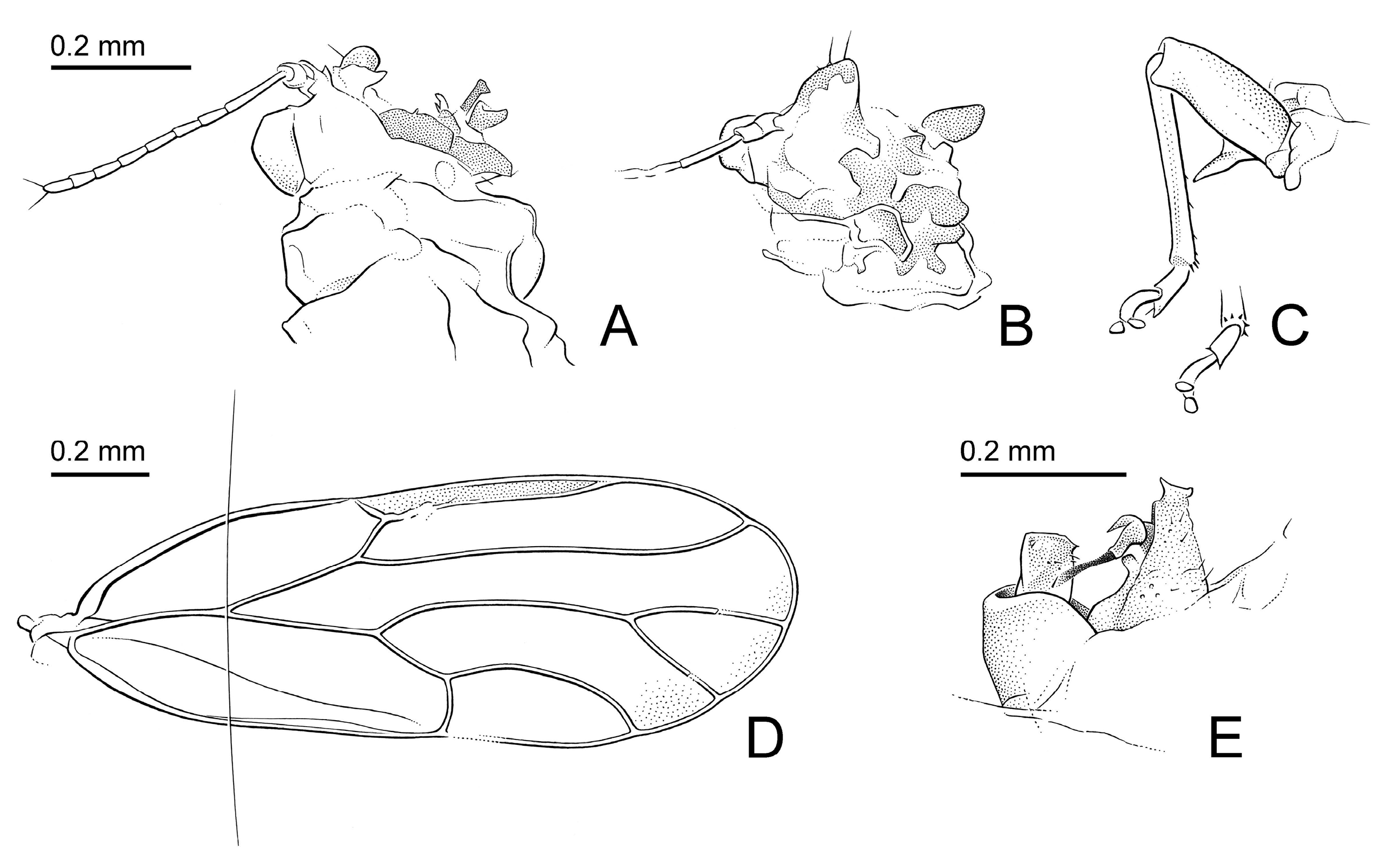
Antennal segment III more than twice as long as segment IV (Figure 4A). Metabasitarsus with two spurs. Pterostigma of forewing (Figure 4D) narrower than adjacent cell r1, at base. Paramere, in lateral view, rectangular (Figure 4E). Apical inflation of distal aedeagal segment moderately sized (Figure 4E)    Primascena subita.
-
Antennal segment III less than twice as long as segment IV (Figure 2A,B). Metabasitarsus with one outer spur. Pterostigma of forewing (Figure 3) wider than adjacent cell r1, at base. Paramere, in lateral view, oval (Figure 5A,C,D,F). Apical inflation of distal aedeagal segment large (Figure 5G,H)    2
2.
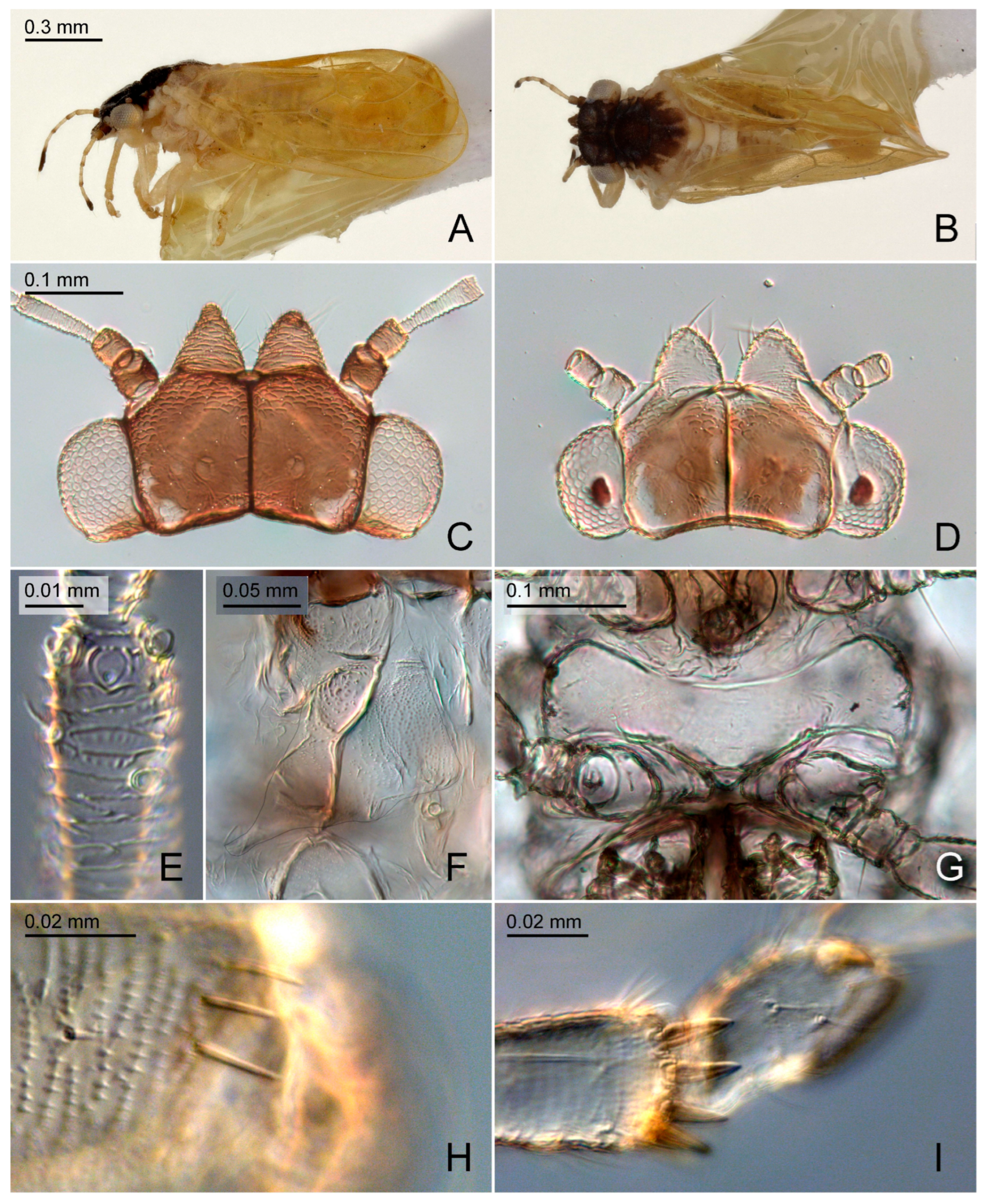
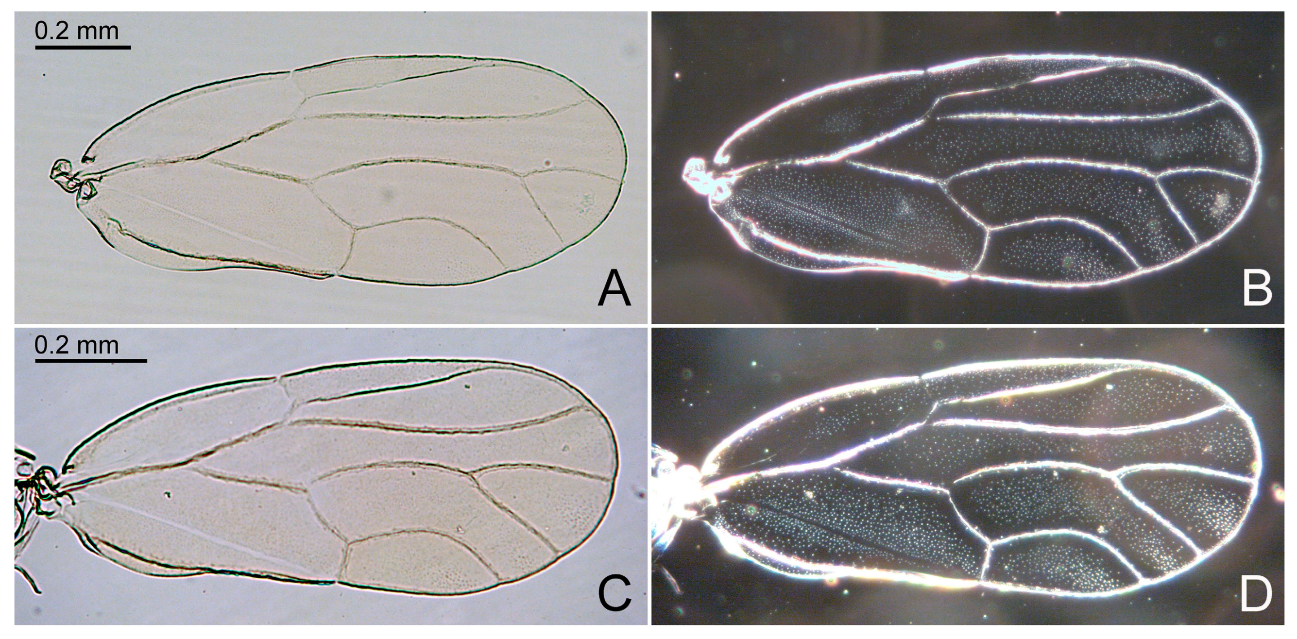
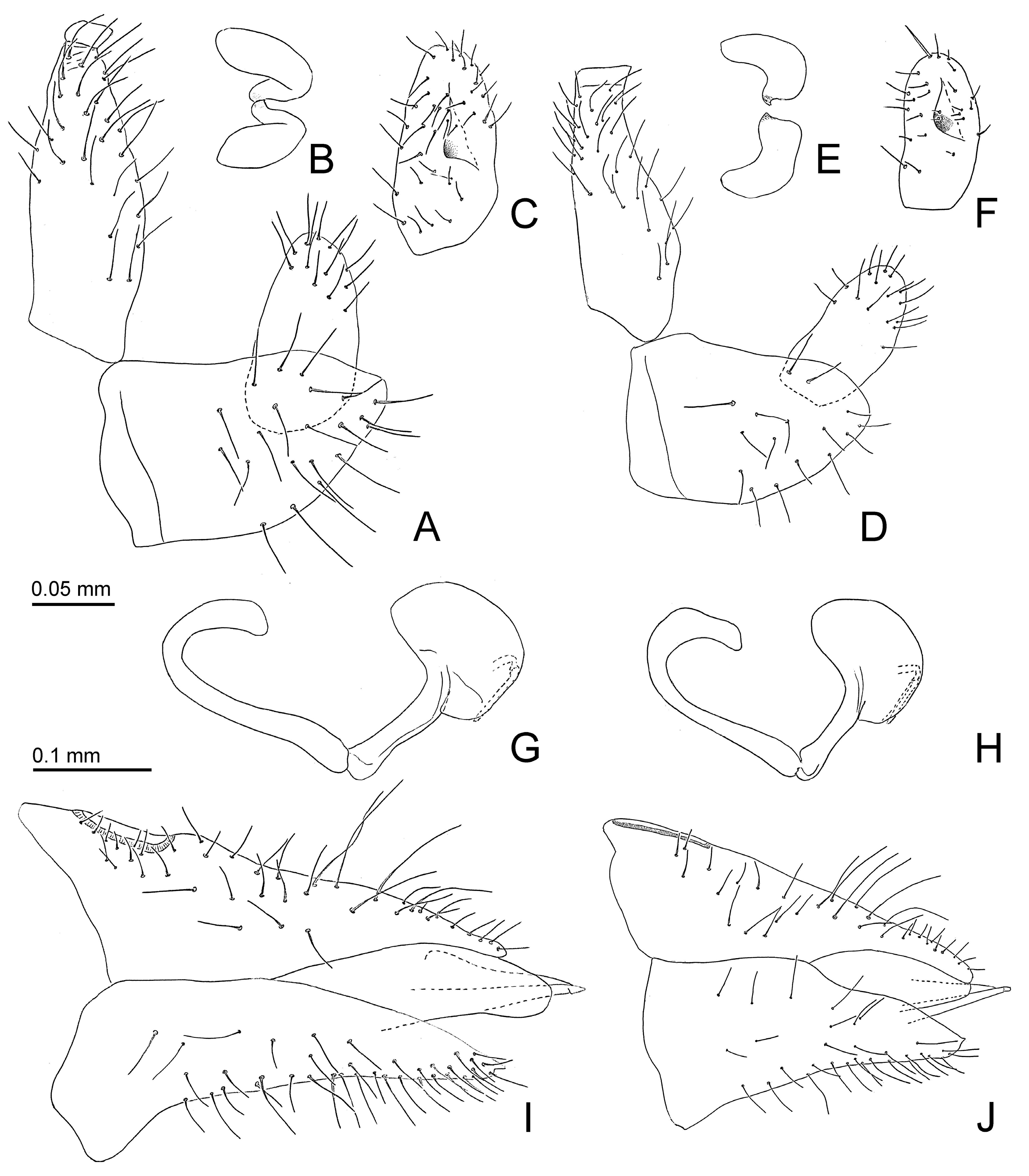
Genal processes dark brown or black (Figure 2C). Forewing 3.0 or more times as long as head width; m1 cell value ≤ 1.4. Paramere, in lateral view, slightly angular apically (Figure 5A); inner face with relatively large process proximal of the middle (Figure 5B,C). Apical inflation of distal aedeagal segment relatively angular (Figure 5G). Dorsal margin of female proctiger, in lateral view, weakly sinuate (Figure 5I). Probable host plant Ruprechtia laxiflora    Primascena empsycha.
-
Genal processes light (Figure 2C). Forewing shorter than 3.0 times head width; m1 cell value ≥ 1.5. Paramere, in lateral view, rounded apically (Figure 5D); inner face with relatively small process distal of the middle (Figure 5E,F). Apical inflation of distal aedeagal segment relatively rounded (Figure 5H). Dorsal margin of female proctiger, in lateral view, almost straight (Figure 5J). Host plant Ruprechtia exploratricis    Primascena ruprechtiae.

3.2.4. Primascena empsycha New Species (Figure 2A–C, Figure 3A,B and Figure 5A–C,G,I)

LSID: urn:lsid:zoobank.org:act:C15FF427-901D-4F1D-99D5-5363A732750F
Diagnosis. Adults. Genal processes dark brown or black. Antennal segment III less than twice as long as segment IV. Metabasitarsus with one outer spur. Forewing 3.0 or more times as long as head width; pterostigma wider than adjacent cell r1, at base m1 cell value ≤ 1.4. Paramere, in lateral view, narrowly oval, slightly angular apically; inner face with relatively large process proximal of the middle. Apical inflation of distal aedeagal segment large, relatively angular. Dorsal margin of female proctiger, in lateral view, weakly sinuate. Immatures unknown.
Additional characters. Adults. Coloration. Whitish. Vertex dark brown or almost black sometimes with small white dot laterally on either side; genal processes dark dorsally. Antenna with apices of segments III, IV, VI, and VIII, and entire segments IX and X dark brown or black. Tip of rostrum black. Conspicuous dark brown or almost black pattern covering entire pronotum except for a yellow longitudinal narrow streak in the middle and a sublateral white spot on either side; also dark entire mesopraescutum, a wide line along fore margin and five long, broad longitudinal stripes on mesoscutum. Forewing straw-colored. Structure. Genal processes 0.6–0.7 times as long as vertex along mid-line. Antenna 1.0–1.1 times as long as head width; segment III with 2–3 apical disc-like sensoria; rhinaria on segments IV, VIII, and IX lacking marginal spines, segment VI with a proximal comb of two spines; relative length of flagellar segments as 1.0:0.6:0.3:0.6:0.4:0.7:0.3:0.5; relative length of segment X and terminal setae as 1.0:1.1:1.2. Metatibia with 7–8 weakly or not sclerotized apical spurs; metabasitarsus with one outer spur. Forewing (Figure 3A,B), 3.0–3.2 times as long as head width; pterostigma, at base, wider than adjacent part of cell r1; cell m1 value = 2.4, cell cu1 value = 1.2–1.4. Terminalia as in Figure 5A–C,G,I. Male proctiger 0.5 times as long as head width; beset with long setae except for antero-basal third. Male subgenital plate elongate, beset with long setae in apical two thirds. Paramere, in lateral view, narrowly oval, slightly angular apically; outer face with long setae in apical half; inner face with large tubercle in the middle bearing an inward and slightly backward directed sclerotized tooth. Proximal segment of aedeagus strongly curved in basal third, straight in apical two thirds; distal segment straight in basal half, forming large reniform inflation in apical half which is incised dorso-basally; sclerotized end tube long and straight. Female terminalia, in lateral view, cuneate. Proctiger 0.9 times as long as head width; dorsal margin, in lateral view, almost straight; apex blunt; irregularly beset with moderately long setae in basal half, very long setae in the third quarter from base, and short setae in apical quarter; circumanal ring 0.3 times as long as proctiger. Subgenital plate, in lateral view, irregularly cuneate, 0.8 times as long proctiger; ventral margin weakly concave; apex rounded, ending in small point. Measurements. Body length (1 ♂, 5 ♀): ♂ 1.4 mm, ♀ 1.7–1.9 mm. Morphological structures (1 ♂, 1 ♀): head width ♂ 0.40 mm, ♀ 0.46 mm; antenna length ♂ 0.44 mm, ♀ 0.46 mm; forewing length ♂ 1.20 mm, ♀ 1.46 mm; ♂ proctiger length 0.18 mm; paramere length 0.14 mm; length of distal aedeagus segment 0.14 mm; ♀ proctiger length 0.42 mm. Fifth instar immature unknown.
Etymology. From the Ancient Greek adjective ἔμψυχος = animate, living (literally, those who possess a living soul), referring to the fact that this is an extant species.
Host plant. Adults were collected on Ruprechtia laxiflora Meisn. (Polygonaceae) which is a likely host.
Biology. Unknown.
Distribution. Brazil (Mato Grosso do Sul).
Type material. Holotype ♂: Brazil, Mato Grosso do Sul, Dourados, Embrapa campus, S22.2738° W54.8182°, 400 m, 8.x.2021, park vegetation, Ruprechtia laxiflora (D. Burckhardt & D.L. Queiroz) #459(13) (UFPR, dry mounted). Paratypes: 1 ♂, 6 ♀, same data as holotype but (NHMB, UFPR, dry and slide mounted, in 70% ethanol).

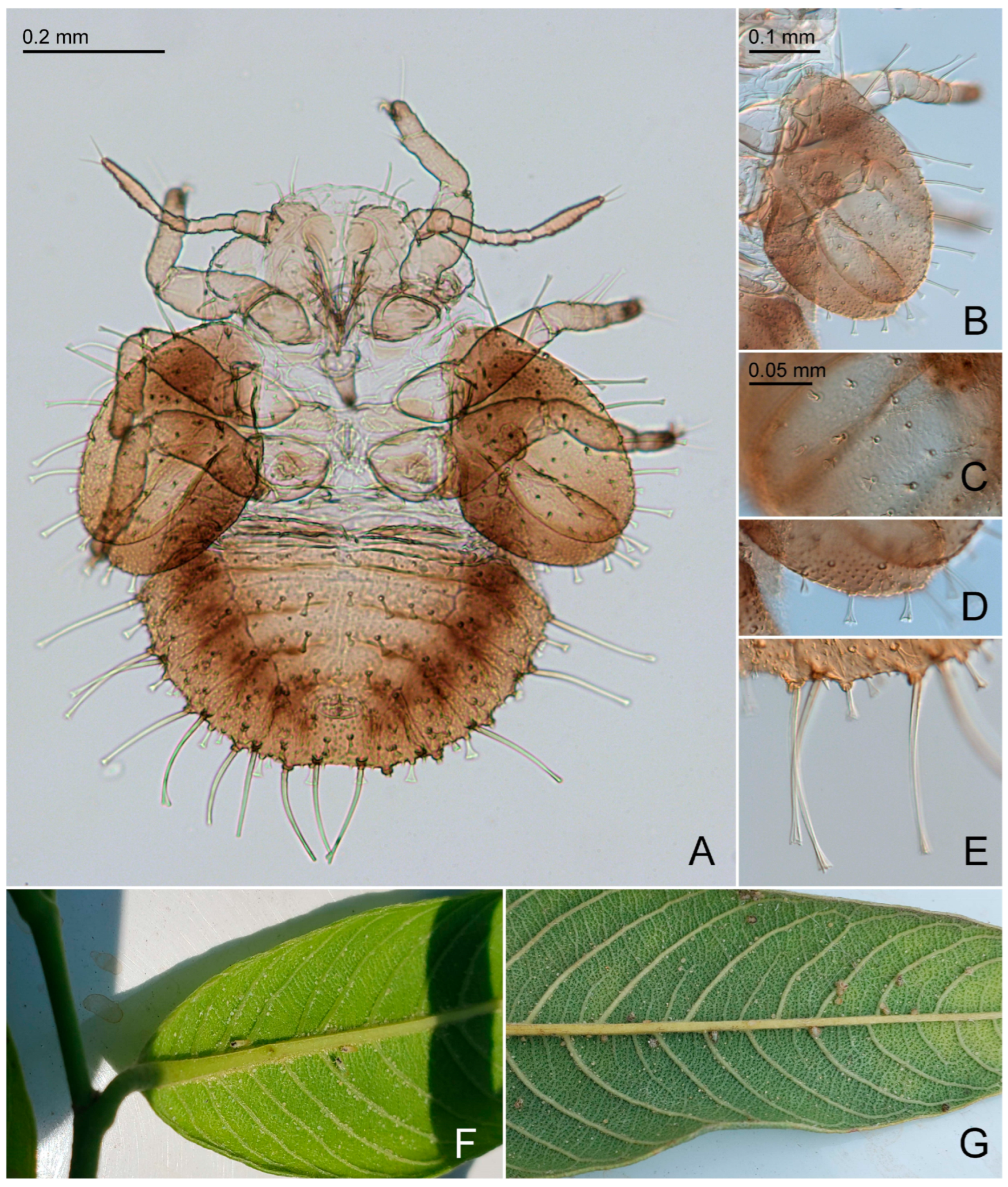
3.2.5. Primascena ruprechtiae New Species (Figure 2D–I, Figure 3C,D, Figure 5D–F,H,J and Figure 6)

LSID: urn:lsid:zoobank.org:pub:96BB823E-9465-4734-83C9-5CFB221ADB15
Diagnosis. Adults. Genal processes dark light. Antennal segment III less than twice as long as segment IV. Metabasitarsus with one outer spur. Forewing shorter than 3.0 head width; pterostigma wider than adjacent cell r1, at base; m1 cell value ≥ 1.5. Paramere, in lateral view, narrowly oval, rounded apically; inner face with relatively small process distal of the middle. Apical inflation of distal aedeagal segment large, relatively rounded. Dorsal margin of female proctiger, in lateral view, almost straight. Immatures. Body 1.0–1.2 times as long as wide. Caudal plate with 16–20 long marginal capitate setae, and around 60 short dorsal funnel-shaped setae.
Additional characters. Adults. Coloration. Whitish. Vertex dark brown or almost black with a sublateral white stripe on either side; genal processes light. Antenna with apices of segments III, IV, VI, and VIII, and entire segments IX and X dark brown or black. Tip of rostrum black. Conspicuous dark brown or almost black pattern covering entire pronotum except for on each a lateral and sublateral white spot on either side, most of the mesopraescutum dark brown or black except for a white lateral spot on either side, and a narrow line along fore margin and five short, narrow longitudinal stripes on mesoscutum. Forewing weakly yellowish. Structure. Genal processes 0.4–0.6 times as long as vertex along mid-line. Antenna 1.0–1.3 times as long as head width; segment III with a subapical and an apical disc-like sensorium and two apical peg-like sensoria (Figure 2E); rhinaria on segments IV, VIII, and IX lacking marginal spines, segment VI with a proximal comb of five spines; relative length of flagellar segments as 1.0:0.6:0.5:0.6:0.5:0.7:0.4:0.4; relative length of segment X and terminal setae as 1.0:2.3:1.8. Metatibia with 6–8 weakly or not sclerotized apical spurs; metabasitarsus with one outer spur. Forewing (Figure 3C,D), 2.7–2.9 times as long as head width; pterostigma, at base, wider than adjacent part of cell r1; cell m1 value = 2.4–2.5, cell cu1 value = 1.5–1.7. Terminalia as in Figure 5D–F,H,J. Male proctiger 0.4 times as long as head width; beset with long setae except for antero-basal third. Male subgenital plate elongate, beset with long setae in apical two thirds. Paramere, in lateral view, narrowly oval, rounded apically; outer face with long setae in apical half; inner face with large tubercle in the middle bearing an inward and slightly backward directed sclerotized tooth. Proximal segment of aedeagus strongly curved in basal third, straight in apical two thirds; distal segment straight in basal half, forming large reniform inflation in apical half which is incised dorso-basally; sclerotized end tube long and straight. Female terminalia, in lateral view, cuneate. Proctiger 0.8–0.9 times as long as head width; dorsal margin, in lateral view, almost straight; apex blunt; irregularly beset with moderately long setae in basal half, very long setae in the third quarter from base, and short setae in apical quarter; circumanal ring 0.3–0.4 times as long as proctiger. Subgenital plate, in lateral view, irregularly cuneate, 0.6–0.7 times as long as proctiger; ventral margin weakly concave; apex rounded, ending in small point. Measurements. Body length (10 ♂, 10 ♀): ♂ 1.4–1.7 mm, ♀ 1.5–1.7 mm. Morphological structures (2 ♂, 2 ♀): head width ♂ 0.36–0.38 mm, ♀ 0.42–0.44 mm; antenna length ♂ 0.46–0.48 mm, ♀ 0.42–0.46 mm; forewing length ♂ 0.98–1.06 mm, ♀ 1.20–1.26 mm; ♂ proctiger length 0.14 mm; paramere length 0.10 mm; length of distal aedeagus segment 0.10–0.12 mm; ♀ proctiger length 0.34–0.38 mm. Fifth instar immature. Coloration. Whitish. Cephalothorax and thoracic abdominal tergites very light brown with irregular slightly darker brown patches. Antenna whitish with segments I, II, and VII brown; apex of segment VII dark brown. Tips of tarsi brown. Forewing pad light brown at base and apical half, otherwise dark brown; hindwing pad dark brown with small area at base light brown. Caudal plate dorsally with broad brown margin, an irregular, moderately wide longitudinal whitish stripe in the middle and irregularly dark brown otherwise; ventrally similar but with more expanded dark pattern. Structure. Body (Figure 6A) 1.0–1.2 times as long as wide across wing pads. Antenna 0.7–0.9 times as long as forewing pad; segments III and V each with a short capitate seta about half or two-thirds as long as diameter of respective segment. Mesotibia with two long funnel-shape setae and two short capitate setae; metatibia with four short to moderately long capitate setae. Caudal plate 0.5–0.6 times as long as wide. Measurements (six specimens). Body length 0.78–1.00 mm; antenna length 0.28–0.32 mm.
Etymology. Named after its host genus Ruprechtia.
Host plant. Ruprechtia exploratricis Sandwith (Polygonaceae).
Biology. Immatures are free-living and develop on the lower leaf face (Figure 6F). Immatures often are close to the larger veins (Figure 6G).
Distribution. Brazil (Mato Grosso do Sul).
Type material. Holotype ♂: Brazil, Mato Grosso do Sul, Corumbá, Polícia militar ambiental, S19.0121° W57.6859°, 110 m, 12–16.x.2021, degraded vegetation near river, Ruprechtia exploratricis (D. Burckhardt & D.L. Queiroz) #470(2) (UFPR, dry mounted). Paratypes: 75 ♂, 87 ♀, 55 immatures, 17 skins, 20 egg, same data as holotype but (NHMB, UFPR, dry and slide mounted, in 70% ethanol); 4 ♂, 4 ♀, same but Assentamento Taquaral, S19.1235° W57.6862°, 220 m, 17.x.2021, deciduous forest, Ruprechtia exploratricis, #478(2) (NHMB, in 70% ethanol); 4 ♂, 25 ♀, 5 skins, same but Corumbá, Cemitério Nelson Chamma, S19.0199° W57.6956°, 130 m, 17.x.2021, park vegetation, Ruprechtia exploratricis, #479(1) (NHMB, in 70% ethanol).

3.2.6. Primascena subita Klimaszewsi, 1998 [13] (Figure 4)

Diagnosis. Adults. Antennal segment III more than twice as long as segment IV. Metabasitarsus with two spurs. Forewing with pterostigma narrower than adjacent cell r1, at base; m1 cell value = 1.5. Paramere, in lateral view, rectangular. Apical inflation of distal aedeagal segment moderately sized. Female and immatures unknown.
Redescription. Coloration. Body and forewing lacking distinct dark pattern. Structure. Head (Figure 4A,B) severely damaged; genal processes developed; compound eyes semicircular, adpressed to head. Clypeus and rostrum missing. Antenna 1.1 times as long as head width; relative length of flagellar segments as 1.0:0.4:0.4:0.5:0.4:0.4:0.4:0.4; relative length of segment X and terminal setae as 1.0:2.3:1.8. Thorax strongly damaged and incomplete. Metacoxa with spur-shaped meracanthus (Figure 4C); metatibia 0.8 times as long as head width, bearing a posteriorly open crown of five evenly spaced, weakly sclerotized apical spurs (Figure 4C); metabasitarsus with two spurs. Forewing (Figure 4D) 3.8 times as long as head width, 2.6 times as long as wide; with pterostigma narrower than adjacent cell r1, at base; m1 cell value = 1.5, cu1 cell value = 3.1. Paramere (Figure 4E), in lateral view, rectangular. Apical inflation of distal aedeagal segment moderately sized, incised at the base dorsally. Female and immatures unknown. Measurements (1 ♂). Head width 0.40 mm; antenna length 0.43 mm; forewing length 1.50 mm; proctiger length 0.15 mm; paramere length 0.10 mm; length of distal aedeagus segment 0.13 mm.
Etymology. From the Latin adjective subitus = sudden, unexpected [13].
Host plant and biology. Unknown.
Distribution. Amber fossil from the Dominican Republic.
Comments. Klimaszewski [13] described the clypeus as “bent downwards” and figured it to be a tubular structure perpendicular to the ventral head face. Klimaszewski’s drawing of the head is difficult to interpret but we think that the structure in question could be one of the genal processes (Figure 5A,B). Our measurements also differ slightly from those by Klimaszewski [13] probably because they were taken at different angles.
Type material. Holotype ♂: Dominican Republic, North Mines, Miocene amber, DR-14-137 (not DR-14-67 as indicated by Klimaszewski [13]) (AMNH).

4. Discussion

The psyllid fauna of Dominican amber is diverse at both the family and genus level and is similar to today’s fauna [10,12]. Burckhardt, et al. [14] reported 19 described species from Dominican amber. Six of these were originally described in extant genera, while another six species were transferred from fossil genera to recent genera due to synonymization. This large proportion of species where the systematic affinity had to be revised (almost one-third!) is a consequence of inaccurate observations and the general poor knowledge of neotropic psyllids. While six of the remaining seven fossil genera listed by Burckhardt, et al. [14] are currently known only from fossils, represented by a single species based on a single specimen, one genus, Primascena, also includes two extant species which are described here. The cladistic analysis strongly supports the monophyly of P. subita with P. empsycha and P. ruprechtiae, and confirms the monophyly of nine other genera of Aphalaroidinae (Figure 1), hence supporting the current classification of the subfamily [4,5]. While Pachyparia and Yangus are represented in the analysis by only one species, Connectopelma (in node 8) is paraphyletic with respect to Panisopelma, though the position of Panisolepma within Connectopelma is only weakly supported.
Primascena is sister to all but Aphalaroida, though with little support (Figure 1). Weak is also the support for nodes 3–5. Node 6, which includes eight genera, is strongly supported by five synapomorphies and two homoplasies with a Bremer support = 2. Within node 6, the relationships between genera are, again, only weakly supported, with the exception of Russelliana + Sphinia. While there are no other cladistic analyses examining the relationships of all recognized genera of Aphalaroidinae, Burckhardt [16] discussed possible relationships among some of the genera within the subfamily and suggested the following grouping: ((Baccharopelma, Connectopelma), (Zonopelma, (Panisopelma, (Russelliana, (Sphinia, (Aphalaroida, Prosopidopsylla)))))). The considerable difference between the two sets of phylogenetic relationships is due to the fact that the latter is derived from a discussion of characters and does not result from a parsimony analysis. Further studies should be conducted, e.g., by including more species, in particular of Pachyparia and Yangus, represented in the analysis by only one species each. Pachyparia is currently monotypic and Yangus is known only from three described Asian species. Several undescribed Afrotropical species of both genera are represented in the collection of the NHMB. In addition to morphological characters, molecular data should also be analyzed to test the present phylogeny.
Psyllids exhibit interesting host plant patterns. They are reasonably host-specific, and related psyllid species often develop on related hosts [2,3,4,6,20]. Aphalaroidinae is atypical in this respect. It comprises the only polyphagous psyllid species apart from the few Bactericera species [40,41]. The host range of Aphalaroidinae, without those of the polyphagous Russelliana solanicola Tuthill, is also surprisingly wide; the hosts belong to 12 plant families of ten orders and include conifers (Cupressaceae) and several groups of eudicots (Table 1). Four genera are entirely associated and two are partially associated with Fabaceae, the family with the largest number of associated psyllid genera. It is mostly confined to the Psyllidae [20], of which it hosts members of seven of the nine recognized subfamilies. The extant species of Primascena are associated with Ruprechtia (Polygonaceae: Eriogonoideae). This family serves as host to 69 psylloid species [5,42]. Most of these hosts are members of the subfamily Polygonoideae, which harbors Aphalaridae (27 Aphalara spp.), Liviidae (9 Eremopsylloides spp., 13 Pachypsylloides spp., 10 Shaerqia spp.), and Triozidae (5 Bactericera spp., 2 Trioza spp.). Only three species of Aphalaroidinae (Psyllidae) develop on members of the subfamily Eriogonoideae: two Primascena spp. on Ruprechtia and one Russelliana species on Chorizanthe. The majority of immature Aphalaroidinae, including Primascena, are free-living. Gall-inducing taxa are all species of Baccharopelma and some species of Connectopelma.
The Aphalaroidinae is predominantly American, with the exception of Pachaparia and Yangus, which are restricted to the Old World (Figure 1, Table 1). The former is known from Africa and the Arabian Peninsula while the latter has been reported from the Oriental Region [5] and is represented by undescribed species from Africa (unpublished NHMB data). Aphalaroida is entirely North American [43] and Freysuila has two North American and one South American species [44]. The other nine genera are confined to the Neotropical region [9,16,22,45]. Russelliana is by far the largest genus in the subfamily and has also the widest distribution. It ranges from the Chilean Far South to Peru, with a few species also occurring in Brazil [46]. It is a characteristic element of Mediterranean sclerophyll scrub. Similar habitats are used by species of the genera Baccharopelma, Ehrendorferiana, Panisopelma, and Sphinia, but they are geographically less widespread than Russelliana. The two species of Zonopelma develop on Misodendrum species parasitizing Nothofagus and occur mostly in temperate rain forests. Primascena, finally, is known so far only from Dominican amber and two extant species from the Brazilian Pantanal, which live in predominantly forested habitats such as riverine or gallery forests and in seasonally semideciduous forests.

5. Conclusions

Homoplasy in morphological characters of adult psyllids is a major problem in phylogenetic reconstruction. This is one of the reasons why fossil psyllids are often difficult to place in a phylogeny. In general, the fauna of Dominican amber is quite similar to the extant fauna at the genus level, but in psyllids almost a third of the species described from Dominican amber are assigned to fossil-only genera. Psyllids are most species-rich in the tropics and south temperate regions, but Afrotropical and Neotropical faunas are only poorly known. To find extant members of these fossil genera is, therefore, not unlikely. Primascena exemplifies this. The discovery of two extant species permitted a careful morphological study of the genus and added information on the female and fifth instar immature. These new morphological data provided the base for a cladistic analysis that supported the monophyly of the fossil and extant species, and confirm its placement in the Aphalaroidinae, previously suggested by a revision of the holotype of P. subita.
Targeted field work is not only important for discovering previously unknown biodiversity, but also to find new host associations. While psyllid subfamilies or tribes are often restricted to a single plant family or order, the host range of Aphalaroidinae is surprisingly broad. Even though Ruprechtia is an unusual psyllid host, it fits the pattern found in Aphalaroidinae.

Supplementary Materials

The following supporting information can be downloaded at: https://www.mdpi.com/article/10.3390/insects15060382/s1, File S1: The character matrix used for the phylogenetic analysis.

Author Contributions

Conceptualization, D.B. and D.L.Q.; methodology, D.B. and D.L.Q.; collecting of material, D.B. and D.L.Q.; investigation, D.B. and D.L.Q.; resources, D.B. and D.L.Q.; data curation, D.B. and D.L.Q.; validation, D.B. and D.L.Q.; formal analysis, D.B.; writing, D.B. and D.L.Q. All authors have read and agreed to the published version of the manuscript.

Funding

This research received no external funding.

Data Availability Statement

Data are contained within the article or Supplementary Materials.

Acknowledgments

We thank David Grimaldi (AMNH) for the loan of the type of Primascena subita, Armin Coray (NHMB) for making the beautiful drawings of Primascena subita (Figure 5), and Catia Urbanetz, Head of Research and Development, and the staff of Embrapa Pantanal for supporting our fieldwork. We are grateful to three anonymous reviewers for their constructive comments and suggestions on the manuscript. We gratefully acknowledge receiving the following collecting permit in Brazil: CNPq, IBAMA/SISBIO (71632-3 41169-12).

Conflicts of Interest

The authors declare no conflicts of interest.

References

  1. Ouvrard, D.; Burckhardt, D.; Greenwalt, D. The Oldest Jumping Plant-Louse (Insecta: Hemiptera: Sternorrhyncha) with Comments on the Classification and Nomenclature of the Palaeogene Psylloidea. Acta Musei Morav. Sci. Biol. Brno 2013, 98, 21–33. [Google Scholar]
  2. Burckhardt, D.; Serbina, L.Š.; Malenovský, I.; Queiroz, D.L.; Aléné, D.C.; Cho, G.; Percy, D.M. Phylogeny and Classification of Jumping Plant Lice of the Subfamily Liviinae (Hemiptera: Psylloidea: Liviidae) Based on Molecular and Morphological Data. Zool. J. Linn. Soc. 2023, 1–35. [Google Scholar] [CrossRef]
  3. Burckhardt, D.; Queiroz, D.L. Neotropical Jumping Plant-Lice (Hemiptera, Psylloidea) Associated with Plants of the Tribe Detarieae (Leguminosae, Detarioideae). Zootaxa 2020, 4733, 1–73. [Google Scholar] [CrossRef] [PubMed]
  4. Burckhardt, D.; Ouvrard, D.; Percy, D.M. An Updated Classification of the Jumping Plant-Lice (Hemiptera: Psylloidea) Based on Molecular and Morphological Evidence. Eur. J. Taxon. 2021, 736, 137–182. [Google Scholar] [CrossRef]
  5. Ouvrard, D. Psyl’list—The World Psylloidea Database. Available online: http://www.hemiptera-databases.com/psyllist (accessed on 3 February 2024).
  6. Burckhardt, D.; Ouvrard, D.; Queiroz, D.L.; Percy, D.M. Psyllid Host-Plants (Hemiptera: Psylloidea): Resolving a Semantic Problem. Fla. Entomol. 2014, 97, 242–246. [Google Scholar] [CrossRef]
  7. Ouvrard, D.; Burckhardt, D.; Cocquempot, C. An Annotated Checklist of the Jumping Plant-Lice (Insecta: Hemiptera: Psylloidea) from the Mercantour National Park, with Seven New Records for France and One New Synonymy. Zoosystema 2015, 37, 251–271. [Google Scholar] [CrossRef]
  8. Hodkinson, I.D.; Bird, J. Sedge and Rush-Feeding Psyllids of the Subfamily Liviinae (Insecta: Hemiptera: Psylloidea): A Review. Zool. J. Linn. Soc. 2000, 128, 1–49. [Google Scholar] [CrossRef]
  9. Burckhardt, D. Ehrendorferiana, a New Genus of Neotropical Jumping Plant Lice (Insecta: Hemiptera: Psylloidea) Associated with Conifers (Cupressaceae). Org. Divers. Evol. 2005, 5, 317–319. [Google Scholar] [CrossRef]
  10. Bekker Migdisova, E.E. Iskopajemije Nasekomije Psillomorfy; Izdatelstvo “Natshka” Ed.; Trudy paleontologitshekogo Instituta: Moscow, Russia, 1985; Volume 206, p. 92. [Google Scholar]
  11. Iturralde-Vinent, M.A.; Macphee, R.D.E. Remarks on the Age of Dominican Amber. Palaeoentomology 2019, 2, 236–240. [Google Scholar] [CrossRef]
  12. Klimaszewski, S.M. New Psyllids (Homoptera, Psylloidea) from Dominican Amber. Acta Biol. Silesiana 1996, 29, 24–44. [Google Scholar]
  13. Klimaszewski, S.M. Further Data Concerning Dominican amber Jumping Plant Lice (Homoptera, Psylloidea). Acta Biol. Silesiana 1997, 30, 19–27. [Google Scholar]
  14. Burckhardt, D.; Drohojowska, J.; Serbina, L.Š.; Malenovský, I. First Record of Jumping Plant Lice of the Family Liviidae (Insecta, Hemiptera, Psylloidea) from Dominican Amber. Neues Jahrb. Für Geol. Und Palaontol. 2024. accepted. [Google Scholar]
  15. Burckhardt, D.; Mifsud, D. Jumping Plant-Lice of the Paurocephalinae (Insecta, Hemiptera, Psylloidea): Systematics and Phylogeny. Contrib. Nat. Hist. 2003, 2, 3–34. [Google Scholar]
  16. Burckhardt, D. Jumping Plant Lice (Homoptera: Psylloidea) of the Temperate Neotropical Region: Part 1. Psyllidae (subfamilies Aphalarinae, Rhinocolinae and Aphalaroidinae). Zool. J. Linn. Soc. 1987, 89, 299–392. [Google Scholar] [CrossRef]
  17. Percy, D.M.; Crampton-Platt, A.; Sveinsson, S.; Lemmon, A.R.; Lemmon, E.M.; Ouvrard, D.; Burckhardt, D. Resolving the Psyllid Tree of Life: Phylogenomic Analyses of the Superfamily Psylloidea (Hemiptera). Syst. Entomol. 2018, 43, 762–776. [Google Scholar] [CrossRef]
  18. Vondráček, K. Jumping Plant-Lice (Psylloidea-Homoptera) of Central Africa. Part 1 (Congo). Acta Entomol. Musei Natl. Pragae 1963, 35, 263–290. [Google Scholar]
  19. Burckhardt, D.; Ouvrard, D. A Revised Classification of the Jumping Plant-Lice (Hemiptera: Psylloidea). Zootaxa 2012, 3509, 1–34. [Google Scholar] [CrossRef]
  20. Ouvrard, D.; Chalise, P.; Percy, D.M. Host-Plant Leaps Versus Host-Plant Shuffle: A Global Survey Reveals Contrasting Patterns in an Oligophagous Insect Group (Hemiptera, Psylloidea). Syst. Biodivers. 2015, 13, 434–454. [Google Scholar] [CrossRef]
  21. Crawford, D.L. A Monograph of the Jumping Plant-Lice or Psyllidae of the New World; Smithsonian Institution United States National Museum, Ed.; Government Printing Office: Washington, DC, USA, 1914; Volume 85. [Google Scholar]
  22. Burckhardt, D.; Espirito-Santo, M.M.; Fernandes, G.W.; Malenovsky, I. Gall-Inducing Jumping Plant-Lice of the Neotropical genus Baccharopelma (Hemiptera, Psylloidea) Associated with Baccharis (Asteraceae). J. Nat. Hist. 2004, 38, 2051–2071. [Google Scholar] [CrossRef]
  23. Šulc, K. Eine neue Panisopelma (Connectopelma sbgn. n.) und eine neue Trioza aus Chile. Sitzungsber. Königl.-Böhm. Ges. Wissensch. Math.-Naturw. Classe 1914, 5, 1–8. [Google Scholar]
  24. Aleman, D.J. Frey-Suila Dugesii. La Nat. 1887, 1, 21–26. [Google Scholar]
  25. Loginova, M.M. Ergebnisse der Zoologischen Nubien-Expedition 1962. Teil XXXIII. Homoptera-Psylloidea. Ann. Naturhist. Mus. Wien 1967, 70, 401–409. [Google Scholar]
  26. Enderlein, G. Panisopelma quadrigibbiceps, eine neue Psyllidengattung aus Argentinien. (Psyllidologica I). Zool. Anz. 1910, 36, 280–281. [Google Scholar]
  27. Tuthill, L.D. Los Psyllidae del Peru Central (Insecta, Homoptera). Rev. Peru. De Entomol. Agric. 1959, 2, 1–27. [Google Scholar]
  28. Blanchard, E.; Zoologia, V. Afidideos. In Historia Fisica y Politica de Chile; Museo de Historia Natural de Santiago: Santiago, Chile, 1852; Volume 7, pp. 306–320. [Google Scholar]
  29. Fang, S.J. Psylloidea of Taiwan: Supplement II. (Homoptera). J. Taiwan Mus. 1990, 43, 103–117. [Google Scholar]
  30. Bastin, S.; Burckhardt, D.; Reyes-Betancort, J.A.; Hernandez-Suarez, E.; Ouvrard, D. A Review of the Jumping Plant-Lice (Hemiptera: Psylloidea) of the Canary Islands, with Descriptions of Two New Genera and Sixteen New Species. Zootaxa 2023, 5313, 1–98. [Google Scholar] [CrossRef]
  31. Ouvrard, D.; Bourgoin, T.; Campbell, B.C. Comparative Morphological Assessment of the Psyllid Pleuron (Insecta, Hemiptera, Sternorrhyncha). J. Morphol. 2002, 252, 276–290. [Google Scholar] [CrossRef]
  32. Drohojowska, J. Thorax Morphology and Its Iiportance in Establishing Relationships within Psylloidea (Hemiptera, Sternorrhyncha); Wydawnictwo Uniwersytetu Śląskiego: Katowice, Poland, 2015; Volume 3414, p. 172. [Google Scholar]
  33. POWO. Plants of the World Online. Facilitated by the Royal Botanic Gardens, Kew. Royal Botanic Gardens, Kew. Available online: http://www.plantsoftheworldonline.org/ (accessed on 23 March 2024).
  34. Zachos, F.E. Species Concepts in Biology, Historical Development, Theoretical Foundations and Practical Relevance; Springer: Berlin/Heidelberg, Germany, 2016; p. 220. [Google Scholar] [CrossRef]
  35. Nelson, G.; Platnick, N.I. Systematics and Biogeography: Cladistics and Vicariance; Columbia University Press: New York, NY, USA, 1981. [Google Scholar]
  36. Burckhardt, D. Insects of Saudi Arabia. Sternorrhyncha: Suborder Psyllodea. Fauna Saudi Arab. 1981, 3, 213–226. [Google Scholar]
  37. Nixon, K.C. Winclada; v.1.00.08; K.C. Nixon: Ithaca, NY, USA, 2002. [Google Scholar]
  38. Goloboff, P.A.; Catalano, S.A. Tnt V.1.5, Including a Full Implementation of Phylogenetic Morphometrics. Cladistics 2016, 32, 221–238. [Google Scholar] [CrossRef]
  39. Lipscomb, D.L. Winclada, a Windows Program for Creating, Editing, and Analyzing Systematic Data Sets: Basic Users Guide; George Washington University, Ed.; George Washington University: Washington, DC, USA, 2002. [Google Scholar]
  40. Hodkinson, I.D. Life Cycle Variation and Adaptation in Jumping Plant Lice (Insecta: Hemiptera: Psylloidea): A Global Synthesis. J. Nat. Hist. 2009, 43, 65–179. [Google Scholar] [CrossRef]
  41. Burckhardt, D.; Lauterer, P. A Taxonomic Reassessment of the Triozid Genus Bactericera (Hemiptera: Psylloidea). J. Nat. Hist. 1997, 31, 99–153. [Google Scholar] [CrossRef]
  42. Burckhardt, D.; Cort, G.D.; Queiroz, D.L. Jumping Plant Lice of the Genus Aphalara (Hemiptera, Psylloidea, Aphalaridae) in the Neotropics. Zookeys 2020, 980, 119–140. [Google Scholar] [CrossRef] [PubMed]
  43. Hodkinson, I.D. New World Legume-Feeding Psyllids of the Genus Aphalaroida Crawford (Insecta: Homoptera: Psylloidea). J. Nat. Hist. 1991, 25, 1281–1296. [Google Scholar] [CrossRef]
  44. Burckhardt, D.; Wyniger, D. The Systematic Position of Psylla rhorodendri Tuthill with Comments on the New World Genus Freysuila Aleman (Hemiptera, Psylloidea, Aphalaroidinae). Mitteilungen Schweiz. Entomol. Ges. 2007, 80, 63–70. [Google Scholar] [CrossRef]
  45. Burckhardt, D.; Ouvrard, D. The Temperate Neotropical Psyllid Genus Sphinia (Hemiptera, Psylloidea). Rev. Fr. d’entomologie 2001, 23, 35–46. [Google Scholar]
  46. Serbina, L.; Burckhardt, D. Systematics, Biogeography and Host-Plant Relationships of the Neotropical Jumping Plant-Louse Genus Russelliana (Hemiptera: Psylloidea). Zootaxa 2017, 4266, 1–114. [Google Scholar] [CrossRef]
Figure 1. Phylogenetic relationships among the known genera of Aphalaroidinae (outgroup = Acizzia hollisi). For Aphalaroida, Baccharopelma, Connectopelma, Panisopelma, Prosopidopsylla, Russelliana, Sphinia, and Yangus, only a selection of the known species is included. The single most parsimonious tree was obtained by cladistic analysis of unweighted characters. The character state changes are shown for each node using the ‘Fast optimization’ option of WinClada. Synapomorphies are represented with black circles and homoplasies with white circles. Bremer support values are indicated in squares at the base of branches and the numbers of nodes are provided in circles. NA = North America.
Insects 15 00382 g001
Figure 2. Primascena spp.: (AC) P. empsycha; (DI) P. ruprechtiae. (A) Habitus, dorsal view; (B) habitus, lateral view; (C,D) head, dorsal view; (E) apical half of antennal segment III; (F) propleurites, dorsad up, anteriad left; (G) mesosternum; (H) apex of metafemur; (I) apex of metatibia and metabasitarsus.
Insects 15 00382 g002
Figure 3. Primascena spp., forewing: (A,B) P. empsycha; (C,D) P. ruprechtiae. (A,C) Forewing: shape and venation (brightfield); (B,D) forewing: distribution of surface spinules (darkfield).
Insects 15 00382 g003
Figure 4. Primascena subita Klimaszewski, 1997; holotype ♂. (A) Head, antenna, and pronotum, dorsal view; (B) head and basal segments of antenna, ventral view; (C) hind leg, outer face and tip of leg, inner face; (D) forewing; (E) male terminalia (drawings by Armin Coray).
Insects 15 00382 g004
Figure 5. Primascena spp., terminalia. (AC,G,I) P. empsycha; (DF,H,J) P. ruprechtiae. (A,D) Male terminalia, in lateral view; (B,E) parameres, dorsal view, left anteriad, right posteriad; (C,F) parameres, inner face, in lateral view; (G,H) aedeagus, lateral view; (I,J) female terminalia, in lateral view.
Insects 15 00382 g005
Figure 6. Primascena ruprechtiae, immatures. (A) Habitus, dorsal view; (B) wing pads, dorsal view; (C) disca of forewing pad, dorsal view; (D) margin of hindwing pad, dorsal view; (E) margin of caudal plate, dorsal view; (F) adults and eggs on abaxial leaf surface; (G) immatures on abaxial leaf surface.
Insects 15 00382 g006
Disclaimer/Publisher’s Note: The statements, opinions and data contained in all publications are solely those of the individual author(s) and contributor(s) and not of MDPI and/or the editor(s). MDPI and/or the editor(s) disclaim responsibility for any injury to people or property resulting from any ideas, methods, instructions or products referred to in the content.

