Article

Histone Acetylation Enhancing Host Melanization in Response to Parasitism by an Endoparasitoid Wasp

1
State Key Laboratory of Ecological Pest Control for Fujian and Taiwan Crops, Fujian Agriculture and Forestry University, Fuzhou 350002, China
2
Fujian Provincial Key Laboratory of Insect Ecology, Department of Plant Protection, Fujian Agriculture and Forestry University, Fuzhou 350002, China
*
Author to whom correspondence should be addressed.
These authors contributed equally to this work.
Insects 2024, 15(3), 161; https://doi.org/10.3390/insects15030161
Submission received: 17 January 2024 / Revised: 18 February 2024 / Accepted: 26 February 2024 / Published: 27 February 2024
(This article belongs to the Section Insect Molecular Biology and Genomics)

Abstract

:

Simple Summary

Endoparasitic wasps are nature’s pest controllers, targeting various harmful insects. They lay their eggs inside these pests and release special substances (like viruses or venoms) that change the way the pest’s immune system works. However, we are still uncovering exactly how these changes happen. Our research looks into how certain wasps, which do not use these viruses, can still affect their host’s immune system. We studied this using a specific wasp–beetle pair as an example. We found that these wasps can control a crucial part of the beetle’s immune response through a process that modifies the beetle’s genetic material, specifically by adjusting certain markers on the genes that are important for immunity. This discovery sheds light on the sophisticated ways wasps can suppress the immune systems of their hosts, making them more effective at controlling pest populations.

Abstract

Endoparasitoids are insects that develop within other insects, employing unique strategies to enhance their offspring’s survival. They inject polydnavirus and/or venom into their hosts along with eggs, effectively suppressing the host’s immune system. Polydnavirus from Braconidae and Ichneumonidae wasps can integrate into the host’s genome to express viral genes using the host’s transcription systems. However, the ability of parasitoids without polydnavirus to manipulate host gene expression remains unclear. Lysine acetylation (LysAc), a post-translational modification critical for gene regulation, is hypothesized to be used by endoparasitoids lacking polydnavirus. We utilized the Chalcidoidea wasp Tetrastichus brontispae, which lacks polydnavirus, as an idiobiont endoparasitoid model to test this hypothesis, with pupae of the nipa palm hispid beetle Octodonta nipae as the host. Parasitism by T. brontispae resulted in the reduced expression of histone deacetylase Rpd3 and elevated levels of LysAc modification at histones H3.3K9 and H3.3K14 through proteomics and LysAc modification omics. The knockdown of Rpd3 increased the expression level of OnPPAF1 and OnPPO involved in the phenoloxidase cascade, leading to melanization in the host body whereby it resembled a mummified parasitized pupa and ultimately causing pupa death. This study enhances our understanding of how endoparasitoids employ histone acetylation to regulate immunity-related genes, offering valuable insights into their survival strategies.

1. Introduction

The employment of Hymenoptera parasitoid wasps as biological control agents against insect pests underscores the critical ecological and practical importance of dissecting parasitoid–host interactions [1]. These endoparasitoids, which develop within their hosts, face the challenge of overcoming complex physiological and immunological defenses through either evasion or neutralization strategies [2,3,4]. The evolutionary arms race between endoparasitoids and their hosts has spurred the development of sophisticated adaptive strategies aimed at modifying the host’s immunity and influencing its development and physiology. Central to these strategies is the deployment of virulence factors, such as venoms [5], polydnaviruses [6,7], and teratocytes [8]. Particularly for those species lacking polydnavirus, venom emerges as a key mechanism for breaching host defenses [2].
Advances in omics technologies have revolutionized our understanding of changes in host gene expression following parasitization, revealing significant alterations in immunity and development. These breakthroughs, alongside targeted functional experiments [9], have emphasized the considerable impact of parasitoids on modulating specific immunity-related host genes. Despite these advancements, fully comprehending the mechanisms through which parasitoids regulate host gene expression remains an ongoing endeavor. Within this context, epigenetic regulation emerges as a pivotal evolutionary mechanism for adaptation to environmental challenges, extending beyond mere genetic mutation [10,11,12]. Recent research has highlighted the ability of parasitoids to alter host epigenetic landscapes, thereby influencing gene expression. Braconidae parasitoids, such as Snellenius manila and Cotesia vestalis, employ polydnavirus- or teratocyte-derived microRNAs (Cve-miR-281-3p from teratocyte, Cve-miR-novel22-5p from CvBv, Sm-miR-199b-5p, and Sm-miR-2989 from SmBV) to regulate genes crucial for host development [13,14], demonstrating the sophisticated mechanisms at play. This backdrop emphasizes the need to explore the capabilities of parasitoids, particularly those lacking polydnavirus or teratocytes, in influencing host gene expression through other epigenetic means.
The symbiotic origins of parasitoid venoms, and their potential evolution through lateral gene transfer (LGT) from ancient viral symbionts, offer a fascinating glimpse into the complex evolutionary history of these interactions [15,16]. This evolutionary perspective, suggesting deep-rooted symbiosis between parasitoids and their viral partners, enriches our understanding of the diverse strategies employed by parasitoids to manipulate their hosts. This is exemplified by the idiobiont ichneumon wasp Pimpla turionellae, whose venom delivery system significantly alters host epigenetic states, thereby affecting global DNA methylation, histone lysine acetylation ratios, and miRNA profiles—a testament to the intricate dynamics governing host gene transcriptional reprogramming [17].
Central to this study is lysine acetylation (LysAc), a key post-translational modification that responds to environmental cues and plays a vital role in reversible epigenetic regulation prior to transcription initiation [18,19]. The equilibrium maintained by histone acetyltransferases (HATs) and histone deacetylases (HDACs) in modulating transcription and gene expression underscores the complexity of epigenetic regulation [20,21,22,23,24,25]. Our investigation focuses on the pupal endoparasitoid wasp Tetrastichus brontispae Ferrière (Hymenoptera: Eulophidae), which, in the absence of polydnavirus, relies on maternal venom as its primary virulence factor [26,27]. T. brontispae targets Chrysomelidae (Coleoptera) pests, including Octodonta nipae and Brontispa longissimi, in palm cultivation [28,29]. Previous studies have validated the significant impact of T. brontispae parasitization on immunity-related genes in O. nipae pupae [30]. Utilizing the O. nipae-T. brontispae system, with the available genomic resource for O. nipae (unpublished data), we aim to explore how T. brontispae might influence host gene expression through histone acetylation modifications. This study hypothesizes that T. brontispae can manipulate the transcriptional reprogramming of immunity-related genes by altering host histone acetylation/deacetylation patterns, thereby achieving effective biological regulation.

2. Materials and Methods

2.1. Insect Rearing

Octodonta nipae were collected from Canary Date Palm (Phoenix canariensis Hortulanorum ex Chabaud) in Fuqing City, Fujian Province, China. T. brontispae were initially sourced from mummified B. longissima pupae provided by the Chinese Academy of Tropical Agricultural Sciences in 2008. These were then cultured in our laboratory using newly exuviated, one-day-old O. nipae pupae as hosts for consecutive generations. Both species were maintained under controlled conditions in an artificial climate incubator at 25 ± 1 °C and 75 ± 5% relative humidity, with a 12 h light:12 h dark photoperiod, as detailed in previous studies [28,31]. The diet of O. nipae consisted of fresh Canary Island date palm leaves (Phoenix canariensis). Concurrently, T. brontispae were propagated using one-day-old O. nipae pupae as hosts. Post-emergence, the adult wasps received 10% sucrose solution for nutrition within a plastic container. For the parasitism experiments, one-day-old adult female T. brontispae, chosen for their optimal parasitism rate, were utilized.

2.2. Analysis of LysAc Modification by Western Blot

Hemolymph and fat body samples were collected from thirty O. nipae pupae at intervals of 24, 48, and 72 h post-parasitization (PP), alongside tissues from an identical number of un-parasitized pupae (UP) serving as controls. Fat body samples underwent total protein extraction using a Protein Extraction Kit (BestBio Biotechnology Co. Ltd., Shanghai, China), whereas hemolymph samples were analyzed directly without prior protein extraction. Protein concentrations were determined using a BCA kit (Labgic Technology Co., Ltd., Beijing, China).
Approximately 15 μg of proteins from each sample underwent Western blot analysis. Proteins were separated via 12% SDS-PAGE and subsequently transferred onto a nitrocellulose filter membrane with Trans-Blot SD (Bio-Rad, Hercules, CA, USA). The membrane was then blocked using 5% skim milk for one hour at room temperature before being washed. Immunoblots were incubated with a pan anti-acetyllysine mouse monoclonal antibody (1:1000 dilution, PTM-101, PTM Biolabs Inc., Hangzhou, China) as the primary antibody, followed by horseradish peroxidase-conjugated goat anti-mouse IgG (1:1000 dilution, Thermo Fisher Scientific, Pleasanton, CA, USA) as the secondary antibody. Signals were detected using an Amersham™ ECL Select™ Western Blotting Detection Reagent (Global Life Sciences Solutions USA LLC, Marlborough, MA, USA) and visualized on an Amersham Imager 600 QC (GE Healthcare, Marlborough, MA, USA).

2.3. Analysis of LysAc Modification through the Combination of 4D Label-Free Proteomics and LysAc Modification Omics

Based on the Western blot results, proteins were extracted from 100 O. nipae pupae at 48 h post-parasitization and from an equivalent number of un-parasitized pupae for comparison. Protein extraction followed the same procedure as previously outlined. Equal amounts of protein from both the PP and UP groups underwent trypsinization, vacuum drying, and desalting procedures at PTM Biolabs Inc., Hangzhou, China.
For proteomic analysis, tryptic peptides were resuspended in solvent A (0.1% formic acid and 2% acetonitrile in water) and loaded onto a custom-made reversed-phase analytical column (25 cm length, 75/100 μm i.d.). Peptides were eluted using a gradient from 6% to 24% solvent B (0.1% formic acid in acetonitrile) over 70 min, increased from 24% to 35% in 14 min, and then, to 80% in 3 min, with a final hold at 80% for 3 min, at a constant flow rate of 450 nL/min using a NanoElute UHPLC system (Bruker Daltonics, Billerica, MA, USA). Following capillary source treatment, peptides were analyzed by a timsTOF Pro mass spectrometer (Bruker Daltonics), with the electrospray voltage set to 1.60 kV. Both precursor and fragment ions were detected across a broad m/z range of 100 to 1700 in MS/MS mode, using parallel accumulation serial fragmentation (PASEF) mode to select precursors with charge states from 0 to 5 for fragmentation. The system completed 10 PASEF-MS/MS scans per cycle, with dynamic exclusion set to 30 s.
For LysAc modification omics, affinity enrichment was conducted. Tryptic peptides in NETN buffer (100 mM NaCl, 1 mM EDTA, 50 mM Tris-HCl, 0.5% NP-40, pH 8.0) were incubated overnight at 4 °C with anti-acetyllysine antibody-conjugated agarose beads (PTM-104, PTM Biolabs Inc.) under gentle agitation. Peptides were eluted with 0.1% trifluoroacetic acid, vacuum-dried, and desalted using C18 ZipTips (Millipore, Burlington, MA, USA) before LC-MS/MS analysis, which proceeded as above.
Data were processed using the MaxQuant search engine (version 1.6.15.0), with tandem mass spectra matched against a comprehensive whole-length transcriptome database (including Tetrastichus brontispae and Octodonta nipae, totaling 62,796 entries) and a concatenated reverse decoy database. Trypsin/P was the designated cleavage enzyme, with up to two missed cleavages allowed. Precursor ion mass tolerance was set at 20 ppm for the initial search, narrowed to 5 ppm for the main search, and fragment ion mass tolerance at 0.02 Da. The fixed modification was carbamidomethyl, and variable modifications included N-terminal acetylation and Met oxidation. The false discovery rate (FDR) was maintained below 1%. Differential expression and LysAc modification analyses between the PP and UP groups utilized fold change (FC) criteria of >1.2 or <1/1.2 and a p-value threshold of <0.05, guiding comprehensive functional enrichment analyses.

2.4. RNA Isolation and Quantitative Real-Time PCR (qRT-PCR)

To investigate the influence of HATs and HDACs during parasitization, eight HAT genes and eight HDAC genes were targeted for qRT-PCR analyses at various intervals (within 48 h) following parasitization. RNA was extracted from both un-parasitized and parasitized pupae using Trizol reagents (Thermo Fisher Scientific) and subsequently converted to cDNA using the TransScript® Uni All-in-One First-Strand cDNA Synthesis SuperMix for qRT-PCR (TransGen, Beijing, China). A minimum of three biological replicates were employed, with each replicate comprising ten pupae. Primer design was facilitated by the online tool Primer3 (V4.1.0) [32], with specifics provided in Supplementary Table S1. qRT-PCR was executed in triplicate per biological replicate on a 7500 Real-Time PCR System utilizing the SYBR Green protocol as previously outlined [33]. The O. nipae ribosomal protein S3 (rpS3) gene was used as an internal control to adjust for potential variances in cDNA concentrations. Expression levels were determined with ABI 7500 system software (V2.0.6), ensuring the inclusion of the cited references.

2.5. Verification of Protein Expression and the LysAc Modification Level in Histone Proteins by Western Blot and qRT-PCR

For Western blot analysis, the protein samples used were the same as those in the omics analyses. Immunoblots were incubated with either anti-Acetyl-Histone H3 (Lys9) rabbit monoclonal antibody (PTM-112RM, PTM Biolabs) or anti-Acetyl-Histone H3 (Lys14) rabbit monoclonal antibody (PTM-113RM, PTM Biolabs) serving as the primary antibodies. The subsequent steps were conducted as previously described, maintaining consistency in the methodology.
For the qRT-PCR analysis, pupa samples were collected at several post-parasitization intervals: 6, 12, 24, 48, 72, and 96 h. Histone H3 sequences were obtained from the comprehensive whole-length transcriptome database of O. nipae. The remaining experimental protocols were carried out in accordance with the methods outlined earlier, ensuring uniformity across different analyses.

2.6. RNA Interference

Based on the insights from qRT-PCR and proteomics analyses, Rpd3, a HDAC, was selected for RNA interference studies. Specific primers, conjugated to 20 bases of the T7 RNA polymerase promoter sequences and designed using E-RNAi [34], were utilized to amplify target DNA fragments of Rpd3 and eGFP (details provided in Supplementary Table S1). Double-stranded RNA (dsRNA) synthesis was carried out using a HiScribe® T7 Quick High Yield RNA Synthesis Kit (New England Biolabs, Ipswich, MA, USA), adhering to the manufacturer’s protocol. The produced dsRNA was quantified with a NanoDrop2000 spectrophotometer (Thermo Fisher Scientific) and its integrity verified by agarose gel electrophoresis. Subsequently, the dsRNA concentration was adjusted to 5000 ng/μL, with 200 nL injected per pupa using a Nanoliter2010 injector (World Precision Instruments, Sarasota, FL, USA). Injections of dsRNA targeting eGFP served as the negative control. Each experimental condition was replicated a minimum of three times, involving ten pupae per replicate. The efficacy of RNAi was assessed 24 h post-injection through qRT-PCR following the previously described methodology.

2.7. Phenotype Analysis

Ten newly molted O. nipae pupae were chosen as a replicate for injections with either dsRpd3 or dseGFP, ensuring three biological replicates per treatment to validate the results. Post-injection, the pupae were incubated under normal conditions. Observations for any phenotypic changes in the treated pupae were meticulously conducted and documented through daily photography up to the point of eclosion. In cases where mortality was observed before eclosion, the rate was accurately recorded, providing critical data for assessing the impact of the dsRNA treatments on pupal development and survival.
Based on observed phenotype changes, the expression levels of crucial genes within the phenoloxidase cascade, specifically OnPPAF1 (O. nipae prophenoloxidase-activating factor 1) [35] and OnPPO1 (O. nipae prophenoloxidase 1) [36], as well as several antimicrobial peptide genes, including attacin, defensin, and lysozyme, were analyzed 24 h post-dsRpd3 injection. The analysis was conducted according to the methodology described previously.

2.8. Capacity Assay of Clearance of Pathogen

Pupae were administered dsRNA treatments for 12 h, followed by a secondary injection of 100 nL Escherichia coli exhibiting green fluorescence (OD600 = 1) for an additional 12 h. From these treated pupae, approximately 3 μL of hemolymph was extracted from a cohort of five, and then, diluted to 30 μL with PBS. This diluted hemolymph sample was spread onto LB-ampicillin medium plates and incubated at 37 °C for 16 h. As a control, an injection of dseGFP was employed. This experiment was replicated three times for each treatment to ensure reliability. Post-incubation, the culture plates were examined under a fluorescence microscope to identify and quantify fluorescent colonies, with their count and area meticulously documented.

2.9. Statistical Analysis

Data are presented as mean ± standard error. The analysis of qPCR data, pathogen clearance capacity, and triglyceride content across treatments was conducted utilizing Student’s t-test in GraphPad Prism version 8.3.0 for Windows (GraphPad Software, San Diego, CA, USA [https://www.graphpad.com, accessed on 29 October 2019]). A p-value of less than 0.05 was considered statistically significant.

3. Results

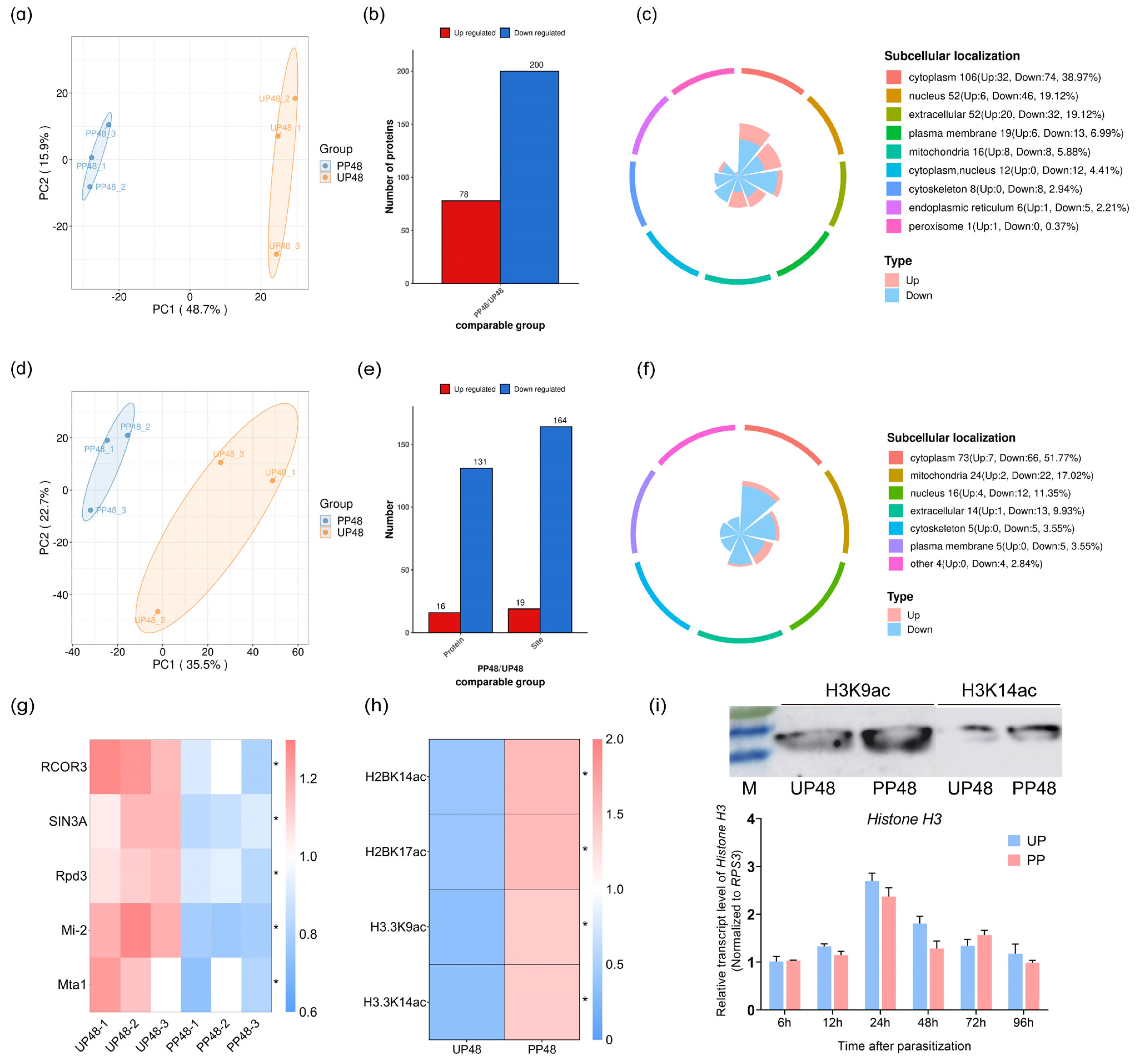
3.1. Initial Analysis of Regulation of Host Protein LysAc through the Parasitism of T. brontispae

Parasitoid wasps strategically target the hemolymph and fat body tissues of their hosts, which are essential for nurturing their offspring. To explore this dynamic, we harvested these tissues from pupae at intervals of 24, 48, and 72 h post-parasitization, alongside tissues from un-parasitized pupae as controls. Proteins extracted from these samples underwent Western blot analysis using a LysAc-specific pan antibody to evaluate the LysAc landscape. The analysis identified two prominent protein bands with molecular weights of 55 and 75 kDa, surpassing the molecular weight of histones, pinpointed at approximately 15 kDa based on our transcriptome data (Figure 1). This discovery underscores the occurrence of LysAc across both histone and non-histone proteins. Significantly, a marked decrease in LysAc modification levels was observed in the fat body tissues at 48 h post-parasitization, highlighting this time point as being crucial for differential LysAc modulation between the UP and PP groups (Figure 1). This critical finding directed our subsequent omics analyses towards the 48 h post-parasitization mark, with the aim of uncovering the intricate mechanisms through which T. brontispae parasitism influences host protein regulation.

3.2. Dynamic Changes in Proteome and LysAc Modifications in Host Pupae after Parasitization

To elucidate the regulatory effects of T. brontispae on LysAc modification, we utilized a comprehensive approach combining 4D-label-free proteomics and LysAc modification omics in O. nipae pupae at 48 h post-parasitization and un-parasitized controls. Principal component analysis (PCA) underscored a high correlation among biological replicates (Figure 2a,d), affirming the reliability of our dataset. The acetylome analysis unveiled 5515 unique LysAc sites across 1933 proteins (Supplementary Table S2). When normalized with proteomic data, 851 proteins with quantitative information were identified as acetylated, featuring 2660 unique LysAc sites (Supplementary Table S2). Applying the criteria of FC > 1.2 or FC < 1/1.2 and a p-value < 0.05 between the PP and NP groups, we identified 78 up-regulated and 200 down-regulated proteins (Figure 2b). To further our investigation, 19 unique sites on 16 proteins were found to be acetylated, whereas 164 sites on 131 proteins were deacetylated (Figure 2e). These differentially expressed and acetylated proteins predominantly resided in the cytoplasm, mitochondria, and extracellular regions (Figure 2c,f). Notably, proteins enriched in histone deacetylase activity and protein deacetylase activity were identified through Molecular Function GO enrichment analysis (Supplementary Table S3), with Rpd3, a HDAC, exhibiting decreased expression in the PP groups (Figure 2g).
KEGG pathway analysis further explored the potential functions of differentially acetylated proteins, revealing activation and enrichment in metabolism-related pathways (e.g., fructose and mannose metabolism, citrate cycle) and cell signaling pathways (e.g., cellular senescence, cGMP-PKG signaling pathway) (Supplementary Table S4). These findings highlight LysAc’s significant role in modulating host physiology following T. brontispae parasitization.
Special attention was given to histone proteins to compare their abundance and the LysAc modification levels between PP and NP. In line with our expectations, parasitization did not affect the abundance of these histone proteins (Supplementary Table S5). However, a notable increase in LysAc acetylation, including H2BK14ac, H2BK17ac, H3.3K9ac, and H3.3K14ac, was observed, with an approximate 4-fold increase in PP (Figure 2h and Table S5). Considering histone H3.3’s varied roles in transcription, genomic stability, and mitosis, Western blot analysis was conducted to confirm the increased acetylation of H3.3K9 and H3.3K14 (Figure 2i and Table S6), with qPCR validating that mRNA levels of histone H3 remained constant throughout the parasitization timeline (Figure 2i).

3.3. Expression Analysis of Host HATs and HDACs Induced by the Parasitism of T. brontispae

Proteomic analysis unveiled a distinctive expression pattern of Rpd3 at 48 h post-parasitization compared to un-parasitized pupae, piquing our interest in Rpd3’s transcriptional dynamics during the early stages of parasitization. A pronounced decrease in Rpd3 mRNA levels was observed as early as 12 h post-parasitization, showing a significant reduction of approximately 0.7-fold in both Rpd3 and its isoforms (Figure 3). In addition to Rpd3, we assessed the mRNA expression levels of other HDACs—including HIS, SAP130, and Sir2—and HATs, namely KAT2A, KAT2B, KAT6B, KAT7, and KAT8.
Throughout the initial parasitization period, Sir2 isoforms exhibited a decrease in mRNA levels by about 0.3-fold and 0.6-fold, respectively, while the transcripts of other HDACs remained largely unchanged (Figure 3). Moreover, a subtle yet significant variation in the abundance of KAT2B and KAT6B was detected between the PP and UP groups (Figure 3). This comprehensive analysis of Rpd3, along with other HDACs and HATs, during early parasitization highlights the nuanced regulatory mechanisms T. brontispae employs to modulate host epigenetic machinery, suggesting the targeted suppression of specific histone-modifying enzymes as a strategic adaptation to facilitate successful parasitism.

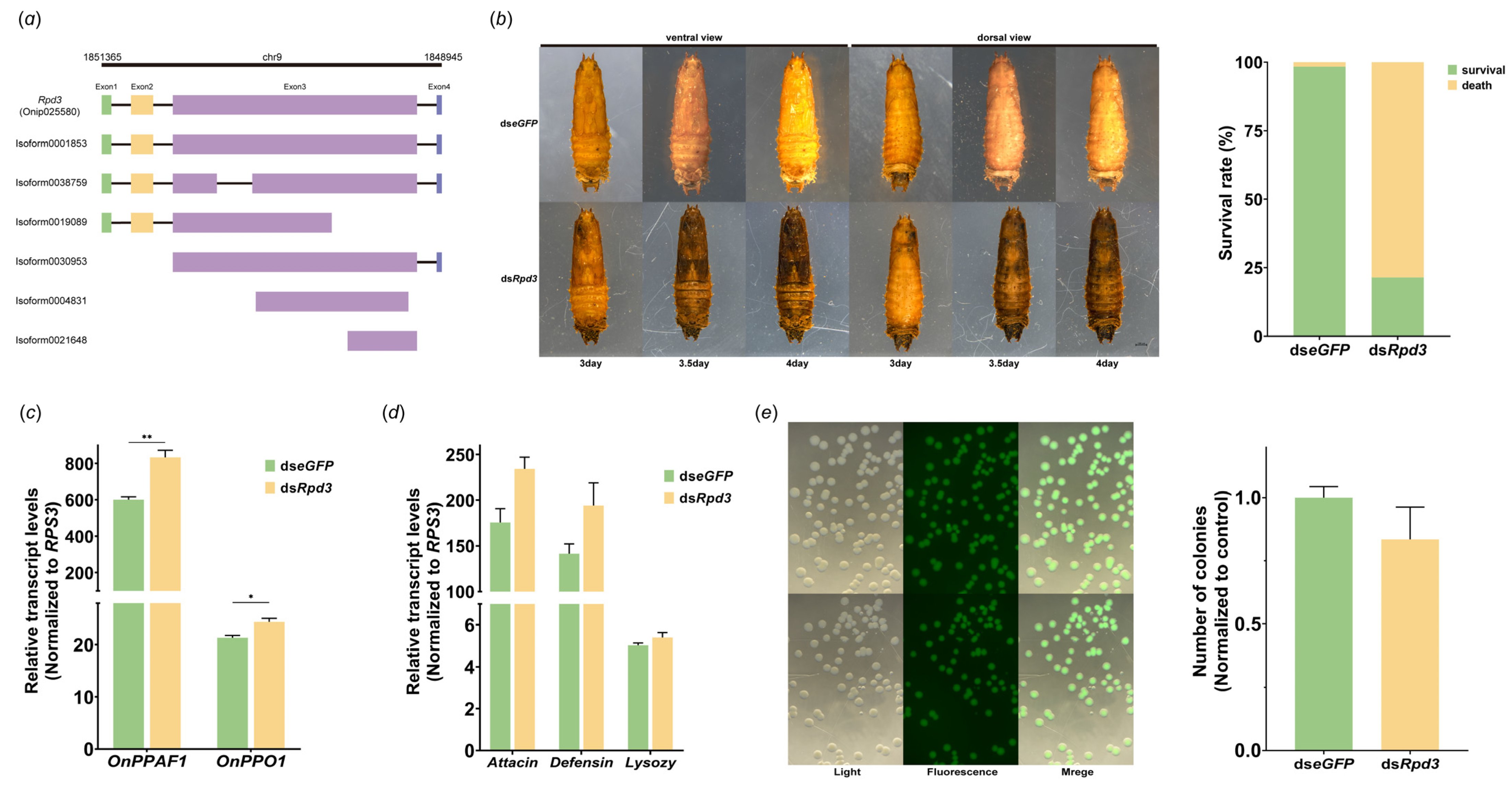
3.4. Role of Rpd3 in Prophenoloxidase Cascade

The parasitization by T. brontispae precipitated significant down-regulation in the expression of Rpd3 at both the protein and transcript levels, leading us to further investigate its role in this biological process. Considering the presence of multiple isoforms of Rpd3, dsRNA was precisely engineered to target a conserved sequence within exon 3 (Figure 4a). Notably, the efficiency of RNA interference was observed to reach 60% at 24 h post-injection with dsRpd3, demonstrating a substantial reduction in Rpd3 expression (Figure S1). Fascinatingly, a marked phenotypic change was observed; the pupae’s heads began to darken at 72 h post-injection, with the discoloration extending to the thorax and eventually leading to the entire body turning black or resulting in death by 96 h post-injection (Figure 4b). This phenotype closely mimics that of mummified parasitized pupae, and calculations revealed the mortality rate to be approximately 80% (Figure 4b).
To elucidate the underlying cause of this dramatic transformation, we analyzed the expression levels of crucial genes within the phenoloxidase cascade, specifically OnPPAF1 and OnPPO1, at 24 h after dsRpd3 injection. The results showed a statistically significant increase in the transcripts of both OnPPAF1 and OnPPO1, albeit modest (OnPPAF1, t4 = 5.702, p = 0.0047; OnPPO1, t4 = 3.756, p = 0.0199) (Figure 4c).
Moreover, given previous reports of clip-domain serine proteases being involved in the synthesis of antimicrobial peptides, we assessed the expression of antimicrobial peptide genes and the hemolymph’s capacity to clear foreign pathogens following Rpd3 knockdown. However, we observed no significant changes in the mRNA levels of these antimicrobial peptide genes or in the hemolymph’s pathogen clearance ability at 24 h following Rpd3 interference (Attacin, t4 = 2.733, p = 0.0523; Defensin, t4 = 1.957, p = 0.1220; Lysozy, t4 = 1.496, p = 0.2089) (Figure 4d,e).

4. Discussion

Endoparasitoids have evolved an intricate repertoire of strategies to modulate host physiological pathways, creating an optimal environment for their offspring’s development [37]. Central to these strategies are virulence factors such as venoms and polydnaviruses, which are adept at altering host physiological responses to favor parasitoid development [38,39]. The integration of polydnaviruses into the host genome, a phenomenon observed in Braconidae and Ichneumonidae wasps, exemplifies a profound level of genomic integration and expression, underscoring a deep symbiotic relationship [40,41,42]. This context provides a unique lens through which the evolutionary origins of parasitoid venoms, possibly resulting from LGT from ancient viral symbionts, can be explored [15,16]. Such a perspective adds depth to our understanding of the co-evolutionary dynamics between parasitoids and their hosts, highlighting the complexity of these biological interactions.
Our study delves into the realm of epigenetic modifications, focusing on histone acetylation and deacetylation, to understand how T. brontispae influences host gene expression. The bracovirus of C. plutellae offers a compelling case of host manipulation through epigenetic modifications, introducing a variant histone H4 with an extended N-terminal tail into the host nucleosome, facilitating transcriptional reprogramming within the host [43,44]. This example underscores the pivotal role of histone modifications in the intricate relationship between parasitoids and their hosts, setting the stage for our investigation into T. brontispae’s epigenetic influence.
Initial analyses using anti-LysAc antibody-based Western blots revealed that the impact of lysine acetylation extends beyond histones to non-histone proteins, affecting vital metabolic and immune pathways post-parasitization. The observed predominance of lysine deacetylation, aligning with known transcription repression mechanisms [45,46], suggests a sophisticated strategy employed by T. brontispae to suppress host immunity.
Our further comparative LysAc proteomics identified an increase in the acetylation levels of the histone proteins H3.3K9 and H3.3K14 in parasitized pupae, highlighting the dynamic interplay between HATs and HDACs in mediating these modifications [23,47]. The down-regulation of Rpd3, a class I HDAC previously identified in Saccharomyces cerevisiae as being involved in the acetylation modification of histone H4K12 [48] and correlated with the acetylation levels of histones H3 and H4 across several biological pathways in Drosophila and Tribolium castaneum [49,50], presents a sophisticated epigenetic regulatory mechanism employed by T. brontispae. This mechanism is further illustrated by the significant differences in the mRNA expression levels of Sir2, KAT2B, and KAT6B between parasitized and un-parasitized groups, emphasizing the nuanced role of Rpd3 and other HDACs and HATs in host–parasitoid interactions.
The physiological roles of Rpd3, from influencing lifespan and memory capacity in Drosophila to developmental stagnation in T. castaneum upon interference [51,52], further contextualize our findings. The knockdown of Rpd3 in O. nipae pupae, resulting in black clumps and subsequent death, resembles mummified parasitized pupae and suggests a link to melanization processes [53]. This observation led us to investigate OnPPAF1 and OnPPO1 expression [35,36], pivotal in the phenoloxidase cascade, which, despite not showing significant fold changes post-Rpd3 silencing, aligns with enhanced host PO activity observed following parasitization [54].
Despite these significant findings, the absence of notable changes in antimicrobial peptide gene expression or pathogen clearance ability following Rpd3 silencing, coupled with the modest fold changes in the expression of other HDACs and HATs, as well as OnPPAF1 and OnPPO1, underscores the complexity of the interactions at play. This complexity suggests that the timeframe of our investigation may not have been sufficient to capture the extensive range of epigenetic and transcriptional shifts induced by T. brontispae parasitization. This observation underscores the necessity for further research to elucidate the temporal dynamics of these regulatory mechanisms. Additionally, future studies should focus on the role of venom as a factor in driving these epigenetic modifications, which could provide deeper insights into the mechanisms by which T. brontispae influences host gene expression.
In summary, this investigation sheds light on a detailed regulatory mechanism by which T. brontispae modulates host prophenoloxidase activity through Rpd3-mediated histone acetylation, emphasizing the critical role of histone acetylation in regulating prophenoloxidase expression. Our findings not only contribute to the understanding of the epigenetic regulatory strategies employed by idiobiont endoparasitoids lacking polydnaviruses and teratocytes, but also highlight the significance of histone acetylation in the regulation of host immune responses, offering new insights into the survival strategies of parasitoid offspring.

Supplementary Materials

The following supporting information can be downloaded at: https://www.mdpi.com/article/10.3390/insects15030161/s1, Figure S1: Interference efficiency of Rpd3; Table S1: Primers for qRT-PCR and RNAi; Table S2: Summary of MS/MS spectrum database search analysis; Table S3: Go enrichment analysis of differentially expressed proteins; Table S4: KEGG pathway enrichment analysis of differentially acetylated proteins; Table S5: Summary of histone lysine acetylation modification. Table S6: Analysis of Western blot result through calculation of integrated density; Original Western blot image of Figure S1: Figure S1—24 and 48 h samples, Figure S1—72 h samples; Original Western blot image of Figure S2i.

Author Contributions

Conceptualization, B.-Z.T.; investigation, K.J., Y.Z., W.C., Y.-W.H., P.C. and G.-M.L.; resources, B.-Z.T. and Y.-M.H.; data curation, K.J., Y.Z., W.C., Y.-W.H., P.C. and G.-M.L.; formal analysis, K.J., Y.Z., W.C., Y.-W.H., P.C. and G.-M.L.; validation, K.J., Y.Z., W.C., Y.-W.H., P.C. and G.-M.L.; writing—original draft preparation, K.J.; writing—review and editing, B.-Z.T. and Y.-M.H.; visualization, B.-Z.T. and K.J.; supervision, B.-Z.T.; project administration, B.-Z.T.; funding acquisition, B.-Z.T. All authors have read and agreed to the published version of the manuscript.

Funding

This work was funded by grants from the National Natural Science Foundation of China [32072492 and 31672086] and the Natural Science Foundation of Fujian Province, China [2020J01595].

Data Availability Statement

The data that support the findings of this study are available from the corresponding author upon reasonable request.

Conflicts of Interest

The authors declare no conflicts of interest. The funders had no role in the design of the study; in the collection, analyses, or interpretation of data; in the writing of the manuscript; or in the decision to publish the results.

References

  1. Godfray, H.C.J. Parasitoids: Behavioral and Evolutionary Ecology; Princeton University Press: Princeton, NJ, USA, 1994. [Google Scholar]
  2. Moreau, S.J.M.; Asgari, S. Venom proteins from parasitoid wasps and their biological functions. Toxins 2015, 7, 2385–2412. [Google Scholar] [CrossRef]
  3. Corley, L.S.; Strand, M.R. Evasion of encapsulation by the polyembryonic parasitoid Copidosoma floridanum is mediated by a polar body-derived extraembryonic membrane. J. Invertebr. Pathol. 2003, 83, 86–89. [Google Scholar] [CrossRef]
  4. Hu, J.; Xu, Q.; Hu, S.; Yu, X.; Liang, Z.; Zhang, W. Hemomucin, an O-glycosylated protein on embryos of the wasp Macrocentrus cingulum that protects it against encapsulation by Hemocytes of the host Ostrinia furnacalis. J. Innate Immun. 2014, 6, 663–675. [Google Scholar] [CrossRef] [PubMed]
  5. Asgari, S.; Rivers, D.B. Venom proteins from endoparasitoid wasps and their role in host-parasite interactions. Annu. Rev. Entomol. 2011, 56, 313–335. [Google Scholar] [CrossRef] [PubMed]
  6. Beckage, N.E. Polydnaviruses as endocrine regulators. In Parasitoid Viruses; Academic Press: Cambridge, MA, USA, 2012; pp. 163–168. [Google Scholar] [CrossRef]
  7. Strand, M.R. Polydnavirus gene products that interact with the host immune system. In Parasitoid Viruses; Academic Press: Cambridge, MA, USA, 2012; pp. 149–161. [Google Scholar] [CrossRef]
  8. Strand, M.R. Teratocytes and their functions in parasitoids. Curr. Opin. Insect Sci. 2014, 6, 68–73. [Google Scholar] [CrossRef] [PubMed]
  9. Ye, X.Q.; Shi, M.; Huang, J.H.; Chen, X.X. Parasitoid polydnaviruses and immune interaction with secondary hosts. Dev. Comp. Immunol. 2018, 83, 124–129. [Google Scholar] [CrossRef] [PubMed]
  10. Skinner, M.K. Environmental epigenetics and a unified theory of the molecular aspects of evolution: A neo-lamarckian concept that facilitates neo-Darwinian evolution. Genome Biol. Evol. 2015, 7, 1296–1302. [Google Scholar] [CrossRef] [PubMed]
  11. Bonduriansky, R.; Crean, A.J.; Day, T. The implications of nongenetic inheritance for evolution in changing environments. Evol. Appl. 2012, 5, 192–201. [Google Scholar] [CrossRef] [PubMed]
  12. Jaenisch, R.; Bird, A. Epigenetic regulation of gene expression: How the genome integrates intrinsic and environmental signals. Nat. Genet. 2003, 33 (Suppl. S3), 245–254. [Google Scholar] [CrossRef] [PubMed]
  13. Tang, C.K.; Tsai, C.H.; Wu, C.P.; Lin, Y.H.; Wei, S.C.; Lu, Y.H.; Li, C.H.; Wu, Y.L. MicroRNAs from Snellenius manilae bracovirus regulate innate and cellular immune responses of its host Spodoptera litura. Commun. Biol. 2021, 4, 52. [Google Scholar] [CrossRef]
  14. Wang, Z.Z.; Ye, X.Q.; Shi, M.; Li, F.; Wang, Z.H.; Zhou, Y.N.; Gu, Q.J.; Wu, X.T.; Yin, C.L.; Guo, D.H.; et al. Parasitic insect-derived miRNAs modulate host development. Nat. Commun. 2018, 9, 2205. [Google Scholar] [CrossRef] [PubMed]
  15. Martinson, E.O.; Martinson, V.G.; Edwards, R.; Mrinalini; Werren, J.H. Laterally transferred gene recruited as a venom in parasitoid wasps. Mol. Biol. Evol. 2016, 33, 1042–1052. [Google Scholar] [CrossRef]
  16. Gilbert, S.F.; McDonald, E.; Boyle, N.; Buttino, N.; Gyi, L.; Mai, M.; Prakash, N.; Robinson, J. Symbiosis as a source of selectable epigenetic variation: Taking the heat for the big guy. Philos. Trans. R. Soc. B-Biol. Sci. 2010, 365, 671–678. [Google Scholar] [CrossRef] [PubMed]
  17. Ozbek, R.; Mukherjee, K.; Uckan, F.; Vilcinskas, A. Reprograming of epigenetic mechanisms controlling host insect immunity and development in response to egg-laying by a parasitoid wasp. Proc. Biol. Sci. 2020, 287, 20200704. [Google Scholar]
  18. Kim, G.W.; Yang, X.J. Comprehensive lysine acetylomes emerging from bacteria to humans. Trends Biochem. Sci. 2011, 36, 211–220. [Google Scholar] [CrossRef] [PubMed]
  19. Hamon, M.A.; Cossart, P. Histone modifications and chromatin remodeling during bacterial infections. Cell Host Microbe 2008, 4, 100–109. [Google Scholar] [CrossRef] [PubMed]
  20. Jensen, O.N. Modification-specific proteomics: Characterization of post-translational modifications by mass spectrometry. Curr. Opin. Chem. Biol. 2004, 8, 33–41. [Google Scholar] [CrossRef]
  21. Mann, M.; Jensen, O.N. Proteomic analysis of post-translational modifications. Nat. Biotechnol. 2003, 21, 255–261. [Google Scholar] [CrossRef]
  22. Cho, Y.; Griswold, A.; Campbell, C.; Min, K.T. Individual histone deacetylases in Drosophila modulate transcription of distinct genes. Genomics 2005, 86, 606–617. [Google Scholar] [CrossRef]
  23. Mukherjee, K.; Fischer, R.; Vilcinskas, A. Histone acetylation mediates epigenetic regulation of transcriptional reprogramming in insects during metamorphosis, wounding and infection. Front. Zool. 2012, 9, 25. [Google Scholar] [CrossRef]
  24. Spange, S.; Wagner, T.; Heinzel, T.; Kramer, O.H. Acetylation of non-histone proteins modulates cellular signalling at multiple levels. Int. J. Biochem. Cell Biol. 2009, 41, 185–198. [Google Scholar] [CrossRef]
  25. Takakura, M.; Nakagawa, R.; Ota, T.; Kimura, Y.; Ng, M.Y.; Alia, A.G.; Okuno, H.; Hirano, Y. Rpd3/CoRest-mediated activity-dependent transcription regulates the flexibility in memory updating in Drosophila. Nat. Commun. 2021, 12, 628. [Google Scholar] [CrossRef] [PubMed]
  26. Zhang, H.J.; Lin, Y.P.; Li, H.Y.; Wang, R.; Fu, L.; Jia, Q.C.; Hou, Y.M.; Tang, B.Z. Variation in parasitoid virulence of Tetrastichus brontispae during the targeting of two host beetles. Int. J. Mol. Sci. 2021, 22, 3581. [Google Scholar] [CrossRef]
  27. Meng, E.; Qiao, T.; Tang, B.Z.; Hou, Y.M.; Yu, W.Z.; Chen, Z.M. Effects of ovarian fluid, venom and egg surface characteristics of Tetrastichus brontispae (Hymenoptera: Eulophidae) on the immune response of Octodonta nipae (Coleoptera: Chrysomelidae). J. Insect Physiol. 2018, 109, 125–137. [Google Scholar] [CrossRef]
  28. Tang, B.; Xu, L.; Hou, Y. Effects of rearing conditions on the parasitism of Tetrastichus brontispae on its pupal host Octodonta nipae. Biocontrol 2014, 59, 647–657. [Google Scholar] [CrossRef]
  29. Chen, Q.; Peng, Z.; Xu, C.; Tang, C.; Lu, B.; Jin, Q.; Wen, H.; Wan, F. Biological assessment of Tetrastichus brontispae, a pupal parasitoid of coconut leaf beetle Brontispa longissima. Biocontrol Sci. Technol. 2010, 20, 283–295. [Google Scholar] [CrossRef]
  30. Tang, B.Z.; Chen, J.; Hou, Y.M.; Meng, E. Transcriptome immune analysis of the invasive beetle Octodonta nipae (Maulik) (Coleoptera: Chrysomelidae) parasitized by Tetrastichus brontispae Ferrière (Hymenoptera: Eulophidae). PLoS ONE 2014, 9, e91482. [Google Scholar] [CrossRef] [PubMed]
  31. Li, J.L.; Zhang, X.; Hou, Y.M.; Tang, B.Z. Effects of multiple mating on the fecundity of an invasive pest (Octodonta nipae): The existence of an intermediate optimal female mating rate. Physiol. Entomol. 2014, 39, 348–354. [Google Scholar] [CrossRef]
  32. Koressaar, T.; Lepamets, M.; Kaplinski, L.; Raime, K.; Andreson, R.; Remm, M. Primer3_masker: Integrating masking of template sequence with primer design software. Bioinformatics 2018, 34, 1937–1938. [Google Scholar] [CrossRef]
  33. Zhang, X.M.; Zhang, H.J.; Liu, M.; Liu, B.; Zhang, X.F.; Ma, C.J.; Fu, T.T.; Hou, Y.M.; Tang, B.Z. Cloning and immunosuppressive properties of an acyl-activating enzyme from the venom apparatus of Tetrastichus brontispae (Hymenoptera: Eulophidae). Toxins 2019, 11, 672. [Google Scholar] [CrossRef]
  34. Horn, T.; Boutros, M. E-RNAi: A web application for the multi-species design of RNAi reagents—2010 update. Nucleic Acids Res. 2010, 38, 332–339. [Google Scholar] [CrossRef]
  35. Zhang, H.; Tang, B.; Lin, Y.; Chen, Z.; Zhang, X.; Ji, T.; Zhang, X.; Hou, Y. Identification of three prophenoloxidase-activating factors (PPAFs) from an invasive beetle Octodonta nipae Maulik (Coleoptera: Chrysomelidae) and their roles in the prophenoloxidase activation. Arch. Insect Biochem. Physiol. 2017, 96, e21425. [Google Scholar] [CrossRef]
  36. Zhang, X.F.; Cui, W.; Wang, M.J.; Zhou, Y.; Fu, T.T.; Jiang, K.; Hou, Y.M.; Tang, B.Z. Role of prophenoloxidase 1 from the beetle Octodonta nipae in melanized encapsulation of a wasp egg. Dev. Comp. Immunol. 2024, 150, 105082. [Google Scholar] [CrossRef]
  37. Pennacchio, F.; Strand, M.R. Evolution of developmental strategies in parasitic hymenoptera. Annu. Rev. Entomol. 2006, 51, 233–258. [Google Scholar] [CrossRef]
  38. Poirie, M.; Carton, Y.; Dubuffet, A. Virulence strategies in parasitoid Hymenoptera as an example of adaptive diversity. C. R. Biol. 2009, 332, 311–320. [Google Scholar] [CrossRef]
  39. Glatz, R.V.; Asgari, S.; Schmidt, O. Evolution of polydnaviruses as insect immune suppressors. Trends Microbiol. 2004, 12, 545–554. [Google Scholar] [CrossRef] [PubMed]
  40. Federici, B.A.; Bigot, Y. Origin and evolution of polydnaviruses by symbiogenesis of insect DNA viruses in endoparasitic wasps. J. Insect Physiol. 2003, 49, 419–432. [Google Scholar] [CrossRef] [PubMed]
  41. Drezen, J.-M.; Chevignon, G.; Louis, F.; Huguet, E. Origin and evolution of symbiotic viruses associated with parasitoid wasps. Curr. Opin. Insect Sci. 2014, 6, 35–43. [Google Scholar] [CrossRef] [PubMed]
  42. Strand, M.R.; Burke, G.R. Polydnaviruses: Evolution and function. Curr. Issues Mol. Biol. 2019, 34, 163–182. [Google Scholar] [PubMed]
  43. Talbert, P.B.; Armache, K.-J.; Henikoff, S. Viral histones: Pickpocket’s prize or primordial progenitor? Epigenet. Chromatin 2022, 15, 21. [Google Scholar] [CrossRef] [PubMed]
  44. Hepat, R.; Song, J.-J.; Lee, D.; Kim, Y. A viral histone H4 joins to eukaryotic nucleosomes and alters host gene expression. J. Virol. 2013, 87, 11223–11230. [Google Scholar] [CrossRef] [PubMed]
  45. Narita, T.; Weinert, B.T.; Choudhary, C. Functions and mechanisms of non-histone protein acetylation. Nat. Rev. Mol. Cell Biol. 2019, 20, 156–174. [Google Scholar] [CrossRef] [PubMed]
  46. Shvedunova, M.; Akhtar, A. Modulation of cellular processes by histone and non-histone protein acetylation. Nat. Rev. Mol. Cell Biol. 2022, 23, 329–349. [Google Scholar] [CrossRef] [PubMed]
  47. Vilcinskas, A. The impact of parasites on host insect epigenetics. Insect Epigenet. 2017, 53, 145–165. [Google Scholar]
  48. Chang, C.S.; Pillus, L. Collaboration between the essential Esa1 acetyltransferase and the Rpd3 deacetylase is mediated by H4K12 histone acetylation in Saccharomyces cerevisiae. Genetics 2009, 183, 149–160. [Google Scholar] [CrossRef]
  49. Foglietti, C.; Filocamo, G.; Cundari, E.; De Rinaldis, E.; Lahm, A.; Cortese, R.; Steinkuhler, C. Dissecting the biological functions of Drosophila histone deacetylases by RNA interference and transcriptional profiling. J. Biol. Chem. 2006, 281, 17968–17976. [Google Scholar] [CrossRef]
  50. Pile, L.A.; Schlag, E.M.; Wassarman, D.A. The SIN3/RPD3 deacetylase complex is essential for G(2) phase cell cycle progression and regulation of SMRTER corepressor levels. Mol. Cell Biol. 2002, 22, 4965–4976. [Google Scholar] [CrossRef]
  51. Rogina, B.; Helfand, S.L.; Frankel, S. Longevity regulation by Drosophila Rpd3 deacetylase and caloric restriction. Science 2002, 298, 1745. [Google Scholar] [CrossRef]
  52. George, S.; Gaddelapati, S.C.; Palli, S.R. Histone deacetylase 1 suppresses Kruppel homolog 1 gene expression and influences juvenile hormone action in Tribolium castaneum. Proc. Natl. Acad. Sci. USA 2019, 116, 17759–17764. [Google Scholar] [CrossRef] [PubMed]
  53. Mortimer, N.T.; Fischer, M.L.; Waring, A.L.; Kr, P.; Kacsoh, B.Z.; Brantley, S.E.; Keebaugh, E.S.; Hill, J.; Lark, C.; Martin, J.; et al. Extracellular matrix protein N-glycosylation mediates immune self-tolerance in Drosophila melanogaster. Proc. Natl. Acad. Sci. USA 2021, 118, e2017460118. [Google Scholar] [CrossRef] [PubMed]
  54. Meng, E.; Tang, B.Z.; Hou, Y.M.; Chen, X.X.; Chen, J.T.; Yu, X.Q. Altered immune function of Octodonta nipae (Maulik) to its pupal endoparasitoid, Tetrastichus brontispae Ferrière. Comp. Biochem. Physiol. B Biochem. Mol. Biol. 2016, 198, 100–109. [Google Scholar] [CrossRef] [PubMed]
Figure 1. Analysis of LysAc modification level in tissues from post-parasitized (PP) and un-parasitized (UP) pupae using Coomassie brilliant blue gel staining (a) and Western blot (b). Hemolymph and fat body samples were collected at 24, 48, and 72 h post-parasitization. Tissues from un-parasitized pupae were collected simultaneously as controls. To ensure accuracy, the quality of an equal amount of protein in each lane was verified through Coomassie brilliant blue gel staining. The samples were then subjected to immunoblot analysis using a pan anti-acetyllysine mouse monoclonal antibody. The band with a molecular weight of approximately 15 kDa, indicative of histone protein, is highlighted with a red triangle, while bands with higher molecular weights correspond to non-histone proteins. Among these, two obvious non-histone bands, 55 and 85 kDa, are highlighted with red arrows. F, fat body samples; H, hemolymph samples.
Insects 15 00161 g001
Figure 2. Dynamic changes in proteome and LysAc modifications in host pupae after parasitization. (a,d) PCA analyses of protein expression (a) and LysAc modifications (d) in two groups: PP48 (proteins from pupae at 48 h post-parasitization) versus UP48 (proteins from un-parasitized pupae at the same time point). (b,e) Summary of differentially expressed proteins and proteins with differential lysine acetylation modification between PP and UP, identified by a fold change (FC) > 1.2 or FC < 1/1.2 and p-value < 0.05. (c,f) Subcellular classification analysis of the differentially expressed proteins (c) and proteins with differential LysAc modification (f). (g) Heat map displaying proteins enriched in the GO term for protein deacetylase activity. (h) Heat map showing the level of LysAc modification in histone proteins. (i) Verification of the LysAc modification level of histone H3.3 via Western blot and qRT-PCR. Protein samples were identical to those used in omics analyses. Immunoblots were probed with the primary antibodies anti-Acetyl-Histone H3 (Lys9) rabbit monoclonal antibody and anti-Acetyl-Histone H3 (Lys14) rabbit monoclonal antibody. RNA samples were extracted at various time points post-parasitization: 6, 12, 24, 48, 72, and 96 h. The transcript level of histone H3 was normalized against the reference gene ribosomal protein S3 (rpS3), with UP samples collected at 6 h serving as the baseline. “*” means significant difference at the 0.05 level between PP and UP.
Insects 15 00161 g002
Figure 3. Transcript levels of HATs and HDACs. Eight HATs (KAT2A, KAT2B, KAT6B, KAT7, and KAT8) and eight HDACs (HIS1, Rpd3, SAP130, and Sir2) were selected for analysis. RNA samples were collected at various time points post-parasitization (6, 12, 24, and 48 h) (PP). Control RNA samples from un-parasitized pupae (UP) were collected simultaneously. Transcript levels were normalized to the reference gene rpS3 and the UP sample collected at 6 h. * and ** indicate significant differences between PP and UP at the levels of p < 0.05 and p < 0.01, respectively.
Insects 15 00161 g003
Figure 4. Involvement of Rpd3 in the regulation of prophenoloxidase cascade. (a) Analysis of alternative splicing of Rpd3. Chr9 represents chromosome 9 in the O. nipae genome, while 1,851,365 and 1,848,945 indicate the locations of Onip025580 (Rpd3) in the genome. Exon1–4 denote the four exons of this gene. (b) Phenotypic variation and survival analysis in O. nipae pupae following Rpd3 knockdown. dseGFP was used as a control. Observations were made at 3, 3.5, and 4 days post-injection of dsRpd3. (c,d) Transcript levels of genes related to the prophenoloxidase cascade (c) and the antimicrobial peptide (d) 24 h post-Rpd3 knockdown. * and ** represent significant differences between PP and UP at the levels of p < 0.05 and p < 0.01, respectively. (e) Evaluation of pathogen clearance in hemolymph after Rpd3 knockdown. Pupae were treated with dsRNA for 12 h, and then, subjected to another injection of E. coli with green fluorescence for another 12 h. Cultured media were examined under a fluorescence microscope, counting and measuring the area of fluorescent colonies.
Insects 15 00161 g004
Disclaimer/Publisher’s Note: The statements, opinions and data contained in all publications are solely those of the individual author(s) and contributor(s) and not of MDPI and/or the editor(s). MDPI and/or the editor(s) disclaim responsibility for any injury to people or property resulting from any ideas, methods, instructions or products referred to in the content.

