Review

Epidemiology of Sandfly-Borne Phleboviruses in North Africa: An Overview

1
Institute of Veterinary Sciences, University Blida 1, Blida 09000, Algeria
2
Laboratory of Biotechnologies Linked to Animal Reproduction, Institute of Veterinary Sciences, University Blida 1, Blida 09000, Algeria
3
Department of Microbiology and Veterinary Pathology, Institut Pasteur of Algeria, Algiers 16000, Algeria
4
Department of Preventive Medicine, Institut Pasteur of Algeria, Algiers 16000, Algeria
5
Center of Research in Agro-Pastoralism, Djelfa 17000, Algeria
*
Author to whom correspondence should be addressed.
Insects 2024, 15(11), 846; https://doi.org/10.3390/insects15110846
Submission received: 27 September 2024 / Revised: 21 October 2024 / Accepted: 25 October 2024 / Published: 29 October 2024
(This article belongs to the Section Medical and Livestock Entomology)

Simple Summary

Phleboviruses transmitted by sandflies had been described in North Africa since seven decades ago. However, knowledge gaps still exist in the region. Therefore, a comprehensive literature search is performed to provide an overview of their evidence and distribution. At least eight phleboviruses transmitted by sandflies are present in North African region, namely Toscana, Sicilian, Naples, Cyprus, Punique, Utique, Saddaguia, and Medjerda Valley viruses. They were detected in sandflies, in animals, or in humans from Morocco, Algeria, Tunisia, Libya, and Egypt. Their infection rate ranged from 0.02% to 0.6% in sandflies and from 4.3% to 43.5% in dogs. Their overall seroprevalence in healthy persons was 15.9%. They were concentrated mainly in the north and might occur together. Sicilian (1.3–21%) and Toscana (3.8–50%) viruses were the most prevalent among humans. In particular, Toscana virus was frequent and involved in clinical cases from Algeria (3.8%) and Tunisia (12.86%). Noticeably, data from Morocco focused on TOSV in sandflies, whereas data from Libya and Egypt were very limited and old, respectively. Phleboviruses transmitted by sandflies are widespread and constantly present in North Africa. This calls for the implementation of phlebovirus diagnosis in patients with febrile and neurological manifestations.

Abstract

Eight sandfly-borne phleboviruses were found to circulate in North Africa. Phleboviruses detected in sandflies were Toscana (TOSV), Sandfly Fever Sicilian (SFSV), Sandfly Fever Naples (SFNV), Cyprus (CYPV), Punique (PUNV), Utique, Saddaguia, and Medjerda Valley (MVV) viruses, yielding an overall infection rate of 0.02–0.6%. Phlebotomus perniciosus and Phlebotomus longicuspis were the most common vector species in the region. TOSV seroprevalence in dogs from Algeria (4.56%) and Tunisia (7.5%) was low and close, unlike SFSV (38.1%) and PUNV (43.5%), which were restricted to Tunisia. SFSV (1.3–21%) and TOSV (3.8–50%) were the most prevalent among humans. TOSV was frequently detected and symptomatically confirmed in both Algeria (3.8%) and Tunisia (12.86%). Other sandfly-borne phleboviruses have also been detected but less importantly, such as SFNV in Morocco (2.9%) and Tunisia (1.1%) and PUNV (8.72%), CYPV (2.9%), and MVV (1.35%) in Tunisia. Their distribution was mainly northern. Overall, 15.9% of the healthy population were seropositive for sandfly-borne phleboviruses, with evidenced cocirculation. Noticeably, studies conducted in Morocco were mostly interested in TOSV in sandflies. Available data from Libya and Egypt were scant or historical. Further elaboration is required to check the sporadic detection of less-prevalent phleboviruses and fully elucidate the epidemiological situation.

1. Introduction

The last few decades have been characterized by the global emergence of many new infectious diseases in the human population caused by arthropod-borne viruses (arboviruses) [1,2]. Viral diseases are spreading quickly to new territories because of commercial transportation, globalization, deforestation, and urbanization. Climate and environmental changes induced by those disruptive human activities facilitate the spread of vectors and cause them to get closer to humans [3,4].
Phlebotomine sandflies (Diptera: Psychodidae) are regarded as important arthropods in human and veterinary medicine, as they harbor pathogens that infect humans and both domestic and wild animals [5,6]. They have a global distribution extending over southern Europe, Asia, Africa, Australia, and central and South America, with peak activity in the warmer months of the year [7]. Despite having a relatively limited geographic dispersal capacity compared with mosquitoes, sandflies exhibit an expansion of activity zones owing to changes in the climate and ecosystem, as exemplified by vector abundance and leishmaniasis cases [7,8]. Humidity is one of the key factors, together with suitable temperature, that define their geographical distribution in vast regions of both the old and new worlds [9]. They are often abundant in peri-urban, rural, and natural environments, which are concentrated near human and domestic animal habitats in suitable areas [10,11]. Therefore, further introduction of pathogens into naïve regions, leading to disease emergence and outbreaks, should be expected [6,7].
Although primarily known as proven vectors of protozoan parasite Leishmania (Le.), sandflies also transmit other pathogens during their blood meal, namely bacteria of the genus Bartonella and viruses belonging to five different families: Rhabdoviridae, Flaviviridae, Reoviridae, Peribunyaviridae, and Phenuiviridae [5,6,12]. Viral pathogens vectored by sandflies mainly belong to the Phlebovirus genus within the Phenuiviridae family (order Bunyavirales), which is by far the most studied [5,13,14,15,16,17]. As provided by the International Committee on Taxonomy of Viruses (ICTV), these viruses are currently classified into species based on the threshold of 95% identity in the amino acid sequence of the ribonucleic acid (RNA)-dependent RNA polymerase (RdRp) protein [13]. Accordingly, Phlebovirus comprises about 70 named viruses, of which Toscana virus (TOSV), Sandfly Fever Naples virus (SFNV), Sandfly Fever Sicilian virus (SFSV), and Cyprus virus (CYPV) are most frequently detected in sandflies [13,18,19,20]. They have been documented as human pathogens, with symptoms spanning from fever to central nervous system involvement [21,22]. SFSV and SFNV are responsible for a self-resolving febrile illness called sandfly or Phlebotomus fever [23,24]. CYPV, a variant of the SFSV, causes an acute febrile disease [25,26]. TOSV displays a neurotropism distinct from other phleboviruses. It is unique as an agent of central nervous system infections that manifest as aseptic meningitis, encephalitis, and meningoencephalitis following the febrile period [21,27].
Sandfly-borne phleboviruses’ distribution and circulation peaks are closely related to vector presence and activity [28]. The Mediterranean countries have been identified as endemic regions for many sandfly-borne phleboviruses [27,29]. They are not only confined to southern Europe and the Middle East: there is significant evidence for their presence in North African countries [22,30,31,32,33,34,35].
The number of newly described sandfly-borne phleboviruses has been steadily growing in recent years [36]. Recent studies conducted in Mediterranean countries showed an increase in phlebovirus circulation with newly discovered viruses [36,37]. For instance, seven phleboviruses representing three distinct serocomplexes were screened in the endemic region of the eastern Mediterranean basin, with various phleboviruses in circulation together [22]. Unfortunately, no up-to-date review is available to summarize the current published data.
Although sandfly-borne phleboviruses had been described in North Africa from seven decades ago, knowledge gaps still exist in the region [38]. Indeed, phleboviruses remain obviously less studied in Africa in comparison to European and Asian countries [15,23,33,34,35,39,40,41]. Despite several reports of acute infections and preliminary findings of seroreactivity in humans and non-human vertebrates, information on phlebovirus exposure remains limited, and their health relevance is largely uncharacterized [22,36,42]. This unsatisfactory state of knowledge could also be due to fragmentary and rather poorly understood data when compared with vast knowledge of well-investigated regions.
As these diseases can easily spread to even uninfected areas, permanent monitoring is needed to control phleboviruses [42]. Understanding epidemiology contributes to public health protection by predicting virus transmission patterns and so preventing infection emergence. Herein, a comprehensive literature search is performed with the aim of providing an overview of the evidence and the distribution in hosts and geographic locations of sandfly-borne phleboviruses across North African region. Relevant online literature was sought on the academic search engines of Google Scholar and PubMed, using the terms sandfly-borne phlebovirus, Morocco, Algeria, Tunisia, Libya and Egypt. Gathered data were mapped with Esri Arcgis pro, Version 3.3, license acquired by Institut Pasteur of Algeria.

2. Sandfly-Borne Phleboviruses in Algeria

2.1. Sicilian Virus

Sandfly-borne phleboviruses were first described in Algeria two decades ago, as attention had been drawn to TOSV in the world. The first sandfly-borne phlebovirus investigated in Algeria was SFSV, after it had been shown to be circulating in Asia, Africa, and southern Europe. In 2006, a viral sequence closely related to that of SFSV was detected in a Phlebotomus (Ph.) ariasi from Larbaa Nath Iraten in the governorate of Tizi Ouzou (Kabylia), northern Algeria. Hence, sera from healthy persons living in the same locality were tested for SFSV immunoglobulin (Ig) G, of which 5% were positive [43].

2.2. Cyprus Virus

The sequence determined from Ph. papatasi trapped in northern Algeria in 2007 was most closely related to CYPV but not to the SFSV-like phlebovirus obtained from Ph. ariasi in the same region in 2006. No virological nor serological data were available to support this sporadic detection of Cyprus-like virus in Algeria [44].

2.3. Naples Virus

In 2007, a combination of entomological and seroprevalence studies provided evidence that a novel phlebovirus distinct from but related to SFNV was present in two areas, Larbaa Nath Iraten and Bou Ismail in the governorates of Tizi Ouzou and Tipaza, respectively, in northern Algeria, and could infect humans. Two sequences obtained from Ph. longicuspis were found to be most closely related to the Poona strain of SFNV and clearly belonged to the species SFNV. Human infection due to this virus was supported by seroprevalence studies indicating rates varying from 10.6 to 21.6%. The indirect immunofluorescence-based seroprevalence studies were carried out on healthy persons and patients attending biological laboratories for any reason in the vicinity of the areas where sandflies were trapped, respectively [44].

2.4. Toscana Virus

One strain of TOSV, named TOSV–Algeria 189, was isolated from sandflies collected in 2013, in Draa El Mizan, governorate of Tizi Ouzou. Phylogenetic studies indicated that it was most closely related with the TOSV strain from Tunisia within lineage A, which also includes Italian, French, and Turkish strains. A seroprevalence study performed on blood donor sera originating from the same area showed that almost 50% possessed TOSV-neutralizing antibodies, a much higher rate than that observed in southern Europe. Together, these results strongly suggested that TOSV was heavily affecting sandfly-exposed people in northern Algeria [31].
A further study has reported the results of the diagnosis of neuro-invasive TOSV infection in Algeria between 2016 and 2018 and described the first isolation of the TOSV strain from human samples in North Africa. TOSV infection was confirmed in 3.8% of hospitalized patients displaying neurological infection, mainly restricted to northern Algeria. TOSV cases were identified in coastal or inland governorates known for high activity of sandfly vectors and where TOSV-specific antibodies were detected in dogs, but not in the region of Kabylia, although it had previously been reported to be highly prevalent in humans there. The isolated strain was recovered from the cerebrospinal fluid (CSF) of a patient living in the governorate of Tlemcen, northwest of Algeria, and was genetically close to the strain isolated from sandflies collected in the region of Kabylia in 2013. These data confirmed that TOSV was endemic in Algeria and represented an important etiologic agent of neuro-invasive disease [34].
Regarding animals, studies were rather interested in dogs, as they were shown to be good markers for virus exposure and proxies for TOSV circulation [45]. In 2014, 4.3% of watchdogs from Tizi Ouzou and Bejaia governorates, northern Algeria, appeared to have neutralizing antibodies against TOSV [46]. A more recent study reported an Enzyme-Linked Immunosorbent Assay (ELISA)-based seroprevalence rate of 20% (14.5–25.5%) and suggested likely evidence for dog transmission and circulation of phleboviruses other than TOSV. Alongside this, a seroprevalence rate of 4.56% (1.65–7.43%) was recorded by a microneutralization test elucidating the exact occurrence of TOSV exposure in dogs. Eventually, these findings demonstrated that TOSV and antigenically related viruses were not only confined to Kabylia, or to northern Algeria, as believed at the outset of this study, but that they were even present in the south [45].
The latest findings provided an update of TOSV geographic distribution and revealed the exposure of different domestic animals other than canines, with a potential role of livestock (p = 0.00731) in the natural cycle. Positive results were obtained in 14.6% of cattle, 17.18% of sheep, 15% of horses, and 3.33% of goats. They originated mainly from the northern center of Algeria, with new areas where the virus was detected for the first time, namely Medea and Ain Defla in the north and Tissemsilt in the northwest [35].

2.5. Punique Virus

The impact of Punique virus (PUNV) on public health is still unknown. However, as a member of the SFNV group, of which the majority are pathogenic to humans, it may involve a potential risk for public health. An entomological study conducted in Kherrata, governorate of Bejaia, in 2020 resulted in the detection of two phleboviruses closely related to PUNV isolated in Tunisia and detected in Algeria. Ph. perniciosus (98.67%) was the dominant species and was identified in both positive pools. The overall prevalence of phlebovirus infection among the collected sandflies was 0.06%. The isolation for the first time of two different strains related to PUNV and the second report in Algeria from two distinct regions confirmed its large circulation in the country [47].

3. In Morocco

3.1. Toscana Virus

TOSV RNA was detected for the first time in Morocco in 2008, in Ph. perniciosus collected from Louata, a locality in Sefrou province, northern Morocco. The rate of infection in the vector Ph. perniciosus was (4/129) higher than rates reported in northern Mediterranean countries. The detected TOSV belonged to the genotype B, which has been previously recognized in the neighboring countries of France, Spain, and Portugal [48].
Subsequently, in the district of El Hanchane near Essaouira, center of Morocco, Ph. sergenti, the most abundant sandfly species (76%), was found to be infected by TOSV. The phylogenetic analysis indicated that the obtained sequence was most closely related to the TOSV France strain. So far, that is the first time that TOSV has been detected in phlebotomine sandflies other than Ph. perniciosus, Ph. perfiliewi, and Se. minuta. Hence, Ph. sergenti was considered to be a potential vector of TOSV [49].
In that same context, TOSV was obtained from Ph. longicuspis in Sefrou province and Ph. sergenti in Azilal province. Phylogenetic analyses indicated that those viruses clustered with TOSV strains circulating in Morocco, Spain, and France, i.e., belonged to genotype B [30].
Correspondingly, a much larger study that allowed exclusive identification of TOSV showed positive results in four provinces, Azilal, Sefrou, Essaouira, and Ouarzazate together, proving its existence in the north, the center, and the south of the country. In addition, TOSV was detected and isolated from three new sandfly vectors: Ph. sergenti, Ph. longicuspis, and Ph. papatasi. Three viruses were isolated from Ph. sergenti in Azilal, whereas two were found in Ph. longicuspis in Sefrou. The overall prevalence of TOSV infection in trapped sandflies was 0.21%. Sequence analysis confirmed that the Moroccan TOSV belonged to genotype B and was close to those isolated in France and Spain [50].
A more recent study reported evidence for TOSV in central Morocco. TOSV was detected in Lalla Laaziza locality from Chichaoua Province, where Ph. sergenti was the most abundant sandfly species (47.6%) [11].

3.2. Naples Virus

According to the unique study involving SFNV, neutralizing antibodies were found to occur in 2.9% people from Itzer in the province of Midelt, central Morocco [38].

3.3. Sicilian Virus

Scarce findings claimed that 5.7% people living in Itzer, province of Midelt, possessed SFSV-neutralizing antibodies (Nt-Ab) together with SFNV’s [38]. Shortly after, wild rodents and insectivora trapped in the north displayed a seroprevalence rate of 9.4% [51].

4. In Tunisia

4.1. Sicilian and Sicilian-like Viruses

The earliest study on this topic presented data on a selected human population in 1975, in which the SFSV-Nt-Ab rate was 1.6% [38]. Then, sera of wild rodents, insectivores, and cheiroptera, trapped in 1980 in different areas, mostly in Eastern Tunisia, were positive for antibodies against SFSV at a rate of 31% [52].
Three decades later, viral sequences corresponding to a novel phlebovirus closely related to but distinct from SFSV were detected in the same vector as PUNV within the region of Utique, northern Tunisia. The infection rate of sandflies was 0.53% for SFS-like viruses. Obtained sequences were related to SFSV, Corfu virus, SFSV Algeria, and SFSV Kabylia and were most closely related to Corfu virus. They were detected mainly from sandfly species belonging to the subgenus Larroussius (Ph. perniciosus and Ph. longicuspis), besides a sandfly species of the subgenus Sergentomyia (Se. minuta) [53].
Afterwards, the presence of SFSV was revealed in the CSF sample of one febrile patient with meningoencephalitis from the Sousse region, situated in the lower semi-arid bioclimatic zone. It showed similarities with SFSV isolated in Algeria. SFSV have infrequently been implicated in central nervous system infections. However, the presence of symptoms and the detection of viral RNA confirmed that the Sousse patient suffered from an acute infection with SFSV. A sporadic unexplained case of meningitis recorded during summer was then attributed to SFSV. Likewise, sporadic cases from Turkey caused by this virus or a SFS-like virus were described [54].
A next study provided evidence that detected phleboviruses in sandflies collected from central Tunisia in 2014 belonged to different clusters corresponding to SFSV and Utique virus (UTIV), as well as Saddaguia virus and TOSV. The development of irrigation in arid bio-geographical areas of central Tunisia may have led to an abundance of Ph. perfiliewi and thereby the emergence of phleboviruses in that region [25].
Moreover, a high rate of SFSV-NT-Ab (38.1%) was observed in dogs from the governorate of Kairouan (central Tunisia), demonstrating that SFSV could infect dogs and circulated at high levels in the region [32].
Again in central Tunisia, a seropositivity of 1.3% was recorded for SFSV among recently assayed blood donors from Kairouan and thereabouts. However, exposure to sandfly-borne phleboviruses other than TOSV was not identified in the Kairouan governorate but was in Sousse and Monastir [55].

4.2. Cyprus Virus

The first and the only seroepidemiology survey testing for human exposure to CYPV concerned the general population of central Tunisia during 2017 and disclosed a seroprevalence rate of 2.9% [55].

4.3. Punique Virus

A molecular analysis carried out in 2008 revealed the presence of a novel virus closely related to SFNV which was named Punique virus as reference to its origin, a Punique ruin site. PUNV was isolated from Ph. perniciosus and detected in Ph. longicuspis from the Utique region. The infection rate of sandflies with SFN-like viruses was 0.13% [53].
Subsequently, sandflies were collected during two other consecutive years from the site in Utique. In 2009, PUNV was detected in Ph. perniciosus, yielding an infection rate of 0.11%, while in 2010, it was found to be carried in unidentified sandflies with an infection rate of 0.05%. The isolated strains were most closely related with the sequence of the PUNV strain isolated in 2008 [33]. The new PUNV strains were obtained together with TOSV that was likely cocirculating [56]. The phenology of sandflies in the site of Utique showed that Ph. perniciosus was the most abundant sandfly species and suggested that it was the main vector of PUNV. PUNV was isolated from sandflies of northern Tunisia during three consecutive years, 2008, 2009, and 2010, supporting the idea that it is endemic in this geographic area [33].
To attempt the determination of the role of PUNV in human infection, a sero-epidemiological study among a population living in the governorate of Bizerte, northern Tunisia, was performed in the same period. PUNV-NT-Ab were detected in 8.72% of tested sera. Accordingly, PUNV could infect humans, but it occurred seldom in a region where the virus circulated at a high level in sandfly populations. Furthermore, it was involved at a much lower rate in human infections in northern Tunisia compared to TOSV [57]. Similarly, PUNV was isolated from Ph. perniciosus collected in Medjez el Bab, located in the governorate of Beja, between 2011 and 2012 [54].
Further evidence that PUNV was present in Tunisia was provided by a neutralization-based seroprevalence in dogs from the governorate of Kairouan, central Tunisia. The results showed the cocirculation in the region of TOSV and SFSV with PUNV, of which the seroprevalence rate was 43.5%. In particular, PUNV much more frequently infected dogs than other phleboviruses [32].

4.4. Naples Virus

According to a recent seroprevalence study focusing on the Tunisian central governorates, exposure to SFNV in blood donors was estimated as 1.1%. Therein, SFNV seroprevalence was the lowest among the five phleboviruses assayed for. Unfortunately, there have been no studies so far that could be considered as a further elaboration [55].

4.5. Toscana Virus

The first report of TOSV circulating in Tunisia aimed to evaluate its implication in neurological disease patients between 2003 and 2009. Specific IgM for TOSV were detected in 10% of patients with neurological diseases that predominated during summer and autumn and originated, for the majority, from the coastal region. Anti-TOSV IgG were detected in 7% of cases, corresponding to previous infection [58]. TOSV was even observed to circulate regularly throughout the study period, which suggested that its circulation was continuous [59].
Further, TOSV-NT-Ab were detected in 41% of out-care patients from the governorate of Bizerte. More importantly, TOSV appeared responsible for the vast majority of human infections by sandfly-borne phleboviruses in northern Tunisia [57].
Later, patients with meningeal syndrome from the four cardinal points of Tunisia were investigated. More than 12% patients were IgM-positive for TOSV, among whom 78% were IgG-positive. Yet 12.86% of CSF samples contained TOSV RNA and were mostly from Tunis. Along with this, Ph. perniciosus and Ph. perfiliewi trapped in northern Tunisia near the habitats of some TOSV-positive patients showed evidence for TOSV. The obtained sequences corresponded to those described in Morocco, France, Italy, and Tunisia. Several unexplained cases of meningitis recorded every summer in Tunisian hospitals were then confirmed as being due to TOSV [54].
In 2010, sandflies from Utique were found to be positive for TOSV (and PUNV), yielding an infection rate of 0.03%. Most of the sandflies belonged to the subgenus Larroussius (98.3%), and Ph. perniciosus was the most abundant species (71.74%). It is therefore probable that TOSV was transmitted by sandfly species of the subgenus Larroussius. The TOSV strain obtained from positive pools was most closely related to strains within the Italian lineage [56].
As for animals, a retrospective study of TOSV and other phleboviruses, namely PUNV and SFSV, was undertaken in dogs from the governorate of Kairouan during 2013. Only 7.5% of dog sera possessed TOSV-NT-Ab. Thus, TOSV was present in dogs from the governorate of Kairouan but at a lower rate compared to PUNV and SFSV. Indeed, this was in contrast with the high rate of TOSV-NT-Ab and low rate of PUNV-NT-Ab reported in humans from the governorate of Bizerte [32].
The first detection of TOSV from sandflies collected from central Tunisia was reported in 2014. It suggested that the abundance of Ph. perfiliewi was associated with the development of irrigation in arid bio-geographical areas of central Tunisia, which may have led to the emergence of phleboviruses [25].
Entomological screening performed in the village of Saddaguia, a highly irrigated area located in the governorate of Sidi Bouzid within arid central Tunisia, between 2014 and 2016 revealed the presence of TOSV, with an infection rate of sandflies varying from 0.05% to 0.22%. The three-year investigation clearly showed the endemic co-circulation of TOSV and Le. infantum. However, no co-infection of TOSV and Le. infantum was detected in any of the sandfly pools investigated. According to the phylogenetic tree topology, the Tunisian TOSV strains isolated in 2015 and those previously described in 2010 were grouped within one phylogenetic cluster (sublineage A) and were very close to each other [60].
On another note, newly reported updates showed that the percentage of IgG positivity for TOSV was 13.3% in asymptomatic blood donors from central Tunisia. It has been by far the most frequently detected virus among the six tested from the order Bunyavirales [55].

4.6. Saddaguia Virus

An entomological investigation performed in 2013 highlighted the presence of a novel phlebovirus in Tunisia, yielding a minimum infection rate of sandflies of 0.6%. This putative novel virus, tentatively called Saddaguia virus (SADV), was clearly distinct from but closely related to Massilia and Granada viruses. SADV was widely distributed in Tunisia. Viral sequences were detected in sandflies collected from Mateur and Borj Youssef in the north and Saddaguia in the center, where Ph. perniciosus, Ph. perfiliewi, and Ph. longicuspis were the most abundant species [24]. A further study performed in the same region showed the coexistence of SADV with TOSV, SFSV, and UTIV in sandflies [25].

4.7. Medjerda Valley Virus

Medjerda Valley virus (MVV) was isolated from Phlebotomus spp. sandflies trapped during the summer of 2010 in the vicinity of the Utique site (northern Tunisia), yielding an infection rate of 0.02%. Genetic analysis indicated that MVV was most closely related to members of the Salehabad virus species, being the first evidence of a virus within the Salehabad virus species in Tunisia. The novel phlebovirus was tentatively named MVV from the eponymous valley located near the trapping site. MVV-NT-Ab were detected in 1.35% of outpatient sera from the same region, which demonstrated that MVV can readily infect humans despite low seroprevalence rates [61].

5. In Libya

Unfortunately, sandfly-borne phleboviruses are very poorly documented in Libya. Antibodies for two types of sandfly viruses were detected in a serological survey conducted in 2008, indicating past exposure. It showed the presence of antibodies in humans against SFNV and SFSV but with very low prevalence rates (0.5% and 0.7%, respectively). Thus, it was simply meant to provide a baseline for further studies [62].
In 2014, local circulation of TOSV was revealed in 25% of patients attending hospital for laboratory investigations in the area of Yafran, located in Jabal Nafusah (northwestern Libya), an endemic area of leishmaniasis [63].

6. In Egypt

Sandfly-borne phlebovirus studies in Egypt were exclusively long-established and rudimentary. They had covered period between the 1950s and early 1990s, when only SFNV and SFSV were reported. They were repeatedly isolated from sandflies (Ph. papatasi) and acutely ill patients, and their seroprevalence in humans was 2–56.3% for SFNV and 2–59.4% for SFSV. Many years later, SFSV was presumptively considered a probable cause for fatal encephalitis, although there was insufficient evidence to support the virus clinical case of the IgM-positive patient [23,64].

7. Discussion

This study provides overall synthesized data on occurrence and recovery of sandfly-borne phleboviruses in North Africa, based on available research and conclusions made in original evidence papers, since 1971. The reviewed data revealed that sandfly-borne phleboviruses are widespread in North Africa, with eight virus species detected in either sandflies, humans, or animals between 1950 and 2020 in the five neighboring countries of Morocco, Algeria, Tunisia, Libya, Egypt together. Sandfly-borne phleboviruses were repeatedly, but unequally, studied in the North African countries. There was remarkably more emphasis on Tunisia, where more numerous current studies were realized, showing evidence for various phlebovirus species, compared to Algeria and Morocco (Table 1). In Libya, there is a scarcity in studies about the occurrence of sandfly-borne phleboviruses. The lack of case reports may be due to weak knowledge of the actual virus epidemiology in the country and shortage of laboratory diagnosis facilities [63]. No current data were available from Egypt. The collected data might be regarded as out of date and need to be updated for more relevance [23]. Thusly, few conclusions could be drawn on these two North African countries.
TOSV, followed by SFSV and PUNV, was subjected to more important findings from North Africa. They were likely most commonly investigated in the region because of their potential pathogenicity [12,15,65], their expansion [32,34,35,45,57], or their historical background [38,43,51,52].
According to the latest review, TOSV was confirmed to circulate all over the Mediterranean area. In addition to North African countries, TOSV has been reported in Portugal, Spain, France, Italy, Turkey, Croatia, and Greece, but also in Balkan countries such as Kosovo, Bosnia Herzegovina, and Bulgaria and in the Mediterranean islands of Elba, Baleares, Malta, Corsica, Sardinia, Cyprus, and Crete. Human seroprevalence of TOSV was usually around 10–24%; however, it exceptionally reached 77.2% in a high-risk population from Italy (Tuscany) and 37.5% in a resident population in Croatia who presented IgM antibodies. The first isolation of TOSV from animals was from a brain sample of a bat (Pipistrellus kuhlii) captured in 1984 in Tuscany, Italy. Then, it has been detected in other mammals and poultry from Portugal, France, Greece, Turkey, and Spain [23,41,66].
Human infections of Sicilian or Sicilian-like phleboviruses have been reported in many countries of the Mediterranean region and the Middle East. Outbreaks or sporadic human cases have been described, for example, in Cyprus, Turkey, Iraq, and Ethiopia. In Europe, cases have been reported from France, Italy, Greece, and Kosovo. Likewise, there were studies in Africa from Sudan and in Asia from Iran, Pakistan, Bangladesh, and Afghanistan. Regarding the circulation of Sicilian phlebovirus among non-human vertebrates, several reports confirmed its detection in different mammal species, including rodents, insectivores, and carnivores in Spain and Italy, bats in Spain, dogs in Greece and Cyprus, and livestock in Kosovo [41].
In Algeria, the phleboviruses detected in sandflies were TOSV, SFSV, CYPV, and PUNV [31,43,44,47], but infection rate was only available for the latter (0.06%), which was almost equal to the rate reported in Tunisia (0.05%) for the same virus [33,47]. Sandflies from Tunisia yielded variable rates of infection with TOSV and PUNV, ranging, respectively, from 0.03% to 0.22% [56,60] and from 0.05% to 0.13% [33,53]. The presence of other phlebovirus species in sandflies from Tunisia was described as well. MVV was less prevalent (0.02%); however, its infection rate remained consistent with the formers [61]. On the contrary, infection rates for SADV (0.6%) and UTIV (0.53%) were superior and appeared to be the highest among the phleboviruses circulating in sandflies in the whole region of North Africa [24,53]. In Morocco, less diversity was documented. TOSV alone was present in sandflies, with an infection rate (0.21%) similar to that reported in sandflies from Tunisia (0.22%) for the same virus [11,30,48,49,50,60].
It is worth underlining that Ph. perfiliewi, Ph. perniciosus, and Ph. longicuspis were the most common phlebovirus vectors in North Africa. They were often involved in the transmission of various phleboviruses such as SFSV, TOSV, PUNV, UTIV, and SADV. In particular, Ph. perniciosus and Ph. longicuspis were known to circulate and harbor phleboviruses in three North African countries (Morocco, Algeria, and Tunisia; Table 1). In fact, the main sandfly vector of TOSV is believed to be Ph. perniciosus [41,42]. It occurs mainly from the humid to arid bioclimatic zone but can also be found in the Saharan bioclimatic zones in the south area and central Sahara, albeit with low density compared to the northern regions. Ph. longicuspis is generally recorded in sympatry with Ph. perniciosus from the subhumid to Sahara bioclimatic zones. Ph. perfiliewi is found in subhumid, arid, and subarid regions at high latitudes. It was sympatric with Ph. longicuspis and Ph. Perniciosus in Morocco, Algeria, and Tunisia. These countries share the same climatic conditions and landscape environments in large borderland areas, allowing the exchange of species either naturally or via human activities [67]. Importantly, weather conditions, namely precipitation, were shown to have a direct influence on the abundance of sandflies [40].
Animal investigations conducted on sandfly-borne phleboviruses in North Africa concerned livestock and small wild mammals, but focused more importantly on dogs (Figure 1) due to their supposed role in phlebovirus transmission cycles [68]. Findings were exclusively serological and provided no evidence for virus replication, which impeded any contribution to reservoir host identification from the region. TOSV seroprevalence rates in dogs from Algeria (4.3%, 4.56%) and Tunisia (7.5%) were low and close, whereas those of SFSV (38.1%) and PUNV (43.5%), whose distribution was geographically restricted to Tunisia, owing to the lack of corresponding documentations in other countries, were significantly more prevalent [32,45,46]. Surprisingly, in Tunisia, SFSV seroprevalence observed recently in dogs (38.1%) was congruent with that obtained from small wild mammals (31%) analyzed four decades ago. According to this same study context applied in Morocco, SFSV seroprevalence (9.4%) was much lower [32,51,52].
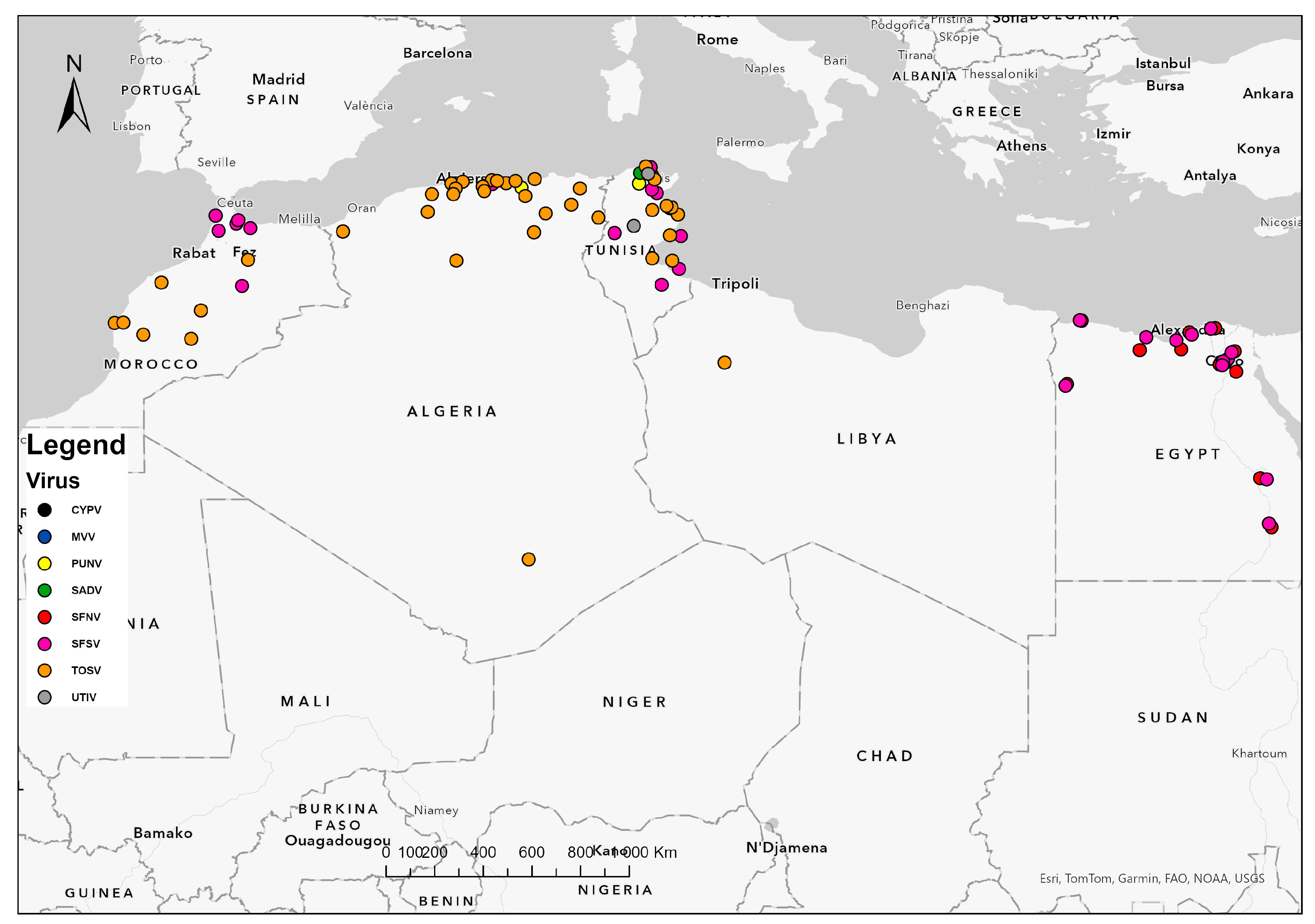
Assessed human sera, either healthy or patients’, were mostly positive for anti-SFSV and anti-TOSV Ab (Figure 1 and Figure 2). Occurrence of antibodies against other sandfly-borne phleboviruses in studied persons was sporadic and lesser, including SFNV (2.9%) in Morocco and (1.1%) Tunisia and PUNV (8.72%), CYPV (2.9%), and MVV (1.35%) in Tunisia [38,55,57,61]. As claimed by an overall study in Tunisia, 15.9% of the healthy population as IgG-positive against sandfly-borne phleboviruses, of which 51% resided in rural areas. Dual reactivity was observed between SFSV and SFCV (0.8%) and between TOSV and SFNV (0.5%) or SFCV (0.5%) [55].
SFSV seroprevalence in North African population varied from 1.3% to 21%, while TOSV occurrence was between 3.8% and 50%, showing a huge gap between confirmed etiologic agent prevalence and asymptomatic infection rate, respectively (Table 1). Particularly, TOSV was identified as an etiological agent of neurological disease in patients from Algeria and Tunisia with notable rates (3.8% and 12.86%, respectively) [34,54]. In contrast, it has never been described in humans from Morocco up to now because no studies were run for that purpose. Comparatively, data on sandfly-borne phleboviruses were lesser in Morocco. Studies conducted on human and non-human vertebrates were very limited and old. Recent investigations were exclusively entomological and concerned TOSV only (Table 1).
The discrepant seroprevalence would be due to the vector distribution among the highly changing sampling areas, which is tightly related to geographical area and environmental factors [31,32,45]. Sandfly-borne phleboviruses occurred in the arid and humid bioclimatic stages but were restricted mainly to the north. It is strongly suggested that phleboviruses are heavily affecting highly populated regions, like the northern and the coastal areas, whose sandfly-exposed inhabitants are at greater risk of infection [31,34]. Their distribution is congruent with entomological data. Sandflies are abundant in peri-urban and rural environments and are often found in close proximity to human and domestic animal habitats, which results in an overlap [35,45].
However, not all persons in a community have equal exposure to sandflies; some people apparently escape infection. Sandflies often show a marked atmosphere preference. This may indicate that some individuals within a community are at greater risk of virus infection than others [38]. In the Maghreb region, a total of 32 species of the genera Phlebotomus and Sergentomyia were reported (15 from Libya, 18 from Tunisia, 23 from Morocco, and 24 from Algeria). They feed on a wide range of vertebrate hosts, and their trophic preferences vary depending on the sandfly species. Multiple feeding patterns were detected, with various combinations of humans and vertebrate hosts [64]. As example, Ph. perniciosus is more anthropophilic compared to Ph. longicuspis, which is more zoophilic, leading to a difference in human versus dog biting rates and subsequently to a difference in the infection status with phleboviruses [32].
More significantly, sandfly fauna density and diversity vary by bioclimatic zones [54]. The number of sandfly vectors, namely Ph. sergenti, Ph. longicuspis, and Ph. perniciosus, showed a positive correlation with altitude. In fact, the biodiversity index and richness were higher in highland areas (912 m above sea level) than in plain areas. Along with the abiotic factor of altitude, the distribution, biodiversity, and richness of sandflies seemed to be primarily influenced by bioclimatic factors such as the effect of soil texture on their habitats [11].
In addition, latest findings showed that environmental changes, mainly due to the intensive development of irrigation systems for agriculture implemented in arid bio-geographical areas, yielded sustained populations of sandflies of the subgenus Larroussius, mainly Ph. perfiliewi, Ph. perniciosus, and Ph. Longicuspis, which may have led to the emergence of phleboviruses in new areas [25,60].
Recent data from elsewhere in the African continent were only available in Kenya, where a novel virus designated Ntepes virus (NPV) was isolated from sandflies and specific neutralizing antibodies were found in 13.9% of humans from the northeast [69].
Furthermore, in Italy, at least six identified phleboviruses co-circulated in sandflies: TOSV, Fermo (FERV), Corfou (CFUV), Ponticelli I (PONV-I), Ponticelli II (PONV-II), and Ponticelli III (PONV-III) [36,40,70]. On the other hand, co-circulation of phleboviruses and Leishmania parasites occurred in sandflies, of which Ph. perfiliewi (98%) was the foremost species [40]. Still, the results of a serological survey performed on domestic animals, against TOSV, FERV, PONV-I, and PONV-III, showed neutralizing activities for at least one of the four phlebovirus strains in 17.3% of dogs, 57.1% of goats, and 18.7% of sheep [71]. For TOSV alone, isolation and high seroprevalence rates (30–50%) were reported in a population from several regions of the country [23].
Four phleboviruses are known to circulate in Portugal: Sicilian, Toscana, Alcube, and Massilia phlebovirus [41]. For TOSV, Portugal was the second country, after Italy, to be considered endemic, with seroprevalence rates of 4.2% and 1.3% in patients with and without neurological symptoms, respectively, and 5.6% of CSF samples from meningitis patients being positive for TOSV infection. The presence of TOSV in animals was corroborated by several seroprevalence studies, which found antibodies in domestic animals such as cats (2.2–4.9%) and dogs (6.2–56.3%), as well as in wild animals, namely wolves (Canis lupus signatus, 2%) and foxes (Vulpes vulpes, 2.7%). Sandfly vectors identified in the region were Ph. perniciosus, Ph. papatasi, Ph. ariasi, and Se. minuta [23,41].
In France, TOSV isolation was achieved from human samples and from Ph. perniciosus sandflies in Marseille and RNA detection from Se. minuta. Alongside this, 8.7–14% of blood donors possessed anti-TOSV IgG. For Sicilian virus, low seroprevalence rates were reported in the southwest (2%) and in Marseille (1%) among blood donors. Wild mammals displayed 0.3% seropositivity [23].
In Spain, TOSV seropositivity rates were lower (5–26%) than those reported in Italy. Seroprevalence studies conducted in domestic animals from Granada showed evidence that they were frequently bitten by infected sandflies (17.7% in goats, 17.9% in cows, 22% in pigs, 32.3% in sheep, 48.3% in dogs, 59.6% in cats, and 64.3% in horses) [23]. More recently, high neutralizing antibody rates were identified in wild animals, represented by the Common Quail (Coturnix coturnix) for SFSV (45.45%) and TOSV (42.45%) [66].
In Greece, serological rates were 13.1–24.7% for SFNV, 2–8.5% for SFSV, and 11–51.7% for TOSV. Peculiarly, neutralizing activity against SFNV and SFSV was noticed in farmers. Also, antibodies against Corfu virus/Sicilian virus were detected in 4% of healthy residents [23].
In Germany, competent vectors of TOSV were distributed in the southwest, and TOSV-neuroinvasive was detected in 4% of cases with suspected viral meningoencephalitis [72].
Far away, in the Middle East, the most frequently observed virus exposure in Turkey was due to TOSV (13.7–22.6%), followed by SFNV (5.2–15.3%), SFSV (12.1–14.7%), and Sandfly Fever Turkey Virus (12.1%) [22,23]. In particular, TOSV seroreactivity among blood donors had reached very high IgG (56%) and IgM (43.6%) antibody levels. Among central nervous system patients, TOSV IgM antibodies were detected in 11.2% of the sera and in 1.76% of the CSF samples, whereas IgG antibodies were detected in 8% of the sera and 3% of the CSF samples. Furthermore, IgM antibodies against SFSV (27.27–36%), SFNV (45.45%), and CYPV (4%) viruses were detected in acute patient sera [23]. Additionally, TOSV sequences were identified in birds’ organs during the screening of avian specimens collected in the Mediterranean coast of the Anatolian peninsula [41].
Nearby, sandfly fever outbreak was reported from northern Lebanon during 2007. SFNV appeared as the main infection and was detected in soldiers returning from Syria and Lebanon in 2019 [73,74].
Seroprevalence rates of 9.4% and 0.8% were found in blood donors from western Saudi Arabia for SFSV and TOSV, respectively. The highest seropositivity rate was among samples collected from animal handlers, suggesting that contact with livestock animals could be a risk factor. Sera from livestock animals showed seropositivity of 53.3% and 4.4% in cows, 27.5% and 7.8% in sheep, 2.2% and 0.0% in goats, and 10.0% and 2.3% in camels for SFSV and TOSV, respectively [75].
Epidemiological patterns observed with sandfly-borne phleboviruses imply that they have the same ecology. Their geographic distribution was quite similar and appeared to parallel that of the vectors occurring in humid zones (Figure 2), i.e., northern areas characterized by Mediterranean climate or highly irrigated arid areas [25,34,35,50].
Interestingly, either cutaneous or zoonotic visceral leishmaniasis, or both, were endemic in most of regions where sandfly-borne phleboviruses circulated, indicating an overlapping geographic distribution. It is important to highlight that most study areas consisted of leishmaniasis foci, which promoted evidence to Leishmania spp. and phlebovirus cocirculation, along with identification of common harboring sandfly species [11,32,33,38,44,46,50,53,56].
Table 1. Summary of published studies of sandfly-borne phleboviruses in North Africa.
CountryVirusYearAreaSite FactorsGovernorateRegionPopulationPrevalenceRef.
Algeria SFSV2006Larbaa Nath
Iraten
NATizi OuzouNorthPh. ariasiNA[43]
Healthy humans5% (IgG)
SFSV2007Bou IsmailNATipazaNorthPh. longicuspisNA[44]
Larbaa Nath
Iraten
NATizi OuzouNorth
SFSV2007Bou IsmailNATipazaNorthOutpatients10.6% (IgG)[44]
Larbaa Nath
Iraten
NATizi OuzouNorthHealthy persons21.6% (IgG)
CYPV2007Bou IsmailNATipazaNorthPh. papatasiNA[44]
Larbaa Nath
Iraten
NATizi OuzouNorth
TOSV2013Draa El Mizan36°32′146” N,
3°50′850” E
380 m altitude
Tizi OuzouNorthUnidentified sandfliesNA[31]
Blood donors50% (Nt-Ab)
TOSV2014Tifra36°39”59” N,
4°22”16” E
BejaiaNorthWatchdogs4.3% (Nt-Ab)[46]
Ouaguenoun36°46”12” N,
4°10”29” E
Tizi OuzouNorth
Azazga36°44”43” N,
4°22”16” E
Tizi OuzouNorth
TOSV *2017–2018NANATlemcen NorthOwned dogs20% (IgG)[45]
NANABlidaNorth
NANAAlgiersNorth
NANAMedeaNorth
NANABouiraNorth
NANASetifNorth
NANABejaiaNorth
NANALaghouatCenter
NANATamanrassetSouth
TOSV2017–2018NANABlidaNorthOwned dogs4.56% (Nt-Ab)[45]
NANAAlgiersNorth
NANABejaiaNorth
NANASetifNorth
NANALaghouatCenter
TOSV2016–2018NANATlemcen NorthHospitalized patients with neurological
infection
3.8% (RNA/Ig)[34]
NANABlidaNorth
NANAMedeaNorth
NANAAlgiersNorth
NANAJijelNorth
NANASetifNorth
NANABatnaNorth
NANABiskraNorth
NANAGuelmaNorth
NANAOum El BouaghiNorth
NANATebessaNorth
TOSV2017NANATissemsiltNorthLivestock3.33–17.18%
(Nt-Ab)
[35]
NANAAin DeflaNorth
NANATipazaNorth
NANABlidaNorth
NANAMedeaNorth
NANAAlgiersNorth
PUNV2020Kherrata36°24′20′′ N, 5°16′37′′ EBejaiaNorthPh. perniciosus0.06% [47]
MoroccoSFNV1976ItzerNAMideltCenterHumans 2.9% (Nt-Ab)[38]
SFSV1976ItzerNAMideltCenterHumans 5.7% (Nt-Ab)[38]
SFSV1979Bab Besen1600 m altitude,
Humid
ChaouenNorthRodents
Insectivora
9.4% (IgG)[51]
Chaouen600 m altitude, Sub-humidChaouenNorth
Talembote400 m altitude, Semi-aridChaouenNorth
Arbaoua130 m altitude, Sub-humidKenitraNorth
Asilah200 m altitude, Sub-humidTangerNorth
TOSV2008Louata 338,319 latitude,
4828 longitude, 800 m altitude
SefrouNorthPh. perniciosus4/129[48]
TOSV2011El Hanchane31°31′11′′ N,
9°26′02′′ W
Essaouira CenterPh. sergentiNA[49]
TOSV2008–2011NANAAzilalCenterPh. sergentiNA[30]
NANASefrouNorthPh. longicuspisNA
TOSVNANABani Hassan:
31°59′50′′ N,
6°59′44′′ W
722 m altitude, Semi-arid
AzilalCenterPh. longicuspis0.18%[50]
Tabia: 32°01′49′′ N
6°47′48′′ W
563 m altitude
Semi-arid
Ph. sergenti
El Hanchane290–300 m altitude
Semi-arid
EssaouiraCenterPh. sergenti0.15%
Ouarzazate1134 m altitude
Arid
OuarzazateSouthPh. papatasi0.09%
Louata674 m altitudeSefrouNorthPh. longicuspis0.33%
Temperate Ph. perniciosus
TOSV2018Lalla Laaziza 31°04′ N, 8°42′ W
>912 m altitude
Chichaoua CenterPh. sergentiNA[11]
TunisiaSFSV1975NANAMixedNAHumans1.6% (Nt-Ab)[38]
SFSV1980El Gharia750 m altitudeBizerteNorthRodents
Insectivora
Cheiroptera
31% (IgG)[52]
Zarzis1 m altitudeMedenineSouth
Kerkennah1–3 m altitudeSfaxCenter
Tataouine190 m altitudeTataouineSouth
Ezzriba 70 m altitudeZaghouanNorth
Moghrane110 m altitudeZaghouanNorth
SFSV2011–2012SousseNASousse NorthPatient with meningo-
encephalitis
NA[54]
SFSV2013Haffouz34°51′ N, 9°29′ E
Arid
Kairouan CenterDogs38.1% (Nt-Ab)[32]
Bouhajla35°24′ N, 9°56′ E
Arid
SFSV2014El Felta35°16′ N, 9°26′ ESidi BouzidCenterPh. perfiliewiNA[25]
SFSV2017NANAMonastirCenterBlood donors1.3% (IgG)[55]
NANASousse
PUNV2008Utique37°2′ N, 10°2′ E
Sub-humid
BizerteNorthPh. perniciosus0.13%[53]
Ph. longicuspis
PUNV2009Utique37°08′ N, 7°74′ EBizerteNorthPh. perniciosus0.11%[33]
PUNV2010Utique37°08′ N, 7°74′ EBizerteNorthUnidentified sandflies0.05%[33]
PUNV2011NANABizerteNorthHumans8.72% (Nt-Ab)[57]
PUNV2011–2012Medjez El BabSemi-aridBejaNorthPh. perniciosusNA[54]
PUNV2013Haffouz34°51′ N, 9°29′ E
Arid
Kairouan CenterDogs43.5% (Nt-Ab)[32]
Bouhajla35°24′ N, 9°56′ E
Arid
UTIV2008Utique37°2′ N, 10°2′ E
Sub-humid
BizerteNorthPh. perniciosus0.53%[53]
Ph. longicuspis
Se. minuta
UTIV2014Saddaguia35°05′ N, 9°25′ ESidi BouzidCenterPh. perfiliewiNA[25]
SFNV2017NANA MonastirCenterBlood donors1.1% (IgG)[55]
NANA Sousse
TOSV2003–2009NANAMahdiaNorthNeurological disease
patients
10% (IgM)
7% (IgG)
[58]
NANAMonastirNorth
NANASousse North
NANASfaxCenter
NANAGabesSouth
NANADjerbaSouth
TOSV2010Utique 37°08′ N, 7°74′ EBizerteNorthPh. perniciosus0.03%[56]
TOSV2011NANABizerteNorthOutpatients41% (Nt-Ab)[57]
TOSV2011–2012NANATunis North Patients with meningeal
syndrome
12.16% (IgM)
78% (IgG)
12.86% (RNA)
[54]
Ph. perniciosusNA
Ph. perfiliewiNA
TOSV2013Haffouz34°51′ N, 9°29′ E
Arid
Kairouan CenterDogs7.5% (Nt-Ab)[32]
Bouhajla35°24′ N, 9°56′ E
Arid
TOSV 2014Saddaguia35°05′ N, 9°25′ ESidi BouzidCenterPh. perfiliewi0.03%[25]
TOSV2014–2016Saddaguia35°05′ N, 9°25′ ESidi BouzidCenterPh. perniciosus0.05–0.22%[60]
Ph. longicuspis
Ph. perfiliewi
TOSV2017NANAKairouanCenterBlood donors13.3% (IgG)[55]
NANAMonastir
NANASousse
SADV2013Borj YoussefBorj Youssef: 36°56′ N, 10°07′ E, Semi-arid ArianaNorthPh. perniciosus0.6%[24]
Utique: 37°08′ N, 7°74′ E, Semi-aridPh. longicuspis
SADV2013MateurMateur: 37°03′ N, 9°28′ E, SubhumidBizerteNorthPh. perfiliewi
Sejnene: 36°56′ N, 9°21′ E, HumidPh. perniciosus
SADV2013SaddaguiaSaddaguia: 35°05′ N, 9°25′ E, AridSidi BouzidCenterPh. perfiliewi
Bouhajla: 35°24′ N, 9°56′ E, AridPh. perniciosus
SADV2014El Felta35°16′ N, 9°26′ ESidi BouzidCenterPh. perfiliewiNA[25]
MVV2010Utique 37°08′ N, 7°74′ EBizerteNorthPhlebotomus spp.0.02%[61]
Outpatients1.35% (Nt-Ab)
CYPV2017NANAMonastirCenterBlood donors2.9% (IgG)[55]
NANASousse
LibyaSFNV2008NANANANAFebrile patients0.5% (IgG)[62]
SFSV2008NANANANAFebrile patients0.7% (IgG)
TOSV2014Yafran 32°08′ N, 12°55′ E 691 m altitude
Semi-arid
Jabal al GharbiNorthOutpatients25% (IgG)[63]
Egypt SFNV1950’sNANANANAFebrile patientsNA[76]
1952–1954QalyubNAQalyubiaNorthHumans31% (Nt-Ab)[38]
1959–1961Metropolitan CairoNACairo
Giza
Qalyubia
NorthPh. papatasiNA[64]
1960–1963GizaNAGizaNorthHumans43.8% (Nt-Ab)[38]
AlexandriaNAAlexandriaNorthHumans21.6% (Nt-Ab)
Baltim-Borg BurulusNAKafr El SheikhNorthHumans6.7% (Nt-Ab)
LuxorNALuxorSouthHumans56.3% (Nt-Ab)
SiwaNAMatruhNorthHumans3.9% (Nt-Ab)
Sidi BarraniNAMatruhNorthHumans9.5% (Nt-Ab)
BahigNAAlexandriaNorthHumans25% (Nt-Ab)
El DabaNAMatruhNorthHumans13.9% (Nt-Ab)
Aswan NAAswanSouthHumans47.3% (Nt-Ab)
1975CairoNACairoNorthHumans6.1% (Nt-Ab)
1984ImbabaNAGizaNorth Hospitalized patients2/55 (IgG)[77]
1991BilbeisNASharqiyaNorthHumans 2% (IgG)[78]
SFSV1952–1954QalyubNAQalyubiaNorthHumans22.6% (Nt-Ab)[38]
1959–1961Metropolitan CairoNACairo
Giza
Qalyubia
NorthPh. papatasiNA[64]
1960–1963GizaNAGizaNorthHumans43.8% (Nt-Ab)[38]
AlexandriaNAAlexandriaNorthHumans27% (Nt-Ab)
Baltim-Borg BurulusNAKafr El SheikhNorthHumans8.9% (Nt-Ab)
LuxorNALuxorSouthHumans59.4% (Nt-Ab)
SiwaNAMatruhNorthHumans2% (Nt-Ab)
Sidi BarraniNAMatruhNorthHumans4.8%(Nt-Ab)
BahigNAAlexandriaNorthHumans6.3% (Nt-Ab)
El DabaNAMatruhNorthHumans8.3% (Nt-Ab)
AswanNAAswanSouthHumans43.6% (Nt-Ab)
1969NANANANAUniversity students23% (IgG)[79]
1975CairoNACairoNorthHumans5.0% (Nt-Ab)[38]
1984ImbabaNAGizaNorth Hospitalized patients1/55 (IgG)[77]
1989BilbeisNASharqiyaNorthSchoolchildren 9% (IgG)[80]
1991BilbeisNASharqiyaNorthHumans 4% (IgG)[78]
*: TOSV and antigenically related viruses; NA: not available; E: east; N: north; W: west; m: meter; Ref.: reference.

8. Conclusions

Sandfly-borne phleboviruses were encountered frequently and widely over North African region. Synthesis findings counted eight evidenced phleboviruses and noted their geographical distribution. Their circulation became permanent, apparently due to virus maintenance by successive passages through vectors and vertebrate hosts. Their occurrence was indeed stated in Libya and Egypt, although respective knowledge was weak and evidently needs to be reinforced and updated. Noticeably, all studies conducted in Morocco were mostly interested in TOSV in sandflies, demonstrating its presence all over the country. Hence, no current data were available for TOSV in humans and animals, nor for any other sandfly-borne phlebovirus. Conversely, various sandfly-borne phleboviruses were found to cocirculate in both Algeria and Tunisia, mainly SFSV, TOSV, and PUNV. Phlebovirus investigations there concerned several vertebrate host species besides the vectors and showed that they were highly concentrated in the north. In addition, they have been recognized as potential etiological agents of neurological disease in humans. Additionally, TOSV activity was repeatedly reported and symptomatically confirmed in Algeria and Tunisia. Therefore, they should be considered for diagnosis in individuals with compatible clinical presentation. Further studies are required, not only to confirm the sporadic detection of less-prevalent phleboviruses, i.e., CYPV, SFNV, and MVV, but to fully elucidate the epidemiological situation in Morocco, Libya, and Egypt as well.

Author Contributions

Conceptualization, I.L. and I.B.; methodology, S.S. and M.B.; validation, I.B. and I.L.; investigation, S.S. and H.M.; writing—original draft preparation, S.S. and H.M.; writing—review and editing, S.S., I.L. and R.G.; visualization, R.G.; supervision, I.B. and M.L. All authors have read and agreed to the published version of the manuscript.

Funding

This research received no external funding.

Data Availability Statement

No new data were created or analyzed in this study.

Conflicts of Interest

The authors declare no conflicts of interest.

References

  1. Atkinson, B.; Hewson, R. Emerging arboviruses of clinical importance in Central Asia. J. Gen. Virol. 2018, 99, 1172–1184. [Google Scholar] [CrossRef] [PubMed]
  2. Krow-Lucal, E.; Lindsey, N.P.; Lehman, J.; Fischer, M.; Staples, J.E. West Nile Virus and Other Nationally Notifiable Arboviral Diseases—United States, 2015. MMWR Morb. Mortal. Wkly. Rep. 2017, 66, 51–55. [Google Scholar] [CrossRef] [PubMed]
  3. Dvorak, V.; Shaw, J.; Volf, P. Parasite biology: The vectors. In The Leishmaniases: Old Neglected Tropical Diseases; Bruschi, F., Gradoni, L., Eds.; Springer International Publishing: Cham, Switzerland, 2018; pp. 31–77. [Google Scholar]
  4. Bichaud, L.; de Lamballerie, X.; Alkan, C.; Izri, A.; Gould, E.; Charrel, R. Arthropods as a source of new RNA viruses. Microb. Pathog. 2014, 77, 136–141. [Google Scholar] [CrossRef]
  5. Depaquit, J.; Grandadam, M.; Fouque, F.; Andry, P.E.; Peyrefitte, C. Arthropod-borne viruses transmitted by Phlebotomine sandflies in Europe: A review. Euro Surveill. 2010, 15, 19507. [Google Scholar] [CrossRef] [PubMed]
  6. Maroli, M.; Feliciangeli, M.D.; Bichaud, L.; Charrel, R.N.; Gradoni, L. Phlebotomine sandflies and the spreading of leishmaniases and other diseases of public health concern. Med. Vet. Entomol. 2013, 27, 123–147. [Google Scholar] [CrossRef] [PubMed]
  7. Ayhan, N.; Charrel, R.N. Of Phlebotomines (Sandflies) and Viruses: A Comprehensive Perspective on a Complex Situation. Curr. Opin. Insect Sci. 2017, 22, 117–124. [Google Scholar] [CrossRef]
  8. Chaves, L.F.; Calzada, J.E.; Valderrama, A.; Saldaña, A. Cutaneous Leishmaniasis and Sand Fly Fluctuations Are Associated with El Niño in Panamá. PLoS Negl. Trop. Dis. 2014, 8, e3210. [Google Scholar] [CrossRef]
  9. Jancarova, M.; Polanska, N.; Volf, P.; Dvorak, V. The role of sand flies as vectors of viruses other than phleboviruses. J. Gen. Virol. 2023, 104, 001837. [Google Scholar] [CrossRef]
  10. Killick-Kendrick, R. The biology and control of phlebotomine sand flies. Clin. Dermatol. 1999, 17, 279–289. [Google Scholar] [CrossRef]
  11. Daoudi, M.; Calzolari, M.; Boussaa, S.; Bonilauri, P.; Torri, D.; Romeo, G.; Lelli, D.; Lavazza, A.; Hafidi, M.; Dottori, M.; et al. Identification of Toscana virus in natural population of sand flies (Diptera: Psychodidae) from Moroccan leishmaniasis foci. J. Infect. Public Health 2022, 15, 406–411. [Google Scholar] [CrossRef]
  12. Ayhan, N.; Charrel, R.N. Sandfly-borne viruses of demonstrated/relevant medical importance. In Vectors and Vector-Borne Zoonotic Diseases; IntechOpen: London, UK, 2018; pp. 1–22. [Google Scholar]
  13. Kuhn, J.H.; Adkins, S.; Alioto, D.; Alkhovsky, S.V.; Amarasinghe, G.K.; Anthony, S.J.; Avšic-Županc, T.; Ayllón, M.A.; Bahl, J.; Balkema-Buschmann, A.; et al. 2020 taxonomic update for phylum Negarnaviricota (Riboviria: Orthornavirae), including the large orders Bunyavirales and Mononegavirales. Arch. Virol. 2020, 165, 3023–3072. [Google Scholar] [CrossRef] [PubMed]
  14. Charrel, R.N.; Gallian, P.; Navarro-Mari, J.M.; Nicoletti, L.; Papa, A.; Sánchez-Seco, M.P.; Tenorio, A.; de Lamballerie, X. Emergence of Toscana virus in Europe. Emerg. Infect. Dis. 2005, 11, 1657–1663. [Google Scholar] [CrossRef] [PubMed]
  15. Moriconi, M.; Rugna, G.; Calzolari, M.; Bellini, R.; Albieri, A.; Angelini, P.; Cagarelli, R.; Landini, M.P.; Charrel, R.N.; Varani, S. Phlebotomine sand fly-borne pathogens in the Mediterranean Basin: Human leishmaniasis and phlebovirus infections. PLoS Negl. Trop. Dis. 2017, 11, e0005660. [Google Scholar] [CrossRef] [PubMed]
  16. Ergunay, K.; Ayhan, N.; Charrel, R.N. Novel and emergent sandfly-borne phleboviruses in Asia Minor: A systematic review. Rev. Med. Virol. 2017, 27, e1898. [Google Scholar] [CrossRef] [PubMed]
  17. Ayhan, N.; Charrel, R.N. An update on Toscana virus distribution, genetics, medical and diagnostic aspects. Clin. Microbiol. Infect. 2020, 26, 1017–1023. [Google Scholar] [CrossRef]
  18. Marklewitz, M.; Tchouassi, D.P.; Hieke, C.; Heyde, V.; Torto, B.; Sang, R.; Junglen, S. Insights into the evolutionary origin of Mediterranean sand fly fever viruses. MSphere 2020, 5, e00598-20. [Google Scholar] [CrossRef]
  19. Abudurexiti, A.; Adkins, S.; Alioto, D.; Alkhovsky, S.V.; Avsic-Zupanc, T.; Ballinger, M.J.; Bente, D.A.; Beer, M.; Bergeron, E.; Blair, C.D.; et al. Taxonomy of the order Bunyavirales: Update 2019. Arch. Virol. 2019, 164, 1949–1965. [Google Scholar] [CrossRef]
  20. Maes, P.; Alkhovsky, S.V.; Bao, Y.; Beer, M.; Birkhead, M.; Briese, T.; Buchmeier, M.J.; Calisher, C.H.; Charrel, R.N.; Choi, I.R.; et al. Taxonomy of the family Arenaviridae and the order Bunyavirales: Update 2018. Arch. Virol. 2018, 163, 2295–2310. [Google Scholar] [CrossRef]
  21. Lambert, A.J.; Hughes, H.R. Clinically Important Phleboviruses and Their Detection in Human Samples. Viruses 2021, 13, 1500. [Google Scholar] [CrossRef]
  22. Polat, C.; Ayhan, N.; Saygan, M.B.; Karahan, S.; Charrel, R.; Ergünay, K. Comprehensive Cross-Sectional Evaluation of Human Sandfly-Borne Phlebovirus Exposure in an Endemic Region. Viruses 2023, 15, 1902. [Google Scholar] [CrossRef]
  23. Alkan, C.; Bichaud, L.; de Lamballerie, X.; Alten, B.; Gould, E.A.; Charrel, R.N. Sandfly-borne phleboviruses of Eurasia and Africa: Epidemiology, genetic diversity, geographic range, control measures. Antivir. Res. 2013, 100, 54–74. [Google Scholar] [CrossRef]
  24. Fares, W.; Charrel, R.N.; Dachraoui, K.; Bichaud, L.; Barhoumi, W.; Derbali, M.; Cherni, S.; Chelbi, I.; de Lamballerie, X.; Zhioua, E. Infection of sand flies collected from different bio-geographical areas of Tunisia with phleboviruses. Acta Trop. 2015, 141, 1–6. [Google Scholar] [CrossRef]
  25. Dachraoui, K.; Fares, W.; Bichaud, L.; Barhoumi, W.; Beier, J.C.; Derbali, M.; Cherni, S.; Lamballerie, X.; Chelbi, I.; Charrel, R.N.; et al. Phleboviruses associated with sand flies in arid bio-geographical areas of Central Tunisia. Acta Trop. 2016, 158, 13–19. [Google Scholar] [CrossRef]
  26. Tezcan, S.; Dincer, E.; Ulger, M.; Ozgur, D.; Erdogan, S.; Ozkul, A.; Emekdas, G.; Ergunay, K. Serological investigation of phlebovirus exposure in blood donors from the Mediterranean Province of Mersin, Turkey. Mikrobiyol. Bul. 2015, 49, 403–413. [Google Scholar] [CrossRef] [PubMed]
  27. Ergunay, K.; Aydogan, S.; Ilhami Ozcebe, O.; Cilek, E.E.; Hacioglu, S.; Karakaya, J.; Ozkul, A.; Us, D. Toscana virus (TOSV) exposure is confirmed in blood donors from Central, North and South/Southeast Anatolia, Turkey. Zoonoses Public Health 2012, 59, 148–154. [Google Scholar] [CrossRef] [PubMed]
  28. Elliott, R.M.; Brennan, B. Emerging phleboviruses. Curr. Opin. Virol. 2014, 5, 50–57. [Google Scholar] [CrossRef]
  29. Ergunay, K.; Saygan, M.B.; Aydogan, S.; Lo, M.M.; Weidmann, M.; Dilcher, M.; Sener, B.; Hascelik, G.; Pinar, A.; Us, D. Sandfly fever virus activity in central/northern Anatolia, Turkey: First report of Toscana virus infections. Clin. Microbiol. Infect. 2011, 17, 575–581. [Google Scholar] [CrossRef] [PubMed]
  30. Es-sette, N.; Ajaoud, M.; Anga, L.; Mellouki, F.; Lemrani, M. Toscana virus isolated from sandflies, Morocco. Parasit. Vectors 2015, 8, 205. [Google Scholar] [CrossRef]
  31. Alkan, C.; Allal-Ikhlef, A.B.; Alwassouf, S.; Baklouti, A.; Piorkowski, G.; de Lamballerie, X.; Izri, A.; Charrel, R.N. Virus isolation, genetic characterization and seroprevalence of Toscana virus in Algeria. Clin. Microbiol. Infect. 2015, 21, 1040.e1–1040.e9. [Google Scholar] [CrossRef]
  32. Sakhria, S.; Alwassouf, S.; Fares, W.; Bichaud, L.; Dachraoui, K.; Alkan, C.; Zoghlami, Z.; de Lamballerie, X.; Zhioua, E.; Charrel, R.N. Presence of sandfly-borne phleboviruses of two antigenic complexes (Sandfly fever Naples virus and Sandfly fever Sicilian virus) in two different bio-geographical regions of Tunisia demonstrated by a microneutralisation-based seroprevalence study in dogs. Parasit. Vectors 2014, 7, 476. [Google Scholar]
  33. Dachraoui, K.; Chelbi, I.; Ben Said, M.; Ben Osman, R.; Cherni, S.; Charrel, R.; Zhioua, E. Transmission Dynamics of Punique Virus in Tunisia. Viruses 2022, 14, 904. [Google Scholar] [CrossRef] [PubMed]
  34. Benbetka, C.; Hachid, A.; Benallal, K.E.; Khardine, F.A.; Ayhan, N.; Bouredjoul, N.; Boulehbal, W.M.; Bellila, D.; Khaldi, A.; Charrel, R. Epidemiology, Isolation, and Genetic Characterization of Toscana Virus in Algerian Patients Displaying Neurological Infection, 2016–2018. IJID Reg. 2023, 7, 193–198. [Google Scholar] [CrossRef] [PubMed]
  35. Sellali, S.; Lafri, I.; Ayhan, N.; Medrouh, B.; Messahel, N.E.; Lafri, M.; Charrel, R.; Bitam, I. Neutralizing based seroprevalence study of Toscana virus in livestock from Algeria. Comp. Immunol. Microbiol. Infect. Dis. 2023, 103, 102075. [Google Scholar] [CrossRef] [PubMed]
  36. Daoudi, M.; Romeo, G.; Marzani, K.; Petrella, A.; Bonilauri, P.; Lelli, D.; Boumezzough, A.; Boussaa, S.; Dottori, M.; Calzolari, M. New Isolation of Ponticelli III Virus (Bunyavirales: Phenuiviridae) in Emilia-Romagna Region, Italy. Viruses 2023, 15, 422. [Google Scholar] [CrossRef] [PubMed]
  37. Calzolari, M.; Russo, S.; Marzani, K.; Dalmonte, G.; Ricchi, M.; Bonilauri, P. Development of a real-time PCR assay for the detection of the phlebovirus fermo virus. Viruses 2023, 15, 2082. [Google Scholar] [CrossRef]
  38. Tesh, R.B.; Saidi, S.; Gajdamovic, S.J.; Rodhain, F.; Vesenjak-Hirjan, J. Serological studies on the epidemiology of sandfly fever in the Old World. Bull. World Health Organ. 1976, 54, 663–674. [Google Scholar]
  39. Ergünay, K.; Polat, C.; Özkul, A. Vector-Borne Viruses in Turkey: A Systematic Review and Bibliography. Antivir. Res. 2020, 183, 104934. [Google Scholar] [CrossRef]
  40. Calzolari, M.; Romeo, G.; Callegari, E.; Bonilauri, P.; Chiapponi, C.; Carra, E.; Rugna, G.; Taddei, R.; Lelli, D.; Dottori, M. Co-circulation of phleboviruses and Leishmania parasites in sand flies from a single site in Italy monitored between 2017 and 2020. Viruses 2021, 13, 1660. [Google Scholar] [CrossRef]
  41. Amaro, F.; Zé-Zé, L.; Alves, M.J. Sandfly-Borne Phleboviruses in Portugal: Four and Still Counting. Viruses 2022, 14, 1768. [Google Scholar] [CrossRef]
  42. Nasiri, Z.; Kalantari, M.; Amin, M.; Daliri, S.; Azizi, K. Phleboviruses (Family: Phenuiviridae) Worldwide, with Emphasis on the Middle East: A Systematic Review Study. J. Health Sci. Surveill. Syst. 2023, 11, 408–417. [Google Scholar]
  43. Izri, A.; Temmam, S.; Moureau, G.; Hamrioui, B.; De Lamballerie, X.; Charrel, R.N. Sandfly fever Sicilian virus, Algeria. Emerg. Infect. Dis. 2008, 14, 795. [Google Scholar] [CrossRef] [PubMed]
  44. Moureau, G.; Bichaud, L.; Salez, N.; Ninove, L.; Hamrioui, B.; Belazzoug, S.; De Lamballerie, X.; Izri, A.; Charrel, R.N. Molecular and serological evidence for the presence of novel phleboviruses in sandflies from northern Algeria. Open Virol. J. 2010, 4, 15. [Google Scholar] [CrossRef] [PubMed]
  45. Sellali, S.; Lafri, I.; Hachid, A.; Ayhan, N.; Benbetka, C.; Medrouh, B.; Messahel, N.E.; Bekara, M.A.; Lafri, M.; Charrel, R.N.; et al. Presence of the sandfly-borne phlebovirus (Toscana virus) in different bio-geographical regions of Algeria demonstrated by a microneutralisation-based seroprevalence study in owned dogs. Comp. Immunol. Microbiol. Infect. Dis. 2022, 88, 101861. [Google Scholar] [CrossRef] [PubMed]
  46. Tahir, D.; Alwassouf, S.; Loudahi, A.; Davoust, B.; Charrel, R.N. Seroprevalence of Toscana virus in dogs from Kabylia (Algeria). Clin. Microbiol. Infect. 2016, 22, e16–e17. [Google Scholar] [CrossRef] [PubMed]
  47. Manseur, H.; Hachid, A.; Khardine, A.F.; Benallal, K.E.; Bia, T.; Temani, M.; Hakem, A.; Sánchez-Seco, M.P.; Bitam, I.; Vázquez, A.; et al. First Isolation of Punique Virus from Sand Flies Collected in Northern Algeria. Viruses 2022, 14, 1796. [Google Scholar] [CrossRef]
  48. Es-Sette, N.; Nourlil, J.; Hamdi, S.; Mellouki, F.; Lemrani, M. First Detection of Toscana Virus RNA From Sand Flies in the Genus Phlebotomus (Diptera: Phlebotomidae) Naturally Infected in Morocco. J. Med. Entomol. 2012, 49, 1507–1509. [Google Scholar] [CrossRef]
  49. Es-Sette, N.; Ajaoud, M.; Bichaud, L.; Hamdi, S.; Mellouki, F.; Charrel, R.N.; Lemrani, M. Phlebotomus sergenti a common vector of Leishmania tropica and Toscana virus in Morocco. J. Vector Borne Dis. 2014, 51, 86–90. [Google Scholar]
  50. Es-Sette, N.; Ajaoud, M.; Charrel, R.N.; Lemrani, M. Epidemiologie moleculaire des phlebovirus dans quatre provinces du Maroc. Bull. Soci. Pathol. Exot. 2016, 109, 143–150. [Google Scholar] [CrossRef]
  51. Chastel, C.; Launay, H.; Bailly-Choumara, H.; Le Lay, G.; Beaucournu, J.C. Arbovirus infections in Morocco: Serosurvey in small wild mammals in the northern part of the country. Bull Soc. Pathol. Exot. Filiales. 1982, 75, 466–475. [Google Scholar]
  52. Chastel, C.; Bach-Hamba, D.; Launay, H.; Le Lay, G.; Hellal, H.; Beaucournu, J.C. Arbovirus infections in Tunisia: New serological survey of small wild mammals. Bull. Soc. Pathol. Exot. Filiales. 1983, 76, 21–33. [Google Scholar]
  53. Zhioua, E.; Moureau, G.; Chelbi, I.; Ninove, L.; Bichaud, L.; Derbali, M.; Champs, M.; Cherni, S.; Salez, N.; Cook, S.; et al. Punique virus, a novel phlebovirus, related to sandfly fever Naples virus, isolated from sand flies collected in Tunisia. J. Gen. Virol. 2010, 91, 1275–1283. [Google Scholar] [CrossRef] [PubMed]
  54. Fezaa, O.; M’ghirbi, Y.; Savellini, G.G.; Ammari, L.; Hogga, N.; Triki, H.; Cusi, M.G.; Bouattour, A. Serological and molecular detection of Toscana and other Phleboviruses in patients and sandflies in Tunisia. BMC Infect. Dis. 2014, 14, 598. [Google Scholar] [CrossRef] [PubMed]
  55. Ayari, R.; Chaouch, H.; Findlay-Wilson, S.; Hachfi, W.; Ben Lasfar, N.; Bellazreg, F.; Dowall, S.; Hannachi, N.; Letaief, A. Seroprevalence and Risk Factors Associated with Phleboviruses and Crimean–Congo Hemorrhagic Fever Virus among Blood Donors in Central Tunisia. Pathogens 2024, 13, 348. [Google Scholar] [CrossRef] [PubMed]
  56. Bichaud, L.; Dachraoui, K.; Piorkowski, G.; Chelbi, I.; Moureau, G.; Cherni, S.; De Lamballerie, X.; Sakhria, S.; Charrel, R.N.; Zhioua, E. Toscana virus isolated from sandflies, Tunisia. Emerg. Infect. Dis. 2013, 19, 322–324. [Google Scholar] [CrossRef] [PubMed]
  57. Sakhria, S.; Bichaud, L.; Mensi, M.; Salez, N.; Dachraoui, K.; Thirion, L.; Cherni, S.; Chelbi, I.; de Lamballerie, X.; Zhioua, E.; et al. Co-Circulation of Toscana virus and Punique virus in Northern Tunisia using a microneutralization-based serological study. PLoS Negl. Trop. Dis. 2013, 7, 2429. [Google Scholar] [CrossRef]
  58. Bahri, O.; Fazaa, O.; Ben Alaya-Bouafif, N.; Bouloy, M.; Triki, H.; Bouattour, A. Role of Toscana virus in meningo-encephalitis in Tunisia. Pathol. Biol. 2011, 59, e125–e127. [Google Scholar]
  59. Sghaier, W.; Bahri, O.; Kedous, E.; Fazaa, O.; Rezig, D.; Touzi, H.; Ben Yahia, A.; Meddeb, Z.; Triki, H. Retrospective study of viral causes of central nervous system infections in Tunisia (2003–2009). Med. Sante Trop. 2012, 22, 373–378. [Google Scholar] [CrossRef]
  60. Fares, W.; Dachraoui, K.; Barhoumi, W.; Cherni, S.; Chelbi, I.; Zhioua, E. Co-circulation of Toscana virus and Leishmania infantum in a focus of zoonotic visceral leishmaniasis from Central Tunisia. Acta Trop. 2020, 204, 105342. [Google Scholar] [CrossRef]
  61. Bichaud, L.; Dachraoui, K.; Alwassouf, S.; Alkan, C.; Mensi, M.; Piorkowski, G.; Sakhria, S.; Seston, M.; Fares, W.; De Lamballerie, X.; et al. Isolation, full genomic characterization and neutralization-based human seroprevalence of Medjerda Valley virus, a novel sandfly-borne phlebovirus belonging to the Salehabad virus complex in northern Tunisia. J. Gen. Virol. 2016, 97, 602–610. [Google Scholar] [CrossRef]
  62. Elahmer, O.R.; Abdelfadeel, M.; Safwat, S.; Smeo, M.N.; Abudher, A.; Sammoud, M.A. A retrospective study to define viral and bacterial etiologies causing acute febrile illness in Libya. In Proceedings of the Conference of Endemic and Emerging Infectious Diseases of Priority in the Middle East and North Africa—Research Opportunities and Biosafety in a Changing Environment, Istanbul, Turkey, 18–21 June 2012. [Google Scholar]
  63. Saadawi, W.K.; Abozaid, F.D.; Almukhtar, M.; Annajar, B.B.; Shaibi, T. Seroprevalence study of Toscana virus in Yafran area, Libya. J. Vector Borne Dis. 2022, 59, 186–189. [Google Scholar] [CrossRef]
  64. Schmidt, J.R.; Schmidt, M.L.; Said, M.I. Phlebotomus fever in Egypt. Isolation of phlebotomus fever viruses from Phlebotomus papatasi. Am. J. Trop. Med. Hyg. 1971, 20, 483–490. [Google Scholar] [CrossRef] [PubMed]
  65. Pierro, A.; Ficarelli, S.; Ayhan, N.; Morini, S.; Raumer, L.; Bartoletti, M.; Mastroianni, A.; Prati, F.; Schivazappa, S.; Cenni, P.; et al. Characterization of antibody response in neuroinvasive infection caused by Toscana virus. Clin. Microbiol. Infect. 2017, 23, 868–873. [Google Scholar] [CrossRef] [PubMed]
  66. Ayhan, N.; Rodríguez-Teijeiro, J.D.; López-Roig, M.; Vinyoles, D.; Ferreres, J.A.; Monastiri, A.; Charrel, R.N.; Serra-Cobo, J. High rates of antibodies against Toscana and Sicilian phleboviruses in common quail Coturnix coturnix birds. Front. Microbiol. 2023, 13, 1091908. [Google Scholar] [CrossRef] [PubMed]
  67. Benallal, K.E.; Garni, R.; Harrat, Z.; Volf, P.; Dvorak, V. Phlebotomine sand flies (Diptera: Psychodidae) of the Maghreb region: A systematic review of distribution, morphology, and role in the transmission of the pathogens. PLoS Negl. Trop. Dis. 2022, 16, e0009952. [Google Scholar] [CrossRef] [PubMed]
  68. Munoz, C.; Ayhan, N.; Ortuno, M.; Ortiz, J.; Gould, E.A.; Maia, C.; Berriatua, E.; Charrel, R.N. Experimental Infection of Dogs with Toscana Virus and Sandfly Fever Sicilian Virus to Determine Their Potential as Possible Vertebrate Hosts. Microorganisms 2020, 8, 596. [Google Scholar] [CrossRef]
  69. Tchouassi, D.P.; Marklewitz, M.; Chepkorir, E.; Zirkel, F.; Agha, S.B.; Tigoi, C.C.; Koskei, E.; Drosten, C.; Borgemeister, C.; Torto, B.; et al. Sand fly–associated Phlebovirus with evidence of neutralizing antibodies in humans, Kenya. Emerg. Infect. Dis. 2019, 25, 681. [Google Scholar] [CrossRef]
  70. Calzolari, M.; Chiapponi, C.; Bellini, R.; Bonilauri, P.; Lelli, D.; Moreno, A.; Barbieri, I.; Pongolini, S.; Lavazza, A.; Dottori, M. Isolation of three novel reassortant phleboviruses, Ponticelli I, II, III, and of Toscana virus from field-collected sand flies in Italy. Parasites Vectors 2018, 11, 84. [Google Scholar] [CrossRef]
  71. Lelli, D.; Scanferla, V.; Moreno, A.; Sozzi, E.; Ravaioli, V.; Renzi, M.; Tosi, G.; Dottori, M.; Lavazza, A.; Calzolari, M. Serological evidence of phleboviruses in domestic animals on the pre-Apennine Hills (northern Italy). Viruses 2021, 13, 1577. [Google Scholar] [CrossRef]
  72. Dersch, R.; Sophocleous, A.; Cadar, D.; Emmerich, P.; Schmidt-Chanasit, J.; Rauer, S. Toscana virus encephalitis in Southwest Germany: A retrospective study. BMC Neurol. 2021, 21, 495. [Google Scholar] [CrossRef]
  73. Malik, M.; Mafi, A.; Mahjour, J.; Opoka, M.; Elhakim, M.; Muntasir, M.O. Novel coronavirus infection in the Eastern Mediterranean Region: Time to act. East Mediterr. Health J. 2013, 19, S31–S38. [Google Scholar] [CrossRef]
  74. Kniha, E.; Obwaller, A.G.; Dobler, G.; Poeppl, W.; Mooseder, G.; Walochnik, J. Phlebovirus seroprevalence in Austrian Army personnel returning from missions abroad. Parasites Vectors 2019, 12, 416. [Google Scholar] [CrossRef] [PubMed]
  75. Al-Numaani, S.A.; Al-Nemari, A.T.; El-Kafrawy, S.A.; Hassan, A.M.; Tolah, A.M.; Alghanmi, M.; Zawawi, A.; Masri, B.E.; Hindawi, S.I.; Alandijany, T.A.; et al. Seroprevalence of Toscana and sandfly fever Sicilian viruses in humans and livestock animals from western Saudi Arabia. One Health 2023, 17, 100601. [Google Scholar] [CrossRef] [PubMed]
  76. Feinsod, F.M.; Ksiazek, T.G.; Scott, R.M.; Soliman, A.K.; Farrag, I.H.; Ennis, W.H.; Peters, C.J.; El Said, S.; Darwish, M.A. Sand fly fever-Naples infection in Egypt. Am. J. Trop. Med. Hyg. 1987, 37, 193–196. [Google Scholar] [CrossRef] [PubMed]
  77. Darwish, M.A.; Feinsod, F.M.; Scott, R.M.; Ksiazek, T.G.; Botros, B.A.; Farrag, I.H.; El Said, S. Arboviral causes of non-specific fever and myalgia in a fever hospital patient population in Cairo. Egypt. Trans. R. Soc. Trop. Med. Hyg. 1987, 81, 1001–1003. [Google Scholar] [CrossRef]
  78. Corwin, A.; Habib, M.; Watts, D.; Darwish, M.; Olson, J.; Botros, B.; Hibbs, R.; Kleinosky, M.; Lee, H.W.; Shope, R.; et al. Community-based prevalence profile of arboviral, rickettsial, and Hantaan-like viral antibody in the Nile River Delta of Egypt. Am. J. Trop. Med. Hyg. 1993, 48, 776–783. [Google Scholar] [CrossRef]
  79. Darwish, M.A.; Ibrahim, A.H. Prevalence of antibodies to arboviruses in Egypt. Results of a serological survey among 1,113 university students. Am. J. Trop. Med. Hyg. 1975, 24, 981–985. [Google Scholar] [CrossRef]
  80. Corwin, A.; Habib, M.; Olson, J.; Scott, D.; Ksiazek, T.; Watts, D.M. The prevalence of arboviral, rickettsial, and Hantaan-like viral antibody among schoolchildren in the Nile river delta of Egypt. Trans. R. Soc. Trop. Med. Hyg. 1992, 86, 677–679. [Google Scholar] [CrossRef]
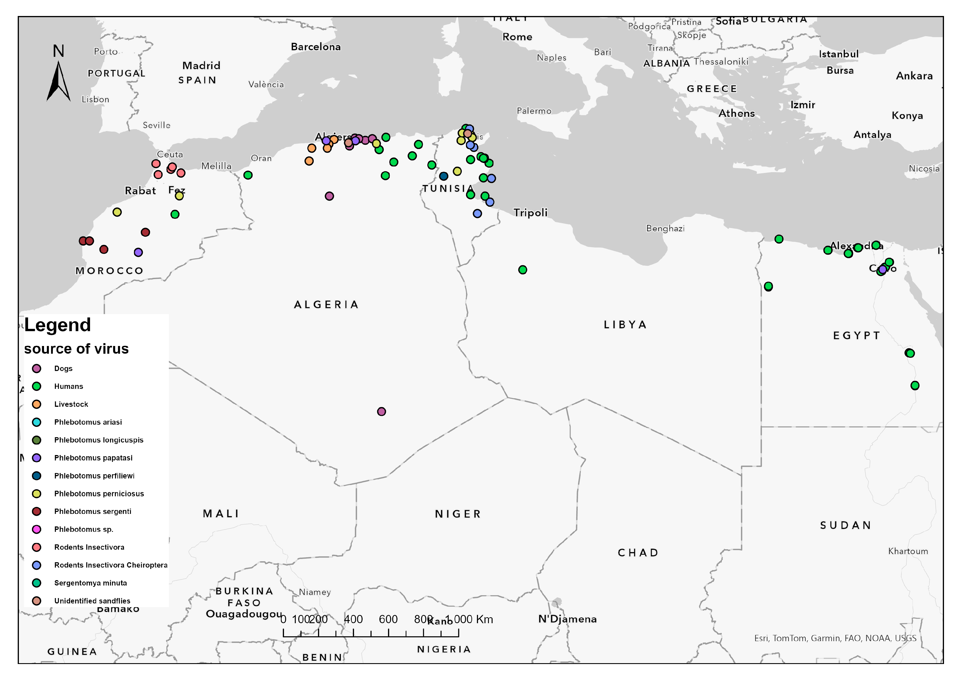
Figure 1. Distribution of detected and isolated sandfly-borne phleboviruses in vectors and vertebrate hosts from North Africa. A map of five North African countries showing geographical location of sandflies and vertebrates infected by sandfly-borne phleboviruses. Dots denote infected areas. Colors indicate species of sandflies, human, and non-human vertebrate hosts from the region.
Insects 15 00846 g001
Figure 2. Geographical distribution of detected and isolated sandfly-borne phleboviruses in North Africa. A map of five North African countries showing geographical distribution of circulating sandfly-borne phleboviruses. Dots denote infected areas. Colors indicate species variety of detected and isolated sandfly-borne phleboviruses in the region. Legend: CYPV: Cyprus virus, MVV: Medjerda Valley virus, PUNV: Punique virus, SADV: Saddaguia virus, SFNV: Sandfly Fever Naples virus, SFSV: Sandfly Fever Sicilian Virus, TOSV: Toscana Virus, UTIV: Utique virus.
Insects 15 00846 g002
Disclaimer/Publisher’s Note: The statements, opinions and data contained in all publications are solely those of the individual author(s) and contributor(s) and not of MDPI and/or the editor(s). MDPI and/or the editor(s) disclaim responsibility for any injury to people or property resulting from any ideas, methods, instructions or products referred to in the content.