Article

Effects of the Entomopathogenic Fungus Mucor hiemalis BO-1 on the Physical Functions and Transcriptional Signatures of Bradysia odoriphaga Larvae

1
College of Agronomy, Liaocheng University, Liaocheng 252000, China
2
College of Plant Protection, Shandong Agricultural University, Tai’an 271018, China
*
Authors to whom correspondence should be addressed.
Insects 2023, 14(2), 162; https://doi.org/10.3390/insects14020162
Submission received: 8 December 2022 / Revised: 25 January 2023 / Accepted: 5 February 2023 / Published: 8 February 2023
(This article belongs to the Section Insect Pest and Vector Management)

Abstract

:

Simple Summary

Mucor hiemalis BO-1 was acutely toxic to Bradysia odoriphaga larvae and was as effective as some chemical pesticides. Mucor hiemalis BO-1 infection inhibited the feeding capacity of Bradysia odoriphaga larvae, reduced food consumption and nutrient contents, and caused a decline in digestive enzyme activity. Transcriptome analysis of diseased B. odoriphaga larvae indicated significant changes in the expression of the genes related to the digestive process, energy metabolism, antioxidant response, and immune processes.

Abstract

Mucor hiemalis BO-1 is an entomopathogenic fungus that infects Bradysia odoriphaga, a destructive root maggot. M. hiemalis BO-1 possesses stronger pathogenicity to the larvae than to other stages of B. odoriphaga, and provides satisfactory field control. However, the physiological response of B. odoriphaga larvae to infection and the infection mechanism of M. hiemalis are unknown. We detected some physiological indicators of diseased B. odoriphaga larvae infected by M. hiemalis BO-1. These included changes in consumption, nutrient contents, and digestive and antioxidant enzymes. We performed transcriptome analysis of diseased B. odoriphaga larvae, and found that M. hiemalis BO-1 showed acute toxicity to B. odoriphaga larvae and was as toxic as some chemical pesticides. The food consumption of diseased B. odoriphaga after inoculation with M. hiemalis spores decreased significantly, and there was a significant decrease in total protein, lipid, and carbohydrates in diseased larvae. Key digestive enzymes (protease, α-amylase, lipase, and cellulase) were significantly inhibited during infection. Peroxidase maintained high activity, and the activity of other antioxidant enzymes (catalase, superoxide dismutase, and glutathione S-transferases) first increased and then decreased. Combined with the transcriptional signatures of diseased B. odoriphaga larvae, M. hiemalis BO-1 infection resulted in decreased food consumption, reduced digestive enzyme activity, and altered energy metabolism and material accumulation. Infection was also accompanied by fluctuations in immune function, such as cytochrome P450 and the Toll pathway. Therefore, our results laid a basis for the further study of the interactions between M. hiemalis BO-1 and B. odoriphaga and promoted the genetic improvement of entomopathogenic fungi.

1. Introduction

Biological control is an important part of integrated pest management (IPM). Successful biological control helps protect the environment and advances harmonious interaction between man and nature [1,2]. Entomopathogenic fungi have been used in biological control programs against several agricultural pests. Some fungal biocontrol agents, such as Beauveria bassiana and Metarhizium anisopliae, have replaced chemical insecticides for the control of agricultural pests [3,4,5]. However, the limitations of entomopathogenic fungi, such as high environment dependence and protracted insecticidal activity, have slowed their development and use. Due to this, only a few fungal species have been successfully developed as biopesticides. Many entomopathogenic fungi possess good pathogenicity in the laboratory, but their field efficacy depends on favorable environmental conditions, such as temperature, humidity, and characteristics of the microorganism [6,7,8]. For example, the optimal temperatures of Beauveria bassiana that provide the best control efficiency range from 25 to 30 °C [9,10]. Hence, it is necessary to find an entomopathogenic fungus with high pathogenicity and broad environmental adaptability.
Most entomopathogenic fungi infect host insects directly by penetration through the cuticle into the hemocoel. Some pathogens can infect host insects via the oral route during food ingestion [11,12]. Once the pathogens invade the hemocoel, they switch to yeast-like hyphal bodies to overcome, evade, or suppress insect immune defenses, and then grow to form mycelia. This ultimately kills the host, and sporulation occurs in the cadaver [3,13]. Generally, in the course of the disease, entomopathogenic fungi can proliferate rapidly by robbing the nutrients of the host and secreting mycotoxins. The mycotoxins damage the free amino acids in the hemolymph, disturb the storage and utilization of nutrients, and interfere with important metabolic enzymes [14,15]. Insects rely on the combined efforts of physical barriers (i.e., cuticle surface and chitin barriers of the gut) and both cellular and humoral components of their innate immune system to protect themselves from invasion and death due to microorganisms [16]. However, the immune response of insects involves high consumption of energy and inhibits the normal life activities of host insects [17,18]. B. bassiana infection elicits immune responses from both the cellular and humoral arms of the insect immune system, including antioxidant enzymes, phenoloxidase (PO) cascade, and cytochrome P450 [19,20]. The ability to infect through both cuticular and oral routes would make fungal pathogens a more difficult challenge to the insects’ immune system and thereby a good candidate for a biopesticide. Entomopathogenic fungi infection can also cause dietary stress since conidia represent indigestible material for larvae. They occupy the digestive tract and limit host access to ingested nutrients [5,11]. Consideration of the mechanisms of pathogenesis is important for the determination of fungal virulence factors that might be manipulated to increase the speed of mycosis and death to levels similar to those produced by chemical insecticides.
Bradysia odoriphaga Yang et Zhang is a root maggot that causes serious losses to vegetables, flowers, and edible fungi [21,22]. The larvae can directly damage plants by feeding on the root and corm tissues and causing wilt or rot. Uncontrolled Bradysia infestation can cause more than 50% yield reduction in vegetables and edible fungi [23]. Since the damage caused by larvae is primarily on the underground parts of plants, they are difficult to control effectively [22,24]. Chemical insecticides remain the most important component in B. odoriphaga larvae control programs. The chemical products include organophosphates (phoxim and chlorpyrifos), carbamates (carbosulfan), and neonicotinoids [25], despite direct and indirect toxic effects on nontarget organisms and humans. However, after many years of application, larval pesticide resistance has developed [21]. In addition, conventional chemical pesticide use is now restricted due to environmental pollution and human health concerns. Therefore, there is a need for alternative biocontrol approaches.
Compared to other agricultural pests, there are few effective biological control agents of B. odoriphaga. The list includes Beauveria bassiana, Bacillus thuringiensis, Stratiolaelaps scimitus (predatory mite) and entomopathogenic nematodes [21,26]. Only B. bassiana, B. thuringiensis, and entomopathogenic nematodes have been used for field control of B. odoriphaga [21]. The field control effects of these biocontrol agents are often unsatisfactory due to poor efficacy and slow action. Zhou et al. (2014) reported that when the dosage of B. bassiana granules (15 billion spores/g) was 4.5 kg/ha, the field control efficiency was 81.15% (7 d after application) and 85.19% (21 d after application) [27]. The field control efficiency of three species of entomopathogenic nematodes (Heterorhabditis bacteriophora, Steinernema carpocapsae, and Steinernema feltiae) to B. odoriphaga was 40–50% at 14 d after application [26]. The discovery and development of a new biological control agent possessing high pathogenicity and strong environmental adaptability against B. odoriphaga will be necessary to improve the control of B. odoriphaga.
Mucor hiemalis BO-1 is a recently discovered entomopathogenic fungus that possesses strong pathogenicity to B. odoriphaga larvae [28]. Field tests confirmed that M. hiemalis BO-1 produced excellent control of B. odoriphaga and had great potential for further evaluation as a biocontrol agent. M. hiemalis was also reported to be pathogenic against other arthropods, such as Latrodectus geometricus Koch [29], Evergestis extimalis Scopoli [30], and Nemouroidea [31]. After larval inoculation with M. hiemalis spores, B. odoriphaga movement slowed, and the body became transparent. During infection, food residue in the alimentary canal decreased [28]. It was hypothesized that M. hiemalis BO-1 infection disrupted the physiological and digestive functions of B. odoriphaga larvae. However, little is known about the physiological responses of B. odoriphaga larvae to pathogen infection and the molecular mechanisms underlying the pathological processes. This lack of information limited the further development of this strain.
In this study, we evaluated the pathogenicity of M. hiemalis BO-1 to fourth-instar larvae of B. odoriphaga. Physiological indicators of diseased B. odoriphaga larvae, such as food consumption, nutrient contents, digestive enzymes, and antioxidant enzymes, were studied to help understand the effects of M. hiemalis BO-1 infection on the physiological functions of B. odoriphaga larvae. The transcriptional signatures of diseased B. odoriphaga larvae were analyzed, and a detailed bioinformatics analysis was conducted on partially important differentially expressed genes (DEGs).

2. Materials and Methods

2.1. Insect Materials

The samples of B. odoriphaga were originally collected from a Chinese chive field in Liaocheng, Shandong, in April 2019. The samples were reared on Chinese chives for more than 20 generations. Eggs, larvae, and pupae were reared in culture dishes (Φ = 9 cm), and newly emerged adults were placed in pairs in oviposition containers (3 cm diameter × 1.5 cm high). Insect samples were maintained in growth cabinets at 23 ± 1 °C with 75 ± 5% relative humidity.

2.2. Entomopathogenic Fungi Strain

Mucor hiemalis BO-1 strain was isolated from infested B. odoriphaga larvae and deposited at China General Microbiological Culture Collection Center (CGMCC, No. 40036). This strain was cultivated on potato dextrose agar (PDA) plate (Φ = 9 cm) in Petri dishes at an optimal temperature of 23 °C. After 10 d, 50 mL 0.1% Tween 80 distilled water was added to the dish, and the surface of the colony was gently and repeatedly scraped with a Petri scalpel. The suspension was filtered using three-layer filter paper, and the filtrate was gathered as a spore suspension. The spore suspension concentration was calculated through blood counting chamber analysis.

2.3. Pathogenicity Assays

Serial spore suspension dilutions (1 × 10³, 1 × 10⁴, 1 × 10⁵, 1 × 10⁶, 1 × 10⁷, and 1 × 10⁸ spores/mL) were made with 0.1% Tween 80 distilled water. Bioassays on 4th-instar larvae were conducted using standard contact and stomach bioassay methods [23]. One piece of filter paper (Φ = 9 cm) was moisturized by dropping 1 mL spore suspension and placed in the culture dish. Fresh diet was placed on the filter paper. Larvae were placed around the fresh diets. Pure water treatment was used as the control. Each treatment contained 5 replicates with 25 individuals. The survival number of B. odoriphaga larvae was recorded daily. The median lethal time (LT50) and median lethal concentration (LC50) values were calculated according to probit analysis using SPSS Statistics 18.0.0 (2009; SPSS Inc., Quarry Bay, Hong Kong, China).

2.4. Effects of M. hiemalis BO-1 on Food Consumption

According to the pathogenicity results, the survival rates of fourth-instar larvae at 24, 48, 72, and 96 h after inoculation were 98.75%, 90%, 78.75%, and 63.75% for 104 spores/mL treatment and 97.5%, 86.25%, 68.75%, and 50% for 105 spores/mL treatment, respectively. At 96 h, the survival rate of infected larvae was close to 50%, and so the two spore concentrations of 104 spores/mL and 105 spores/mL were used for the evaluation tests on the treated larvae. Newly emerged 4th-instar larvae were exposed to an M. hiemalis BO-1 spore suspension (1 × 10⁴ and 1 × 10⁵ spores/mL). The food consumption of larvae was measured using the method described by Zhao et al. (2018) [23] with slight modifications. A total of 50 larvae were transferred to fresh Petri dishes, and fresh Chinese chive rhizomes were weighed and used to feed the larvae. The diets were changed every 24 h, and the remaining rhizomes were reweighed. The food consumption of every larva was calculated according to the variation in Chinese chive rhizome weight/the larva number. Every treatment contained five replicates. The 0.1% Tween 80 treatment was used as the control.

2.5. Biochemical Assay of Nutrient Contents in Larvae

Sample preparation: The larvae were treated with 1 × 10⁴ and 1 × 10⁵ spores/mL of M. hiemalis BO-1 spore suspension. After 24, 48, 72, and 96 h, groups of 100 surviving larvae were weighed and used for the analysis of biochemical indicators. Every treatment contained three replicates.
Lipid amount: The 100 larvae were homogenized and extracted with 1.5 mL chloroform–methanol (2:1) solution. After centrifugation at 5000 rpm for 10 min, the supernatant was dried at 40 °C. The residuum was dissolved with 1.0 mL H2SO4 (98%) and incubated at 90 °C for 10 min. Then, 50 μL of each sample was mixed with 250 μL of vanillin, and the absorbance was read at 530 nm.
Carbohydrate amount: The 100 larvae were homogenized in cold 800 μL of trichloroacetic acid (10%), followed by centrifugation at 8000 rpm for 10 min. Then, 100 μL of the supernatant and 210 μL of 10% trichloroacetic acid were mixed, and 1.8 mL of 0.2% anthrone was added to the mixture. The mixture was placed in a boiling water bath for 10 min. The absorbance was read at 630 nm. A standard curve was established with glucose.
Protein content: The 100 larvae were homogenized in 0.05 M phosphate buffer. The crude homogenates were centrifuged at 5000× g for 15 min. The protein concentration of the supernatant was determined by the Bradford assay [32].

2.6. Enzyme Activity Assay

Sample preparation: Larvae were treated by the method described in Section 2.4. After 12, 24, 36, 48, 72, and 96 h, the surviving larvae were dissected on ice, and the entire digestive tracts of 50 larvae were collected as the sample for digestive enzymes assay. At each test interval, 30 surviving larvae were collected as the sample for the antioxidant enzyme activity assay. All the samples were homogenized in 0.05 M cold phosphate buffer containing 1% polyvinylpyrrolidone (pH 7.8, 0–4 °C). The crude homogenates were centrifuged at 10,000× g for 10 min at 4 °C. The supernatants were collected, and these represented the enzyme samples. Each treatment had five replicates. Protein concentrations of each sample were determined using the Bradford assay [32].
Digestive enzyme activity assay: The digestive enzyme activities were detected using the α-Amylase Assay Kit, Lipase Assay Kit, Protease Assay Kit, and Cellulase (CL) Assay Kit (Nanjing Jiancheng Bioengineering Institute, Nanjing, China). For the protease activity assay, casein was used as the substrate, and tyrosine was detected as the product of enzymatic reactions. The amount (nmol) of tyrosine produced per mg protein catalyzing casein decomposition per min at 660 nm and 37 °C was defined as the protease activity. For the α-amylase activity assay, soluble starch was used as the substrate, and maltose was detected as the product of enzymatic reactions. The α-amylase activity was defined as the amount of maltose produced by soluble starch decomposition catalyzed by per mg protein per min at 540 nm and 37 °C. For lipase activity assay, 4-nitrophenyl butyrate was used as the substrate, and p-nitrophenol was detected as the product of enzymatic reactions. The amount of p-nitrophenol that was produced by the enzymatic reaction catalyzed by per mg protein per min at 405 nm and 37 °C was defined as the lipase activity. For the cellulase activity assay, cellulose was used as the substrate, and reducing sugar (glucose) was detected as the product by the color reaction at 540 nm. The amount of glucose that was produced by the enzymatic reaction catalyzed by per mg protein per min at 540 nm and 37 °C was defined as the cellulase activity.
Antioxidant enzyme activity assay: The antioxidant enzyme activities were detected using the Catalase (CAT) Assay Kit, Peroxidase (POD) Assay Kit, Superoxide Dismutase (SOD) Assay Kit, and Glutathione S-Transferase (GST) Assay Kit (Solarbio Science Technology Company, Beijing, China). The operation sequences were performed following the kit instructions. The amount (μmol) of H2O2 decomposition at 240 nm per min per mg protein was defined as one unit of CAT activity. One unit of POD activity was defined as the amount that catalyzes 1 μmol of guaiacol oxidation at 470 nm per minute per mg protein. One unit of SOD activity was defined as the amount of enzyme that caused 50% inhibition of the nitro blue tetrazolium reduction at 550 nm. GST activity was tested using 1-chloro-2,4-dinitrobenzene (CDNB) as substrate. The reaction mixture contained 75 μL of 0.6 mM CDNB, 150 μL of 6 mM reduced glutathione (GSH), and 50 μL of enzyme solution. The OD value was monitored at 340 nm and 27 °C for 5 min.

2.7. Transcriptome Analysis

Sample preparation: Fourth-instar B. odoriphaga larvae were infested with an M. hiemalis BO-1 spore suspension (1 × 10⁵ spores/mL). After 48 h, 100 surviving larvae were collected as the sample. The treatment without spores was used as the control. Each treatment contained two replicates. All of the samples were immediately immersed in liquid nitrogen. Total RNA was extracted using the Total RNA Isolation TRIzol Kit (Thermo Fisher Scientific Biotech, Shanghai, China). The RNA quality was monitored on 1% agarose gel, and RNA purity was confirmed using a NanoPhotometer spectrophotometer (Table S1). First-strand cDNA was synthesized from mRNA after enrichment by fragmentation with buffer and oligo (dT) magnetic beads. The double-stranded cDNA was purified, which was then eluted with EB buffer for end-repair and addition of poly (A). The library was sequenced on an Illumina HiSeqTM 3000 (Illumina, Beijing Novogene Biotech Co., Ltd. (Beijing, China). The raw data were deposited in the NCBI Short Read Archive (SRA) database with accession number: PRJNA922622.
Transcriptome analysis: To clean up the mRNA derived from the fungus or other microbiota in larvae, clean reads were mapped to the B. odoriphaga genome (Bioproject ID: PRJNA612767) using Hisat 2.0.5 software designed by the Center for Computational Biology of Johns Hopkin University (WA, USA) [33]. The mapped reads were normalized using the DESeq 1.18.0 package. Gene-expression levels were calculated as fragments per kilobase per million (FPKM) using cufflinks 2.1.1 software. Differentially expressed genes (DEGs) were determined based on their expression abundance in different treatment groups. The difference in gene expression between the treatment groups was deemed significant if the p-value < 0.05. Hierarchical clustering analysis of DEGs was performed to cluster genes that exhibited the same or similar expression levels. Gene Ontology (GO) was applied to analyze the function of DEGs based on the GO database and to determine the biological implication of genes in the significant or representative profiles. The identification of pathways for DEGs was according to the Kyoto Encyclopedia of Genes and Genomes (KEGG) database. The resulting p-values were adjusted using the false discovery rate (FDR) algorithm. Then, pathway categories with p-values < 0.01 and FDRs < 0.05 were reported.
Quantitative RT-PCR analysis: To validate the results of the transcriptome analysis, 12 DEGs were analyzed using qRT-PCR. New biological samples were prepared according to the methods described in Section 2.4. Total RNA isolation and cDNA synthesis were performed, and the fluorescence quantitative PCR primer sequences of these genes are listed in Supplementary Table S2. Real-time PCR was performed using the One Step SYBR® Prime Script TMPT-PCR kit (Takara, Dalian, China). The housekeeping gene β-actin was used as the internal control. Transcript levels were determined in relation to that of β-actin using the 2−△△Ct method.

2.8. Statistical Analysis

We tested the other variables for homogeneity of group variances using Levene’s test and normality using the Kolmogorov–Smirnov test before statistical analyses. The significant difference analysis was performed with SPSS Statistics 18.0.0 (2009; SPSS Inc., Quarry Bay, Hong Kong, China).

3. Results

3.1. Pathogenicity of M. hiemalis BO-1

M. hiemalis BO-1 was highly pathogenic to B. odoriphaga larvae (Table 1). The larvae reached a high mortality rate in a short time, even at a low spore inoculum concentration. At observation times 3 d and 5 d, the median lethal concentrations (LC50) were 2.180 × 10⁶ and 0.965 × 10⁵ spores/mL, respectively. When the spore concentrations were 1 × 10⁴, 1 × 10⁶, and 1 × 10⁸ spores/mL, the median lethal times (LT50) were 4.79, 2.99, and 2.43 d, respectively.

3.2. Effects of M. hiemalis Infection on Food Consumption and Nutrient Contents of B. odoriphaga

Food consumption: The food consumption of B. odoriphaga larvae was significantly decreased at 48 h after inoculation with M. hiemalis spores (Figure 1A). High spore-concentration treatment (1.0 × 10⁵ spores/mL) inhibited food consumption more than the low-concentration treatment (1.0 × 10⁴ spores/mL). At 72 h after the initial inoculation, the food consumption decreased by 48.19% (1.0 × 10⁴ spores/mL treatment) and 66.97% (1.0 × 10⁵ spores/mL treatment). However, at 120 h, the food consumption of the 1.0 × 10⁵ spores/mL treatment group was higher than that of the 1.0 × 10⁴ spores/mL treatment group.
Nutrient contents: M. hiemalis infection inhibited nutrient accumulation in the larval body or accelerated nutrient consumption (Figure 1B–D). During normal larval development, proteins, lipids, and carbohydrates gradually accumulate. After M. hiemalis infection, the total protein amount stopped increasing, although the larvae continued feeding (Figure 1B). At 48 h after inoculation, the lipid amounts in the treated groups reached a maximum, and there were no significant differences between the control and treatment groups. The lipid amount of the two inoculation groups decreased rapidly, and significant differences were discovered at 72 and 96 h compared to the control group (Figure 1C). The carbohydrate amount of the two inoculation groups gradually decreased after infection and was lower than the control group at 72 and 96 h (Figure 1D). Significant dosage effects showed that the higher the spore concentration, the more obvious the decrease in the three nutrient contents.

3.3. Effects of M. hiemalis Infection on Digestive Enzyme Activities

The effects of M. hiemalis infection on the digestive enzyme activities of B. odoriphaga larvae are shown in Figure 2. The activities of protease, lipase, α-amylase, and cellulase significantly decreased with increased infection time. After 36 h, all of the digestive enzyme activities of the inoculation groups had decreased significantly compared to the control group. However, at 24 h, the decreases in lipase, α-amylase, and cellulase enzymatic activities were less obvious than the decrease in protease activity. At 72 h after inoculation, the protease activities of the two inoculation groups decreased by 32.91% (104 spores/mL) and 57.34% (105 spores/mL) compared to the control group. The α-amylase activities decreased by 72.58% and 85.12%, cellulase activities decreased by 63.89% and 78.40%, and lipase activities decreased by 41.09% and 69.24%. Significant dosage effects were also observed. The higher the spore concentration, the more obvious the decrease in the four digestive enzyme activities.

3.4. Effects of M. hiemalis Infection on Antioxidant Enzyme Activities

The activities of CAT, POD, SOD, and GST in B. odoriphaga 4th-instar larvae were determined after infection with M. hiemalis spores (Figure 3). POD activity increased after infection; the activity increased by 42.51% (1.0 × 10⁴ spores/mL) and 66.47% (1.0 × 10⁵ spores/mL) compared to the control group at 36 h, and 19.83% (1.0 × 10⁴ spores/mL) and 41.90% (1.0 × 10⁵ spores/mL) at 72 h. However, CAT, SOD, and GST activities fluctuated after infection. Their trends were similar, with an initial increase followed by a decline. The CAT activity increased by 84.22% (1.0 × 10⁵ spores/mL) and 33.57% (1.0 × 10⁴ spores/mL) at 24 h after infection and decreased by 47.78% (1.0 × 10⁵ spores/mL) and 36.33% (1.0 × 10⁴ spores/mL) at 72 h after infection. The SOD and GST activity of the 1.0 × 10⁵ spores/mL treatment group increased by 27.88% (SOD) and 36.04% (GST) at 24 h after infection but decreased by 45.08% (SOD) and 51.66% (GST) at 72 h.

3.5. Transcriptome Analysis

Quality assessment of the transcriptome: The control 2 library produced the most data (51,677,542 clean reads), while the treatment 2 library produced the fewest clean reads (44,925,166). All libraries exhibited good quality, with 94.19% to 94.33% of the clean reads meeting base call quality at the Q30 standard. According to the alignments of sequences in the unigene library, the statistics of mapped reads in each sample are shown in Table 2. The mapped ratio was 87.89 to 89.39%.
Identification and annotation analysis of DEGs: Based on the DEG analysis, 377 DEGs between M. hiemalis infection and control treatment were identified (Table S3). Of these, 214 unigenes were upregulated, and 163 unigenes were downregulated. For GO analysis, we annotated DEGs into three GO categories: biological process (BP), cellular component (CC), and molecular function, which were divided into 44 subgroups (Figure 4A). For the BP category, the two main enriched terms were the cellular process and metabolic process, and the number of DEGs was 81 and 82. For the CC category, the main enriched terms were the cell and cell part, and the number of DEGs in both terms was 95. For the molecular function (MF) category, the main enriched terms were catalytic activity and binding, and the number of DEGs was 106 and 72. The biochemical pathways of the DEGs were investigated using the KEGG database. The top 10 enriched KEGG pathways were the PPAR signaling pathway, metabolic pathways, lysosome, fatty acid metabolism, phagosome, ferroptosis, glycerolipid metabolism, fatty acid degradation, metabolism by cytochrome P450, and steroid biosynthesis (Figure 4B, Table S4). In the metabolic pathways, 23 DEGs were identified (Table S5). Genes including lipid catabolic processes (HA402-011554, HA402-013349), carbohydrate metabolic processes (HA402-007273, HA402-007340, HA402-005954), glutathione metabolic processes (HA402-015641, HA402-005674), glucuronosyltransferase activity (HA402-015641, HA402-015641), or phospholipid metabolic processes (HA402-011554) were partially repressed. Genes including trehalose metabolic processes (HA402-003401, HA402-006219, HA402-013707), monosaccharide metabolic processes (HA402-009149), lipid biosynthetic processes (HA402-002025), dopamine metabolic processes (HA402-005954, HA402-001421), or doxorubicin metabolic processes (HA402-001272) were upregulated.
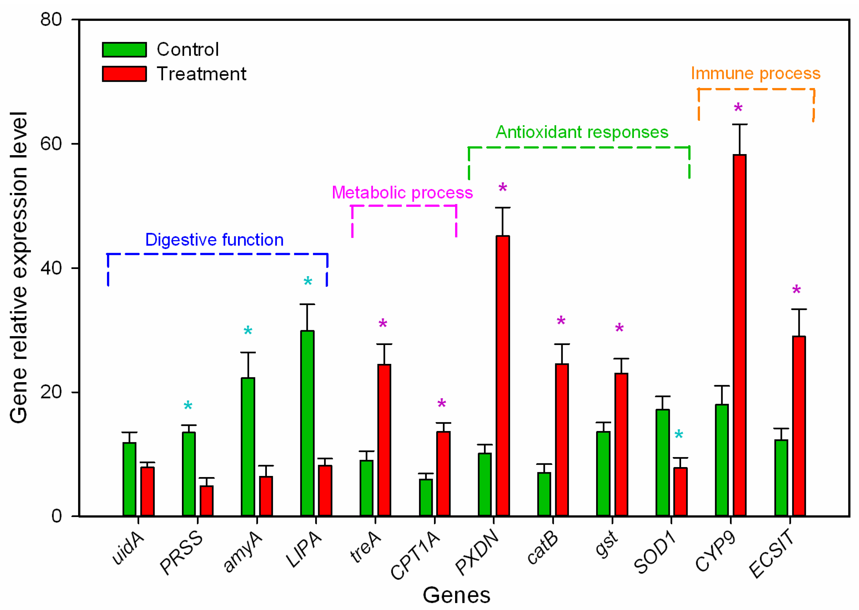
Reverse transcription (RT-PCR) validation: To further validate the gene expression profiles, 12 candidate differentially regulated genes (listed in Table 3) with different biological functions were analyzed using RT-PCR. The DEGs (amyA, PRSS, and LIPA) related to the digestion and carbohydrate metabolic process were down-regulated in B. odoriphaga larvae. This indicated that M. hiemalis infection decreased the insects’ ability to digest nutrients (lipids, starch, and protein). The relative expression of treA and CPT1A related to the metabolic process were up-regulated by 1.72- and 1.29-fold compared to the control group, respectively. The DEGs (PRDX5, catB, and gst) related to the response to the oxidative stress process were up-regulated, while SOD1 was down-regulated indicating high antioxidant capacity (Figure 5). The DEGs (CYP9 and ECSIT) related to drug metabolic and innate immune responses were up-regulated by 2.24- and 1.37-fold indicating significant immune responses caused by M. hiemalis infection. These RT-PCR results were similar to the RNA-seq data (detail shown in Table 3).

4. Discussion

Entomopathogenic fungi represent a novel way to control the root maggot B. odoriphaga. M. hiemalis BO-1 is a newly identified entomopathogenic fungus with high pathogenicity to B. odoriphaga. This fungus has good potential for commercial development and practical use [28]. Our bioassay results revealed that M. hiemalis BO-1 produced high mortality in B. odoriphaga larvae. When the spore concentration was 1.0 × 10⁵ to 1.0 × 10⁶ spores/mL, the median lethal time (LT50) was 2.99 to 3.79 d (Table 1). This was comparable to some neonicotinoid and organophosphorus pesticides [21,22]. A 3 d post-treatment interval is often used to evaluate the activity of chemical insecticides against B. odoriphaga [21,23]. The test time of most biological and “reduced-risk” insecticides is five days or more, and this includes insect growth regulators and botanical insecticides [22,34]. However, the toxicity of known entomopathogens to B. odoriphaga is lower than chemical insecticides. Zhou et al. (2014) reported that the LT50 of B. bassiana (1.0 × 10⁷ spores/mL) against B. odoriphaga larvae was 14.51 d [27]. The test time used to evaluate the infection ability of entomopathogenic nematodes to B. odoriphaga larvae was 10 d [35]. To improve the efficacy of control agents, some studies have applied a mixture of chemicals and biocontrol agents [26].
During infection, food residue in the alimentary canal of B. odoriphaga larvae decreased, and the larval body became transparent. M. hiemalis BO-1 infection disrupted the physiological and digestive functions of B. odoriphaga [28]. In the present study, larval food consumption decreased significantly during M. hiemalis BO-1 infection, and the nutrient substances (proteins, lipids, and sugars) were reduced (Figure 1). Previous studies reported that Beauveria spp. and Metarhizium spp. infection reduced feeding of the host insects. These species included Anopheles gambiae [36], Locusta migratoria [5], Dichroplus maculipennis [4], and Spodoptera littoralis [37]. The significant reduction in nutrient substances could result from the decrease in digestion and absorption and an increase in energy consumption of B. odoriphaga. The decrease in digestive enzyme activity and increase in antioxidant enzyme activity in the present study supported this possibility (Figure 2 and Figure 3). These data suggest that most of the ingested food is being converted to energy for combatting the invading pathogen and little food is being channeled toward larval growth. In addition, the production of toxic substances by the entomopathogenic fungus can disrupt insect food digestion and absorption [38]. A few entomopathogenic fungi have been reported to invade the insect body through the digestive system, where they disrupt larval feeding at the beginning of infection.
Entomopathogenic fungi can bypass the host defense system by several mechanisms. Entomopathogens produce toxins that stimulate oxidative mechanisms in insects, and this suggested that pathogens disable antioxidative enzymes to avoid the protective responses of insects to oxidative stress [15,39]. Karthi et al. (2018) reported that the infection by Aspergillus flavus, an entomopathogenic fungus, resulted in a significant increase in antioxidant enzyme activity (CAT, POD, and SOD) but a significant decline in phenoloxidase activity [17]. Our results indicated that M. hiemalis BO-1 infection triggers the POD enzyme level of B. odoriphaga. The SOD, CAT, and GST enzymes increased significantly at 12 to 24 h after infection (Figure 3), which indicated the importance of these enzymes in the defense against the oxidative stress caused by entomopathogenic fungi infection. However, there was a significant decline in SOD, CAT, and GST enzyme activities at 48 h after infection, and during this time, the infected larvae began to die (Figure 3). This indicated a decrease in the defense ability of insects, which could be attributed to the damage to physiological functions caused by entomopathogenic fungi [40]. A high concentration of fungal inoculum produced more significant effects (Figure 3). Similarly, Qasim et al. (2021) reported that SOD, POD, and GST enzymatic activity of Diaphorina citri Kuwayama adult and nymphal populations decreased significantly at 5 d posttreatment (fungal exposure) [20]. We conclude that entomopathogenic fungi had various effects on the antioxidant enzymes responses of insects such as Aedes aegypti [41], Leptinotarsa decemlineata [42], Chilo suppressalis [43], and Periplaneta americana [44]. Moreover, at 96 h after infection, a striking decrease in antioxidant enzyme activities (SOD and GST), digestive enzyme activities (protease, α-amylase, lipase, and cellulase), and nutrient substances (proteins, lipids, and sugars) was observed. This phenomenon may result from the severe biological stress caused by M. hiemalis infection, which is consistent with the pathogenicity results that the survival rate of infected larvae decreased dramatically from 72 h to 120 h after inoculation. In addition to the above results on digestive enzymes, our present study suggests that M. hiemalis BO-1 produces mycotoxins that disturb the physiology of B. odoriphaga.
Fungal infection is a complex process involving many factors as well as the activation of immune responses in the host insects [45]. To gain insight into the interactions between the insect host and the fungus, we explored the transcriptional response of B. odoriphaga larvae after infection with M. hiemalis BO-1, using RNA-seq technology. We identified 377 DEGs in infected B. odoriphaga larvae. Of these, 214 DEGs were up-regulated, and 163 DEGs were down-regulated. Transcriptional responses of B. odoriphaga to various conditions have been reported. Compared to a previous study [46], fewer DEGs were identified in the non-parametric transcriptome analysis of B. odoriphaga responses to insecticide stress (benzothiazole) in our study, and the quantity of DEGs identified in the transcriptome analysis using B. odoriphaga genome data was similar to that in previous studies [47,48]. Therefore, our transcriptome analysis based on the B. odoriphaga genome database should be more precise. Gene ontology (GO) analysis revealed that DEGs were especially enriched in terms associated with BP and CC. The 10 enriched biochemical pathways of the DEGs according to the KEGG database comprised the PPAR signaling pathway, metabolic pathways, lysosome, fatty acid metabolism, phagosome, ferroptosis, glycerolipid metabolism, fatty acid degradation, metabolism by cytochrome P450 and steroid biosynthesis. The RT-PCR results of the key targeted genes were consistent with the transcriptome results, for example, up-regulation of antioxidant genes (PXDN, gst, and SOD1), immune-related genes (CYP9 and ECSIT) and energy metabolism genes (treA and CPT1A), and down-regulation of digestion and absorption-related genes (uidA, amyA, PRSS, and LIPA) (Figure 5 and Table 3). Our transcriptome analysis and physiological assay results confirmed that M. hiemalis BO-1 infection disturbed the physiological function of B. odoriphaga larvae, especially for energy metabolism, digestive absorption, and immune responses. Previous studies also reported that entomopathogenic fungi caused various effects on the metabolic and immune processes of insects such as Bombyx mori infected by Beauveria bassiana and Helicoverpa zea [49,50], Diaphorina citri infected by Cordyceps fumosorosea [51], and Aedes albopictus and Culex pipiens infected by Pythium guiyangense [52]. Interestingly, M. hiemalis BO-1 infection caused significant effects on the digestion and absorption pathway of B. odoriphaga larvae, which was possibly due to the unique pathogenic mechanism of M. hiemalis BO-1, such as causing infection via the enteric canal, damaging the intestinal structure, and then disturbing ingestion. The entomopathogenic bacteria Bacillus thuringiensis Aizawai GC-91 damaged the midgut cells of Diatraea saccharalis larvae [53]. The internal tissues of Culex quinquefasciatus larvae (midgut wall, skeletal muscles, and the cuticle-secreting epidermis) were destroyed by the entomopathogenic fungi, Aspergillus clavatus [54]. Pythium guiyangense, an oomycete entomopathogenic fungus, uses cuticle penetration and ingestion of mycelia into the digestive system to infect Culex pipiens pallens larvae [52]. Some studies also reported that the gut bacterial community of host insects can be altered by entomopathogenic fungi, and this accelerates larval mortality. Examples include Dendroctonus valens and Beauveria bassiana [55], Anopheles stephensi and Beauveria bassiana [56]. The disease symptoms, the results of food consumption, nutrient content and digestive enzyme activity suggest that M. hiemalis BO-1 may have similar infective functions. However, the interaction between entomological fungi and host insects changes with the infection process. The immune process of the insect is enhanced during the early stages of infection. However, as the disease progresses, the insect’s physiology is disrupted, and its immune processes become ineffective [50,57]. To understand the detailed interaction between M. hiemalis BO-1 and B. odoriphaga, the temporospatial modulation analysis of gene regulatory pathways should be further investigated.

5. Conclusions

This study showed that M. hiemalis BO-1 was highly pathogenic to B. odoriphaga larvae. Infected larvae died quickly, and the lethal efficiency was higher than other entomopathogenic fungi. M. hiemalis BO-1 infection resulted in decreased food consumption and digestive enzyme activity, disturbed energy metabolism, and material accumulation. Infection was also accompanied by fluctuations in immune function. The results suggested that infection of the intestinal canal and damage to the digestive function are important infection and pathogenic strategies of M. hiemalis BO-1.

Supplementary Materials

The following supporting information can be downloaded at: https://www.mdpi.com/article/10.3390/insects14020162/s1, Table S1. Quality of the RNA isolated from the biological samples used for transcriptome analysis; Table S2. Primers used in reverse transcriptase-quantitative polymerase chain reaction (RT-qPCR); Table S3. Differentially expressed genes identified in Bradysia odoriphaga infected by Mucor hiemalis BO-1; Table S4. Detailed information for the KEGG enrichment analysis of the DEGs in the Bradysia odoriphaga transcriptome; Table S5. Detail information in metabolic pathway enriched in Bradysia odoriphaga infected by Mucor hiemalis BO-1.

Author Contributions

G.Z.: conceptualization, data curation, formal analysis, funding acquisition, investigation, methodology, project administration, writing—original draft, writing—review and editing. M.X., H.Z. and P.C.: conceptualization, investigation, methodology, project administration, resources, writing—original draft, writing—review and editing. W.D. and L.J.: conceptualization, investigation, methodology, project administration, resources, software, supervision. All authors have read and agreed to the published version of the manuscript.

Funding

This research was funded by “Shandong provincial Natural Science Foundation (ZR201911130528)”, Chinese University Student Innovation Project “S202110447001”, and “National Natural Science Foundation of China (No. 32001929)”.

Data Availability Statement

The data about biological and physiological indexes are available at https://doi.org/10.5061/dryad.z34tmpgjg accessed on 16 November 2022, and the RNA-seq data are available at NCBI SRA, accession number PRJNA922622.

Acknowledgments

We thank LetPub (www.letpub.com accessed on 16 November 2022) for its linguistic assistance during the preparation of this manuscript.

Conflicts of Interest

The authors declare no conflict of interest.

References

  1. Paulitz, T.C.; Bélanger, R.R. Biological control in greenhouse systems. Annu. Rev. Phytopathol. 2001, 39, 103–133. [Google Scholar] [PubMed]
  2. Chailleux, A.; Bearez, P.; Pizzol, J.; Amiens-Desneux, E.; Ramirez-Romero, R.; Desneux, N. Potential for combined use of parasitoids and generalist predators for biological control of the key invasive tomato pest Tuta absoluta. J. Pest Sci. 2013, 86, 533–541. [Google Scholar] [CrossRef]
  3. Lewis, M.W.; Robalino, I.V.; Keyhani, N.O. Uptake of the fluorescent probe FM4–64 by hyphae and haemolymph-derived in vivo hyphal bodies of the entomopathogenic fungus Beauveria bassiana. Microbiology 2009, 155, 3110–3120. [Google Scholar] [CrossRef] [PubMed]
  4. Pelizza, S.A.; Mariottini, Y.; Russo, L.M.; Vianna, M.F.; Scorsetti, A.C.; Lange, C.E. Beauveria bassiana (Ascomycota: Hypocreales) introduced as an endophyte in corn plants and its effects on consumption, reproductive capacity, and food preference of Dichroplus maculipennis (Orthoptera: Acrididae: Melanoplinae). J. Insect Sci. 2017, 17, 53. [Google Scholar] [CrossRef]
  5. Wang, Z.; Jia, M.; Zhang, Y.; Du, G.; Cao, G.; Nong, X.; Liu, C.; Zhang, Z. Effects of different inhibitors on digestive enzyme and detoxification enzyme in midgut of locusta Migratoria manilensis during Metarhizium anisopliae infection. Chin. J. Biol. Control 2016, 32, 756–761. [Google Scholar] [CrossRef]
  6. Ekesi, S.; Maniania, N.; Ampong-Nyarko, K. Effect of temperature on germination, radial growth and virulence of Metarhizium anisopliae and Beauveria bassiana on Megalurothrips sjostedti. Biocontrol Sci. Technol. 1999, 9, 177–185. [Google Scholar] [CrossRef]
  7. Fargues, J.; Vidal, C.; Smits, N.; Rougier, M.; Boulard, T.; Mermier, M.; Nicot, P.; Reich, P.; Jeannequin, B.; Ridray, G. Climatic factors on entomopathogenic hyphomycetes infection of Trialeurodes vaporariorum (Homoptera: Aleyrodidae) in Mediterranean glasshouse tomato. Biol. Control 2003, 28, 320–331. [Google Scholar] [CrossRef]
  8. Mwamburi, L.A.; Laing, M.D.; Miller, R.M. Effect of surfactants and temperature on germination and vegetative growth of Beauveria bassiana. Braz. J. Microbiol. 2015, 46, 67–74. [Google Scholar] [CrossRef]
  9. Fargues, J.; Goettel, M.; Smits, N.; Ouedraogo, A.; Rougier, M. Effect of temperature on vegetative growth of Beauveria bassiana isolates from different origins. Mycologia 1997, 89, 383–392. [Google Scholar] [CrossRef]
  10. Shimazu, M. Effects of temperature on growth of Beauveria bassiana F-263, a strain highly virulent to the Japanese pine sawyer, Monochamus alternatus, especially tolerance to high temperatures. Appl. Entomol. Zool. 2004, 39, 469–475. [Google Scholar] [CrossRef] [Green Version]
  11. Mannino, M.C.; Huarte-Bonnet, C.; Davyt-Colo, B.; Pedrini, N. Is the insect cuticle the only entry gate for fungal infection? Insights into alternative modes of action of entomopathogenic fungi. J. Fungi 2019, 5, 33. [Google Scholar] [CrossRef]
  12. Islam, W.; Adnan, M.; Shabbir, A.; Naveed, H.; Abubakar, Y.S.; Qasim, M.; Ali, H. Insect-fungal-interactions: A detailed review on entomopathogenic fungi pathogenicity to combat insect pests. Microb. Pathog. 2021, 159, 105122. [Google Scholar] [CrossRef]
  13. Fan, Y.; Liu, X.; Keyhani, N.O.; Tang, G.; Pei, Y.; Zhang, W.; Tang, S. Regulatory cascade and biological activity of Beauveria bassiana oosporein that limits bacterial growth after host death. Proc. Natl. Acad. Sci. USA 2017, 114, E1578–E1586. [Google Scholar] [CrossRef] [PubMed]
  14. Ding, J.N.; Zhang, H.H.; Chi, D.F. Effects of a pathogenic Beauveria bassiana (Hypocreales: Cordycipitaceae) strain on detoxifying and protective enzyme activities in Xylotrechus rusticus (Coleoptera: Cerambycidae) larvae. Fla. Entomol. 2015, 98, 1148–1156. [Google Scholar] [CrossRef]
  15. Hussain, A.; Rizwan-Haq, M.; Al-Ayedh, H.; Ahmed, S.; Al-Jabr, A. Effect of Beauveria bassiana infection on the feeding performance and antioxidant defence of red palm weevil, Rhynchophorus ferrugineus. BioControl 2015, 60, 849–859. [Google Scholar] [CrossRef]
  16. Bitencourt, R.; Salcedo-Porras, N.; Umana-Diaz, C.; Costa Angelo, I.; Lowenberger, C. Antifungal immune responses in mosquitoes (Diptera: Culicidae): A review. J. Invertebr. Pathol. 2021, 178, 107505. [Google Scholar] [CrossRef]
  17. Karthi, S.; Vaideki, K.; Shivakumar, M.S.; Ponsankar, A.; Thanigaivel, A.; Chellappandian, M.; Senthil-Nathan, S. Effect of Aspergillus flavus on the mortality and activity of antioxidant enzymes of Spodoptera litura Fab. (Lepidoptera: Noctuidae) larvae. Pestic. Biochem. Phys. 2018, 149, 54–60. [Google Scholar] [CrossRef]
  18. Ramirez, J.L.; Dunlap, C.A.; Muturi, E.J.; Barletta, A.B.; Rooney, A.P. Entomopathogenic fungal infection leads to temporospatial modulation of the mosquito immune system. PLoS Neglect. Trop. D 2018, 12, e0006433. [Google Scholar] [CrossRef]
  19. Rafaluk-Mohr, C.; Wagner, S.; Joop, G. Cryptic changes in immune response and fitness in Tribolium castaneum as a consequence of coevolution with Beauveria bassiana. J. Invertebr. Pathol. 2018, 152, 1–7. [Google Scholar] [CrossRef]
  20. Cabral, S.; de Paula, A.; Samuels, R.; da Fonseca, R.; Gomes, S.; Silva, J.R.; Mury, F. Aedes aegypti (Diptera: Culicidae) immune responses with different feeding regimes following infection by the entomopathogenic fungus Metarhizium anisopliae. Insects 2020, 11, 95. [Google Scholar] [CrossRef] [Green Version]
  21. Chen, C.; Wang, C.; Guo, J.; Xu, G.; Shi, X.; Song, D. Neonicotinoid insecticide resistance in Bradysia odoriphaga Yang et Zhang. Chin. J. Appl. Entomol. 2016, 53, 1250–1254. [Google Scholar] [CrossRef]
  22. Zhu, G.; Luo, Y.; Xue, M.; Zhao, H.; Sun, X.; Wang, X. Effects of feeding on different host plants and diets on Bradysia odoriphaga population parameters and tolerance to heat and insecticides. J. Econ. Entomol. 2017, 110, 2371–2380. [Google Scholar] [CrossRef]
  23. Zhao, Y.; Wang, Q.; Ding, J.; Wang, Y.; Zhang, Z.; Liu, F.; Mu, W. Sublethal effects of chlorfenapyr on the life table parameters, nutritional physiology and enzymatic properties of Bradysia odoriphaga (Diptera: Sciaridae). Pestic. Biochem. Phys. 2018, 148, 93–102. [Google Scholar] [CrossRef]
  24. Yang, Y.; Luo, L.; Tian, L.; Zhao, C.; Niu, H.; Hu, Y.; Shi, C.; Xie, W.; Zhang, Y. Function and characterization analysis of BodoOBP8 from Bradysia odoriphaga (Diptera: Sciaridae) in the recognition of plant volatiles and sex pheromones. Insects 2021, 12, 879. [Google Scholar] [CrossRef] [PubMed]
  25. Ma, X.D.; Xue, M.; Li, Z.X.; Zhao, H.P.; Ji, G.X. Toxic effects of five insect growth regulators on chive gnat Bradysia odoriphaga. J. Plant Prot. 2015, 42, 271–277. [Google Scholar] [CrossRef]
  26. Wu, H.; Gong, Q.; Fan, K.; Sun, R.; Xu, Y.; Zhang, K. Synergistic effect of entomopathogenic nematodes and thiamethoxam in controlling Bradysia odoriphaga Yang and Zhang (Diptera: Sciaridae). Biol. Control 2017, 111, 53–60. [Google Scholar] [CrossRef]
  27. Zhou, X.; Ma, T.; Zhuang, Q.; Zhang, A.; Zhang, S.; Yu, Y. Indoor bioassay of Beauveria bassiana against Bradysia odoriphaga and evaluation on control effect in field. Shandong Agric. Sci. 2014, 46, 117–120. [Google Scholar] [CrossRef]
  28. Zhu, G.; Ding, W.; Xue, M.; Zhao, Y.; Li, M.; Li, Z. Identification and pathogenicity of a new entomopathogenic fungus, Mucor hiemalis (Mucorales: Mucorales), on the root maggot, Bradysia odoriphaga (Diptera: Sciaridae). J. Insect Sci. 2022, 22, 2. [Google Scholar] [CrossRef]
  29. Bibbs, C.S.; Vitoreli, A.M.; Benny, G.; Harmon, C.L.; Baldwin, R.W. Susceptibility of Latrodectus geometricus (Araneae: Theridiidae) to a Mucor strain discovered in north central Florida, USA. Fla. Entomol. 2013, 96, 1052–1061. [Google Scholar] [CrossRef]
  30. Ma, Y.H.; Lai, Y.P. Identification and pathogenicity evaluation of pathogenic fungi from Evergestis extimalis. Acta Agric. Univ. Jiangxiensis 2022, 44, 1112–1121. [Google Scholar] [CrossRef]
  31. Chen, J.X.; Wei, J.L.; Qian, Y.H.; Ma, H.C.; Wu, J.R. Identification of Nemouroidea (Plecoptera) pathogen in Shangri-La, Yunnan. J. Environ. Entomol. 2021, 43, 122–129. [Google Scholar]
  32. Bradford, M. A rapid and sensitive method for the quantitation of microgram quantities of protein utilizing the principle of protein-dye binding. Anal. Biochem. 1976, 72, 248–254. [Google Scholar] [CrossRef]
  33. Kim, D.; Langmead, B.; Salzberg, S. HISAT: A fast spliced aligner with low memory requirements. Nat. Methods 2015, 12, 357–360. [Google Scholar] [CrossRef] [PubMed]
  34. Li, H.; Zhao, Y.; Wang, Q.; Liu, F.; Mu, W. Toxic characters and toxicity of hexaflumuron against Bradysia odoriphaga Yang et Zhang (Diptera: Sciaridae) at different developmental stages. J. Plant Prot. 2016, 43, 670–676. [Google Scholar] [CrossRef]
  35. Ma, J.; Chen, S.; Moens, M.; Han, R.; De Clercq, P. Efficacy of entomopathogenic nematodes (Rhabditida: Steinernematidae and Heterorhabditidae) against the chive gnat, Bradysia odoriphaga. J. Pest Sci. 2013, 86, 551–561. [Google Scholar] [CrossRef]
  36. Ondiaka, S.N.; Masinde, E.W.; Koenraadt, C.J.; Takken, W.; Mukabana, W.R. Effects of fungal infection on feeding and survival of Anopheles gambiae (Diptera: Culicidae) on plant sugars. Parasites Vectors 2015, 8, 35. [Google Scholar] [CrossRef]
  37. Shaurub, E.S.H.; Reyad, N.F.; Mohamed, A.A. Pathogen-mediated modulation of host metabolism and trophic interactions in Spodoptera littoralis larvae. Entomol. Exp. Appl. 2020, 168, 956–966. [Google Scholar] [CrossRef]
  38. Vinayaga Moorthi, P.; Balasubramanian, C.; Selvarani, S.; Radha, A. Efficacy of sub lethal concentration of entomopathogenic fungi on the feeding and reproduction of Spodoptera litura. SpringerPlus 2015, 4, 681. [Google Scholar] [CrossRef]
  39. Zhang, L.B.; Feng, M.G. Antioxidant enzymes and their contributions to biological control potential of fungal insect pathogens. Appl. Microbiol. Biotechnol. 2018, 102, 4995–5004. [Google Scholar] [CrossRef]
  40. Li, L.J.; Liu, X.M.; Guo, Y.P.; Ma, E.B. Activity of the enzymes of the antioxidative system in cadmium-treated Oxya chinensis (Orthoptera Acridoidae). Environ. Toxicol. Pharm. 2005, 20, 412–416. [Google Scholar] [CrossRef]
  41. Noskov, Y.A.; Polenogova, O.V.; Yaroslavtseva, O.N.; Belevich, O.E.; Yurchenko, Y.A.; Chertkova, E.A.; Glupov, V.V. Combined effect of the entomopathogenic fungus Metarhizium robertsii and avermectins on the survival and immune response of Aedes aegypti larvae. PeerJ 2019, 7, e7931. [Google Scholar] [CrossRef] [PubMed] [Green Version]
  42. Kryukov, V.; Kryukova, N.A.; Tomilova, O.G.; Vorontsova, Y.; Chertkova, E.; Pervushin, A.L.; Yaroslavtseva, O.N. Comparative analysis of the immune response of the wax moth Galleria mellonella after infection with the fungi Cordyceps militaris and Metarhizium robertsii. Microb. Pathog. 2020, 141, 103995. [Google Scholar] [CrossRef]
  43. Shamakhi, L.; Zibaee, A.; Karimi-Malati, A.; Hoda, H. Simultaneous effects of thermal stress and fungal infection on lipid peroxidation and antioxidant system of rice-striped stem borer, Chilo suppressalis Walker (Lepidoptera: Crambidae). Biol. Rhythm Res. 2020, 51, 225–237. [Google Scholar] [CrossRef]
  44. Chaurasia, A.; Lone, Y.; Wani, O.; Gupta, U.S. Effect of certain entomopathogenic fungi on oxidative stress and mortality of Periplaneta americana. Pestic. Biochem. Phys. 2016, 127, 28–37. [Google Scholar] [CrossRef]
  45. Gillespie, J.P.; Bailey, A.M.; Cobb, B.; Vilcinskas, A. Fungi as elicitors of insect immune responses. Arch. Insect Biochem. 2000, 44, 49–68. [Google Scholar] [CrossRef]
  46. Cui, K.; Zhao, Y.; He, L.; Ding, J.; Li, B.; Mu, W.; Liu, F. Comparison of transcriptome profiles of the fungus Botrytis cinerea and insect pest Bradysia odoriphaga in response to benzothiazole. Front. Microbiol. 2020, 11, 1043. [Google Scholar] [CrossRef]
  47. Chen, H.; Lin, L.; Xie, M.; Zhang, G.; Su, W. De novo sequencing and characterization of the Bradysia odoriphaga (Diptera: Sciaridae) larval transcriptome. Comp. Biochem. Phys. D 2015, 16, 20–27. [Google Scholar] [CrossRef]
  48. Cheng, J.; Su, Q.; Xia, J.; Yang, Z.; Zhang, Y. Comparative transcriptome analysis of differentially expressed genes in Bradysia odoriphaga Yang et Zhang (Diptera: Sciaridae) at different acute stress temperatures. Genomics 2020, 112, 3739–3750. [Google Scholar] [CrossRef]
  49. Hou, C.; Qin, G.; Liu, T.; Geng, T.; Gao, K.; Pan, Z.; Guo, X. Transcriptome analysis of silkworm, Bombyx mori, during early response to Beauveria bassiana challenges. PLoS ONE 2014, 9, e91189. [Google Scholar] [CrossRef]
  50. Black, J.L.; Clark, M.K.; Sword, G.A. Physiological and transcriptional immune responses of a non-model arthropod to infection with different entomopathogenic groups. PLoS ONE 2022, 17, e0263620. [Google Scholar] [CrossRef]
  51. Qasim, M.; Xiao, H.; He, K.; Omar, M.A.A.; Hussain, D.; Noman, A.; Li, F. Host-pathogen interaction between Asian citrus psyllid and entomopathogenic fungus (Cordyceps fumosorosea) is regulated by modulations in gene expression, enzymatic activity and HLB-bacterial population of the host. Comp. Biochem. Phys. C 2021, 248, 109112. [Google Scholar] [CrossRef]
  52. Shen, D.; Tang, Z.; Wang, C.; Wang, J.; Dong, Y.; Chen, Y.; Xia, A. Infection mechanisms and putative effector repertoire of the mosquito pathogenic oomycete Pythium guiyangense uncovered by genomic analysis. PloS Genet. 2019, 15, e1008116. [Google Scholar] [CrossRef] [PubMed]
  53. Daquila, B.V.; Scudeler, E.L.; Dossi, F.C.; Moreira, D.R.; Pamphile, J.A.; Conte, H. Action of Bacillus thuringiensis (Bacillales: Bacillaceae) in the midgut of the sugarcane borer Diatraea saccharalis (Fabricius, 1794) (Lepidoptera: Crambidae). Ecotox. Environ. Safe 2019, 184, 109642. [Google Scholar] [CrossRef]
  54. Bawin, T.; Seye, F.; Boukraa, S.; Zimmer, J.Y.; Raharimalala, F.N.; Ndiaye, M.; Francis, F. Histopathological effects of Aspergillus clavatus (Ascomycota: Trichocomaceae) on larvae of the southern house mosquito, Culex quinquefasciatus (Diptera: Culicidae). Fungal Biol.-UK 2016, 120, 489–499. [Google Scholar] [CrossRef]
  55. Xu, L.; Deng, J.; Zhou, F.; Cheng, C.; Lu, M. Gut microbiota in an invasive bark beetle infected by a pathogenic fungus accelerates beetle mortality. J. Pest Sci. 2019, 92, 343–351. [Google Scholar] [CrossRef]
  56. Wei, G.; Lai, Y.L.; Wang, G.D.; Chen, H.; Li, F.; Wang, S.B. Insect pathogenic fungus interacts with the gut microbiota to accelerate mosquito mortality. Proc. Natl. Acad. Sci. USA 2017, 114, 5994–5999. [Google Scholar] [CrossRef]
  57. Bai, J.; Xu, Z.; Li, L.; Ma, W.; Ma, L. Temporospatial modulation of Lymantria dispar immune system against an entomopathogenic fungal infection. Pest Manag. Sci. 2020, 76, 3982–3989. [Google Scholar] [CrossRef]
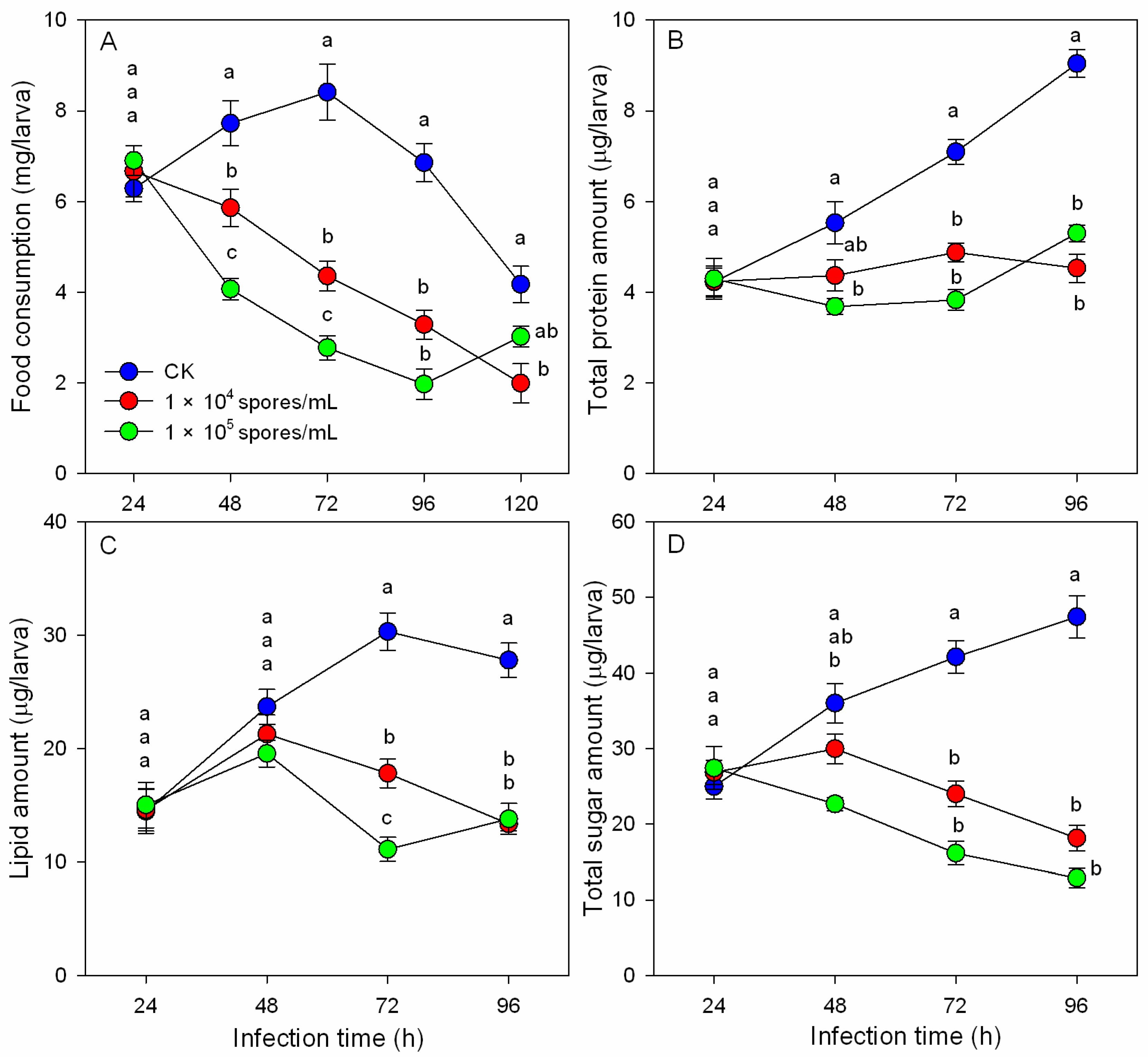
Figure 1. Effects of M. hiemalis infection on food consumption (A) and nutrient contents ((B), protein amount; (C), lipid amount; (D), sugar amount) of B. odoriphaga. Data in the figure are the mean ± SE. Different letters over the same column indicate significant differences between different spore concentration treatments at the p < 0.05 level by one-way ANOVA. Different letters (a and b) over the same column indicate significant differences between different spore concentration treatments at the p < 0.05 level as indicated by one-way ANOVA.
Insects 14 00162 g001
Figure 2. Digestive enzyme activities ((A): protease, (B): α-amylase, (C): lipase, and (D): cellulase) of B. odoriphaga larvae after infection with M. hiemalis spores. Data in the figure are the mean ± SE. Different letters over the same column indicate significant differences between different spore concentration treatments at the p < 0.05 level by one-way ANOVA. Different letters (a and b) over the same column indicate significant differences between different spore concentration treatments at the p < 0.05 level as indicated by one-way ANOVA.
Insects 14 00162 g002
Figure 3. Antioxidant enzyme activities ((A): CAT, (B): POD, (C): SOD, and (D): GST) of B. odoriphaga larvae after infection with M. hiemalis spores. Data in the figure are the mean ± SE. Different letters over the same column indicate significant differences between different spore concentration treatments at the p < 0.05 level by one-way ANOVA.
Insects 14 00162 g003
Figure 4. GO term enrichment analysis for the DEGs and the distribution of pathways annotated in the Kyoto Encyclopedia of Genes and Genomes (KEGG) data library in B. odoriphaga larvae after infection with M. hiemalis spores.
Insects 14 00162 g004
Figure 5. Real-time polymerase chain reaction (RT-PCR) analysis of gene expression in B. odoriphaga larvae after infection with M. hiemalis spores. RT-PCR data calculated with the 2−∆∆Ct method. The value of the relative expression level on the y-axis was calculated according to log(2−∆∆Ct). *, p < 0.05, as determined by one-way analysis of variance (ANOVA).
Insects 14 00162 g005
Table 1. Pathogenicity of M. hiemalis BO-1 against B. odoriphaga 4th-instar larvae.
Infection Time
(d)
Regression Equation
(y = ax + b)
LC50 Median Lethal Concentration (spores/mL)95% Confidence Interval
(spores/mL)
R2
3y = −10.802x + 8.6632.180 × 1061.099~4.736 × 1060.892
4y = −9.888x + 7.1431.891 × 1051.001~3.836 × 1050.983
5y = −11.187x + 7.8040.965 × 1050.551~1.729 × 1050.940
6y = −7.384x + 4.7982.914 × 1041.438~6.742 × 1040.999
Spore concentration
(spores/mL)
Regression equation
(y = ax + b)
LT50 Median lethal time (d)95% Confidence interval
(d)
R2
1.00 × 10⁴y = −7.149x + 4.6614.7874.476~5.1460.995
1.00 × 10⁵y = −7.004x + 8.6633.7913.518~4.0850.988
1.00 × 10⁶y = −7.672x + 3.8452.9862.753~3.2070.887
1.00 × 10⁷y = −8.143x + 3.6052.6182.407~2.8160.938
1.00 × 10⁸y = −7.796x + 3.2092.4322.223~2.6270.898
Table 2. Aligning statistics of clean reads with assembled unigenes.
SamplesTotal ReadsClean ReadsQ30(%)GC Content
(%)
Mapped ReadsMapped Ratio
(%)
Control-17,485,207,66750,812,19294.2241.9245,067,12888.69
Control-27,558,357,48051,677,54294.3342.0246,192,51589.39
Treatment-17,141,260,17949,670,66294.1942.2143,655,54587.89
Treatment-26,615,063,32844,925,16694.2742.0139,841,70688.68
Table 3. Differently expressed genes of B. odoriphaga larvae infected by M. hiemalis BO-1 by transcriptome analysis.
Gene IDGene NameGO Biological ProcessGO FunctionKEGG Pathwayslog2 Ratio
(T/C)
p-Value
HA402_000028uidAGO0005975: Carbohydrate
metabolic process
Beta-glucosidase activityko04973: Carbohydrate
digestion and absorption
−2.053.96 × 10-4
HA402_001933PRSSGO0007586: DigestionSerine-type endopeptidase activityko04974: Protein digestion
and absorption
−1.858.96 × 10-6
HA402_001429amyAGO0005975: Carbohydrate
metabolic process
Alpha-amylase activityko04973: Carbohydrate
digestion and absorption
−2.405.38 × 10-6
HA402_001695LIPAGO0016042: Lipid
digestion process
Lipase activityko00100: Steroid
biosynthesis
−3.081.53 × 10-6
HA402_003401treAGO0005991: Trehalose
metabolic process
Trehalase activityko01100: Metabolic
pathways
1.962.97 × 10-13
HA402_014097CPT1AGO0006635: Fatty acid
beta-oxidation
Palmitoleoyltransferase activityko01212: Fatty acid
metabolism
2.542.97 × 10-13
HA402_012831PRDXGO0006979: Response to
oxidative stress
Peroxidase activityko04146: Peroxisome1.564.69 × 10-4
HA402_001366catBGO0042744: Hydrogen
peroxide catabolic process
Catalase activityko04146: Peroxisome2.314.24 × 10-3
HA402_005674gstGO0006979: Response to
oxidative stress
Glutathione transferase
activity
ko00480: Glutathione
metabolism
−2.322.18 × 10-7
HA402_011746SOD1GO0019430: Removal of
superoxide radicals
Superoxide dismutase
activity
ko04146: Peroxisome−1.591.11 × 10-5
HA402_008470CYP9GO0017144: Drug
metabolic process
cytochrome P450
activity
ko00982: Drug metabolism—cytochrome P4502.674.25 × 10-6
HA402_001791ECSITGO0045087: Innate immune
response
Hydrolase activityko04624: Toll and Imd
signaling pathway
1.275.32 × 10-4
Disclaimer/Publisher’s Note: The statements, opinions and data contained in all publications are solely those of the individual author(s) and contributor(s) and not of MDPI and/or the editor(s). MDPI and/or the editor(s) disclaim responsibility for any injury to people or property resulting from any ideas, methods, instructions or products referred to in the content.