Article

Selection and Comparative Gene Expression of Midgut-Specific Targets for Drosophila suzukii

1
USDA Agricultural Research Service, Horticultural Crops Research Unit, Corvallis, OR 97331, USA
2
Department of Agricultural Convergence Technology, Jeonbuk National University, Jeonju 54596, Republic of Korea
3
Department of Biochemistry, Molecular Biology, Entomology and Plant Pathology, Mississippi State University, Mississippi State, MS 39762, USA
*
Author to whom correspondence should be addressed.
Insects 2023, 14(1), 76; https://doi.org/10.3390/insects14010076
Submission received: 24 November 2022 / Revised: 3 January 2023 / Accepted: 8 January 2023 / Published: 12 January 2023
(This article belongs to the Special Issue Insect Genome and Transcriptome Data)

Abstract

:

Simple Summary

Spotted-wing drosophila (SWD), Drosophila suzukii, a major pest of small fruits and cherries, is often managed with conventional insecticides. RNA interference (RNAi) technology has been investigated to develop an alternative control method for SWD. Our previous RNAi work showed that the RNAi efficacy on SWD was limited because double-stranded RNA (dsRNA) was degraded by the midgut nuclease (= dsRNA − degrading enzyme) before passing through the midgut membrane of the fly. To overcome this obstacle, RNAi directly targets the midgut genes, thus eliminating the need for dsRNA to pass through the midgut membrane. The primary focus of this study is to identify the fly adult midgut’s genes. We compared differential gene expressions between the midgut and the rest of the whole-body using transcriptomic analysis. We found that 1921 genes were upregulated, and 1834 genes were downregulated in the midgut. We chose ten midgut-specifically upregulated genes involved in various biological functions and confirmed their gene expressions. Particularly, the midgut membrane proteins found in this study would be potential targets for developing RNAi and other biological tools for controlling SWD in the future.

Abstract

Spotted-wing drosophila (SWD), Drosophila suzukii, is a destructive and invasive pest that attacks most small fruits and cherries. The current management for SWD involves the use of conventional insecticides. In an effort to develop a biologically based control option, the application of RNA interference (RNAi) has been investigated. To develop an RNAi approach, suitable targets must be identified, and an efficient delivery method must be developed for introducing the double-stranded RNA (dsRNA) in the midgut. In D. suzukii, we previously found that dsRNA nucleases actively degrade dsRNA molecules in the midgut. In this study, we focused on identifying biological targets focused on the midgut membrane. The profile of midgut-specific genes was analyzed and compared with the genes expressed in the whole-body using transcriptome analysis. Differential gene expression analysis revealed that 1921 contigs were upregulated and 1834 contigs were downregulated in the midgut when compared to genes from other body tissues. We chose ten midgut-specifically upregulated genes and empirically confirmed their expressions. We are particularly interested in the midgut membrane proteins, including G protein-coupled receptors (GPCRs) such as diuretic hormone 31 (DH31) receptor, neuropeptide F (NPF) recepror, toll-9, adhesion receptors, methuselah (mth), and gustatory receptor, because insect GPCRs have been offered great potential for next-generation pest management.

1. Introduction

Spotted-wing drosophila (SWD), Drosophila suzukii, is a worldwide pest fly. Females have a serrated ovipositor, a morphological adaptation [1] that enables them to lay eggs into ripening fruits before harvest. Moreover, D. suzukii exhibits a wide host range in its introduced and native range, infesting most small fruits and cherries in Asia, Europe, and North America [2], and now they have spread to subtropical areas including South America, Australia, and Africa [3,4]. Therefore, small fruit industries around the world are facing economic losses due to increased spending on pest management in the field.
Despite many potential adverse effects, current control methods for D. suzukii rely primarily on conventional chemical insecticides. Recent research has sought to develop alternative control methods to replace or at least reduce chemical insecticide inputs [5,6], but they cannot fully protect small fruits from fly damage. The availability of various insect genomes and RNA sequences facilitates a genome-based approach to identifying biological targets for insect control. Since the genome of D. suzukii was published [7], a few case studies analyzing the functional transcriptome of its antennae were reported [8,9,10]. A comprehensive genome study was recently achieved by the long-read sequencing [11]. Moreover, related to pest management, transcriptome studies facilitate finding target genes for RNAi and can be applied towards developing sterile insect technique (SIT) and CRISPR/Cas9 technology reviewed by [12].
Insect neuropeptides (NPs) and G protein-coupled receptors (GPCRs) are involved in a variety of critical physiological processes such as homeostasis, feeding, digestion, excretion, circulation, reproduction, and metamorphosis during insect life stages [13,14]. Therefore, insect NPs and their GPCRs have been proposed as potential targets for decades [15,16,17]. Recently, the application of RNA interference (RNAi) has been investigated to develop a new biological alternative, a more species- and gene-specific method for controlling pests. A few trials for D. suzukii RNAi with potential targets evaluated the RNAi effects against larvae and adults [18,19,20,21]. However, the RNAi efficacy with the target genes was limited because the dsRNA molecules introduced in the midgut are degraded before being transported into hemocoel [22]. There are some options to overcome this obstacle: (1) encapsulation of dsRNAs using nanoparticles or exosomes, and (2) direct-targeting the midgut membrane for RNAi, thus eliminating the need for dsRNA to pass through the midgut membrane, and (3) modification of dsRNA structures to avoid/reduce the degradation [23,24].
The digestive tract, especially the midgut, is an important alimentary canal where various mechanical and physiological processes occur, including food digestion, nutrient absorption, and detoxification [25]. The importance of the gut is evident by the many studies of gene expression profiles of the gut using various advanced technologies during the past decades. Moreover, studies of microbes, insecticide resistance, immunity, and nutrient absorption have been accelerated to understand gut physiology and its application [26,27].
Regardless of extensive gene sequencing data, basic research on the gene expression of the midgut and the rest of body and differential data comparisons in D. suzukii is limited. Therefore, the primary focus of this study is to identify the adult midgut membrane genes for potential targets with particular interest in membrane-bound proteins, including GPCRs. Differential gene expressions of the midgut and whole-body of D. suzukii were analyzed using transcriptome. The selected genes showing relatively high expression in the midgut were validated by qRT-PCR. In addition, gene ontology analysis was performed to identify the midgut-specific genes involved in various biological functions and differential expression patterns. This midgut transcriptome data might be useful to those who work on gut physiology, gut microbes, insecticide resistance, and the selection of biological targets to develop D. suzukii control methods.

2. Materials and Methods

2.1. Insects

Drosophila suzukii were maintained using standard rearing methods [28] at the Horticultural Crops Research Unit, USDA ARS in Corvallis, OR, USA, and 3 to 5-day-old adults were used in all the experiments in this study.

2.2. Isolation of Total RNA

Approximately 20–30 midguts were dissected from 3 to 5-day-old adults and collected in a 1.5 mL tube on dry ice. Samples were homogenized in a lysis buffer using a plastic pestle. Total RNA was isolated using PureLink RNA Kit (Thermo Fisher Scientific, Waltham, MA, USA) according to the manufacturer’s instructions. The total RNA was quantified on a NanoDrop 2000 spectrophotometer (Thermo Fisher Scientific) and stored at −20 °C until use. For comparison, total RNA was isolated from the whole-body of female adults using the same kit above. The total RNA isolated from the female midgut and whole-body were used for transcriptome sequencing and differential gene expression analysis. The RNA concentrations obtained after the extraction were adjusted to 60 ng/μL in 50 μL for both tissues. The quality of RNA was analyzed using Agilent 2100 Bioanalyzer (Agilent Technologies, Santa Clara, CA, USA) based on RNA Integrity Number (RIN).

2.3. Illumina Sequencing

The cDNA libraries were prepared using TruSeq Stranded Total RNA Library Prep Kit (Illumina, San Diego, CA, USA). Illumina sequencing was performed by Psomagen, Inc. (Rockville, MD, USA) using the Illumina HiSeq 2000 platform. Briefly, ribosomal RNA was removed from total RNA, and the remaining RNA was purified, fragmented, and primed for cDNA synthesis. The first strand of cDNA was synthesized with reverse transcriptase and priming with random hexamers. The RNA template of the first strand cDNA was replaced by incorporating dUTP in place of dTTP to generate the second strand cDNA. The cDNA then underwent an end repair process, adenylation of 3’ ends, and subsequent ligation of the adapter. The quantified and qualified libraries for both samples were sequenced using an Illumina HiSeq 2000. RNA-Seq raw reads are available in the GenBank SRA database (BioProject accession No PRJNA903844).

2.4. Anlaysis of up- or down-Regulated Genes

Based on the expression values as fragment per kilobase of transcript per million mapped (FPKM) reads up- or down-regulated genes were identified among the midgut and whole-body samples (MG and WB) after transformation, normalization, and fold change (fc) comparisons. The FPKM values were obtained by the Reference Annotation Based Transcript (RABT) method using -G option of Cufflinks. Raw FPKM values were transformed into Log2(FPKM+1) values in order to make the range with low values widely distributed, and then normalized by quantile normalization method using preprocessCore in R package in order to reduce systematic bias among the samples. Up- or down-regulated genes with |fc| ≥ 2 were regarded as up- or down-regulated genes. If the FPKM value was 0 (i.e., not changed) in either of the two samples, it was not included in the analysis. However, they were retrieved from the original raw data and manually added to the DEG analysis to identify tissue-specific genes.

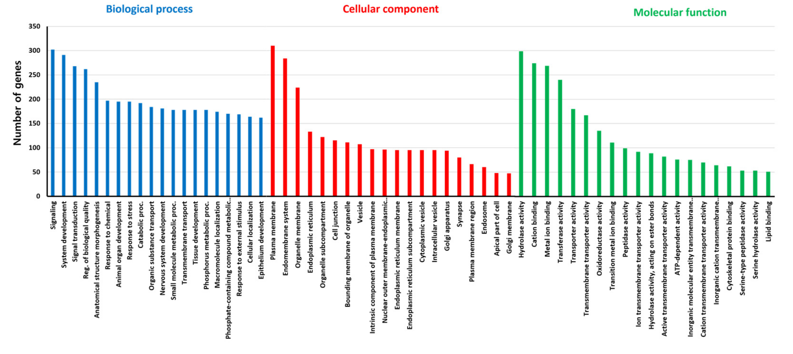
2.5. Functional Annotation and Gene Ontology Analysis

For the two tissue samples, if more than one FPKM value was 0, it was not included in the analysis. Therefore, from the total of 13,583 contigs, 4712 were excluded, and only 8871 contigs were used for statistical analysis. The functional annotation of the 13,583 official gene set (OGS) was performed by sequence similarity searches against the NCBI non-redundant protein sequences (nr) database using BLASTx algorithm with a cut-off E-value of 1 × 10⁻⁵ run by Geneious 8.1.5 software (Biomatters Ltd., Auckland, New Zealand). In cases where there were no clear “hits,” the sequences were searched again using BLASTn. The BLAST hits were grouped into major gene families based on their putative functions in fly biology. The organism information obtained by the BLAST hit was collected to confirm the sequence similarity of the transcripts to closely related species. Gene ontology analysis was performed via ShinyGO 0.76 (http://bioinformatics.sdstate.edu/go/, accessed on 15 November 2022). Biological processes, cellular components, and molecular functions are examined.

2.6. Manual Annotation of the Midgut Transcriptome

Regarding 1921 upregulated genes, we searched for the DS10 numbers on SpottedWingFlyBase (http://spottedwingflybase.org/, accessed on 15 November 2022). Then, the C.G. number, FBgn, and symbol were found in the D. melanogaster FlyBase (https://flybase.org/, accessed on 15 November 2022). Functional analysis and InterPro information were also noted in Supplementary S1.

2.7. Quantitative Real-Time PCR (qRT-PCR)

To validate midgut-specifically upregulated genes from the fly pool, we selected some putatively important midgut genes, including dsRNases, GPCRs, and neuropeptides, and verified their expression patterns by qRT-PCR using samples isolated from different sets of flies. To isolate total RNAs, three different replications of the midgut pool from 3 to 5-day-old adults were dissected as described above. cDNAs were synthesized from 500 ng total RNA for the midgut and whole-body samples, respectively, using Verso cDNA Synthesis Kit (Thermo Fisher Scientific), according to the manufacturer’s instructions. The synthesized cDNAs were stored at −20 °C until qRT-PCR. Gene-specific primers (Table S4) were designed to amplify 90–120 bp portions of target genes and two reference genes. The qRT-PCR reaction mixture was prepared in 20 μL with 2X PowerUp SYBR Green Master Mix (Thermo Fisher Scientific), deionized H2O, and 0.25 μM primer pairs. The reaction conditions were 95 °C for 15 min and then 40 cycles of 95 °C for 15 s and 60 °C for 1 min, run in StepOnePlus™ Real-Time PCR System (Thermo Fisher Scientific). This was followed by a melting curve analysis over the range of 60–95 °C and sequencing of the qRT-PCR products to check the amplification specificity. A minimum of three repetitions of each standard were done using different preparations of D. suzukii cDNA. Five different housekeeping genes were tested for primer efficiency, and Rpn2 and Rpt6 were selected as reference genes in the analysis because they were less variable than other housekeeping genes previously tested [9]. The geometrical mean (geomean) of Rpn2 and Rpt6 was used for the qRT-PCR experiment, and the delta delta Ct method was applied.

2.8. Statistical Analysis

Student’s t-test was used to compare the relative mRNA level differences between control and treatment groups to test the relative mRNA expression. A p-value of 0.05 or less between groups was considered significantly different.

3. Results and Discussion

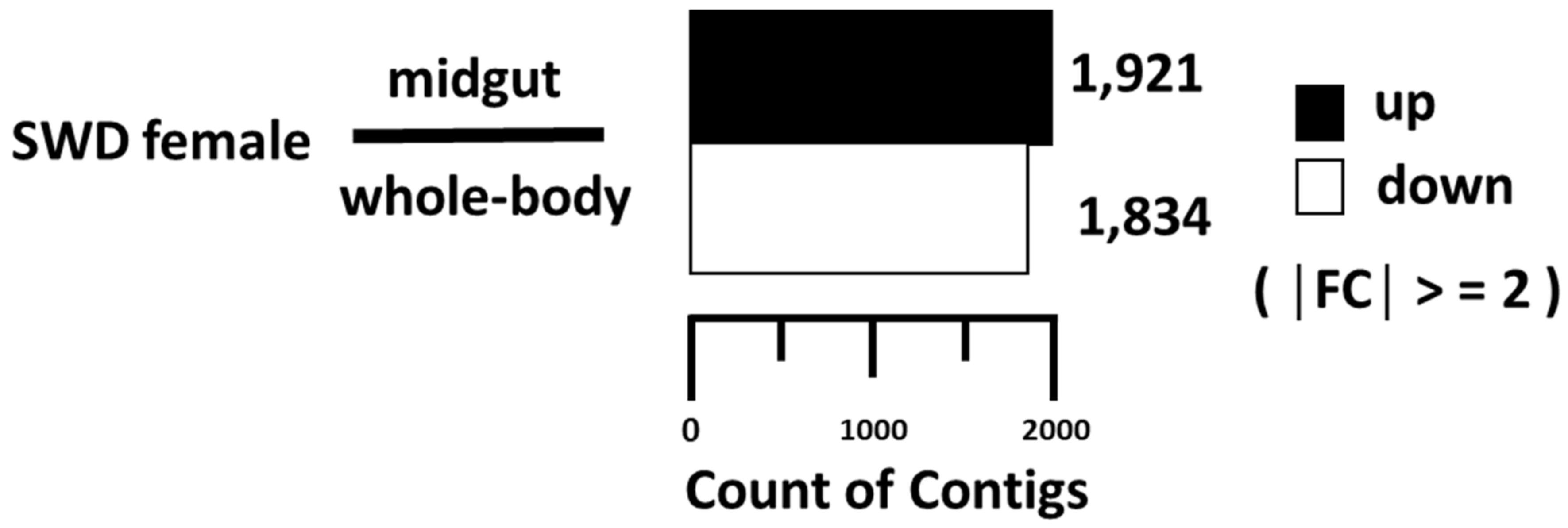
3.1. Midgut Transcriptome Analysis

Transcriptomes of midgut and whole-body generated over 5.6 G bases, resulting in 54 and 59 million reads, respectively (Table S1). Based on the D. suzukii official gene set (OGS) provided in the SpottedWingFlyBase (http://spottedwingflybase.org, accessed on 15 November 2022), over 78.9% and 78.8% of reads from the midgut and whole-body transcriptomes, respectively, were mapped to the reference genome and assembled into 13,583 contigs (Table S2).
For expression analysis, contigs with zero value of FPKM in either transcriptome were excluded from analysis, leaving 8871 contigs to be further analyzed. The distributions of the contig expression levels were similar between the midgut and whole-body (Figure 1).
The analysis of up- or down-regulated genes between the two transcriptomes revealed 1921 contigs that were upregulated and 1834 contigs that were downregulated in the midgut compared to the whole-body (Figure 1), whereas the rest (6881 contigs) were similarly expressed between the two samples. To provide a functional overview of the transcriptional differences between midgut and whole-body, gene ontology (G.O.) enrichment analysis was performed (Figure 2). According to the ontology data, signaling-related genes are dominantly occupied in the biological process, and membrane-related genes (plasma membrane and endomembrane system) are highly present in cellular components. In molecular function analysis, hydrolase activity, cation, and metal ion binding take large portions.
The most upregulated genes of the midgut compared to whole-body are listed in Table S3. Sequences of DS10 contigs were searched against FlyBase (version FB2022_05; http://flybase.org/, accessed on 15 November 2022) to find homologous genes in D. melanogaster using the BLASTn program with the BLOSUM62 matrix at the threshold value 0.01. Hence, corelated CG numbers, molecular functions, and biological functions were noted (Table S3). Moreover, InterProScan data was added to classify the protein family of each gene. Using the Drosophila Gene Expression Tool (DGET), a search program of RNA-Seq-based expression profiles (www.flyrnai.org/tools/dget/web/, accessed on 15 November 2022), all the genes were checked. Most of the genes were highly expressed in the digestive system [29] (Table S3). These genes showed a relatively higher expression pattern in the digestive system than in other body parts.

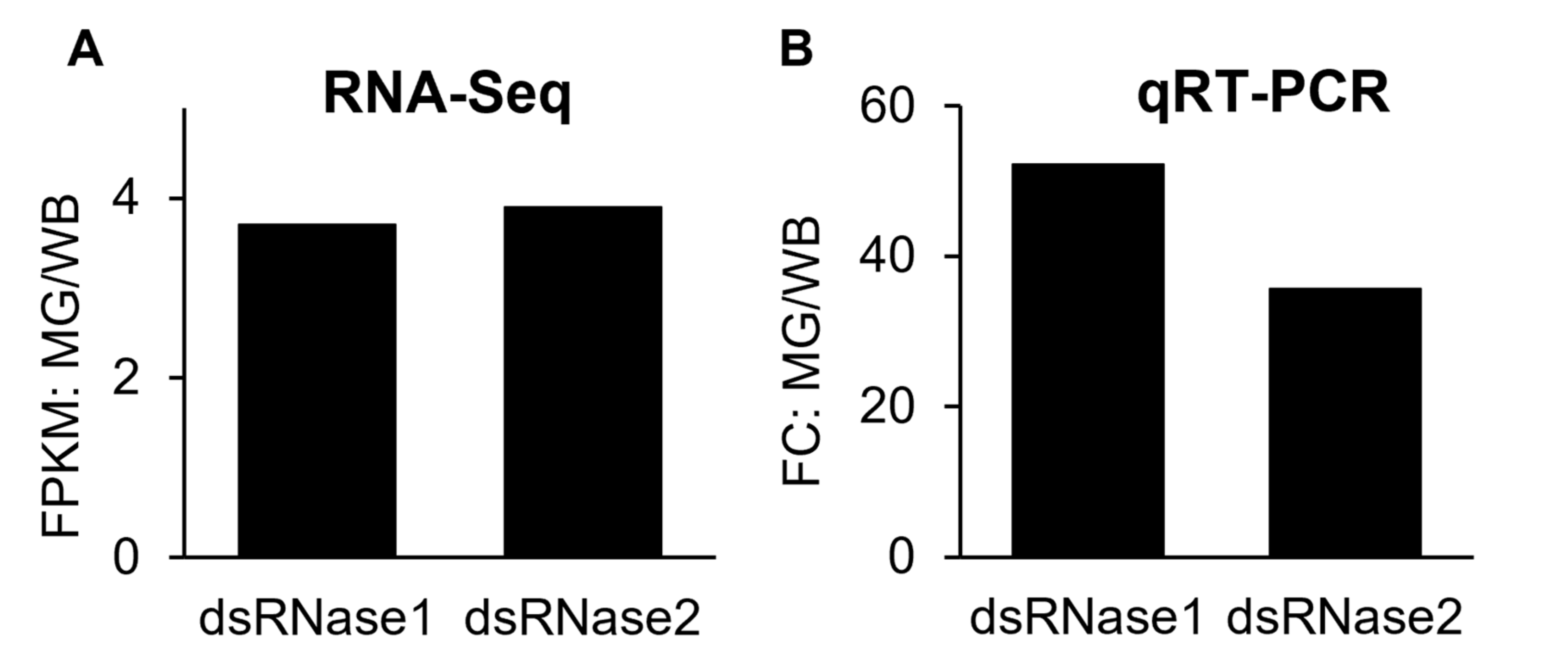
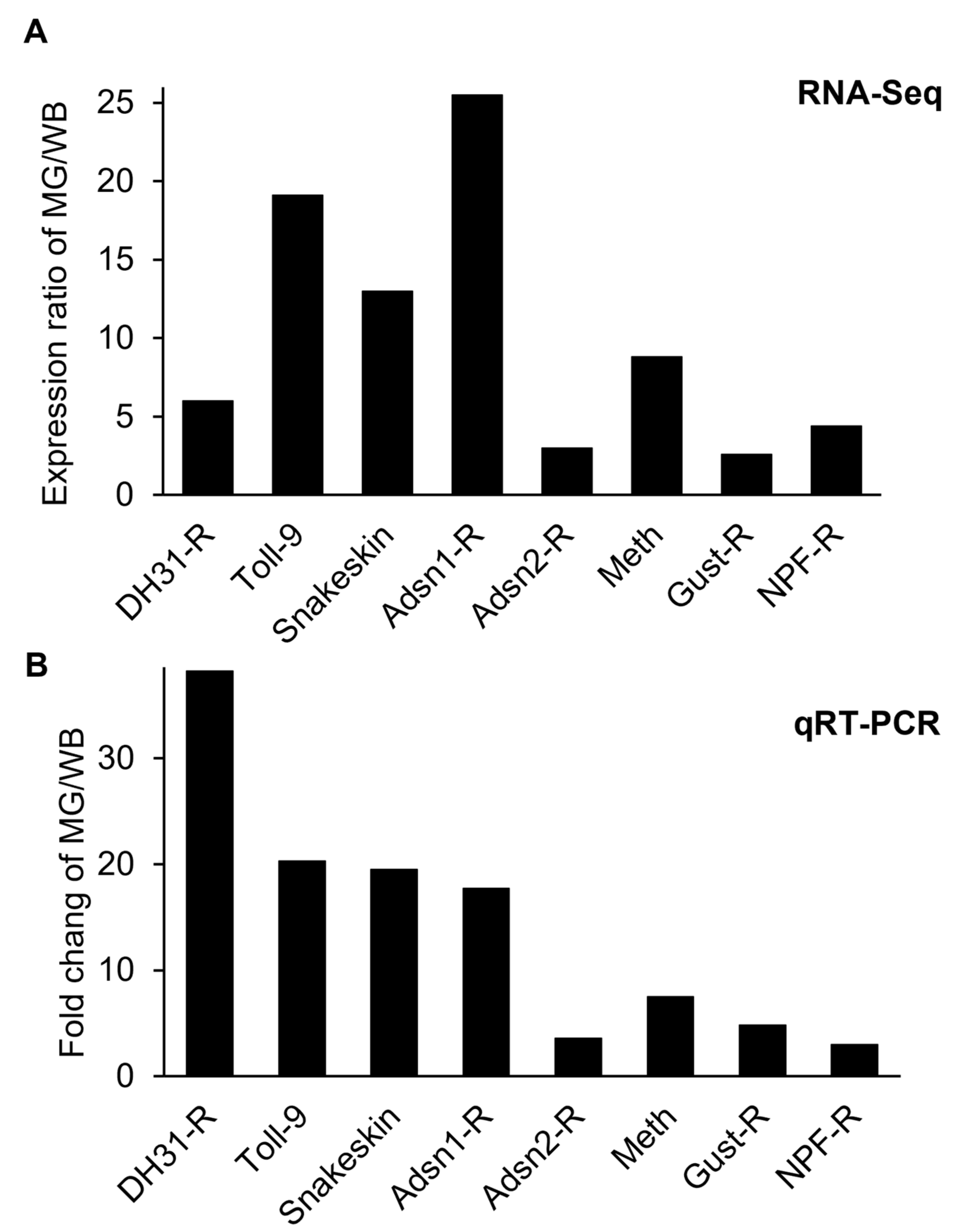
3.2. Validation of the Midgut-Specifically Upregulated Genes

In order to validate FPKM values of transcriptome data, ten genes were selected from the upregulated gene group, confirmed by qRT-PCR, and compared to transcriptome data (Figure 3 and Figure 4). Besides the qRT-PCR confirmation, to overcome the limit of experimental replications and to avoid false-positive-effects, the upregulated genes from the midgut of D. suzukii were compared with D. melanogaster’s expression database (Table S3). Those ten genes were selected by previous literature to prove the midgut highly expressed characteristics described below. The midgut is the primary digestive system in insects for the uptake of biological molecules, including nutrients and bioactive compounds (i.e., dsRNA and small peptides) transportation into the target tissues or cells through the hemocoel. From the midgut transcriptome analysis, we focused on identifying potential biological targets to be applied for RNA interference (RNAi) or receptor interference (Receptor-i) for D. suzukii management. RNAi for dipteran pests is challenging with low efficiency of RNAi due to limited dsRNA delivery and degradation of dsRNA by the midgut nuclease (=dsRNase) prior to transport into target cells through the midgut [19,22,30].
Considering other insects, it is common knowledge that expression patterns of dsRNases are relatively high in midgut compared to other body tissues [31,32,33,34,35]. In our previous study, two D. suzukii dsRNase genes, DrosudsRNase1 (DS10_00008824, GenBank Accession No. MW984608) and DrosudsRNase2 (DS10_00008823, GenBank Accession No. MW984609), were identified, functionally expressed in Sf9 cells derived from Spodoptera frugiperda, and characterized for enzyme activity [22]. Both dsRNA degradation enzymes are active in the midgut; dsRNase1 is relatively more active than dsRNase2 in terms of its gene expression level and in vitro degradation activities [22]. From the RNA-Seq analysis, the FPKM expressions of the dsRNase1 & 2 genes in the midgut were at similar levels, which are 3.7 and 3.9 times greater than whole-body, respectively (Supplementary S1). In the qRT-PCR result, their relative mRNA levels (=FC values) of midgut tissues are 35.6–52.2 times greater than whole-body. The previous and current qRT-PCR results confirmed that dsRNase1 is a dominant enzyme in D. suzukii.

3.3. Midgut-Specifically Upregulated Membrane Proteins and GPCRs

Except for the two dsRNA enzymes above, the other eight membrane proteins selected in this study have not yet been identified and characterized in D. suzukii. DS10_00008095 is orthologous to the D. melanogaster CG6981 (Snakeskin), which plays an important role in the gut: intestinal epithelial cell development, the establishment of endothelial barrier, anterior midgut development, and septate junction assembly. Expression of Snakeskin in the midgut is significantly high compared to the whole-body, which ranges 13.0–19.5 times from RNA-Seq and qRT-PCR results of D. suzukii. As a pest management tactic, dsRNA targeting the Snakeskin gene of corn rootworm induced larval growth inhibition and eventual mortality [36]. Midgut-specific genes can be targeted for RNAi-mediated pest control because they can cause an instant effect, such as fasting and mortality, by interfering with the midgut, where digestive substances are absorbed.
Over decades, insect GPCRs have been proposed as potential targets for next-generation pest management because they involve various critical physiological processes at all insect life stages [15,37,38]. GPCRs are membrane-bound proteins consisting of seven transmembrane domains. Especially, the GPCRs upregulated in the midgut mediate various endocrine signals including neurohormones into cells for digestive processes, activation of digestive enzymes and gustatory receptors, recognition of nutritional levels, and water balance [16,38]. These could be potential targets for D. suzukii control. The midgut transcriptome profile selected the seven most upregulated GPCRs in the midgut (Figure 5), as described in detail below.
DS10_00007171 (GenBank Accession No. XP_036670585) is a toll-like receptor. Toll receptors, in general, play a crucial role in the innate immune system and interact with microbes. Moreover, epithelial toll-like receptors are specialized in gut homeostasis, nutrition absorption, and gut-related diseases [39]. Expression levels of the toll receptor (=Toll-9) from the RNA-Seq and qRT-PCR results in the midgut are 19.1–20.3 times greater than the whole-body (Figure 4), indicating Toll-9 might be a midgut-specific gene in D. suzukii. The biological function(s) of this gene in the midgut remains to identify in the future study. In insects, the Bombyx mori Toll-9 gene is engaged in the local gut immune response [40]. Of 14 putative B. mori toll genes, B. mori Toll9-1 is upregulated explicitly in the midgut compared to other body parts [40].
DS10_00006592 is orthologous to CG17415 of D. melanogaster, which is related to a member of the calcitonin-like receptor (CLR) and identified as the diuretic hormone 31 receptor (DH31-R) [41]. DH receptors belong to the Class B GPCR group signaling pathway using cAMP as a second messenger instead of Ca2+ [16,42]. In the qRT-PCR result, the expression of DH31-R showed midgut is almost 40 times (fold change ratio of MG/WB) greater than whole-body, indicating DH31-R might be involved in the digestion of D. suzukii. The physiological function of the DH31 receptor is expected to regulate fluid secretion from foods in the midgut through Malpighian tubules in Drosophila [41]. DH31-R would be a good target because water regulation is critical in dipteran insects. The other diuretic hormone receptor in insects is the DH44 receptor (DS10_00002631). Its expression in the D. suzukii midgut was as high as that of DH31-R from the transcriptome analysis.
DS10_00013361 gene (GenBank Accession No. XP_036670319) refers to methuselah (mth) that belongs to the Class B GPCR group, related to increasing the lifespan with increased resistance to heat and oxidative stress, and starvation in Drosophila mutants [43,44]. The gene has been evaluated to understand its potential functions, including its response to insecticide stresses and its role in the regulation of longevity [44], pointing out a potential target for new management of D. suzukii. The gene expressions of the midgut measured by RNA-Seq and qRT-PCR are 8.8–7.5 times greater than whole-body, which is significant.
Both DS10_00012673 and DS10_00012139 (GenBank Accession No. XP_016939533 and XP_016935592) were identified for adhesion GPCRs, one of the glycoproteins, E-cadherin, that functions as part of a dynamic membrane-spanning macromolecular complex to facilitate adhesion mechanically between epithelial cells of the midgut [45,46]. In addition, the receptor extends to multiple functions, including cell recognition, sorting, boundary formation, maintenance, and coordination of cell movements. There are different adhesion receptors; both receptors belong to Ca2+-dependent Type 1 classical cadherins, like housekeeping genes, which are expressed in almost all tissues [45]. In D. suzukii midgut, two adhesion GPCRs are found, that adhesion1 receptor (Adsn1-R) is dominant and expressed at 17.7–25.5 times greater than whole-body from RNA-Seq and qRT-PCR analyses, respectively. The expression of Adsn1-R is ~5 times greater than Adsn2-R in the midgut, which would be a better target for the fly. However, the specific functions of the two GPCRs are unclear in Drosophila.
DS10_00008044 (GenBank Accession No. XP_036675103) was identified with the receptor of neuropeptide F (NPF), which has various functions including food search, intake, and odor perception important in the regulation of feeding activities in insects [13,14]. In Drosophila, the first NPF receptor (NPF-R) exhibits a similar pattern of localization in larvae, as the NPF ligand gene that is expressed in the brain and in the midgut for regulation of feeding and digestion [47]. As expected, the NPF-R was highly expressed in the midgut of D. suzukii, 3.0–4.4 times greater than whole-body from both analyses, suggesting its role in modulating food intake and ingestion with other neuropeptides in the midgut. Interfering with the specific receptor would be critical to the physiological function of the NPF in the midgut.
DS10_00005654 (GenBank Accession No. XP_016941187) was identified as a gustatory receptor 43a (Gr43a), one of the most conserved gustatory receptors across insect species. The specific gustatory receptor is highly expressed in the midgut, 2.6–4.8 times greater than whole-body in D suzukii. The receptor is usually expressed in the brain and plays a critical role in sensing internal fructose levels in Drosophila [48,49]. Activation of this receptor is mediated by the neuropeptide corazonin, as a neurotransmitter, in the fly [49]. The Gr43a neurons projecting to the midgut and expression of Gr43a in the gastrointestinal system are found across different insect groups that may regulate food transport and/or secretion of neuropeptides/hormones in response to sugar consumption [49]. Like the NPF-R, disruption of the Gr43a pathway would be critical to the sugar metabolism and/or nutritional value of food for the fly survival.
All the upregulated genes for biological targets were provided in this study (See Supplementary S1). Due to the contig-level assembly process and deficient D. suzukii annotation information, duplicated annotations exist, and many unknown genes are listed. Although the physiological processes of the highly expressed genes selected in this study are inferred from other species, further studies are needed to verify their specific functions in D. suzukii. While this RNA-Seq data may not give the exact values for gene expression levels, it not only provides overall gene expression patterns for the study of midgut physiology, but also for gut microbes, insecticide resistance, and identifying biological targets for D. suzukii management.

Supplementary Materials

The following supporting information can be downloaded at: https://www.mdpi.com/article/10.3390/insects14010076/s1, Table S1. Raw data of the midgut and whole-body transcriptome results. Table S2. Mapped data stats. # of processed reads: # of cleaned reads after trimming. Number of mapped reads: # of reads mapped to reference. # of suppressed reads by multiple mapping: Number of reads removed due to multiple mapping. Overall read mapping ratio: Number of total mapped reads / # of total processed reads. Table S3. The most upregulated genes in the midgut of Drosophila suzukii are found in Drosophila melanogaster. Table S4. List of the primers used in this study. Supplementary S1: Upregulated genes in Midgut. Deposited in figshare with DOI (10.6084/m9.figshare.21711476).

Author Contributions

Conceptualization, M.-Y.C.; methodology, J.-S.Y., S.-J.A. and M.-Y.C.; software, J.-S.Y. and S.-J.A.; validation, J.-S.Y., S.-J.A. and M.-Y.C.; formal analysis, J.-S.Y., S.-J.A.; investigation, M.-Y.C.; resources, M.-Y.C.; data curation, J.-S.Y., S.-J.A. and M.-Y.C.; writing—J.-S.Y., S.-J.A. and M.-Y.C.; supervision, M.-Y.C.; administration, M.-Y.C.; funding acquisition, J.-S.Y. and M.-Y.C. All authors have read and agreed to the published version of the manuscript.

Funding

This research received no external funding.

Informed Consent Statement

Not applicable.

Data Availability Statement

The data presented in this study are available in [insert article or Supplementary Material here].

Acknowledgments

We thank Jana Lee for the fly colony, and Ruth Martin for review of an earlier version of the manuscript. This paper was supported by research funds for newly appointed professors of Jeonbuk National University in 2021. This work was supported by the National Research Foundation of Korea (NRF) grant funded by the Korea government (MEST) (No. 2022R1C1C1007039). This study was supported in part by Northwest Center for Small Fruits Research, Commissions of Oregon and Washington Blueberry, Red-raspberry, Blackberry, Washington Tree Fruit Research, and Oregon Cherry, and base funding USDA ARS CRIS 2072-22000-044-00D.

Conflicts of Interest

The authors declare no conflict of interest.

References

  1. Atallah, J.; Teixeira, L.; Salazar, R.; Zaragoza, G.; Kopp, A. The making of a pest: The evolution of a fruit-penetrating ovipositor in Drosophila suzukii and related species. Proc. Biol. Sci. 2014, 281, 20132840. [Google Scholar] [CrossRef] [PubMed] [Green Version]
  2. Asplen, M.K.; Anfora, G.; Biondi, A.; Choi, D.S.; Chu, D.; Daane, K.M.; Gibert, P.; Gutierrez, A.P.; Hoelmer, K.A.; Hutchison, W.D.; et al. Invasion biology of spotted wing Drosophila (Drosophila suzukii): A global perspective and future priorities. J. Pest Sci. 2015, 88, 469–494. [Google Scholar] [CrossRef]
  3. Lee, J.C.; Bruck, D.J.; Curry, H.; Edwards, D.; Haviland, D.R.; Van Steenwyk, R.A.; Yorgey, B.M. The susceptibility of small fruits and cherries to the spotted-wing drosophila, Drosophila suzukii. Pest Manag. Sci. 2011, 67, 1358–1367. [Google Scholar] [CrossRef] [PubMed]
  4. Dos Santos, L.A.; Mendes, M.F.; Kruger, A.P.; Blauth, M.L.; Gottschalk, M.S.; Garcia, F.R. Global potential distribution of Drosophila suzukii (Diptera, Drosophilidae). PLoS ONE 2017, 12, e0174318. [Google Scholar] [CrossRef] [PubMed] [Green Version]
  5. Klick, J.; Yang, W.Q.; Lee, J.C.; Bruck, D.J. Reduced spray programs for Drosophila suzukii management in berry crops. Int. J. Pest Manag. 2016, 62, 368–377. [Google Scholar] [CrossRef]
  6. Rice, K.B.; Short, B.D.; Leskey, T.C. Development of an Attract-and-Kill Strategy for Drosophila suzukii (Diptera: Drosophilidae): Evaluation of Attracticidal Spheres Under Laboratory and Field Conditions. J. Econ. Entomol. 2017, 110, 535–542. [Google Scholar] [CrossRef]
  7. Chiu, J.C.; Jiang, X.; Zhao, L.; Hamm, C.A.; Cridland, J.M.; Saelao, P.; Hamby, K.A.; Lee, E.K.; Kwok, R.S.; Zhang, G.; et al. Genome of Drosophila suzukii, the spotted wing drosophila. G3 2013, 3, 2257–2271. [Google Scholar] [CrossRef] [Green Version]
  8. Crava, C.M.; Sassu, F.; Tait, G.; Becher, P.G.; Anfora, G. Functional transcriptome analyses of Drosophila suzukii antennae reveal mating-dependent olfaction plasticity in females. Insect Biochem. Mol. Biol. 2019, 105, 51–59. [Google Scholar] [CrossRef]
  9. Ahn, S.J.; Oh, H.W.; Corcoran, J.; Kim, J.A.; Park, K.C.; Park, C.G.; Choi, M.Y. Sex-biased gene expression in antennae of Drosophila suzukii. Arch. Insect Biochem. Physiol. 2020, 104, e21660. [Google Scholar] [CrossRef]
  10. Deng, D.; Xing, S.; Liu, X.; Ji, Q.; Zhai, Z.; Peng, W. Transcriptome analysis of sex-biased gene expression in the spotted-wing Drosophila, Drosophila suzukii (Matsumura). G3 2022, 12, jkac127. [Google Scholar] [CrossRef]
  11. Paris, M.; Boyer, R.; Jaenichen, R.; Wolf, J.; Karageorgi, M.; Green, J.; Cagnon, M.; Parinello, H.; Estoup, A.; Gautier, M.; et al. Near-chromosome level genome assembly of the fruit pest Drosophila suzukii using long-read sequencing. Sci. Rep. 2020, 10, 11227. [Google Scholar] [CrossRef]
  12. Tait, G.; Mermer, S.; Stockton, D.; Lee, J.; Avosani, S.; Abrieux, A.; Anfora, G.; Beers, E.; Biondi, A.; Burrack, H.; et al. Drosophila suzukii (Diptera: Drosophilidae): A decade of research towards a sustainable integrated pest management program. J. Econ. Entomol. 2021, 114, 1950–1974. [Google Scholar] [CrossRef]
  13. Nassel, D.R.; Zandawala, M. Recent advances in neuropeptide signaling in Drosophila, from genes to physiology and behavior. Prog. Neurobiol. 2019, 179, 101607. [Google Scholar] [CrossRef]
  14. Nassel, D.R.; Zandawala, M. Endocrine cybernetics: Neuropeptides as molecular switches in behavioural decisions. Open Biol. 2022, 12, 220174. [Google Scholar] [CrossRef]
  15. Audsley, N.; Down, R.E. G protein coupled receptors as targets for next generation pesticides. Insect Biochem. Mol. Biol. 2015, 67, 27–37. [Google Scholar] [CrossRef]
  16. Caers, J.; Verlinden, H.; Zels, S.; Vandersmissen, H.P.; Vuerinckx, K.; Schoofs, L. More than two decades of research on insect neuropeptide GPCRs: An overview. Front. Endocrinol. 2012, 3, 151. [Google Scholar] [CrossRef] [Green Version]
  17. Yeoh, J.G.C.; Pandit, A.A.; Zandawala, M.; Nassel, D.R.; Davies, S.A.; Dow, J.A.T. DINeR: Database for Insect Neuropeptide Research. Insect Biochem. Mol. Biol. 2017, 86, 9–19. [Google Scholar] [CrossRef]
  18. Murphy, K.A.; Tabuloc, C.A.; Cervantes, K.R.; Chiu, J.C. Ingestion of genetically modified yeast symbiont reduces fitness of an insect pest via RNA interference. Sci. Rep. 2016, 6, 22587. [Google Scholar] [CrossRef] [Green Version]
  19. Taning, C.N.T.; Christiaens, O.; Berkvens, N.; Casteels, H.; Maes, M.; Smagghe, G. Oral RNAi to control Drosophila suzukii: Laboratory testing against larval and adult stages. J. Pest Sci. 2016, 89, 803–814. [Google Scholar] [CrossRef]
  20. Ahn, S.J.; Donahue, K.; Koh, Y.; Martin, R.R.; Choi, M.Y. Microbial-Based Double-Stranded RNA Production to Develop Cost-Effective RNA Interference Application for Insect Pest Management. Int. J. Insect Sci. 2019, 11, 1179543319840323. [Google Scholar] [CrossRef]
  21. Abrieux, A.; Chiu, J.C. Oral delivery of dsRNA by microbes: Beyond pest control. Commun. Integr. Biol. 2016, 9, e1236163. [Google Scholar] [CrossRef] [PubMed] [Green Version]
  22. Yoon, J.S.; Ahn, S.J.; Flinn, C.M.; Choi, M.Y. Identification and functional analysis of dsRNases in spotted-wing drosophila, Drosophila suzukii. Arch. Insect Biochem. Physiol. 2021, 107, e21822. [Google Scholar] [CrossRef] [PubMed]
  23. Zhu, K.Y.; Palli, S.R. Mechanisms, Applications, and Challenges of Insect RNA Interference. Annu. Rev. Entomol. 2020, 65, 293–311. [Google Scholar] [CrossRef] [PubMed] [Green Version]
  24. Abbasi, R.; Heschuk, D.; Kim, B.; Whyard, S. A novel paperclip double-stranded RNA structure demonstrates clathrin-independent uptake in the mosquito Aedes aegypti. Insect Biochem. Mol. Biol. 2020, 127, 103492. [Google Scholar] [CrossRef] [PubMed]
  25. Lemaitre, B.; Miguel-Aliaga, I. The digestive tract of Drosophila melanogaster. Annu. Rev. Genet. 2013, 47, 377–404. [Google Scholar] [CrossRef] [Green Version]
  26. Bing, X.; Gerlach, J.; Loeb, G.; Buchon, N. Nutrient-Dependent Impact of Microbes on Drosophila suzukii Development. mBio 2018, 9, e02199-17. [Google Scholar] [CrossRef] [Green Version]
  27. Solomon, G.M.; Dodangoda, H.; McCarthy-Walker, T.; Ntim-Gyakari, R.; Newell, P.D. The microbiota of Drosophila suzukii influences the larval development of Drosophila melanogaster. PeerJ 2019, 7, e8097. [Google Scholar] [CrossRef] [Green Version]
  28. Woltz, J.M.; Donahue, K.M.; Bruck, D.J.; Lee, J.C. Efficacy of commercially available predators, nematodes and fungal entomopathogens for augmentative control of Drosophila suzukii. J. Appl. Entomol. 2015, 139, 759–770. [Google Scholar] [CrossRef]
  29. Hu, Y.; Comjean, A.; Perrimon, N.; Mohr, S.E. The Drosophila Gene Expression Tool (DGET) for expression analyses. BMC Bioinform. 2017, 18, 98. [Google Scholar] [CrossRef] [Green Version]
  30. Whyard, S.; Singh, A.D.; Wong, S. Ingested double-stranded RNAs can act as species-specific insecticides. Insect Biochem. Mol. Biol. 2009, 39, 824–832. [Google Scholar] [CrossRef]
  31. Almeida Garcia, R.; Lima Pepino Macedo, L.; Cabral do Nascimento, D.; Gillet, F.X.; Moreira-Pinto, C.E.; Faheem, M.; Moreschi Basso, A.M.; Mattar Silva, M.C.; Grossi-de-Sa, M.F. Nucleases as a barrier to gene silencing in the cotton boll weevil, Anthonomus grandis. PLoS ONE 2017, 12, e0189600. [Google Scholar] [CrossRef] [Green Version]
  32. Cooper, A.M.W.; Song, H.; Shi, X.; Yu, Z.; Lorenzen, M.; Silver, K.; Zhang, J.; Zhu, K.Y. Molecular Characterizations of Double-Stranded RNA Degrading Nuclease Genes from Ostrinia nubilalis. Insects 2020, 11, 652. [Google Scholar] [CrossRef]
  33. Liu, J.; Swevers, L.; Iatrou, K.; Huvenne, H.; Smagghe, G. Bombyx mori DNA/RNA non-specific nuclease: Expression of isoforms in insect culture cells, subcellular localization and functional assays. J. Insect Physiol. 2012, 58, 1166–1176. [Google Scholar] [CrossRef]
  34. Peng, Y.; Wang, K.; Chen, J.; Wang, J.; Zhang, H.; Ze, L.; Zhu, G.; Zhao, C.; Xiao, H.; Han, Z. Identification of a double-stranded RNA-degrading nuclease influencing both ingestion and injection RNA interference efficiency in the red flour beetle Tribolium castaneum. Insect Biochem. Mol. Biol. 2020, 125, 103440. [Google Scholar] [CrossRef]
  35. Peng, Y.; Wang, K.; Zhu, G.; Han, Q.; Chen, J.; Elzaki, M.E.A.; Sheng, C.; Zhao, C.; Palli, S.R.; Han, Z. Identification and characterization of multiple dsRNases from a lepidopteran insect, the tobacco cutworm, Spodoptera litura (Lepidoptera: Noctuidae). Pestic. Biochem. Physiol. 2020, 162, 86–95. [Google Scholar] [CrossRef]
  36. Hu, X.; Richtman, N.M.; Zhao, J.Z.; Duncan, K.E.; Niu, X.; Procyk, L.A.; Oneal, M.A.; Kernodle, B.M.; Steimel, J.P.; Crane, V.C.; et al. Discovery of midgut genes for the RNA interference control of corn rootworm. Sci. Rep. 2016, 6, 30542. [Google Scholar] [CrossRef] [Green Version]
  37. Birgul Iyison, N.; Shahraki, A.; Kahveci, K.; Duzgun, M.B.; Gun, G. Are insect GPCRs ideal next-generation pesticides: Opportunities and challenges. FEBS J 2021, 288, 2727–2745. [Google Scholar] [CrossRef]
  38. Choi, M.Y.; Vander Meer, R.K. GPCR-Based Bioactive Peptide Screening Using Phage-Displayed Peptides and an Insect Cell System for Insecticide Discovery. Biomolecules 2021, 11, 583. [Google Scholar] [CrossRef]
  39. Arnot, C.J.; Gay, N.J.; Gangloff, M. Molecular mechanism that Induces activation of Spatzle, the ligand for the Drosophila Toll receptor. J. Biol. Chem. 2010, 285, 19502–19509. [Google Scholar] [CrossRef] [Green Version]
  40. Liu, J.; Smagghe, G.; Swevers, L. Transcriptional response of BmToll9-1 and RNAi machinery genes to exogenous dsRNA in the midgut of Bombyx mori. J. Insect Physiol. 2013, 59, 646–654. [Google Scholar] [CrossRef]
  41. Johnson, E.C.; Shafer, O.T.; Trigg, J.S.; Park, J.; Schooley, D.A.; Dow, J.A.; Taghert, P.H. A novel diuretic hormone receptor in Drosophila: Evidence for conservation of CGRP signaling. J. Exp. Biol. 2005, 208, 1239–1246. [Google Scholar] [CrossRef] [PubMed] [Green Version]
  42. Zandawala, M. Calcitonin-like diuretic hormones in insects. Insect Biochem. Mol. Biol. 2012, 42, 816–825. [Google Scholar] [CrossRef] [PubMed]
  43. Lin, Y.-J.; Seroude, L.; Benzer, S. Extended life-span and stress resistance in the Drosophila mutant methuselah. Science 1998, 282, 943–946s. [Google Scholar] [CrossRef] [Green Version]
  44. Cao, C.; Sun, L.; Du, H.; Moural, T.W.; Bai, H.; Liu, P.; Zhu, F. Physiological functions of a methuselah-like G protein coupled receptor in Lymantria dispar Linnaeus. Pestic. Biochem. Physiol. 2019, 160, 1–10. [Google Scholar] [CrossRef] [PubMed]
  45. Halbleib, J.M.; Nelson, W.J. Cadherins in development: Cell adhesion, sorting, and tissue morphogenesis. Genes Dev. 2006, 20, 3199–3214. [Google Scholar] [CrossRef] [Green Version]
  46. Wu, S.K.; Yap, A.S. Patterns in space: Coordinating adhesion and actomyosin contractility at E-cadherin junctions. Cell Commun. Adhes. 2013, 20, 201–212. [Google Scholar] [CrossRef] [Green Version]
  47. Garczynski, S.F.; Brown, M.R.; Shen, P.; Murray, T.F.; Crim, J.W. Characterization of a functional neuropeptide F receptor from Drosophila melanogaster. Peptides 2002, 23, 773–780. [Google Scholar] [CrossRef]
  48. Miyamoto, T.; Slone, J.; Song, X.; Amrein, H. A fructose receptor functions as a nutrient sensor in the Drosophila brain. Cell 2012, 151, 1113–1125. [Google Scholar] [CrossRef] [Green Version]
  49. Miyamoto, T.; Amrein, H. Diverse roles for the Drosophila fructose sensor Gr43a. Fly 2014, 8, 19–25. [Google Scholar] [CrossRef]
Figure 1. Up- and down-regulated gene counts in Drosophila suzukii female midgut compared to its whole-body. 1921genes were up-regulated, and 1834 genes were down-regulated.
Insects 14 00076 g001
Figure 2. Gene ontology terms of Drosophila suzukii midgut-specific up-regulated genes for the three main biological categories.
Insects 14 00076 g002
Figure 3. Expression of dsRNase genes in the midgut of Drosophila suzukii as measured by RNA-Seq (A) and qRT-PCR (B). Ratios of FPKM (fragment per kilobase of transcript per million mapped reads) and FC (fold change) of the midgut (MG)/whole-body (WB).
Insects 14 00076 g003
Figure 4. Expression of membrane protein genes in the midgut of Drosophila suzukii as measured by RNA-Seq (A) and qRT-PCR (B). Ratios of FPKM (fragment per kilobase of transcript per million mapped reads) and FC (fold change) of the midgut (MG)/whole-body (WB). DH31-R: diuretic hormone 31 receptor; Adsn1&2-R: adhesion receptor1 and 2; Meth: methuselah; Gust-R: gustatory receptor; NPF-R: neuropeptide F receptor.
Insects 14 00076 g004
Figure 5. Scatter plot of the normalized gene expression levels of GPCRs between midgut and whole-body. Gene expression levels of different genes families are highlighted in different colors on the gray background scatter plot of total genes (8871) expressed in both midgut and whole-body. Two diagonal lines in the plot refer to │fold change (fc)│ = 2. Snakeskin is a midgut membrane protein, not a GPCR.
Insects 14 00076 g005
Disclaimer/Publisher’s Note: The statements, opinions and data contained in all publications are solely those of the individual author(s) and contributor(s) and not of MDPI and/or the editor(s). MDPI and/or the editor(s) disclaim responsibility for any injury to people or property resulting from any ideas, methods, instructions or products referred to in the content.

