Article

Observation of the Antimicrobial Activities of Two Actinomycetes in the Harvester Ant Messor orientalis

1
Guangdong Laboratory for Lingnan Modern Agriculture, Red Imported Fire Ant Research Center, South China Agricultural University, Guangzhou 510642, China
2
Sendelta International Academy, Shenzhen 518000, China
3
Department of Material Science and Engineering, College of Engineering, Carnegie Mellon University, Pittsburgh, PA 15213-2683, USA
4
College of Agronomy and Biotechnology, Hebei Normal University of Science & Technology, Qinhuangdao 066600, China
*
Authors to whom correspondence should be addressed.
Insects 2022, 13(8), 691; https://doi.org/10.3390/insects13080691
Submission received: 2 July 2022 / Revised: 24 July 2022 / Accepted: 28 July 2022 / Published: 31 July 2022
(This article belongs to the Special Issue Insect Microbiome and Immunity)

Abstract

:

Simple Summary

Observations in the animal room have shown that the seeds stored by harvester ants, although in a damp environment, are less likely to mold. It was hypothesized that harvester ants may use actinomycetes to protect their seed stores, given that leafcutter ants use actinomycetes as producers of defensive substances. Two actinomycetes were isolated from the harvester ant Messor orientalis. The fermentation broth of the actinomycetes showed significant inhibitory effects on the three indicator fungi. Coculture experiments supported the observed inhibitory effects. The antifungal activities of actinomycetes in harvester ants were revealed. This research provides a significant theoretical reference for the abovementioned hypothesis and for the potential agricultural applications of these actinomycetes for multiple crops.

Abstract

Observations have shown that seeds collected by harvester ants are less likely to mold. Based on evolutionary analysis and other research, it was hypothesized that harvester ants could apply actinomycetes to protect seeds, similar to the protection of mutualistic fungi by leafcutter ants. Two actinomycetes were successfully isolated from the harvester ant Messor orientalis. The taxonomic status of the actinomycetes was determined by 16S rRNA sequence analysis and biochemical experimental observations. Their inhibitory effects on plant pathogens were measured. One of the bacteria was identified as Brachybacterium phenoliresistens and denoted as B. phenoliresistens MO. The other belonged to the genus Microbacterium. It was named Microbacterium sp. Growth rate determination and coculture experiments were performed to explore the inhibitory effect of actinomycetes on indicator plant pathogens. The inhibition rates of the actinomycetes toward Peronophythora litchii and Rhizoctonia solani were 100% in media containing 30% or more fermentation broth, and they also showed an inhibitory effect on Colletotrichum siamense. The coculture experiment supported this result by showing that the growth of P. litchii and R. solani was inhibited in the presence of actinomycetes. Therefore, the results of this study show the agricultural application potential of these bacteria and may provide a reference for research on the symbiosis of harvester ants with actinomycetes.

1. Introduction

Symbiotic microbes exist in a vast majority of insects [1], with actinomycetes accounting for a large proportion of the microbes found in insects [2]. Actinomycetes can help insects adapt to their habitats and resist natural enemies, and they even play a dominant role in the food digestion process [3,4,5]. Various metabolites of symbiotic actinomycetes derived from insects have shown bacteriostatic activity. Lu et al. isolated a wide range of metabolites from the actinomycete Streptomyces violaceoruber BYC-01, which showed inhibitory effects on fungi obtained from termite nests. A single compound, fogacin, was extracted from the fermentation broth of this strain via distillation and ethyl acetate extraction [6,7,8]. Insect-derived Streptomyces species exhibit high inhibitory activity [9]. Streptomyces species symbiotically associated with Dendroctonus frontalis produce the secondary metabolites frontalamides and mycangimycin [9,10,11]. Mycangimycin inhibits the beetle’s antagonistic fungus Ophiostoma minus, while frontalamides have general antifungal activity [9,10,11]. Sceliphrolactam, an antifungal compound isolated from Streptomyces, was found to be associated with a mud dauber wasp [9,12]. These results are promising, and there is a wide variety of insects in the world, so research on insect-associated microorganisms has high application potential, and several abundant sources of active metabolites have yet to be explored. Ants play an indispensable regulatory role in the ecosystem [13]. Studies have shown that leafcutter ants place parasitic fungal spores near symbiotic actinomycetes, e.g., Nocardiopsis, until these spores lose their infectivity [14]. The coexistence of Streptomyces sp. with strong bacteriostatic properties has been reported in invasive fire ants (Solenopsis invicta Buren) [15]. These studies showed that actinomycetes in insects may provide abundant resources for the development of antimicrobial agents. These symbioses are best exemplified in fungus-growing ants [5,8,16], carpenter ant [17,18,19], solitary digger wasps [12], and southern pine beetles [11].
A study by Tarsa et al. revealed a negative correlation between the occurrence of seed-collecting ants and that of plant pathogens [20]. Harvester ants, i.e., Messor Forel, collect and store plant seeds in their nests, which may affect microbial composition [21]. Observations in the animal room have shown that the seeds stored by harvester ants are less likely to mold, but the underlying mechanism remains unclear. Based on analyses from previous studies, actinomycetes in harvester ants may play an important role in this phenomenon.
According to Kang, 10–15% of agricultural production in the world is lost due to improper storage and diseases, among other reasons [22], and 70–80% of the total loss is attributed to plant pathogenic fungi. Thus, to mitigate the loss of grain to plant pathogenic fungi, effective fungicides must be developed. The recent development of fungicides involves the use of plant extracts and the isolation of new compounds from microbial metabolites [23]. Actinomycetes are highly applicable in biological control because their metabolites possess strong bacteriostatic properties [24,25].
In this study, actinomycetes were isolated from Messor orientalis, and two actinomycete species belonging to the genera Microbacterium and Brachybacterium were analyzed. The antimicrobial activity of symbiotic actinomycetes against fungi such as Colletotrichum siamense was assayed. The results obtained in this work may provide a scientific reference for the development of new fungicides and aid future research on seed protection mechanisms.

2. Materials and Methods

2.1. Collection of Ant Samples and Indicator Fungi

The ant samples were collected by local collectors in Wujiaqu, Xinjiang, China, in 2020. The ants were kept in test tubes in a storage box, which was placed in an animal culture room at a constant temperature of 27 °C. The ants were morphologically identified as M. orientalis.
Three plant pathogens, i.e., C. siamense (collection code: CSGD18001), Peronophythora litchii (collection code: PLGD18001), and Rhizoctonia solani (collection code: RSGD18001), were kindly provided by the Plant Fungi Laboratory, South China Agricultural University.

2.2. Isolation of Actinomycetes

The collected ants were provided with water and Phalarit canariensis seeds as food every day. To avoid the influence of disturbance during collection on the community of symbiotic actinomycetes, we let the ant colony stabilize indoors for two weeks before the isolation of actinomycetes. Twenty-five harvester ants from 4 different colonies (6–7 ants per colony) were washed with sterilized water and 70% alcohol. Then, the ants in each group were rinsed twice with sterilized water. The ants were ground with 1 mL water to obtain a liquid ground-ant sample.
Dilutions of the liquid ground-ant sample (10−2, 10−3, and 10−4) were plated on sterilized Gauze’s No. 1 agar medium (every 150 mL was amended with 25 mg of cycloheximide and 8 mg of nalidixic acid). Each dilution was repeated 3 times. Then, the samples were sealed and kept at 28 °C for 7 days. The actinomycete colonies obtained were then inoculated on sterilized Gauze’s No. 1 medium and cultured at 28 °C for 7 days.

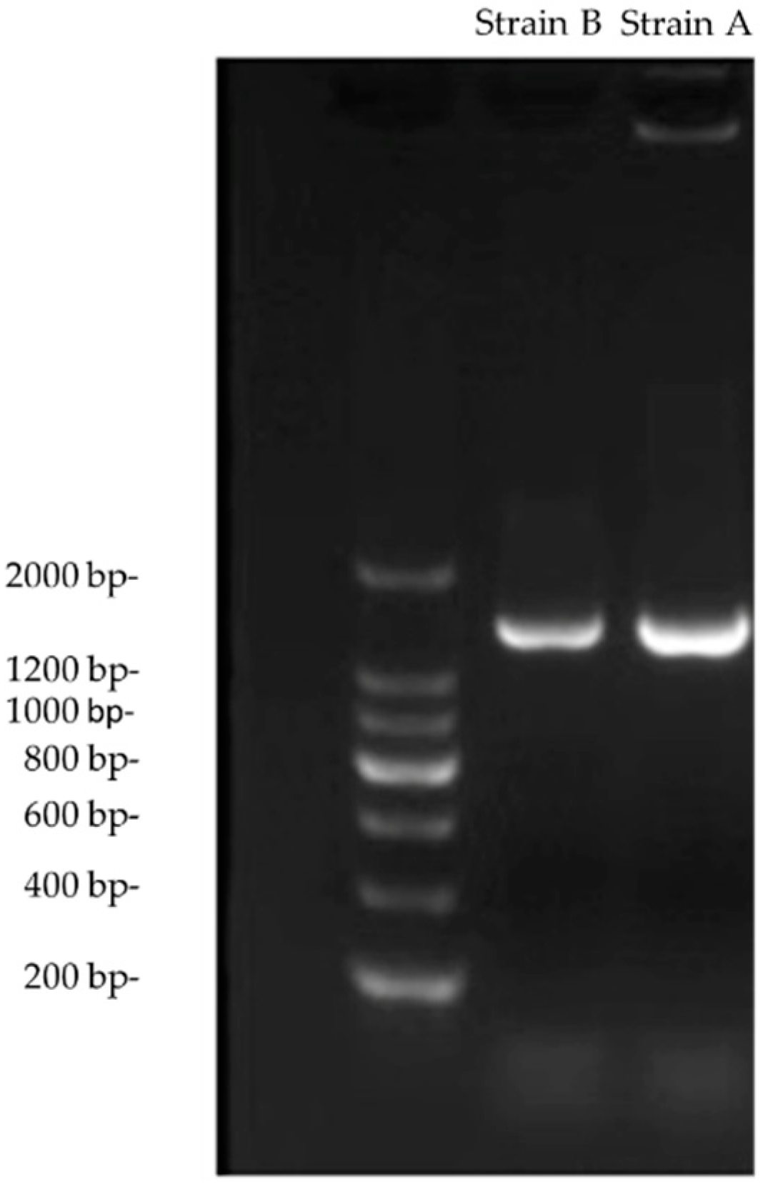
2.3. 16S rRNA Gene Sequencing and Biochemical Identification

A single colony of each actinomycete was used to extract the pure genomic DNA using the Bacterial DNA Extraction Kit (Tiangen Biotech (Beijing) Co., Ltd.) according to the manufacturer’s instructions. Primers 27F (5′-AGAGTTTGATCCTGGCTCAG-3′) and 1492R (5′-GGTTACCTTGTTACGACT-3′) were used as universal primers for bacterial 16S rRNA amplification. The total PCR volume was 25 μL, including 12.5 μL of 2×Taq PCR Mastermix (Tiangen Biotech (Beijing) Co., Ltd., Beijing, China), 1 μL of each primer (10 µM), 1.5 μL of DNA template, and 9 μL of ddH2O. The PCR protocol is shown in Table 1.
The PCR products were sent to RuiBiotech for Sanger sequencing. Reads greater than 1400 bp in length were used for the database analysis. The sequencing results were compared using NCBI nucleotide BLAST (https://blast.ncbi.nlm.nih.gov/Blast.cgi, accessed on 20 July 2022). Then, 16S rRNA sequences with the highest similarity to those of the isolated strains and typical strains in the same genus were obtained for phylogenetic analysis. The phylogenetic trees evaluated with 1000 bootstrap replications were inferred using the maximum likelihood method based on the Tamura 3-parameter model in MEGA7 (GenBank accession number: OM665406; OM665407).
Inositol, maltose, dextrose, rhamnose, sucrose, Neisser-fructose, hydrogen sulfide production, xylose, mannitol, and raffinose identification tubes purchased from Huancai Microbial Technology were used to test the substrate utilization of the isolated strains. A total of 50 μL of activated broth was added to the identification tubes and then cultured at 37 °C for 24 h. Substrate utilization data for other species in similar genera were obtained for comparison with the data for the isolated strains. Gram staining of the colonies was conducted and then observed microscopically.

2.4. Bioassay of the Fungal Inhibition Effect

A single colony of actinomycetes was activated in liquid BHI medium (37 °C ± 1 °C, 160 r/min) for 24 h. Then, 1 mL of the activated broth was added to a 250 mL flask that contained 100 mL of soybean powder fermentation broth. The mixture was cultured in a shaker (28 °C ± 1 °C, 160 r/min) for 7 days. The fermentation broth was centrifuged (4 °C, 8000 r/min, 20 min) and filtered using a 0.22 µm filter membrane to obtain the aseptic filtrate. Various amounts of filtrate (1 mL, 1.5 mL, and 2 mL) were added to sterilized PDA medium (4 mL, 3.5 mL, and 3 mL) on a plate with a diameter of 5.5 cm. Then, the medium was allowed to cool to 55 °C. The control PDA plate was supplemented with various amounts of sterile water (1 mL, 1.5 mL, and 2 mL). Plant pathogenic fungal plugs with a diameter of 0.5 cm were placed on PDA medium. For each actinomycete sample, the experiment was repeated three times on media containing each fermentation broth filtrate dilution for each fungus. The average diameters of the fungal colonies in the experimental and control groups were recorded. The bacteriostatic rate of the actinomycete against the three-indicator plant pathogenic fungi was calculated according to the following equation:
Inhibition   rate % = ( ADc D ) ( ADt D ) ( ADc D )   × 100 %
where ADc represents the average colony diameter in the control group, ADt represents the average colony diameter in the treatment group, and D represents the diameter of the fungal plugs.
The inhibitory effect on phytopathogens was shown directly by the confrontation culture method. Circular plugs of fungi (diameter = 0.5 cm) were placed in one-quarter of the plates after the actinobacterial plugs were placed in the other quarter of the plates for 7 days at 28 °C.
The Kruskal–Wallis (KW) nonparametric analysis of variance was used to compare the different treatments. The Mann–Whitney U test for multiple comparisons among the different groups was used if the results of the Kruskal–Wallis test showed significant differences at a significance level of 0.05.

3. Results

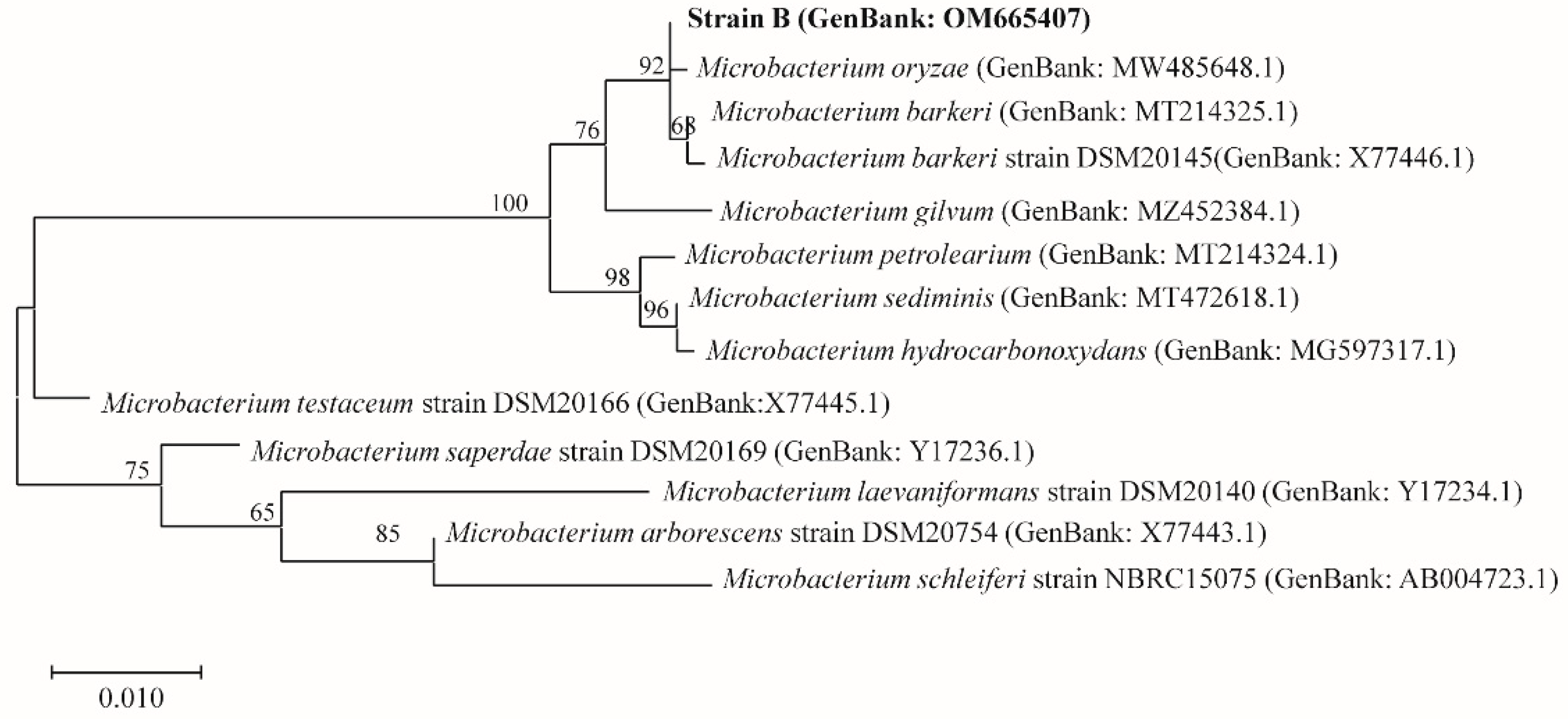
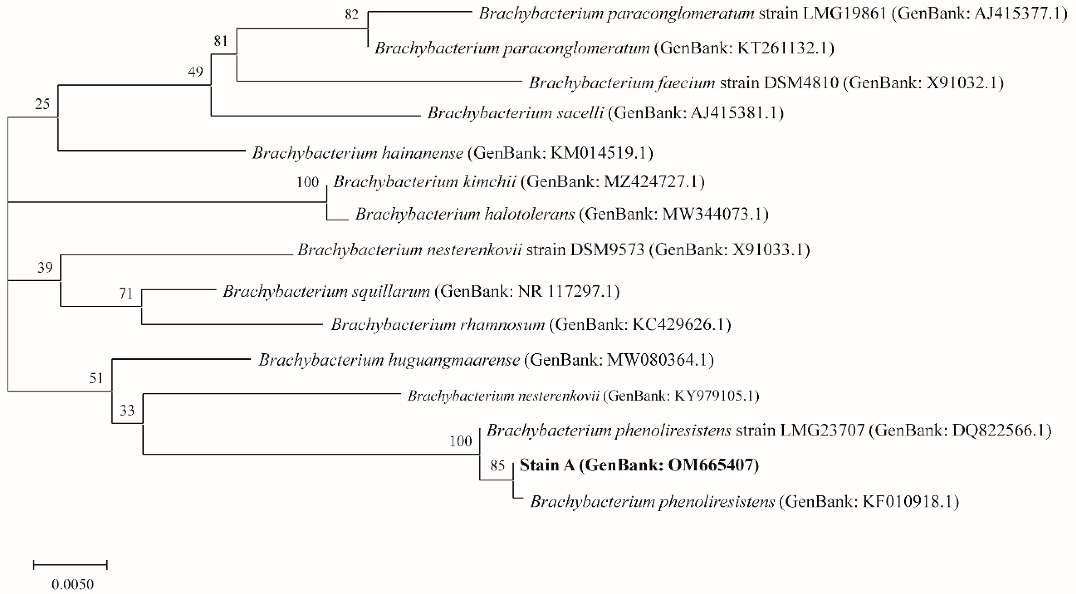
3.1. Identification of Actinomycetes

Two strains of bacteria, A and B, were identified in M. orientalis based on morphological observation and 16S rRNA sequencing. Figure 1 shows the results obtained from the 1.5% agarose gel electrophoresis of the PCR products. Clear bands corresponding to a length of 1500 bp were observed.
The 16S rRNA sequences (GenBank accession number: OM665406; OM665407) of the two bacteria were compared using NCBI nucleotide BLAST. Based on the results, these two bacteria were identified as actinomycetes belonging to the genera Microbacterium and Brachybacterium. The similarity between strain A and Brachybacterium phenoliresistens was 99.86%. The similarity between strain B and Microbacterium barkeri was 99.34%. The physiological and biochemical characteristics of strains A and B are shown in Table 2 and Table 3, respectively. The results indicated that these two actinomycetes were successfully identified. However, it is notable that the rhamnose utilization capacity of strain B differed from that of M. barkeri.
Figure 2 shows the phylogenetic tree that was constructed based on 20 known strains of Brachybacterium and strain A (shown as Brachybacterium sp.). As shown in the phylogenetic tree, B. phenoliresistens and strain A were closely related, with high repeatability. Figure 3 shows the phylogenetic tree that was constructed based on 11 strains with the highest similarity to strain B (shown as Microbacterium sp.), according to BLAST. The findings showed that strain B and M. barkeri were closely related.
Based on the analysis, strain A was preliminarily considered to be a Brachybacterium strain, probably a strain of B. phenoliresistens. Hence, strain A is denoted Brachybacterium sp. MO. Strain B belongs to the Microbacterium genus. However, strain B has yet to be identified by multiphase classification and identification, so it is denoted as Microbacterium sp.

3.2. Study of Fungal Inhibitory Activity

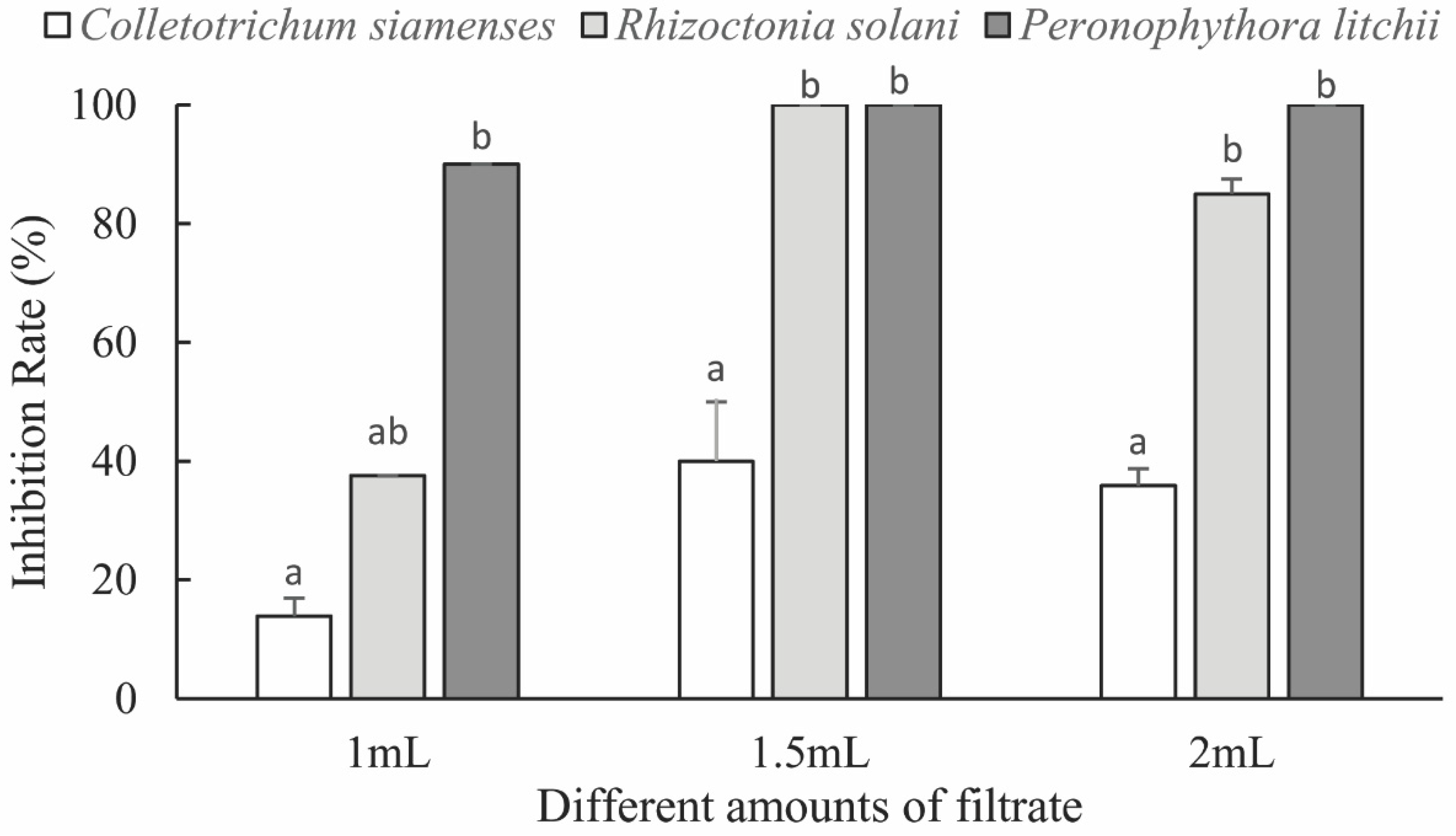
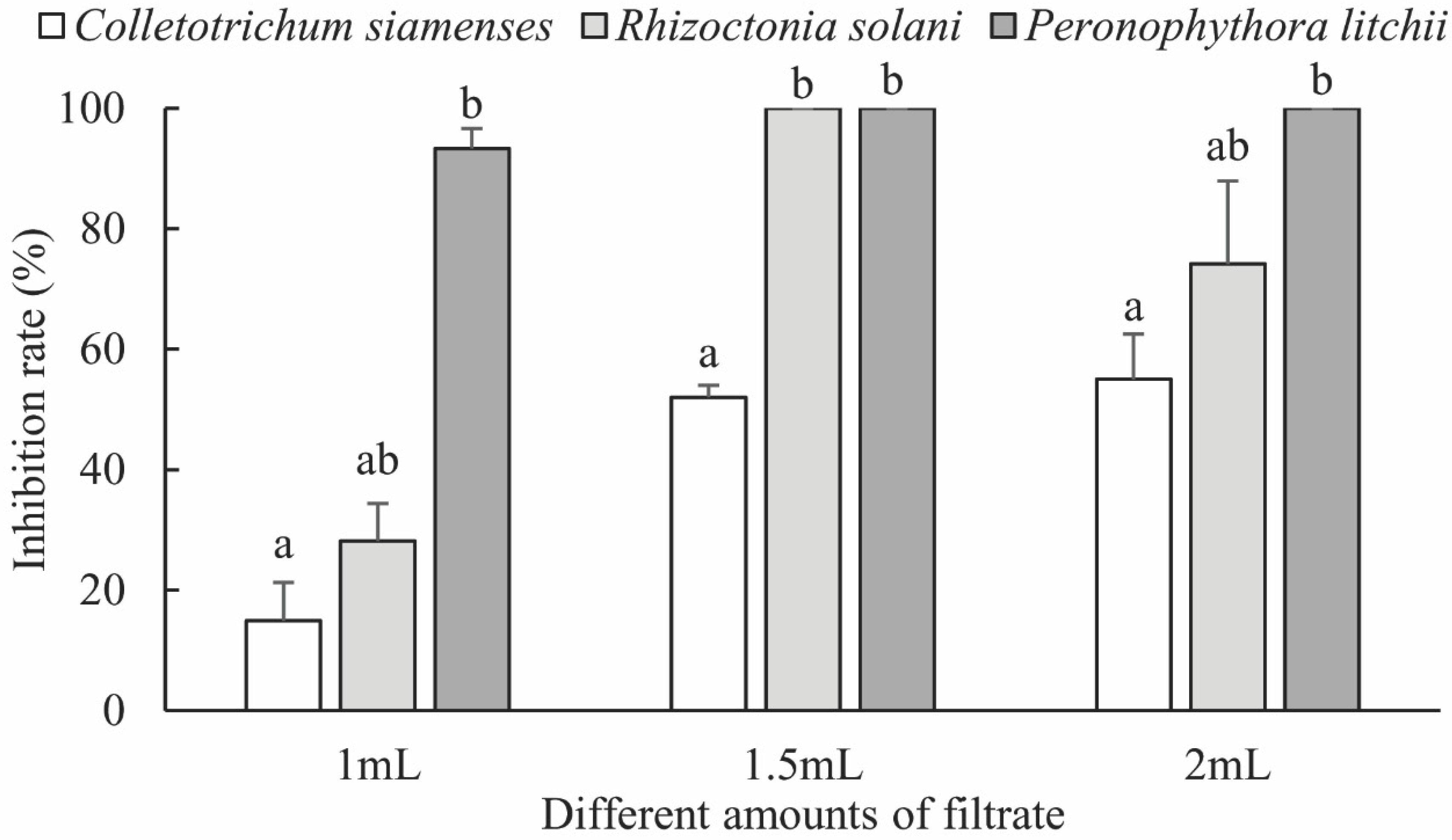
3.2.1. Fungal Inhibitory Activity of the Fermentation Broth

The inhibitory effects of B. phenoliresistens MO and Microbacterium sp. on three types of plant pathogenic fungi with different fermentation broth concentrations are shown in Figure 4 and Figure 5, respectively. Significant inhibitory effects were exhibited by these two actinomycete fermentation broths on the three plant pathogens. As the fermentation broth filtrate concentration increased, the inhibitory effect of the bacteria on plant pathogenic fungi increased. As shown in Figure 4 and Figure 5, the inhibitory rates exhibited by these two actinomycetes on R. solani and P. litchii were both 100%. However, the inhibitory effects of both actinomycetes on C. siamense were weaker. To illustrate these results visually, digital photographs of the pathogenic fungi on the fermentation broth filtrate plate are presented in Figures S1 (C. siamense), S2 (R. solani), and S3 (P. litchii) in the Supporting material. The growth of these plant pathogenic fungi on the fermentation filtrate plate was inhibited.

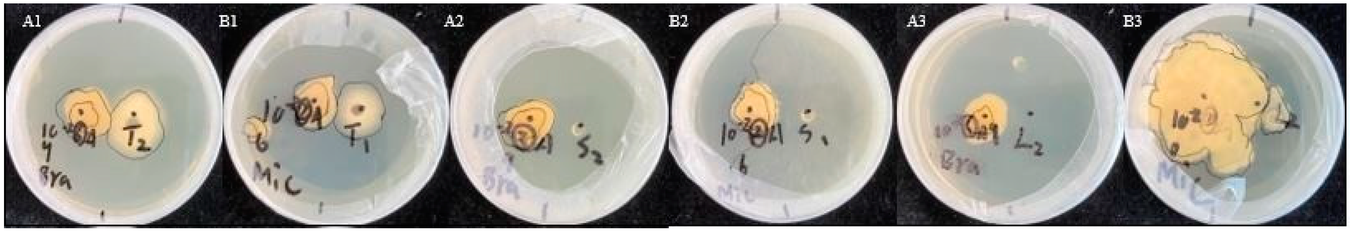
3.2.2. Inhibitory Activity of Actinomycete Colonies

On the bacterial plate, the growth of the pathogen was almost unaffected by live B. phenoliresistens MO and Microbacterium sp., which was consistent with the results obtained from the fermentation broth inhibition experiment. P. litchii growth was significantly inhibited by strains A and B. Mycelial growth around live strain B was relatively unaffected, while germination could not occur around live strain A, as shown in Figure 6.

4. Conclusions

Two actinomycete strains were isolated from the harvester ants. Based on the 16S rRNA and substrate utilization analysis, strain A belongs to the Brachybacterium genus, and it is very likely that B. phenoliresistens. Hence, strain A is denoted B. phenoliresistens MO. Strain B belongs to the Microbacterium genus. Because there were differences in the substrate utilization results, strain B must be further classified. It is denoted here as Microbacterium sp.
The inhibitory effects of the 2 actinomycetes on plant pathogenic fungi were assayed. When the actinomycete fermentation broth concentration was higher than 30%, the inhibition rates on the indicator fungi, i.e., P. litchii and R. solani, were significantly high, wherein a 100% inhibition rate was recorded. The inhibitory effects of these two actinomycetes on C. siamense were also notable, indicating prospects for agricultural applications. Through coculturing live bacteria, we obtained results consistent with a previous conclusion, showing that the two strains exhibited strong inhibitory effects on P. litchii and R. solani.

5. Discussion

Rice sheath blight caused by R. solani is one of the three major rice diseases and is the major disease in rice-producing areas in Asia. As a result, the damage caused by R. solani has led to significant agricultural losses in China each year [26]. In addition, infestation by P. litchii in litchi during storage and transport can result in great losses each year [27]. In this work, two strains of actinomycetes were used on these two types of plant pathogenic fungi at certain concentrations, and an inhibition rate of 100% was observed; therefore, the inhibitory effect of the live bacterial cultures on the pathogenic fungi was confirmed. However, the exact mechanism remains unclear. This result is possibly related to the bacterial fermentation products. In general, the inhibitory rates exhibited by the actinomycetes on P. litchii were higher than those of the other two pathogenic fungi, which suggests that P. litchi is more sensitive to actinomycetes [15]. The 100% pathogenic fungus inhibition rate may indicate the potential agricultural applications of these actinomycetes for multiple crops. Multiple methods—HPLC, metabolomic profiling, and gas chromatography—can be used to study the chemical characteristics, including toxicity, light degradability, and stability, of antifungal metabolites to determine whether actinomycetes can be used as fungicides against R. solani and P. litchii. As such, this work indicates a potential means of reducing the economic loss caused by damage to rice and litchi by R. solani and P. litchii.
Microbacterium is an abundant component of bacterial communities in the soil, insects, and leaf material of plants [8]. In previous studies, M. testaceum KU313 isolated from stored rice grains was antagonistic to Aspergillus flavus and Penicillium spp. [28]. Microbacterium sp. LGMB471 isolated from the medicinal plant Vochysia divergens inhibited the development of Phyllosticta citricarpa [29]. Microbacterium sp. isolated from tomato plants inhibited the growth of Alternaria alternata, Corynespora cassiicola, and Stemphylium lycopersici [30]. Among these, antifungal compounds, 5-methyl-2-phenyl-1H-indole produced by strain KU143, 7-O-β-D-glucosyl-genistein and 7-O-β-D-glucosyl-daidzein produced by strain LGMB471, were also discovered [28,29]. Brachybacterium exists in various environments, such as soil (poultry deep litter, contaminated sand), roots, fermented food, and animals [31]. It was reported that B. paraconglomeratum YEBPT2 isolated from banana contributed to antifungal activity against Fusarium oxysporum f.sp. cubense (Foc), and nine bioactive metabolites were identified as diethyl hydrazine, carbonic acid, nitrosopyrrolidine, 4H-pyran, valeric acid, butanoic acid, trioxsalen, deoxy-d-mannoic acid, and amino caprylic acid [32].
Ants are one of the most successful terrestrial species [33]. Many researchers devoted to screening the high biotechnological potential of ant-associated microorganisms, as well as the significant ecological impact of microbial secondary metabolites [5,8,9,13,14,15,16,17,18,34,35,36]. Nonetheless, most studies have mainly focused on ant–fungus–actinomycete tripartite mutualism evolved by leaf-cutting ants, which use antifungal microbial secondary metabolites produced by actinobacteria (Streptomyces spp., Nocardia spp., Pseudonocardia spp., Amycolatopsis spp., etc.) to control pathogens in their fungal gardens [5,9,16,35,36,37,38,39]. The rich diversity of antimicrobial secondary metabolites plays a driving role in shaping the ecosystems of leaf-cutting ants. Only by revealing the chemical nature of antibiotics can we begin to fully understand the complex interactions between multi-organismic partners. In this work, B. phenoliresistens MO. and Microbacterium sp. exhibited pronounced antifungal properties; however, the possible symbiotic relationship between harvester ants and actinomycetes remains unclear. The genera Microbacterium isolated from gardens and starter cultures of Atta could play disease-suppressing or other unknown roles [8], while no specific function was shown for Brachybacterium isolated from the abdomen of Leucocoprinus gongylophorus [40]. To better understand the ecological role of microorganisms associated with Messor orientalis, it is crucial to analyze the chemical composition and evaluate the biological activity of their metabolites. In further work in this area, we wish to identify more species of functional actinomycetes from Messor orientalis by using diverse isolation methods and media. In addition, we will further examine the relationship between harvester ants and actinomycetes to understand whether the ants and microbes have a mutually beneficial relationship.

Supplementary Materials

The following supporting information can be downloaded at: https://www.mdpi.com/article/10.3390/insects13080691/s1, Figure S1: Growth of Colletotrichum siamense on various inhibitory media. (A) Brachybacterium phenoliresistens MO, (B) Microbacterium sp., (D) control; Figure S2: Growth of Rhizoctonia solani on various inhibitory media. (A) Brachybacterium phenoliresistens MO, (B) Microbacterium sp., (D) control; Figure S3: Growth of Peronophthora litchii on various inhibitory media. (A) Brachybacterium phenoliresistens MO, (B) Microbacterium sp., (D) control.

Author Contributions

Conceptualization, Y.X., Y.W., and S.C.; methodology, Y.W., J.Y., Y.L., and S.C.; formal analysis, Y.X., S.C., J.Y., Y.L., and Y.W.; writing—original draft preparation, Y.X., S.C., and Y.W.; writing—review and editing, Y.X., S.C., J.Y., Y.L., and Y.W.; funding acquisition, Y.X. All authors have read and agreed to the published version of the manuscript.

Funding

This work was funded by the Shenzhen Science and Technology Program (KCXFZ20201221173608022) and the Laboratory of Lingnan Modern Agriculture Project (NZ2021022).

Institutional Review Board Statement

Not applicable.

Informed Consent Statement

Not applicable.

Data Availability Statement

The data presented in this study are available on request from the corresponding author.

Acknowledgments

The authors wish to thank Yu Peng and Yuhang Zhou, Sendelta International Academy, for their support. Yiyang Wu thanks Yuqi Zhang and Boxiang Wang for their suggestions.

Conflicts of Interest

All authors declare that there are no conflict of interest.

References

  1. Baumann, P.; Baumann, L.; Lai, C.; Rouhbakhsh, D.; Moran, N.; Clark, M. Genetics, physiology, and evolutionary relationships of the genus Buchnera: Intracellular symbionts of aphids. Annu. Rev. Microbiol. 1995, 49, 55–94. [Google Scholar] [CrossRef]
  2. Kikuchi, Y. Endosymbiotic bacteria in insects: Their diversity and culturability. Microbes. Environ. 2009, 24, 195–204. [Google Scholar] [CrossRef]
  3. Schӓfer, A.; Konrad, R.; Kuhnigk, T. Hemicellulose-degrading bacteria and yeasts from the termite gut. J. Appl. Bacteriol. 1996, 5, 471–478. [Google Scholar] [CrossRef]
  4. Bignell, D.E.; Oskarsson, H.; Anderson, J.M. Association of actinomycete-lik bacteria with soil-feeding termites (Termitidae, Termitinae). Appl. Environ. Microbiol. 1979, 2, 339–342. [Google Scholar] [CrossRef]
  5. Currie, C.R.; Scott, J.A.; Summerbell, R.C.; Malloch, D. Fungus-growing ants use antibiotic-producing bacteria to control garden parasites. Nature 2003, 398, 701–704. [Google Scholar] [CrossRef]
  6. Guo, Z.K.; Zhang, G.F.; Jiao, R.H.; Shen, Y.; Xu, Q.; Tan, R.X.; Ge, H.M. Actinotetraoses A–H: Tetrasaccharide derivatives from a grasshopper-associated Amycolatopsis sp. HCal. Planta Med. 2012, 78, 988–994. [Google Scholar] [CrossRef] [PubMed]
  7. Lu, Y.H.; Li, S.; Zhou, D.X.; Zhang, Y.L. Isolation and identification of termination antagonistic actinomycetes BYC 01 and its active metabolites. Acta Microbiol. Sinica. 2014, 7, 754–759. [Google Scholar] [CrossRef]
  8. Mueller, U.G.; Dash, D.; Rabeling, C.; Rodrigues, A. Coevolution between attine ants and actinomycete bacteria: A reevaluation. Evolution 2008, 62, 2894–2912. [Google Scholar] [CrossRef]
  9. Chevrette, M.G.; Carlson, C.M.; Ortega, H.E.; Thomas, C.; Ananiev, G.E.; Barns, K.J.; Book, A.J.; Cagnazzo, J.; Carlos, C.; Flanigan, W.; et al. The antimicrobial potential of Streptomyces from insect microbiomes. Nat. Commun. 2019, 10, 516. [Google Scholar] [CrossRef] [PubMed]
  10. Blodgett, J.A.V.; Oh, D.C.; Cao, S.G.; Currie, C.R.; Kolter, R.; Clardy, J. Common biosynthetic origins for polycyclic tetramate macrolactams from phylogenetically diverse bacteria. Proc. Natl. Acad. Sci. USA 2010, 107, 11692–11697. [Google Scholar] [CrossRef] [PubMed]
  11. Scott, J.J.; Oh, D.C.; Yuceer, M.C.; Klepzig, K.D.; Clardy, J.; Currie, C.R. Bacterial protection of beetle-fungus mutualism. Science 2008, 322, 63. [Google Scholar] [CrossRef] [PubMed]
  12. Poulsen, M.; Oh, D.C.; Clardy, J.; Currie, C.R. Chemical analyses of wasp-associated Streptomyces bacteria reveal a prolific potential for natural products discovery. PLoS ONE 2011, 6, e16763. [Google Scholar] [CrossRef] [PubMed]
  13. Weber, N.A. Fungus-growing ants: A symbiotic relationship exists between an insect and a plant, involving an effective culturing technique. Science 1966, 3736, 587–604. [Google Scholar] [CrossRef]
  14. Guo, L.F. Diversity of Symbiotic Actinomycetes from Camponotus janponicus Mayr and Their Antibacterial Activity. Master’s Thesis, Northeast Agricultural University, Harbin, China, 2016. [Google Scholar]
  15. Zhang, X.Y.; Zhao, X.F.; Xu, Y.J.; Cheng, D.F.; Lu, Y.Y. Identification and culture of Streptomyces sp. DF-5 isolated from Solenopsis invicta and the antifungal activity of its fermentation broth to plant pathogenic fungi. Acta J. Environ. Entomol. 2018, 4, 917–924. [Google Scholar]
  16. Oh, D.C.; Poulsen, M.; Currie, C.R.; Clardy, J. Dentigerumycin: A bacterial mediator of an ant-fungus symbiosis. Nat. Chem. Biol. 2009, 5, 391–393. [Google Scholar] [CrossRef]
  17. Baranova, A.A.; Chistov, A.A.; Tyurin, A.P.; Prokhorenko, I.A.; Korshun, V.A.; Biryukov, M.V.; Alferova, V.A.; Zakalyukina, Y.V. Chemical ecology of Streptomyces albidoflavus strain A10 associated with carpenter ant Camponotus vagus. Microorganisms 2020, 8, 1948. [Google Scholar] [CrossRef]
  18. Akalyukina, Y.V.; Birykov, M.V.; Lukianov, D.A.; Shiriaev, D.I.; Komarova, E.S.; Skvortsov, D.A.; Kostyukevich, Y.; Tashlitsky, V.N.; Polshakov, V.I.; Nikolaev, E.; et al. Nybomycin-producing Streptomyces isolated from carpenter ant Camponotus vagus. Biochimie 2019, 160, 93–99. [Google Scholar] [CrossRef]
  19. Osterman, I.A.; Wieland, M.; Maviza, T.P.; Lashkevich, K.A.; Lukianov, D.A.; Komarova, E.S.; Zakalyukina, Y.V.; Buschauer, R.; Shiriaev, D.I.; Leyn, S.A.; et al. Tetracenomycin X inhibits translation by binding within the ribosomal exit tunnel. Nature Chem. Biol. 2020, 16, 1071–1077. [Google Scholar] [CrossRef]
  20. Tarsa, C.; McMillan, A.; Warren, R.J. Plant pathogenic fungi decrease in soil inhabited by seed-dispersing ants. Insect. Soc. 2018, 65, 315–321. [Google Scholar] [CrossRef]
  21. MacMahon, J.A.; Mull, J.F.; Crist, T.O. Harvester ants (Pogonomyrmex spp.): Their community and ecosystem influences. Annu. Rev. Ecol. Syst. 2000, 31, 265–291. [Google Scholar] [CrossRef]
  22. Kang, Z.S. Current status and development strategy for research on plant fungal diseases in China. Plant Prot. 2010, 3, 9–12. [Google Scholar] [CrossRef]
  23. Li, B.D.; Shen, C.R. The fungicide resistance of plant pathogens and solutions. Acta Phytopathol. Sinica 1994, 24, 193–196. [Google Scholar] [CrossRef]
  24. Goodfellow, M.; Williams, S.T. Ecology of Actinomycetes. Ann. Rev. Microbiol. 1984, 1, 189–216. [Google Scholar] [CrossRef]
  25. Solecka, J.; Zajko, J.; Postek, M.; Rajnisz, A. Biologically active secondary metabolites from Actinomycetes. Cent. Euro. J.Bio. 2012, 7, 373–390. [Google Scholar] [CrossRef]
  26. Zuo, S.M.; Wang, Z.B.; Chen, X.J.; Gu, F.; Zhang, Y.F.; Chen, Z.X.; Pang, X.B. Evaluation of resistance of a novel rice germplasm YSBR1 to sheath blight. Acta Agron. Sin. 2009, 04, 608–614. [Google Scholar] [CrossRef]
  27. Kong, G.H.; Feng, D.N.; Li, W.; Lian, S.L.; Xi, P.G.; Jiang, Z.D. Research progress in studies on the downy blight disease in litchi. J. Fruit Sci. 2021, 38, 603–612. [Google Scholar] [CrossRef]
  28. Mannaa, M.; Kim, K.D. Biocontrol activity of volatile-producing Bacillus megaterium and Pseudomonas protegens against Aspergillus and Penicillium spp. Predominant in stored rice grains: Study II. Mycobiology 2018, 46, 52–63. [Google Scholar] [CrossRef]
  29. Savi, D.C.; Shaaban, K.A.; Gos, F.M.W.; Thorson, J.S.; Glienke, C.; Rohr, J. Secondary metabolites produced by Microbacterium sp. LGMB471 with antifungal activity against the phytopathogen Phyllosticta citricarpa. Folia Microbiol. 2019, 64, 453–460. [Google Scholar] [CrossRef] [PubMed]
  30. Lopez, S.M.Y.; Pastorino, G.N.; Balatti, P.A. Volatile organic compounds profile synthesized and released by endophytes of tomato (Solanum lycopersici L.) and their antagonistic role. Arch. Microbiol. 2021, 203, 1383–1397. [Google Scholar] [CrossRef]
  31. Lapidus, A.; Pukall, R.; LaButtii, K.; Copeland, A.; Del Rio, T.G.; Nolan, M.; Chen, F.; Lucas, S.; Tice, H.; Cheng, J.F.; et al. Complete genome sequence of Brachybacterium faecium type strain (Schefferle 6–10(T)). Stand. Genom. Sci. 2009, 1, 3–11. [Google Scholar] [CrossRef]
  32. Ravi, S.; Sevugapperumal, N.; Nallusamy, S.; Shanmugam, H.; Mathiyazhagan, K.; Rangasamy, A.; Subbiah, K.A.; Ganesan, M.V. Differential bacterial endophytome in Foc-resistant banana cultivar displays enhanced antagonistic activity against Fusarium oxysporum f.sp. cubense (Foc). Environ. Microbiol. 2022, 24, 2701–2715. [Google Scholar] [CrossRef]
  33. Schultz, T.R. In search of ant ancestors. Proc. Natl. Acad. Sci. USA 2000, 97, 14028–14029. [Google Scholar] [CrossRef]
  34. Carr, G.; Derbyshire, E.R.; Caldera, E.; Currie, C.R.; Clardy, J. Antibiotic and antimalarial quinones from fungus-growing ant-associated Pseudonocardia sp. J. Nat. Prod. 2012, 75, 1806–1809. [Google Scholar] [CrossRef]
  35. Caldera, E.J.; Poulsen, M.; Suen, G.; Currie, C.R. Insect symbioses: A case study of past, present, and future fungus-growing ant research. Env. Entomol. 2009, 38, 78–92. [Google Scholar] [CrossRef]
  36. Schoenian, I.; Spiteller, M.; Ghaste, M.; Wirth, R.; Herz, H.; Spiteller, D. Chemical basis of the synergism and antagonism in microbial communities in the nests of leaf-cutting ants. Proc. Natl. Acad. Sci. USA 2011, 108, 1955–1960. [Google Scholar] [CrossRef]
  37. Haeder, S.; Wirth, R.; Herz, H.; Spiteller, D. Candicidin-producing Streptomyces support leaf-cutting ants to protect their fungus garden against the pathogenic fungus Escovopsis. Proc. Natl. Acad. Sci. USA 2009, 106, 4742–4746. [Google Scholar] [CrossRef]
  38. Ronque, M.U.V.; Lyra, M.L.; Migliorini, G.H.; Bacci, M., Jr.; Oliveira, P.S. Symbiotic bacterial communities in rainforest fungus-farming ants: Evidence for species and colony specificity. Sci. Rep. 2020, 10, 10172. [Google Scholar] [CrossRef]
  39. Seipke, R.F.; Barke, J.; Brearley, C.; Hill, L.; Yu, D.W.; Goss, R.J.; Hutchings, M.I. A single Streptomyces symbiont makes multiple antifungals to support the fungus farming ant Acromyrmex octospinosus. PLoS ONE 2011, 6, e22028. [Google Scholar] [CrossRef]
  40. Zani, R.D.A.; Ferro, M.; Bacci, M. Three phylogenetically distinct and culturable diazotrophs are perennial symbionts of leaf-cutting ants. Ecol. Evol. 2021, 11, 17686–17699. [Google Scholar] [CrossRef]
Figure 1. PCR amplification and gel electrophoresis of strains A and B.
Insects 13 00691 g001
Figure 2. Molecular phylogenetic analysis of strain A (Brachybacterium sp.) using the maximum likelihood method.
Insects 13 00691 g002
Figure 3. Molecular phylogenetic analysis of strain B (Microbacterium sp.) using the maximum likelihood method.
Insects 13 00691 g003
Figure 4. Inhibitory effect of Brachybacterium phenoliresistens against three plant pathogens with different amounts of filtrate. For each amount of filtrate, bars with the same letter are not significantly different (p > 0.05, Mann–Whitney U test).
Insects 13 00691 g004
Figure 5. Inhibitory effect of Microbacterium sp. against three plant pathogens with different amounts of filtrate. For each amount of filtrate, bars with the same letter are not significantly different (p > 0.05, Mann–Whitney U test).
Insects 13 00691 g005
Figure 6. Cogrowth of actinomycetes and fungi. (A) Brachybacterium phenoliresistens MO. (B) Microbacterium sp. (1) Colletotrichum siamense, (2) Rhizoctonia solani, and (3) Peronophthora litchii.
Insects 13 00691 g006
Table 1. Actinomycete 16S rRNA amplification PCR protocol.
StepReaction Temperature (°C)Reaction Time (min)
Initialization9415
Denaturing940.5 a
Annealing550.5 a
Elongation721 a
Stop7210
a Step was repeated 30 times.
Table 2. Physiological and biochemical characteristics of strain A. Strains: 1, B. phenoliresistens LMG 23707T; 2, B. saceli DSM 14566T; 3, B. alimentarium CCM 4520T; 4, B. freconis DSM 14564T; 5, B. paraconglomeratum DSM 46361T; 6, B. faecium CCM 4372T. Abbreviations: +, positive; (+), weakly positive; −, negative; ND, not determined.
CharacteristicStrain A123456
H2S production+++
Acid production from:
D-fructose+ND+++
Maltose+++++(+)
D-mannose+ND(+)(+)++
L-rhamnose++(+)(+)++
Sucrose+++(+)
D-xylose+++
Galactose+ND+++++
Table 3. Physiological and biochemical characteristics of strain B. Strains: 1, M. barkeri DSM 20145T; 2, M. chocolatum IFO 3758T; 3, M. hominis IFO 15708T; 4, M. thalassium IFO 16060T, IFO 16061; 5, M. halophilum IFO 16062T; 6, M. laevaniformans IFO 15709T. Abbreviations: +, positive; −, negative; ND, not determined.
CharacteristicStrain B123456
H2S productionND+++
Utilization of:
Maltose+ND+++++
D-mannoseND+++++
Acid production from:
L-rhamnose++
Sucrose+ND+++
D-xyloseND+
Galactose+++
Publisher’s Note: MDPI stays neutral with regard to jurisdictional claims in published maps and institutional affiliations.

