Article

Characterization of a Vitellogenin Receptor in the Bumblebee, Bombus lantschouensis (Hymenoptera, Apidae)

1
College of Animal Science and Technology, Yunnan Agricultural University, Kunming 650201, China
2
Key Laboratory for Insect-Pollinator Biology of the Ministry of Agriculture and Rural Affairs, Institute of Apicultural Research, Chinese Academy of Agricultural Sciences, Beijing 100093, China
3
Nanchuan Bureau of Animal Husbandry and Veterinary, Chongqing 408400, China
*
Authors to whom correspondence should be addressed.
Insects 2019, 10(12), 445; https://doi.org/10.3390/insects10120445
Submission received: 18 October 2019 / Revised: 9 December 2019 / Accepted: 10 December 2019 / Published: 12 December 2019

Abstract

:
The vitellogenin receptor (VgR) belongs to the low-density lipoprotein receptor (LDLR) family, responsible for mediating the endocytosis of vitellogenin (Vg) into the ovaries to promote ovarian growth and oviposition. Here, we cloned and measured VgR gene expression characteristics in the bumblebee Bombus lantschouensis. RNA interference was used to validate VgR function. The results showed that the full length of the BLVgR cDNA was 5519 bp, which included a 5280 bp open reading frame encoding 1759 amino acids (AAs). Sequence alignment revealed that the protein contained 12 LDLa, 5 EGF, 2 EGF-CA and 10 LY domains. Phylogenetic analysis showed that BLVgR and the VgR of Bombus terrestris clustered together and the tree of bumblebees (Bombus) appeared as one clade next to honeybees (Apis). Transcript expression analysis showed that BLVgR was expressed in all tested tissues and showed the highest abundance in the ovaries. BLVgR expression was present in all developmental stages. However, the expression level in larvae was extremely low. In addition, the expression of BLVgR was significantly upregulated after egg laying in both workers and queens. In new emerging workers injected with 5 µg of VgR dsRNA, the expression level of BLVgR was 4-fold lower than that in the GFP dsRNA-injected group after 72 h. Furthermore, BLVgR silencing significantly reduced the number of eggs laid (3.67 ± 1.96 eggs) and delayed the first egg-laying time (16.31 ± 2.07 days) in worker microcolonies when compared to dsGFP (37.31 ± 4.09 eggs, 8.15 ± 0.22 days) and DEPC-treated water injected controls (16.42 ± 2.24 eggs, 10.00 ± 0.37 days). In conclusion, the BLVgR gene and its reproductive function were explored in the bumblebee B. lantschouensis. This gene plays an important role in egg laying time and egg number.

1. Introduction

Bumblebees are one of the most efficient pollinators of many wild plants and crops, especially legumes and Solanaceae plants [1]. Moreover, they play important roles in maintaining the balance of natural ecosystems [2]. Since the 1980s, bumblebees have been used commercially to pollinate greenhouse crops around the world and have conferred significant economic benefits [3,4]. As a result, artificial bumblebee rearing has become important. Reproductive regulation is an important biological process that is vital to bumblebee artificial mass rearing. Vitellogenin (Vg) and the vitellogenin receptor (VgR) are two reproductively critical proteins that significantly affect the fecundity of bumblebees. Therefore, full elucidation of the characteristics of VgR will aid understanding of oviposition regulation.
Insect VgR belongs to the low-density lipoprotein receptor (LDLR) gene superfamily and plays a critical role in oocyte development by mediating the endocytosis of the major yolk protein precursor Vg [5]. Vitellogenin is synthesized in the fat body and is internalized by competent oocytes through membrane-bound receptors [6,7]. The main function of Vg is to provide nutriment for embryos. Moreover, it can protect workers and the queen from oxidative stress that contributes to longevity [8,9]. Interestingly, recent studies have shown that Vg expression also affects the behavior of social insects, as it is related to division of labor [9,10]. Insect VgRs and all other LDLR family receptors share five structural domains: the ligand-binding domain (LBD), made up of cysteine-rich repeats; the epidermal growth factor (EGF)-precursor homology domain, characterized by a recurrent ‘YWXD’ motif (YWXD repeats); the O-linked sugar domain of unknown function; the transmembrane domain anchoring the receptor in the plasma membrane; and the cytoplasmic domain containing an internalization signal [5]. In 1986, the VgR protein from the oocytes of the locust Locusta migratoria was first identified and purified [11]. Since then, it has been identified in a variety of economic insects [12,13] and agricultural pests [14,15,16,17]. In 2004, the cloning of the first hymenopteran VgR cDNA from the imported fire ant Solenopsis invicta (SiVgR) was reported, and this VgR was upregulated by methoprene, a juvenile hormone (JH) analog [18]. In 2006, RNAi technology was first used to analyze VgR function in the cockroach Blattella germanica [19]. However, the first silencing of hymenoptera VgR with RNAi was achieved in 2009 [20]. The expression characteristics of VgR from the honeybee Apis mellifera in the queen and workers were also studied, and the results suggested that VgR is not caste or tissue specific. It was detected in all sexes and outside the ovary [21]. To date, the characteristics of VgR have not been explored in bumblebees.
RNA interference is a technology for posttranscriptional gene silencing that has been developed as a powerful tool for studying gene function in a variety of organisms [22]. A variety of efficient means to transport double-stranded RNA (dsRNA) into organisms has been developed including microinjection, feeding and transgenic plant expression [23,24,25]. RNAi has achieved prodigious progress in insect pest control and economic insect biology research [20,25]. In recent years, RNAi has been used to study the regulation of target gene expression, caste differentiation and disease control in honeybees [26,27,28]. However, there have been no reports on reproductive inhibition by using RNAi in bumblebees.
The bumblebee Bombus lantschouensis is a polylectic and primitively eusocial bee that is widely distributed in medium-elevation mountains and plateaus in northern China [29]. It is an important pollinator of wild plants and crops and has been selected as the native species applied to pollinate greenhouse crops. A recent study indicates that the pollination efficiency of this species exceeds that of honeybees. Interestingly, B. lantschouensis prefers to visit plants with less pollen and nectar than honeybees [30]. The molecular mechanisms of reproduction, such as the female fertility regulation, are a vital aspect of artificial mass rearing. Vitellogenin and the vitellogenin receptor are important genes for insect fecundity [6]. Therefore, it is necessary to explore the effect of BLVgR on female reproduction in B. lantschouensis.
Understanding the molecular mechanisms of reproductive regulation is critical for bumblebee artificial rearing. In the present study, the Vg transporter protein VgR gene of a bumblebee (B. lantschouensis) was cloned, and its expression profile was checked in different tissues, developmental stages and reproductive statuses. RNAi was used to validate the function of BLVgR in ovarian activation, first egg laying time and number of eggs laid. These results help us to understand the basic knowledge of VgR in bumblebee commercial rearing.

2. Materials and Methods

2.1. Samples

Bumblebees (B. lantschouensis) were collected from the rearing room at the Institute of Apicultural Research, Chinese Academy of Agricultural Science, Beijing, China. Bumblebees were sampled from thirty independent colonies that were raised in an artificial breeding room (in constant darkness with a temperature of 28 ± 0.5 °C and 60 ± 5% relative humidity) and fed fresh frozen pollen and a 50% sugar solution every other day [31]. Different tissues from 18 egg-laying queens were sampled. The tissues were as follows: antenna (AN), head (HD), thorax (TH), leg (LG), epidermis (EP), midgut (MG), ovary (OV), fat body (FB) and venom gland (VG). All tissues were dissected and frozen in liquid nitrogen immediately. Tissues from six bees as a biological replicate were pooled, and total RNA was extracted. Samples from three different development stages except the egg stage of bumblebee workers, were obtained individually at the following stages: within 48 h of larval hatching (Lar), pupae with an unpigmented body cuticle and white eyes (Pw), pink eyes (Pp), and brown eyes (Pb), pupae with dark brown eyes and a light-pigmented thorax (Pbl), and pupae with brown eyes and a dark-pigmented cuticle (Pbd). Eggs were sampled within 24 h, eighteen eggs were collected (six eggs in each biological replicate). The expression level of VgR in the ovary was measured at egg-laying and non-egg-laying statuses of queens and workers. Each reproductive status had three samples [32]. All samples were stored at −80 °C until use.

2.2. RNA Extraction and Synthesis of cDNA

Total RNA was extracted from all samples with TRIzol reagent (Invitrogen, Carlsbad, CA, USA) following the manufacturer’s instructions. The purity of the RNA was assessed using a NanoDrop 2000 spectrophotometer (Thermo Fisher Scientific, Waltham, MA, USA) at 260/280 nm, and RNA integrity was screened by 1.5% (w/v) agarose gel electrophoresis. The first strand of cDNA was synthesized according to the instructions of the Reverse Transcription kit (Takara, Dalian, China). The reaction conditions were as follows: 42 °C for 30 min, 99 °C for 5 min, and 5 °C for 5 min, after which the product was stored at −20 °C until use.

2.3. Molecular Cloning of BLVgR

To obtain a specific fragment of the BLVgR cDNA, primers (Table 1) were designed and synthesized according to the predicted VgR sequence of Bombus terrestris (GenBank accession number: LOC100649042). The PCR amplification conditions were as follows: 94 °C for 3 min, 35 cycles of 94 °C for 30 s, 55 °C for 50 s and 72 °C for 1 min, and 72 °C for 10 min. The PCR products were ligated into the pMD19-T vector (Takara Bio Inc., Dalian, China), which was then transformed into Trans1-T1 E. coli competent cells (Transgen Biotech, Beijing, China). Ampicillin resistance and white-blue screening were used to select the positive clone. Positive colonies were sent for sequencing by the Sino GenoMax Company (Beijing, China) using the Sanger method.
Full-length cDNA amplification was performed using a SMARTer rapid amplification of cDNA ends (RACE) amplification kit (Clontech, Mountain View, CA, USA) according to the manufacturer’s recommendations with the appropriate primers (Table 1). PCR was carried out with 5 cycles of 94 °C for 30 s and 72 °C for 3 min; 5 cycles of 94 °C for 30 s, 70 °C for 30 s and 72 °C for 3 min; and 27 cycles of 94 °C for 30 s, 68 °C for 30 s and 72 °C for 3 min. The amplified DNA fragment was cloned and sequenced to both ends.

2.4. Sequence Analysis

Sequences were assembled, and the open reading frame (ORF) was predicted using BioEdit 7.2.5 software (http://www.mbio.ncsu.edu/BioEdit/bioedit.html). The molecular weight (MW), theoretical isoelectric point (pI), properties of the amino acid residues, and protein hydropathy were assessed using the European Molecular Biology Open Software Suite (EMBOSS) (http://emboss.sourceforge.net/) [33]. The online TMHMM-2.0 program was used to analyze the protein transmembrane domain (http://www.cbs.dtu.dk/services/TMHMM-2.0/) [34]. The signal peptide position was predicted using SignalP-5.0 (http://www.cbs.dtu.dk/services/SignalP-5.0/) [35]. We download homologous VgR amino acid sequences from GenBank, including those from the following species: B. terrestris (XP_003402703.1), Bombus impatiens (XP_012241122.1), Apis cerana (PBC31775.1), Apis mellifera (XP_026295652.1), Apis dorsata (XP_006610571.1), Apis florea (XP_012350206.1), Solenopsis invicta (NP_001291525.1), Nasonia vitripennis (XP_008217630.1), Drosophila melanogaster (NP_996433.1), Drosophila sechellia (XP_002042769.1), Drosophila yakuba (XP_002100545.2), Drosophila erecta (XP_001978195.1), Drosophila ananassae (XP_001966853.1), Drosophila persimilis (EDW29780.1), Drosophila pseudoobscura (XP_001354886.2), Drosophila willistoni (XP_002064208.2), Drosophila virilis (XP_002054982.2), Aedes aegypti (AAC28497.1), Anopheles gambiae (XP_310672.5), Blattella germanica (CAJ19121.1), Rhyparobia maderae (BAE93218.1), Periplaneta americana (BAC02725.2), and Acyrthosiphon pisum (XP_016657813.1). The SMART program (http://smart.embl-heidelberg.de/) was used to identify conserved domains [36]. Evolutionary analyses were conducted in MEGA X [37]. The VgR sequence of A. pisum was used as an outgroup. The evolutionary history was inferred by using the maximum likelihood method and general reversible chloroplast plus frequency model, and the reliability of the tree was assessed with 1000 bootstrap replications. Branches corresponding to partitions reproduced in less than 50% bootstrap replicates are collapsed [38].

2.5. BLVgR Expression Analysis Using Quantitative RT-PCR

The expression levels of BLVgR mRNA in different tissues, developmental stages and reproductive status of workers and queens, as well as the results of different RNAi treatments, were analyzed by real-time quantitative PCR using a Stratagene Mx3000 real-time PCR system (Agilent, USA). First-strand cDNA samples, were diluted (1:10 v/v) with DEPC-treated water. Amplification was carried out in a 20 µL reaction volume containing 10 µL of 10 × TB Green Master Mix (Takara, Dalian, China), 2 µL cDNA, and 0.5 µL of each 10 µm primer (Table 1). Quantitative measurements were normalized using β-actin and GAPDH. The qPCR conditions were as follows 95 °C for 30 s, followed by 40 cycles of 95 °C for 5 s, and 63 °C for 1 min. Each assay was performed in triplicate and repeated with three independent samples. The relative quantities of BLVgR transcripts were calculated using the comparative Ct method [39].

2.6. RNA Interference

To synthesize double-stranded RNA (dsRNA), a specific fragment was amplified with two T7 promoter primers (Table 1). The amplification conditions were predenaturing at 94 °C for 5 min, followed by 35 cycles of denaturing at 94 °C for 30 s, annealing at 58 °C for 1 min and extension at 72 °C for 1 min, with a final extension at 72 °C for 10 min. The GFP gene (ACY56286) and DEPC-treated water were used as a negative control [17,23,40]. Double stranded RNA was synthesized using the T7 RiboMAX™ Express RNAi System (Promega, Madison, WI, USA) according to the manufacturer’s suggestions. The dsRNA was dissolved in the appropriate volume of DEPC-treated water and stored at −80 °C.
Newly emerged workers (within 24 h) from five colonies were injected with 10 µL (500 ng/µL) of a dsRNA solution containing dsVgR (N = 93), dsGFP (N = 93) or DEPC-treated water (N = 114). The bees were injected through the conjunction between the 5th and 6th abdominal sternum using a FemtoJet micro-injector [41]. After injection, three bees were selected randomly to form a micro-colony individually (21 × 14 × 7 cm contained four colonies to share the food). To obtain the optimal dose of microinjection, eighteen microcolonies including nine colonies of dsVgR and nine colonies of dsGFP were used to test three different concentrations (0.2, 1 and 5 µg) of dsRNA with three biological replicates. The expression of VgR in the ovary was used to measure the silencing efficiency with qPCR at 72 h after the injection. The concentration of 5 µg was the best silencing efficiency and this concentration was used to evaluate the effect of silencing on egg laying. The number of eggs laid within 15 days and the first egg laying time were counted and recorded based on 13 microcolonies of dsVgR (39 workers), 13 microcolonies of dsGFP (39 workers) and 19 microcolonies of DEPC-treated water (57 workers) after injection. The laying status was checked every day. When the worker laid eggs, we removed the eggs and recorded the number of eggs. The microcolonies that remained were used to observe the development of the ovaries.

2.7. Data Analysis

All data are presented as mean ± SE. Before the statistical test, the data were tested for normality. The difference of treatments was analyzed by one-way analysis of variance (ANOVA) followed by Tukey post hoc test with R-project software version 3.5.1 (https://cran.r-project.org).

3. Results

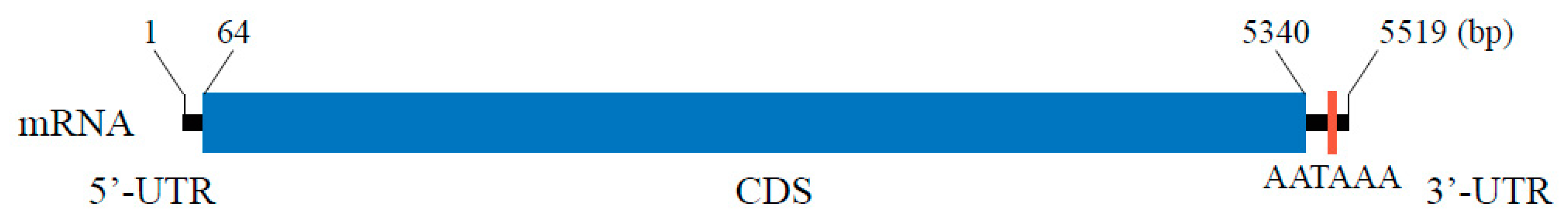
3.1. Full-Length cDNA of BLVgR

The specific amplification fragments and the 3′ end and 5′ end RACE sequences were merged with BioEdit. The results showed that the full-length cDNA of BLVgR (GenBank ID: MN217253) was 5519 bp and contained a 63 bp 5′ UTR and 179 bp 3′ UTR with a polyadenylation signal (AATAAA) (Figure 1). The ORF was composed of 5280 nucleotides encoding 1759 amino acids. The initiation codon (ATG) and stop codon (TAG) were present at positions 64–66 and 5341–5343, respectively. The predicted molecular weight of BLVgR was 198.131 kDa, and the theoretical pI was 5.9. Moreover, BLVgR contained a signal peptide with a potential cleavage site between amino acid residues 27 and 28.

3.2. Sequence and Phylogenetic Analysis of BLVgR

The analysis of conserved domains showed that BLVgR is a member of the LDLR superfamily of receptors, and the BLVgR protein was most similar to the VgR protein sequence of B. terrestris (XP_003402703.1), followed by that of B. impatiens (XP_012241122.1), as expected due to their similar taxonomic positions (Figure 2). BLVgR exhibited two ligand-binding domains (LBDs) with four class A cysteine-rich repeats (LDLa) in the first domain (LBD1) and eight repeats in the second domain (LBD2). Each LBD was followed by an epidermal growth factor (EGF)-like domain. The first EGF-like domain contained a calcium-binding domain, seven LY domains and three EGF domains, whereas only a calcium-binding domain, three LY domains and two EGF domains were found in the second. The second EGF-like domain was followed by a transmembrane domain and a cytoplasmic domain (Figure 2). Therefore, this sequence is considered to be the full-length cDNA sequence of B. lantschouensis.
To inspect the evolutionary relationships between BLVgR and other VgRs from different species, a phylogenetic tree of VgRs was constructed (Figure 2). As expected, VgRs of bumblebees (Bombus) appeared as one clade next to honeybees (Apis), and an ant (Solenopsis) was closer to the bees than the parasitic wasp (Nasonia).

3.3. Expression Patterns of BLVgRs in Different Tissues, Developmental Stages and Reproductive Status

The BlVgR transcript was detected in all tissues, and the highest transcript levels were detected in the ovary. Except in the ovary, the expression level was not significantly different between different tissues (p > 0.05) (Figure 3a). Between the various developmental stages, relatively higher transcript abundance of BLVgR was observed in the eggs than in the other developmental stages. However, it dropped to the lowest level at the larval stage. There were no significant differences between any pupal stages (Figure 3b). For both castes, queens and workers, individuals who were laying eggs had significantly higher transcript levels of BLVgR than non-laying individuals (p < 0.01; Figure 3c,d). Therefore, BLVgR was positively correlated with an enhanced female reproductive status.

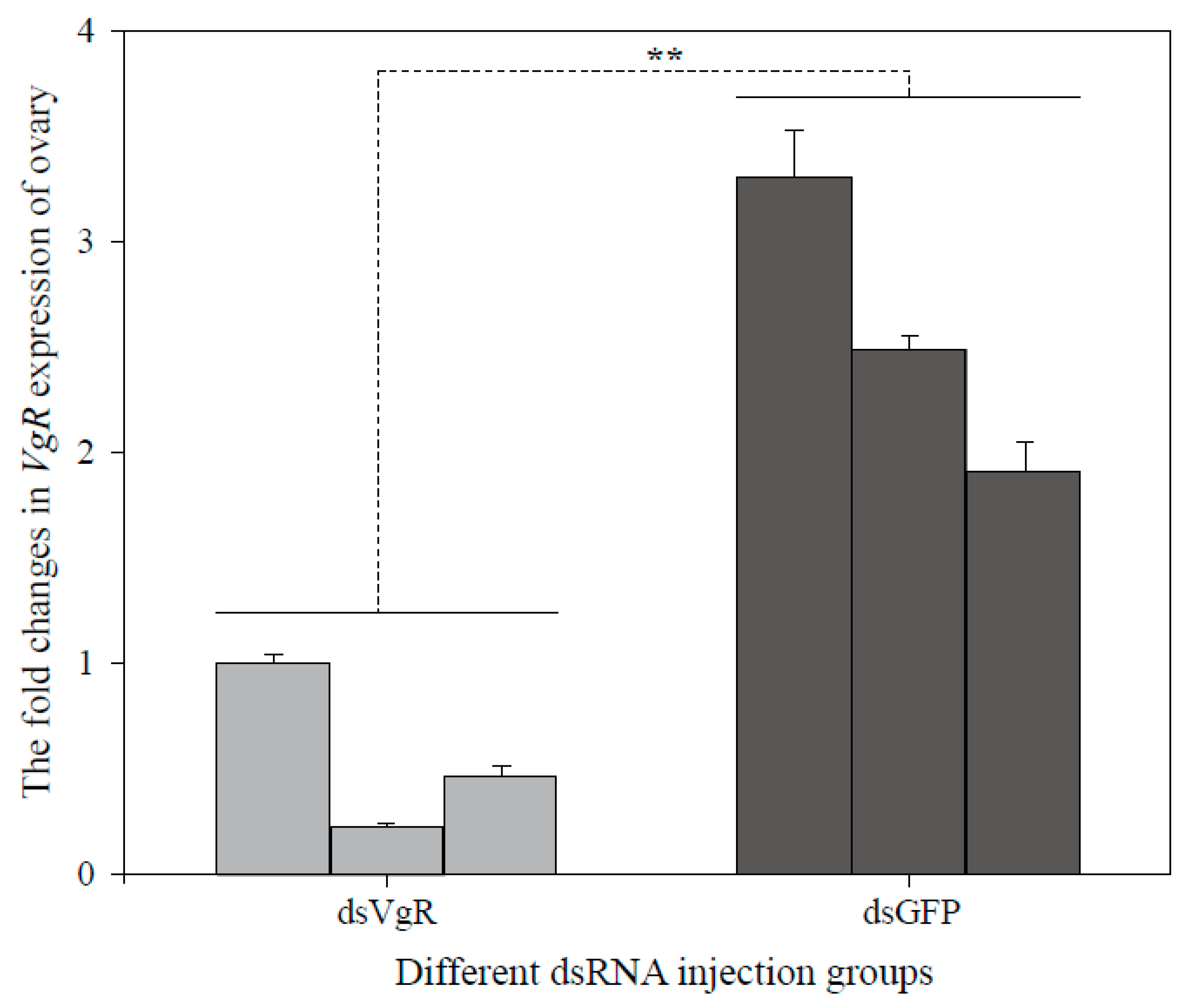
3.4. RNA Interference

To explore the efficiency of dsRNA injection, BLVgR expression levels were measured by qPCR. The BLVgR transcript levels of dsVgR-injected workers were significantly decreased by 4-fold compared to those of the dsGFP group at 72 h after dsRNA injection (Figure 4). The number of eggs laid by workers within 15 days after injection was significantly decreased in the dsVgR group compared to the dsGFP (F = 54.685, df = (1, 24), p < 0.01) and DEPC-treated water (F = 16.1448, df = (1, 24), p < 0.01). The numbers of eggs were lower by 74.5% and 90.1% in the workers injected with dsVgR compared to the DEPC-treated water and dsGFP groups, respectively (Figure 5a). Moreover, the time until first egg laying in the dsVgR-treated workers was significantly longer than in the dsGFP (F = 15.287, df = (1, 24), p < 0.01) and DEPC-treated groups (F = 12.854, df = (1, 30), p < 0.01) (16.31 ± 2.07, 8.15 ± 0.22, and 10.00 ± 0.37 days, respectively). Colonies treated with dsVgR were delayed 8 d and 6 d compared to the colonies treated with dsGFP and DEPC-treated water (Figure 5b). The dissection of ovaries showed the degree of ovary swelling was lower compared to that in the dsGFP groups (Figure 6).

4. Discussion

The understanding of the molecular mechanisms of insect reproductive regulation has reached a new level, especially in social insects [20]. The bumblebee, B. lantschouensis is a social bee that is endemic to northern China and has been selected for artificial rearing and crop pollination [29,32,42]. The development of bee colonies is highly dependent on the ovary activation in the queen, in which the biosynthesis and transport of Vg are vital [43]. VgR as a transporter of Vg, is essential for ovary activation. Moreover, VgR has been demonstrated to not only mediate the transport of Vg but also play an important role in vitellogenesis [18,44]. In the present study, the complete VgR cDNA sequence of B. lantschouensis was identified, which, to our knowledge, represents the first report of a VgR from a bumblebee. It was demonstated that BLVgR affects the first egg-laying duration and the number of eggs laid in workers. These findings will benefit from studying the mechanism of vitellogenin transport and absorption in bumblebees.
The domains present in BLVgR indicate that BLVgR is a member of the LDLR family (Figure 2). BLVgR was shown to contain two ligand-binding domains with four and eight LDLa repeats, respectively, and the BLVgR LDLa repeats present an architecture that is closer to the VgR architecture of A. mellifera, S. invicta and N. vitripennis [18,21]. However, the number of LDLa repeats in Hymenoptera differ from that in other insects. For example, there are four and seven LDLa repeats in Lepidoptera [12,15], five and eight repeats in Diptera and Blattaria [45]. Additionally, BLVgR contains two EGF-like domains, both of which include a calcium-binding site similar to the VgRs of A. mellifera, S. invicta and D. melanogaster. The O-linked sugar domain is in the last EGF-like domain and the TM domain [5,46]. In this study, no O-linked sugar domain was found in BLVgR, which is consistent with the VgRs of B. dorsalis and S. invicta [18,47], and is different from A. mellifera, L. maderae, and N. lugens [17,21,45]. This indicates that the O-linked sugar domain motif is not the common functional domain of vertebrate VgRs [48].
For long time, it was assumed that vitellogenin receptor expression is highly tissue specific and is restricted to the ovaries of the females of insects [15,17,45]. However, it was found that the AmVgR of A. mellifera is expressed not only in the ovarian tissues of reproductive females but also in males [21]. This situation contrasts with observations made in S. invicta in which VgR expression is limited to the ovaries [20]. In the present study, BLVgR was expressed in all tested tissues and showed the highest abundance in the ovaries, which may be related to the pleiotropic biological functions of its ligand Vg (Figure 3c). Besides the reproductive process, it has been demonstrated that vitellogenin is also involved in regulating the longevity of the queen and workers through epidemic inhibition of the antioxidant response [49]. In addition, it is also related to the regulation of JH titer and co-opted to regulate division of labor in workers [8]. However, recent studies have found that the LDLR family receptor has the function of signal transmission, which directly transmits signals on the target cell’s plasma membrane by interacting with the cytoplasmic adaptor and the scaffold protein [50,51]. Therefore, the tissue-specific expression of BLVgR may be related to the alternative function of its ligand and the multi-function of the receptor.
The expression of BLVgR gene was irregular throughout stages before emerging. The high expression at the egg, PW and Pbd stage suggest that it is essential to metamorphosis in these stages. In addition, its almost disappearance in the larval stage means the function of VgR is minor. Interestingly, our results are consistent with the Vg expression changes of the bumblebee, Bombus hypocrita [52]. The expression of Vg mRNA in females was detected at the Pw stage, and increased during the entire pupal development with a peak at the Pbd stage. This suggests that the transcriptional changes of VgR is related to it ligand concentration.
BLVgR expression was closely correlated with the ovarian development of adult females. Significant differences in the expression of BLVgR were observed in different egg-laying statuses. The levels of BLVgR transcripts were increased in the ovaries of egg-laying queens and workers in comparison to virgin queens and naïve workers (Figure 3c,d). In A. mellifera, the AmVgR transcript level was increased in the ovaries of egg-laying queens in comparison to virgin queens as well as workers [21]. In A. aegypti, VgR transcript levels in the ovaries rapidly increase after adult eclosion and continue to rise as the ovaries become vitelogenic, reaching peak levels 24 h after a blood meal [53]. However, in cockroaches, the developmental profile of VgR shows the highest levels in the immature ovaries of final-instar female nymphs and in the early previtellogenic period [45]. Moreover, the SiVgR transcript is present at higher levels in virgin alate females than in reproductively active queens in S. invicta [18]. The different spatiotemporal dynamics of VgR may be caused by the different functions of VgR in different organisms such as observed in A. mellifera [21].
RNAi is a powerful tool for exploring gene function [16]. In 2009, the first study achieving successful posttranscriptional silencing of VgR was reported in Hymenoptera [20]. In bumblebees, RNAi is an effective method for gene function verification. Kim et al. demonstrated expression regulation by reducing Bi-Tf or Bi-FerHCH levels in the fat body via RNA interference in bumblebee [54]. Silencing of VgR in S. invicta led to a significant decrease in SiVgR expression in queen ovaries derived from dsVgR-injected pupae and stunted oocytes showing no VgR signal; the blocking effect continued for at least 10 days upon the eclosion of virgin queens [20]. Interestingly, the silencing of VgR in cockroaches significantly decreased NlVgR transcript and protein levels after injection with dsRNA, and inhibited Vg endocytosis in the oocytes. Moreover, silencing NlVgR causes a delay in ovarian development and inhibition of oviposition [17]. In this study, newly emerged workers and 5 µg dsVgR per bee were selected as the appropriate stage and the optimal dose for microinjection. After dsVgR treatment, the mRNA expression of BLVgR was significantly lower than in the dsGFP group (Figure 4). As expected, the number of eggs laid in the dsVgR-treated groups was lower than that in the controls (Figure 5a). In contrast, the time to the first egg laying was longer (Figure 5b). Similar results were observed in H. armigera [15] and N. lugens [17]. However, the difference in egg production between the dsGFP group and the DEPC group was significant in our study. This may be caused by the injection of dsGFP that lead to the high expression of immune genes and the high expression of immune genes will benefit egg laying [55,56].
In summary, we have demonstrated that RNAi can successfully silence BLVgR to affect the fertility of female bumblebees. However, this study examined the function of VgR in worker bees without examining queens. Both workers and queens develop from diploid fertilized eggs and share the same genes. It has been reported in B. terrestris that the microcolonies (groups of three workers isolated from the queen) can be used to estimate egg numbers, the delay before egg laying, larval production, and adult offspring fitness [57]. Microcolonies are also used to measure the lethal and sublethal effects of insecticidal proteins expressed in transgenic plants, as well as growth efficiency associated with different pollen types in bumblebees [58,59]. In conclusion, regardless of essential characteristics, this study clearly indicates that VgR plays an important role in female oviposition and ovarian maturation in bumblebees.

5. Conclusions

In conclusion, we identified the cDNA and analyzed the molecular characteristics of BLVgR from the bumblebee B. lantschouensis. This gene is structurally conserved compared with the homologous genes of other species. The expression patterns of BLVgR in different tissues, development stages, and reproductive status were identified. In addition, the function of BLVgR was verified by RNAi technology. The injection of dsVgR significantly reduced VgR expression, and worker ovarian development was affected, oviposition was delayed, and egg production was remarkably decreased. Our study provides insights into the reproductive regulation of fecundity in bumblebees. Therefore, the results of the present study provide basic knowledge of the reproductive biology of bumblebees.

Author Contributions

Conceptualization, L.D., J.H., and J.W.; methodology, L.D., M.W., and J.L.; software, L.D., J.H., and J.W.; validation, L.D., J.H., and S.H.; formal analysis, L.D.; data curation, L.D. and J.H.; writing—original draft preparation, L.D., J.H., and M.W.; writing—review and editing, L.D., J.H., S.H., and J.W.; visualization, L.D. and J.H.; supervision, J.W. and J.L.; project administration, J.H. and J.W.; and funding acquisition, J.H. and J.W.

Funding

This project was financially supported in part by the National Natural Science Foundation of China (U1603108), the China Agriculture Research System (CARS-44) and the Agricultural Science and Technology Innovation Program, CAAS (CAAS-ASTIP-2019-IAR).

Acknowledgments

We would like to thank Lei Han for her kindness in guiding the experiment.

Conflicts of Interest

The authors declare no conflict of interest.

References

  1. Velthuis, H.; Doorn, A. A century of advances in bumblebee domestication and the economic and environmental aspects of its commercialization for pollination. Apidologie 2006, 37, 421–451. [Google Scholar] [CrossRef] [Green Version]
  2. Pywell, R.F.; Warman, E.A.; Carvell, C.; Sparks, T.H.; Dicks, L.V.; Bennett, D.; Wright, A.; Critchley, C.N.R.; Sherwood, A. Providing foraging resources for bumblebees in intensively farmed landscapes. Biol. Conserv. 2005, 121, 479–494. [Google Scholar] [CrossRef]
  3. Roldan Serrano, A.; Guerra-Sanz, J.M. Quality fruit improvement in sweet pepper culture by bumblebee pollination. Sci. Hortic. 2006, 110, 160–166. [Google Scholar] [CrossRef]
  4. Morandin, L.A.; Laverty, T.M.; Kevan, P.G. Effect of bumble bee (Hymenoptera: Apidae) pollination intensity on the quality of greenhouse tomatoes. J. Econ. Entomol. 2001, 94, 172–179. [Google Scholar] [CrossRef]
  5. Tufail, M.; Takeda, M. Insect vitellogenin/lipophorin receptors: Molecular structures, role in oogenesis, and regulatory mechanisms. J. Insect Physiol. 2009, 55, 88–104. [Google Scholar] [CrossRef]
  6. Raikhel, A.S.; Dhadialla, T.S. Accumulation of yolk proteins in insect oocytes. Annu. Rev. Entomol. 1992, 37, 217–251. [Google Scholar] [CrossRef]
  7. Engelmann, F. Insect vitellogenin: Identification, biosynthesis, and role in vitellogenesis. Adv. Insect Physiol. 1979, 14, 49–108. [Google Scholar]
  8. Amdam, G.V.; Norberg, K.; Fondrk, M.K.; Page, R.E. Reproductive ground plan may mediate colony-level selection effects on individual foraging behavior in honey bees. Proc. Natl. Acad. Sci. USA 2004, 101, 11350–11355. [Google Scholar] [CrossRef] [Green Version]
  9. Corona, M.; Libbrecht, R.; Wurm, Y.; Riba-Grognuz, O.; Keller, L. Vitellogenin underwent subfunctionalization to acquire caste and behavioral specific expression in the harvester ant Pogonomyrmex barbatus. PLoS Genet. 2013, 9, e1003730. [Google Scholar] [CrossRef]
  10. Cardoso Junior, C.; Oldroyd, B.; Ronai, I. Vitellogenin expression in the ovaries of adult honeybee workers provides insights into the evolution of reproductive and social traits. bioRxiv 2019, 547760. [Google Scholar] [CrossRef] [Green Version]
  11. Röhrkasten, A.; Ferenz, H. Properties of the vitellogenin receptor of isolated locust oocyte membranes. Int. J. Invertebr. Reprod. Dev. 1986, 10, 133–142. [Google Scholar] [CrossRef]
  12. Wu, H.; Jiang, F.; Guo, J.; Yi, J.; Liu, J.; Cao, Y.; Lai, X.; Zhang, G. Molecular characterization and expression of vitellogenin and vitellogenin receptor of Thitarodes pui (Lepidoptera: Hepialidae), an insect on the Tibetan Plateau. J. Insect Sci. 2018, 18, 1–7. [Google Scholar] [CrossRef] [PubMed]
  13. Lin, Y.; Meng, Y.; Wang, Y.X.; Luo, J.; Katsuma, S.; Yang, C.W.; Banno, Y.; Kusakabe, T.; Shimada, T.; Xia, Q.Y. Vitellogenin receptor mutation leads to the oogenesis mutant phenotype scanty vitellin of the silkworm, Bombyx Mori. J. Biol. Chem. 2013, 288, 13345–13355. [Google Scholar] [CrossRef] [PubMed] [Green Version]
  14. Kim, H.R.; Katagiri, C.; Nagao, E.; Chino, H. Purification and characterization of vitellogenin from the American cockroach, Periplaneta americana. Comp. Biochem. Physiol. B Comp. Biochem. 1992, 103, 963–967. [Google Scholar] [CrossRef]
  15. Zhang, W.; Ma, L.; Xiao, H.; Xie, B.; Smagghe, G.; Guo, Y.; Liang, G. Molecular characterization and function analysis of the vitellogenin receptor from the cotton bollworm, Helicoverpa armigera (Hübner) (Lepidoptera, Noctuidae). PLoS ONE 2016, 11, e0155785. [Google Scholar] [CrossRef] [Green Version]
  16. Tufail, M.; Takeda, M. Molecular cloning, characterization and regulation of the cockroach vitellogenin receptor during oogenesis. Insect Mol. Biol. 2005, 14, 389–401. [Google Scholar] [CrossRef]
  17. Lu, K.; Shu, Y.; Zhou, J.; Zhang, X.; Zhang, X.; Chen, M.; Yao, Q.; Zhou, Q.; Zhang, W. Molecular characterization and RNA interference analysis of vitellogenin receptor from Nilaparvata lugens (Stål). J. Insect Physiol. 2015, 73, 20–29. [Google Scholar] [CrossRef]
  18. Chen, M.E.; Lewis, D.K.; Keeley, L.L.; Pietrantonio, P.V. cDNA cloning and transcriptional regulation of the vitellogenin receptor from the imported fire ant, Solenopsis invicta Buren (Hymenoptera: Formicidae). Insect Mol. Biol. 2004, 13, 195–204. [Google Scholar] [CrossRef] [Green Version]
  19. Ciudad, L.; Piulachs, M.D.; Bellés, X. Systemic RNAi of the cockroach vitellogenin receptor results in a phenotype similar to that of the Drosophila yolkless mutant. FEBS J. 2006, 273, 325–335. [Google Scholar] [CrossRef]
  20. Lu, H.; Vinson, S.B.; Pietrantonio, P.V. Oocyte membrane localization of vitellogenin receptor coincides with queen flying age, and receptor silencing by RNAi disrupts egg formation in fire ant virgin queens. FEBS J. 2009, 276, 3110–3123. [Google Scholar] [CrossRef]
  21. Guidugli-Lazzarini, K.R.; Do Nascimento, A.M.; Tanaka, É.D.; Piulachs, M.D.; Hartfelder, K.; Bitondi, M.G.; Simões, Z.L.P. Expression analysis of putative vitellogenin and lipophorin receptors in honey bee (Apis mellifera L.) queens and workers. J. Insect Physiol. 2008, 54, 1138–1147. [Google Scholar] [CrossRef] [PubMed]
  22. Hannon, G.J. RNA interference. Nature 2002, 418, 244–251. [Google Scholar] [CrossRef] [PubMed]
  23. Amdam, G.V.; Simões, Z.L.; Guidugli, K.R.; Norberg, K.; Omholt, S.W. Disruption of vitellogenin gene function in adult honeybees by intra-abdominal injection of double-stranded RNA. BMC Biotechnol. 2003, 3, 1–8. [Google Scholar] [CrossRef] [PubMed] [Green Version]
  24. Turner, C.T.; Davy, M.W.; MacDiarmid, R.M.; Plummer, K.M.; Birch, N.P.; Newcomb, R.D. RNA interference in the light brown apple moth, Epiphyas postvittana (Walker) induced by double-stranded RNA feeding. Insect Mol. Biol. 2006, 15, 383–391. [Google Scholar] [CrossRef]
  25. Mao, Y.B.; Cai, W.J.; Wang, J.W.; Hong, G.J.; Tao, X.Y.; Wang, L.J.; Huang, Y.P.; Chen, X.Y. Silencing a cotton bollworm P450 monooxygenase gene by plant-mediated RNAi impairs larval tolerance of gossypol. Nat. Biotechnol. 2007, 25, 1307–1313. [Google Scholar] [CrossRef]
  26. Costa, C.P.; Elias-Neto, M.; Falcon, T.; Dallacqua, R.P.; Martins, J.R.; Bitondi, M.M.G. RNAi-mediated functional analysis of bursicon genes related to adult cuticle formation and tanning in the honeybee, Apis mellifera. PLoS ONE 2016, 11, e0167421. [Google Scholar] [CrossRef]
  27. Marco, A.D.; Guidugli-Lazzarini, K.R.; Do, N.A.; Simoes, Z.L.; Hartfelder, K. RNAi-mediated silencing of vitellogenin gene function turns honeybee (Apis mellifera) workers into extremely precocious foragers. Naturwissenschaften 2008, 95, 953–961. [Google Scholar] [CrossRef]
  28. Garbian, Y.; Maori, E.; Kalev, H.; Shafir, S.; Sela, I. Bidirectional transfer of RNAi between honey bee and Varroa destructor: Varroa gene silencing reduces Varroa population. PLoS Pathog. 2012, 8, e1003035. [Google Scholar] [CrossRef] [Green Version]
  29. An, J.D.; Huang, J.X.; Shao, Y.Q.; Zhang, S.W.; Wang, B.; Liu, X.Y.; Wu, J.; Williams, P.H. The bumblebees of North China (Apidae, Bombus Latreille). Zootaxa 2014, 3830, 1–89. [Google Scholar] [CrossRef] [Green Version]
  30. Zhou, Z.; Zhang, H.; Liang, C.; Zou, Y.; Dong, J.; Yuan, X.; Huang, J.; An, J. Foraging preference of the honeybee Apis mellifera and the bumblebee Bombus lantschouensis (Hymenoptera: Apidae) in peach greenhouse. Acta Entomol. Sin. 2015, 58, 1315–1321. [Google Scholar]
  31. Gurel, F.; Gosterit, A. Effects of different stimulation methods on colony initiation and development of Bombus terrestris L. (Hymenoptera: Apidae) queens. Appl. Entomol. Zool. 2008, 43, 113–117. [Google Scholar] [CrossRef]
  32. Han, L.; Ding, G.; Liu, Y.; Huang, J.; Wu, J. Characterization of sphingomyelin phosphodiesterase expression in bumblebee (Bombus lantschouensis). J. Insect Sci. 2018, 18, 1–7. [Google Scholar] [CrossRef] [PubMed] [Green Version]
  33. Rice, P.; Longden, I.; Bleasby, A. EMBOSS: The European molecular biology open software suite. Trends Genet. 2000, 16, 276–277. [Google Scholar] [CrossRef]
  34. Krogh, A.; Larsson, B.; von Heijne, G.; Sonnhammer, E.L.L. Predicting transmembrane protein topology with a hidden markov model: Application to complete genomes. J. Mol. Biol. 2001, 305, 567–580. [Google Scholar] [CrossRef] [Green Version]
  35. Almagro, A.J.; Tsirigos, K.D.; Sonderby, C.K.; Petersen, T.N.; Winther, O.; Brunak, S.; von Heijne, G.; Nielsen, H. SignalP 5.0 improves signal peptide predictions using deep neural networks. Nat. Biotechnol. 2019, 37, 420–423. [Google Scholar] [CrossRef]
  36. Ivica, L.; Tobias, D.; Peer, B. SMART 7: Recent updates to the protein domain annotation resource. Nucleic Acids Res. 2011, 40, D302–D305. [Google Scholar]
  37. Sudhir, K.; Glen, S.; Li, M.; Christina, K.; Koichiro, T. MEGA X: Molecular evolutionary genetics analysis across computing platforms. Mol. Biol. Evol. 2018, 6, 1547–1549. [Google Scholar]
  38. Felsenstein, J. Confidence limits on phylogenies: An approach using the bootstrap. Evolution 1985, 4, 783–791. [Google Scholar] [CrossRef]
  39. Livak, K.J.; Schmittgen, T.D. Analysis of relative gene expression data using real-time quantitative PCR and the 2(-Delta Delta C (T)) method. Methods 2001, 25, 402–408. [Google Scholar] [CrossRef]
  40. Chen, J.; Zhang, D.; Yao, Q.; Zhang, J.; Dong, X.; Tian, H.; Chen, J.; Zhang, W. Feeding-based RNA interference of atrehalose phosphate synthase gene in the brown planthopper, Nilaparvata lugens. Insect Mol. Biol. 2010, 19, 777–786. [Google Scholar] [CrossRef]
  41. Amdam, G.V.; Norberg, K.; Hagen, A.; Omholt, S.W. Social exploitation of vitellogenin. Proc. Natl. Acad. Sci. USA 2003, 100, 1799–1802. [Google Scholar] [CrossRef] [Green Version]
  42. Dong, J.; Li, J.; Huang, J.; Wu, J. Identification of suitable reference genes for miRNA quantitation in bumblebee (Hymenoptera: Apidae) response to reproduction. Apidologie 2019, 50, 40–50. [Google Scholar] [CrossRef] [Green Version]
  43. Dohanik, V.T.; Gonçalves, W.G.; Oliveira, L.L.; Zanuncio, J.C.; Serrão, J.E. Vitellogenin transcytosis in follicular cells of the honeybee Apis mellifera and the wasp Polistes simillimus. Protoplasma 2018, 255, 1703–1712. [Google Scholar] [CrossRef]
  44. Boldbaatar, D.; Battsetseg, B.; Matsuo, T.; Hatta, T.; Umemiya-Shirafuji, R.; Xuan, X.; Fujisaki, K. Tick vitellogenin receptor reveals critical role in oocyte development and transovarial transmission of Babesia parasite. Biochem. Cell Biol. 2008, 86, 331–344. [Google Scholar] [CrossRef]
  45. Tufail, M.; Takeda, M. Molecular cloning and developmental expression pattern of the vitellogenin receptor from the cockroach, Leucophaea maderae. Insect Biochem. Mol. Biol. 2007, 37, 235–245. [Google Scholar] [CrossRef]
  46. Chen, S.; Lee, T.; Ou, Y. Incorporating significant amino acid pairs to identify O-linked glycosylation sites on transmembrane proteins and non-transmembrane proteins. BMC Bioinform. 2010, 11, 536. [Google Scholar] [CrossRef] [Green Version]
  47. Cong, L.; Yang, W.; Jiang, X.; Niu, J.; Shen, G.; Ran, C.; Wang, J. The essential role of vitellogenin receptor in ovary development and vitellogenin uptake in Bactrocera dorsalis (Hendel). Int. J. Mol. Sci. 2015, 16, 18368–18383. [Google Scholar] [CrossRef] [Green Version]
  48. Hiramatsu, N.; Chapman, R.W.; Lindzey, J.K.; Haynes, M.R.; Sullivan, C.V. Molecular characterization and expression of vitellogenin receptor from white perch (Morone americana). Biol. Reprod. 2004, 70, 1720–1730. [Google Scholar] [CrossRef] [Green Version]
  49. Piulachs, M.D.; Guidugli, K.R.; Barchuk, A.R.; Cruz, J.; Simoes, Z.L.; Belles, X. The vitellogenin of the honey bee, Apis mellifera: Structural analysis of the cDNA and expression studies. Insect Biochem. Mol. Biol. 2003, 33, 459–465. [Google Scholar] [CrossRef]
  50. Herz, J.; Gotthardt, M.; Willnow, T.E. Cellular signalling by lipoprotein receptors. Curr. Opin. Lipidol. 2000, 11, 161–166. [Google Scholar] [CrossRef]
  51. Gotthardt, M.; Trommsdorff, M.; Nevitt, M.F.; Shelton, J.; Richardson, J.A.; Stockinger, W.; Nimpf, J.; Herz, J. Interactions of the low density lipoprotein receptor gene family with cytosolic adaptor and scaffold proteins suggest diverse biological functions in cellular communication and signal transduction. J. Biol. Chem. 2000, 275, 25616–25624. [Google Scholar] [CrossRef] [PubMed] [Green Version]
  52. Li, J.; Huang, J.; Cai, W.; Zhao, Z.; Peng, W.; Wu, J. The vitellogenin of the bumblebee, Bombus hypocrita: Studies on structural analysis of the cDNA and expression of the mRNA. J. Comp. Physiol. B 2010, 180, 161–170. [Google Scholar] [CrossRef] [PubMed]
  53. Cho, K.H.; Raikhel, A.S. Organization and developmental expression of the mosquito vitellogenin receptor gene. Insect Mol. Biol. 2001, 10, 465–474. [Google Scholar] [CrossRef] [PubMed]
  54. Kim, B.Y.; Lee, K.S.; Yoon, H.J.; Kim, I.; Li, J.; Sohn, H.D.; Jin, B.R. Expression profile of the iron-binding proteins transferrin and ferritin heavy chain subunit in the bumblebee Bombus ignitus. Comp. Biochem. Physiol. B Biochem. Mol. Biol. 2009, 153, 165–170. [Google Scholar] [CrossRef]
  55. Nunes, F.M.F.; Aleixo, A.C.; Barchuk, A.R.; Bomtorin, A.D.; Grozinger, C.M.; Simões, Z.L.P. Non-target effects of green fluorescent protein (GFP)-derived double-stranded RNA (dsRNA-GFP) used in honey bee RNA interference (RNAi) assays. Insects 2013, 4, 90–103. [Google Scholar] [CrossRef] [Green Version]
  56. Duncan, E.J.; Hyink, O.; Dearden, P.K. Notch signalling mediates reproductive constraint in the adult worker honeybee. Nat. Commun. 2016, 7, 12427. [Google Scholar] [CrossRef] [Green Version]
  57. Genissel, A.; Aupinel, P.; Bressac, C.; Tasei, J.N.; Chevrier, C. Influence of pollen origin on performance of Bombus terrestris micro-colonies. Entomol. Exp. Appl. 2010, 104, 329–336. [Google Scholar] [CrossRef]
  58. Babendreier, D.; Reichhart, B.; Romeis, J.; Bigler, F. Impact of insecticidal proteins expressed in transgenic plants on bumblebee microcolonies. Entomol. Exp. Appl. 2010, 126, 148–157. [Google Scholar] [CrossRef]
  59. Dance, C.; Botías, C.; Goulson, D. The combined effects of a monotonous diet and exposure to thiamethoxam on the performance of bumblebee micro-colonies. Ecotoxicol. Environ. Saf. 2017, 139, 194–201. [Google Scholar] [CrossRef]
Figure 1. mRNA structure of BLVgR in the bumblebee (Bombus lantschouensis). The numbers indicate the position in the sequence. The full-length BLVgR cDNA sequence is 5519 bp. The 5′ UTR and 3′ UTR are 63 and 179 bp. A polyadenylation signal (AATAAA) is found at the end of the mRNA sequence.
Insects 10 00445 g001
Figure 2. Diagrammatic phylogenetic relationship and structure comparisons of BLVgR with sequences from other insects: (a) Phylogenetic analysis of BLVgR with the vitellogenin receptors of insects from different orders. The phylogenetic tree was constructed by using MEGA X with a statistical method of maximum-likelihood. The VgR sequence of A. pisum was used as an outgroup. The numbers indicate bootstrap support values (%) based on 1000 replicates. Branches corresponding to partitions reproduced in less than 50% bootstrap replicates are collapsed. (b) Comparison of BLVgR modular domains with those of other insects. LDLa, low-density lipoprotein receptor domain class A (hexagon with green); EGF, epidermal growth factor-like domain (pentagon with green); EGF_CA, calcium-binding EGF-like domain (pentagon with yellow); LY, low-density lipoprotein receptor YWTD protein (hexagon with purple); transmembrane domain (tetragon with blue).
Insects 10 00445 g002
Figure 3. Analysis of relative BLVgR expression in different tissues, developmental stages and reproductive status in bumblebees (B. lantschouensis). (a) Analysis of relative BLVgR expression in different tissues: antenna (AN), head (HD), thorax (TH), leg (LG), epidermis (EP), midgut (MG), ovary (OV), fat body (FB), and venom gland (VG) (F = 91.811, df = (8, 72), p < 0.05). (b) Analysis of relative BLVgR expression at different developmental stages: Egg (within 24 h of laying), Lar (within 48 h of larvae after hatching), Pw (white-eyed pupae with an unpigmented cuticle), Pp (pink-eyed pupae with an unpigmented cuticle), Pb (brown-eyed pupae with an unpigmented cuticle), Pbl (brown-eyed pupae with thoracic pigmentation), Pbd (brown eyes with a dark-pigmented cuticle), and Pdd (dark-eye pupae with a dark-pigmented cuticle) (F = 18.373, df = (7, 64), p < 0.05). (c) Differences in the expression of the BLVgR gene in B. lantschouensis workers with different reproductive status. Three egg-laying and non-egg-laying workers were sampled. The expression level of BLVgR was significantly increased in egg-laying workers compared with non-egg-laying workers (F = 95.588, df = (1, 16), p < 0.01). (d) Differences in the expression of the BLVgR gene in queens with different reproductive status. Three egg-laying and non-egg-laying queens were sampled. The expression level of BLVgR was significantly increased in egg-laying queens compared with non-egg-laying queens (F = 48.977, df = (1, 16), p < 0.01). The asterisks above the error bar indicate that the expression levels were significantly different (p < 0.01).
Insects 10 00445 g003
Figure 4. The transcript level of VgR in ovaries of bumblebee workers that were subjected to RNA interference. The expression levels of BLVgR were determined by qPCR following the injection of dsVgR after 72 h. Three microcolonies of each treatment (dsVgR and dsGFP) were sampled. The dsGFP treatment group was used as a control. The expression level of VgR was significantly decreased (** p < 0.01).
Insects 10 00445 g004
Figure 5. The fecundity including the number of eggs laid and the first egg-laying time was evaluated after dsRNA RNAi-mediated knockdown of VgR. The total number of eggs laid in each microcolony was counted within 15 days after injection. The number of microcolonies in dsVgR, dsGFP and DEPC-treated water groups was 13, 13, and 19. (a) The difference of egg laying number under different treatments. Eggs were removed every day and the number was counted. (b) The difference of first egg laying time under different treatments. Egg laying status was checked every day. When the first egg was observed, the first egg laying time was recorded. Different letters represent a significant level (p < 0.01).
Insects 10 00445 g005
Figure 6. Ovary development status was evaluated after the RNAi-mediated knockdown of BLVgR. Newly emerged adult workers were injected with dsGFP and dsVgR. Ovaries were dissected under dissection microscopy and photographed with a NIKON (D750) camera 7 days after the injection. The ovary on the top was from the dsGFP group, and the ovary on the bottom was from the dsVgR group.
Insects 10 00445 g006
Table 1. Primers used for the identification and analysis of the B. lantschouensis VgR (BLVgR).
PurposeNamePrimer Sequence (5′–3′)
cDNA cloningVgRFACTCATGTTTGTGCCAACCTG
VgRRTTGGATGGTACAGATCGAAGG
RACE5′VgR9FTCGAGGTGCAAGCATGAGGATCAGTT
3′VgR9RGCGTTTCTTTGGCATGTTACGCTCT
UPM-LongCTAATACGACTCACTATAGC
UPM-ShortTCACCGCATTCATCTTCC
Real-time PCRFVgRFGTGTGCCTGTTATCTAATGCTGAT
FVgRRTTCATCTTCACCGTTAGGACAATC
β-actinFCGACTACCTCATGAAGATT
β-actinRCGACGTAACAAAGTTTCTC
GAPDH-FGCTGGAGCTGAATATGTTGTAGAATC
GAPDH-RAGTAGTGCAGGAAGCATTAGAGATAACT
RNAiRNAiVgR1FGTTTCAATGTAAAAACGGCGACT
RNAiVgR1RTCGTTCTTTGGACAATCTGTAACG
T7RNAiVgR1FGGATCCTAATACGACTCACTATAGGGTTTCAATGTAAAAACGGCGACT
T7RNAiVgR1RGGATCCTAATACGACTCACTATAGGTCGTTCTTTGGACAATCTGTAACG
GFP-FCCACAAGTTCAGCGTGTCCG
GFP-RAAGTTCACCTTGATGCCGTTCT
T7GFP-FGGATCCTAATACGACTCACTATAGCCACAAGTTCAGCGTGTCCG
T7GFP-RGGATCCTAATACGACTCACTATAGAAGTTCACCTTGATGCCGTTCT

