Review

Granulomatosis with Polyangiitis-Mimicking Advanced Gynecological Cancer: A Case Report and Systematic Review of the Literature

by
Augusto Pereira
1,*,
Javier F. Magrina
2,
Paul M. Magtibay
2,
Beatriz G. Stamps
2,
Elena Muñoz-Nuñez
1 and
Tirso Perez-Medina
1
1
Department of Gynecologic Surgery, Puerta de Hierro University Hospital, 28222 Madrid, Spain
2
Department of Medical and Surgical Gynecology, Mayo Clinic Hospital, Phoenix, AZ 85054, USA
*
Author to whom correspondence should be addressed.
J. Pers. Med. 2022, 12(2), 289; https://doi.org/10.3390/jpm12020289
Submission received: 26 December 2021 / Revised: 6 February 2022 / Accepted: 13 February 2022 / Published: 16 February 2022
(This article belongs to the Special Issue Diagnostics and Interventional Procedures in Gynecological Tumors)

Abstract

:
(1) Background: Granulomatosis with polyangiitis (GPA) is a necrotizing vasculitis that mimics gynecologic cancer. In GPA patients, the genitourinary system is affected in <1%. The objective of the study was to provide a systematic review of the literature of GPA patients with gynecological involvement. (2) Methods: PubMed and Embase were searched from inception to July 2021 for GPA patients with gynecological involvement Medical Subject Headings (MeSH) and free-text terms. Exclusion criteria were other language, review articles, pregnancy, fertility, or male patients. Data were extracted on clinical evolution, symptoms, examinations findings, diagnosis delay, treatment, outcome, patient status, and follow-up. (3) Results: Seventeen studies included data from patients with GPA and primary or relapsed gynecological involvement. 68% of the authors of this review thought the patient had cancer. The main gynecological symptom is bleeding, but exclusive gynecologic symptomatology is rare (ENT: 63%, lungs: 44%, kidneys-urinary tract: 53%). GPA could affect all areas of the genital tract, but the most frequent location is the uterine cervix. Medical treatment for GPA is effective. (4) Conclusions: GPA of the female genital tract must be considered when biopsies of an ulcerated malignant-appearing cervical or vaginal mass are negative for malignancy even when they are unspecific. Rheumatology consultation is indicated.

1. Introduction

Since January 2011, the Board of Directors of the American College of Rheumatology, the American Society of Nephrology, and the European League Against Rheumatism recommended that the name “Wegener’s granulomatosis” be changed to “granulomatosis with polyangiitis” (GPA) [1].
The prevalence is estimated between 1/6400–42,000 worldwide with an annual incidence between 1/84,000–475,000 [2]. GPA is a rare antineutrophil cytoplasmic antibody (ANCA)-associated vasculitis characterized by necrotizing inflammation of vessels, resulting in tissue ischemia. GPA is a multiorgan disease. Ear, nose, and throat (ENT) symptoms are present in 50–95% of patients, bronchopulmonary symptoms in 60–80%, and renal disease in 60–80%. Other sites of involvement with a lower incidence are skin in 10–50% of patients, peripheral nerves in 25%, central nervous system in 10%, and ocular in 7–8%. Patients with genitourinary involvement, cervix, urethra, and vagina, are extremely rare, <1% of patients with GPA [3].
Diagnosis remains delayed in acute settings in spite of a better understanding of this disease. It is usually based on clinical findings, imaging studies, and biochemical tests, as well as on the detection of serum antineutrophil cytoplasmic autoantibody (ANCA)-associated vasculitis, mainly PR3-ANCA, a biopsy should be performed to confirm the diagnosis [4].
GPA is defined, according to the Chapel Hill criteria, as a necrotizing granulomatous inflammation that affects the respiratory tract, with necrotizing vasculitis of small and medium vessels (for example, capillaries, veins, arterioles, and arteries). The etiology is unknown, but it is accepted that the immune system is involved in this pathogenesis. In the vast majority of cases, the combination of suggestive clinical characteristics and the presence of cytoplasmic-staining ANCA may be sufficient. If the are diagnostic doubts, histologic confirmation such as lung biopsy is needed [5].
Typically, women presenting with a large cervical or vaginal mass and unilateral ureteral obstruction have a locally advanced gynecologic malignancy. The diagnosis is usually straightforward, with a simple biopsy and pathologic assessment of the mass, followed by a metastatic workup when a malignancy is confirmed. However, in rare cases, biopsies are negative for malignancy, resulting in a diagnostic dilemma requiring further investigation. We report a patient referred to our institution for a presumed advanced vaginal carcinoma. Her history was remarkable for a previous hysterectomy for benign disease more than 30 years prior. Imaging confirmed a left ureteral obstruction, and biopsies were negative for malignancy. A final diagnosis of GPA was established when the patient had a CR to medical management.
Our objective is to provide a systematic review of the literature of GPA patients with gynecological involvement and to emphasize that a specific tissue diagnosis of GPA is not necessary to implement medical treatment when other conditions are unlikely to be responsible for the presenting pathology.

Case Report

A 75-year-old white woman presented to Mayo Clinic in Arizona with a 1-year history of pelvic pain, intermittent hematuria, vaginal bleeding, and an undiagnosed and untreated large pelvic mass infiltrating the upper vaginal half, obstructing the left ureter. Her history was important for a total abdominal hysterectomy and bilateral salpingo-oophorectomy 36 years earlier for fibroids. Her other past medical history was unremarkable. Biopsies of the vaginal mass performed elsewhere were reviewed and found to be granulation tissue with necrosis, negative for malignancy. A laparoscopy performed at an outside institution was non-diagnostic. The patient was monitored for more than a year by her local gynecologist without further workup or therapy despite worsening symptoms. A repeat laparoscopy was recommended when she elected to come to our institution for consultation.
General physical examination was negative other than an ulcerated, exophytic mass extending along the anterior vaginal wall from the vaginal cuff to the urethra-vesical junction. The mass was firm and fixed to the left pubic ramus. The clinical diagnosis was advanced vaginal carcinoma. Multiple superficial and deep biopsies were obtained and showed nonspecific acute and chronic inflammation with necrosis and granulomatous inflammation. No evidence was found of vasculitis or malignancy.
Computed tomography (CT) showed a 4-cm vaginal mass involving the vaginal cuff, bladder base, and obstruction of the left ureter with hydronephrosis. Cystoscopy revealed a bulging trigone from an extrinsic mass with a non-visualization of the left ureteral meatus. Bladder biopsies showed necrotizing granulomatous inflammation. A percutaneous, antegrade left ureteral stent was placed to drain the obstructed kidney.
Further histologic stains for acid-fast, fungal organisms and cytokeratin were negative. Tissue cultures were negative for fungus and actinomyces. Reference blood tests included c-ANCA test, serum fungal serologies; interferon g (QuantiFERON; Qiagen, Hilden, Germany); antinuclear antibody and extractable nuclear antigen antibody profiling; and C3, C4, total hemolytic complement, rapid plasma reagin, and cyclic citrullinated peptide antibodies. The sedimentation rate was 17 mm/hour, and C-reactive protein concentration was 6.9 mg/L. Head and chest CT scans were negative.
Findings did not confirm but raised the suspicion of GPA in the absence of other plausible conditions. Biopsies revealed granulomatous necrosis without polyangiitis. Blood test results were inconclusive. There was no history of GPA and absence of pulmonary or renal involvement. The patient was referred to the Division of Rheumatology, Allergy, and Clinical Immunology, and a therapeutic trial of 4 doses (375 mg/m2) of weekly intravenous rituximab was offered to the patient, affirming that GPA was the most likely albeit not confirmed diagnosis. After the 4-week trial, CT revealed a marked reduction in the vaginal mass. The patient underwent a change in treatment to an increasing weekly oral dose of methotrexate, to a maximum of 20 mg, and 1 mg of oral folic acid daily. At 6 months, repeat CT showed complete regression of the vaginal mass, a normal bladder, and normal ureters. Cystoscopy was also normal, and the ureteral stent was removed. Pelvic examination was negative other than vaginal adhesions at the site of previous vaginal involvement. The patient continues to be asymptomatic with no evidence of recurrence after 53 months of follow-up.

2. Materials and Methods

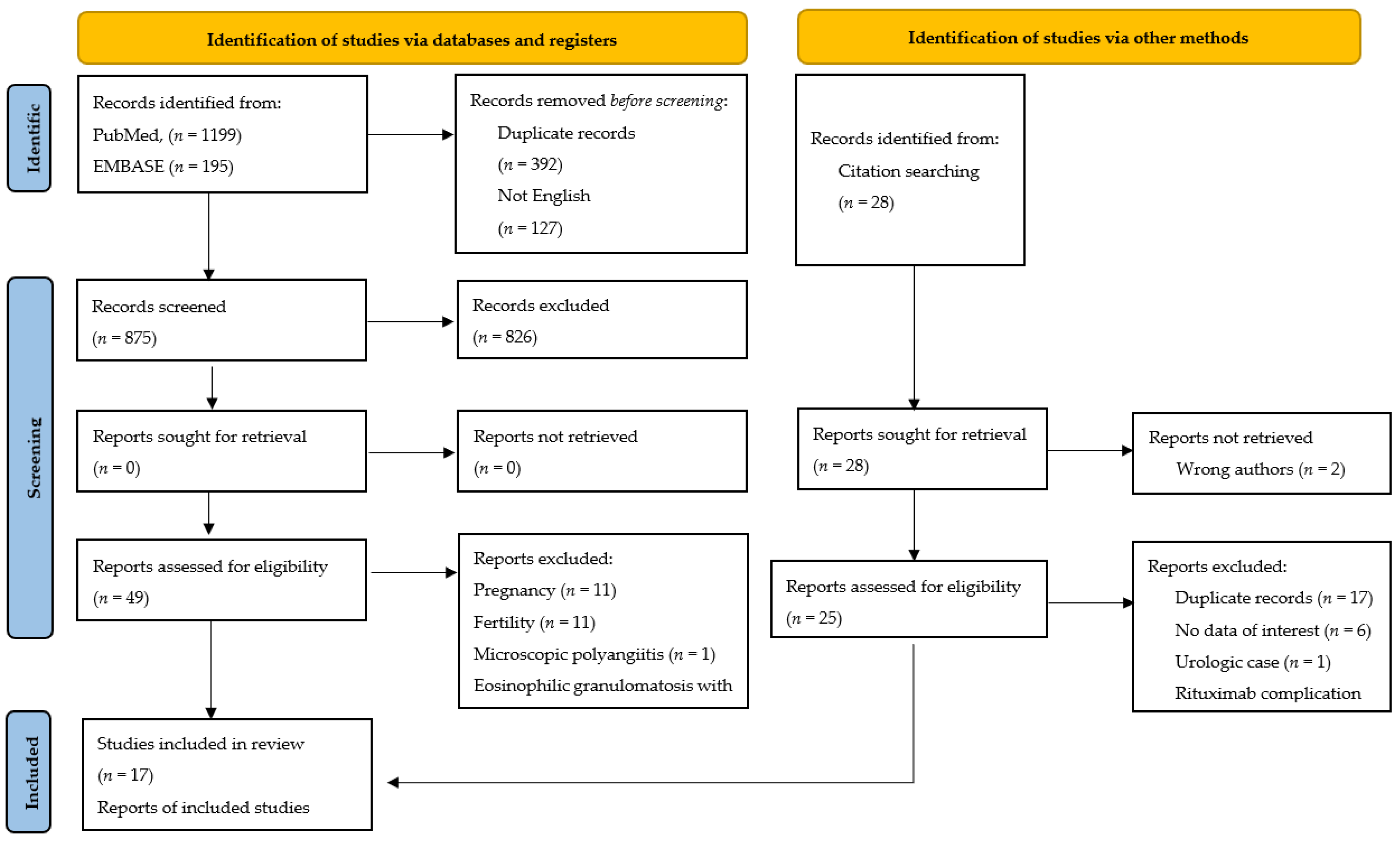
2.1. Search Strategy

A systematic literature review was undertaken. Search terms were used as free terms and as Medical Subject Headings (MeSH) or Emtree terms (indexed on Pubmed or Embase from inception to July 2021): (a) The following free terms were used on all databases: “granulomatosis with polyangiitis” OR “Wegener granulomatosis”, AND “cervix”, “uterus”, “vagina”, “vulva”, “ovary”, “adnexa”, “adnexal” OR “gynecologic disease”. The following MeSH terms were used: “granulomatosis with polyangiitis”, OR “Wegener granulomatosis”, AND, “cervix uteri”, “uterus”, “vagina”, “vulva”, “ovary”, “adnexa uteri”, “adnexal uteri”, “gynecologic disease, female”, “uterine hemorrhage”, “metrorrhagia”, “gynecologic neoplasm, female”, “pelvic neoplasm”, “gynecology”, “genital female, disease”, “female urogenital, disease”, OR “genital neoplasm, female”, “genitalia, female”. The following Emtree terms were used: “granulomatosis with polyangiitis” OR “Wegener’s granulomatosis”, AND, “gynecologic disease” “gynecology” OR “female genital tract tumor”. Hand-searching of citations was carried out on case series and reviewed studies to allow us to identify references that might have been missed in previous searches.

2.2. Selection Criteria

The databases searched were PubMed from 1957 to July 2021 and Embase from 2011 to July 2021. Articles were included if describing patients primarily located in the female genital tract, case series, English language, and had an abstract. Exclusion criteria were other language, review articles, pregnancy or fertility-related issues, or male patients.

2.3. Data Collection and Analysis

All articles were screened on the basis of title and abstract (AP, JFM). The following data were extracted: title, author, year, journal, number of cases, age, clinical evolution, the initial presentation of symptoms, ENT, urologic and gynecological symptoms, lung findings, examinations findings, diagnosis delay, type of treatment, outcome, time to CR or PR, first and second recurrence, interval to recurrence, patient status, and follow-up. All data were included in an EXCEL spreadsheet, and the final selection of the articles was downloaded for full review (AP, JFM); any disagreement was resolved by discussion.
All retrieved articles were case reports with or without literature review; due to the high heterogeneity of data obtained, a descriptive narrative review was planned instead of a meta-analysis. The risk of bias was minimized by adhering to the PRISMA statements [6]. In addition, we described the flow diagram of study search and systematic review with reasons for being excluded, removed or not retrieved, using PRISMA guidelines. All data included in the study (in total 19 patients from 17 studies) were compiled in 4 tables according to the patients with GPA and primary gynecological involvement, and gynecological relapse.

2.4. Definitions

In order to unify the criteria for our study, diagnosis of GPA was established based on pathological reports and/or c-ANCA test. Diagnosis delay was defined as the time from the initial presentation of symptoms to diagnosis. GPA treatment consists of an “initial induction phase” to achieve long-standing remission of the active disease, followed by a “maintenance phase” to extend remission and avoid recurrence. Among the GPA treatments described in the literature, glucocorticoids in combination with either rituximab, cyclophosphamide, methotrexate, or azathioprine have been proposed better than monotherapy with prednisone or methylprednisolone. Complete remission (CR) was defined as the absence of active disease, and partial remission was referred to as the persistence of signs and symptoms of active disease. Recurrence is defined after a previous GPA diagnosis followed by months of disease-free. Follow-up was measured in months from the time of primary treatment until death or last contact.

3. Results

3.1. Main Findings

A total of 1422 references (1199 Pubmed + 195 Embase + 25 Free terms + 3 hand searching) were identified from electronic databases in the search performed on 21 July 2021. One thousand three hundred and seventy-three citations were excluded based on the title and/or abstract, duplicate entries, and written in languages other than English, thereby 49 potentially relevant articles were included for subsequent evaluation. Most of these studies were excluded due to pregnancy (11), fertility (11), or related to other aspects of gynecology (5).
The pathological diagnosis of vasculitis in patients with GPA, microscopic polyangiitis (MPA), or eosinophilic granulomatosis with polyangiitis (EGPA, Chug-Strauss syndrome) is difficult, sometimes not possible. We excluded five patients with genital tract involvement from our review, one with MPA and four with EGPA [7,8,9,10,11].
Once studies with patients with MPA or EGPA (5) were removed, a total of 17 studies fulfilled all inclusion criteria and were selected for full-text evaluation and systematic review [12,13,14,15,16,17,18,19,20,21,22,23,24,25,26,27,28]. After reviewing the 17 articles, 18 patients were identified as GPA with gynecological involvement (two patients reported by Stone et al.) [12]. A flow diagram of the study search is shown in Figure 1.
A total of nineteen patients, including the present case, constitute the basis of the systematic review. Among them, 11 patients (58%) had primary gynecologic involvement, and 8 had genital recurrences (42%). In the primary GPA group, one patient had a cutaneous recurrence at 12 months and is included as recurrence [23].
The median age of the cohort was 55 years (range 32–82), and the median diagnostic delay was 12 months (range 0–60). Initial presentation of primary GPA with exclusively gynecologic symptoms occurred in only two patients (18%). The main gynecological symptom was genital bleeding (16/19, 84%). The coexistence of symptoms in other organs was as follows: ERN symptoms (12/19, 63%), pulmonary (8/19, 44%), and urologic (10/19, 53%). The results are shown in Table 1 and Table 2.
Isolated genital involvement and multi-site involvement were present in 10 and in 9 out of 19 patients, respectively (43%). The most affected areas of the genital tract were the uterine cervix (15/19, 79%) and the vagina (8/19, 42%). The distribution of affected areas was as follows: cervix (7), cervix-vaginal (6), vagina (1), vulva (1), ovary (1), cervix-ovary (1), cervix-uterus (1), and vagina-urethra (1). The clinical presentation included a mass (8), granuloma, nodule, or ulcer (3 respectively), and necrosis (2).
In thirteen of the 19 patients (68%) [14,15,16,17,18,20,21,22,24,25,26,27,28], including our case, the authors considered gynecologic cancer as the first possibility of diagnosis. Among them, 11 (58%) thought they were dealing with cervical cancer [14,15,16,17,18,21,22,24,25,26,27,28].

3.2. Therapy for GPA

The most commonly used regimen in induction therapy was glucocorticoids + cyclophosphamide (9 of 14, 64%). Data on treatment efficacy were obtained from 12 patients, CR was achieved in 9 patients (75%), six of them treated with glucocorticoid + cyclophosphamide regimen. Maintenance therapy was available from 14 patients; the most commonly used regimens were glucocorticoids + cyclophosphamide (4), glucocorticoids + methotrexate (4), and glucocorticoids + azathioprine (3). There were 6/8 CR (75%), and all regimens used had similar results. Rituximab was used for primary and recurrent disease in five studies for induction and maintenance therapy. CR was obtained in all patients with primary disease, including our patient. The results of the different treatments are summarized in Table 3 and Table 4.

3.3. Outcomes

The median follow-up of the cohort was 32.5 months (range 1–180). The median time to the first recurrence was 36 months (range 10–180 months), and the median time to second recurrence was 40 months (range 18–96 months). The mortality rate was 16% (3/19). Two patients died within 43 days from diagnosis and the third one within 5 years. The cause of death was unknown. This section may be divided into subheadings. It should provide a concise and precise description of the experimental results, their interpretation, as well as the experimental conclusions that can be drawn.

4. Discussion

Our systematic review identified 19 patients with GPA and genital tract involvement [12,13,14,15,16,17,18,19,20,21,22,23,24,25,26,27,28]. Among them 11/19 (58%) had primary disease, and 8/19 (42%) had recurrence. A primary diagnosis of GPA is more difficult when the genital tract is the first and only manifestation of the disease, especially when the pathological examination is inconclusive, such as in our patient. The diagnosis is usually, but now always, easier with a previous history of GPA elsewhere in the body [12,21,22,23,24,25,26,27,28].
The most common clinical presentation of primary GPA is that of a gynecologic malignancy, as noted in 68% of the studies reviewed [2,16,17,18,19,22,23,24,26,27,28,29,30]. Some of these patients received incorrect treatments such as surgical excision, anti-tuberculosis treatment, and/or radiotherapy [13,14,15,18,20,21,24,28].
Most patients have additional symptomatology in addition to genital symptoms (ENT (63%), lungs (44%), kidneys-urinary tract (53%)), exclusive gynecologic symptomatology is rare and only noted in 18% of patients [14,22].
Isolated cervical involvement in GPA is uncommon (less than 1%)5 and in our systematic review was observed in seven patients. Cervical disease usually develops as a late manifestation after a mean interval of 12 years and is commonly (79%) associated with vaginal disease (Table 1 and Table 2). The most common symptom is bleeding (84%), and the most common diagnosis is cervical cancer due to the presence of necrosis or a necrotic mass that bleeds on slight touch (Table 1 and Table 2).
Our patient presented with vaginal bleeding and intermittent hematuria and was thought to have advanced vaginal cancer. A diagnosis of GPA was suspected but not established until she responded to a short therapeutic trial of rituximab. Usually, histological analysis of biopsies from affected organs confirms the diagnosis, showing necrotizing vasculitis and granulomatous inflammation. Its hallmark features include necrotizing granulomatous inflammation and pauci-immune vasculitis in small- and medium-sized blood vessels. Although tissue for diagnosis and/or prognosis is usually obtained from a skin or kidney biopsy, histological lesions of the airways and lung parenchyma are more cost-effective and characterized by intravascular and extravascular necrotizing granulomas with abundant multinucleated giant cells and necrotizing vasculitis involving small arteries and veins. In the present case, inconclusive biopsies and blood test results, the absence of other organ involvement, and the existence of only one criterion—intermittent hematuria—rather than the minimum of two criteria required for a clinical GPA diagnosis created a diagnostic challenge. Necrotizing deep tissue infections (e.g., mycobacterial, fungal) and other vasculitides have similar histologic features but without necrotizing vasculitis. After infectious processes and other vasculitides were ruled out, a diagnosis of GPA was considered. Consultation with the rheumatology service was then obtained.
In the systematic review, ANCA testing plays a critical role in the diagnosis and classification of vasculitides, even as debate about their ultimate importance in the pathogenesis and pathophysiology of these conditions continues. Two types of antineutrophil cytoplasmic autoantibody (ANCA) assays are in wide use: (a) Indirect immunofluorescence assay, using alcohol-fixed buffy coat leukocytes; (b) Enzyme-linked immunosorbent assay (ELISA), using purified specific antigens. There are several types of ANCA antibodies depending on the specific protein to which they bind to CD20, cANCA to proteinase 3 (PR3), and pANCA to myeloperoxidase (MPO). The latter are the ones that could be found in another vasculitis than GPA; however, the presence of cANCA is very specific (95%) and sensitive (88%) for GPA. In more than 95% of patients with active disease, cANCA is detected.
According to the current literature, an induction regimen consisting of glucocorticoids in combination with rituximab or cyclophosphamide is recommended. Following CR, rituximab has been the maintenance therapy of choice [17] since 2009, when it was first used [26].
Significant morbidity results from the disease and its treatment, such end-stage renal disease and adrenal or multiorgan failure [12,19,20,21,24,26,29]. As indicate above the mortality rate of gynecological disease is 16% [15,21,24], as compared to slightly higher than 25% [2]. A higher recurrence rate is observed with a diagnosis delay of 12 months or longer. However, there is not a single patient with genital involvement and without a uterus (previous hysterectomy or performed at diagnosis) who has experienced a recurrence, including our patient [14,15,18,19].
The main limitations of this review are related to the high heterogeneity of the published data, the lack of definitions in the manuscripts reviewed (delay in diagnosis, recurrence, follow-up), incomplete outcome data (CR or PR) and loss of follow-up, variability in the diagnostic approach and in the medication used during the therapeutic regimen followed (induction and/or maintenance). In addition, we are aware of the loss of two cases (vulva and cervix) as they are written in a language other than English [30,31]. In this context, we consider that a meta-analytic approach is not feasible and could even contain biases. The main strength of our study is that it provides a better understanding of what is already known about patients with GPA and gynecologic involvement, suggesting some clues for diagnosis and medical decision making but also pointing out the need for future research. Future research should take into account the limitations described above.

5. Conclusions

GPA is a rare chronic condition that can infrequently affect the female genital tract. A diagnosis of GPA must be considered when biopsies of a malignant-appearing cervical or vaginal necrotic or ulcerated mass are negative for malignancy, even with the unspecific pathological examination, with or without other organ involvement. Treatment for GPA is effective.

Author Contributions

A.P. and J.F.M. conceived the study question and collected the data. J.F.M., P.M.M. and B.G.S. drafted the first version of the manuscript, and it was revised and approved by A.P., J.F.M., P.M.M., B.G.S., E.M.-N. and T.P.-M. All authors have read and agreed to the published version of the manuscript.

Funding

This research received no external funding.

Institutional Review Board Statement

The Mayo Clinic Institutional Review Board approved the study.

Informed Consent Statement

Not applicable.

Data Availability Statement

Data presented in this manuscript are available from the corresponding authors on reasonable request.

Conflicts of Interest

The authors declare that there are no conflicts of interest.

References

  1. Falk, R.J.; Gross, W.L.; Guillevin, L.; Hoffman, G.S.; Jayne, D.R.; Jennette, J.C.; Kallenberg, C.G.; Luqmani, R.; Mahr, A.D.; Matteson, E.L.; et al. American College of Rheumatology; American Society of Nephrology; European League Against Rheumatism. Granulomatosis with polyangiitis (Wegener’s): An alternative name for Wegener’s granulomatosis. Arthritis Rheum. 2011, 63, 863–864. [Google Scholar] [CrossRef]
  2. Available online: https://www.orpha.net/consor/cgi-bin/Disease_Search.php?lng=EN&data_id=759&Disease_Disease_Search_diseaseGroup=Wegener&Disease_Disease_Search_diseaseType=Pat&Disease(s)/group%20of%20diseases=Granulomatosis-with-polyangiitis&title=Granulomatosis%20with%20polyangiitis&search=Disease_Search_Simple (accessed on 14 September 2021).
  3. Hoffman, G.S.; Kerr, G.S.; Leavitt, R.Y.; Hallahan, C.W.; Lebovics, R.S.; Travis, W.D.; Rottem, M.; Fauci, A.S. Wegener granulomatosis: An analysis of 158 patients. Ann. Intern. Med. 1992, 116, 488–498. [Google Scholar] [CrossRef]
  4. Sowida, M. Granulomatosis polyangiitis. BMJ Case Rep. 2019, 12, e228693. [Google Scholar] [CrossRef] [PubMed]
  5. Comarmond, C.; Cacoub, P. Granulomatosis with polyangiitis (Wegener): Clinical aspects and treatment. Autoimmun. Rev. 2014, 13, 1121–1125. [Google Scholar] [CrossRef] [PubMed]
  6. Page, M.J.; McKenzie, J.E.; Bossuyt, P.M.; Boutron, I.; Hoffmann, T.C.; Mulrow, C.D.; Shamseer, L.; Tetzlaff, J.M.; Akl, E.A.; Brennan, S.E.; et al. The PRISMA 2020 statement: An updated guideline for reporting systematic reviews. BMJ 2021, 29, 372:n71. [Google Scholar]
  7. Gran, J.T.; Berner, A.; Kloster-Jensen, A.; Bostad, L. Microscopic polyangiitis diagnosed at hysterectomy. Clin. Exp. Rheumatol. 1999, 17, 611–613. [Google Scholar]
  8. von Maltzahn, M.; Kinloch, M.; Truchan, M.; Taylor-Gjevre, R.M. Vasculitic adnexal involvement in eosinophilic granulomatosis with polyangiitis (Churg-Strauss angiitis). J. Clin. Rheumatol. 2014, 20, 341–342. [Google Scholar] [CrossRef]
  9. Sukhdeo, A.; Bertouch, J. Gynecological involvement in eosinophilic granulomatosis with polyangiitis. Int. J. Rheum. Dis. 2016, 19, 932–935. [Google Scholar] [CrossRef]
  10. Famokunwa, B.; Ockrim, J.; Manson, J.J. Eosinophilic granulomatosis with polyangiitis presenting with a vesicovaginal fistula. Rheumatology 2017, 56, 1080. [Google Scholar] [CrossRef] [Green Version]
  11. Swain, C.A.; Sherry, T.R.; Tyson, N. Childhood-Onset Eosinophilic Granulomatosis with Polyangiitis with a Vulvar Granuloma: A Case Report and Review of the Literature. J. Pediatr. Adolesc. Gynecol. 2019, 32, 425–428. [Google Scholar] [CrossRef]
  12. Stone, J.H.; Millward, C.L.; Criswell, L.A. Two genitourinary manifestations of Wegener’s granulomatosis. J. Rheumatol. 1997, 24, 1846–1848. [Google Scholar] [PubMed]
  13. Hendrix, Y.; Wuite-Eversdijk, M.P.; Meijer, J.W.; Kraayenbrink, A.A.; Richter, C. An unusual case of vaginal blood loss. Eur. J. Obstet. Gynecol. Reprod. Biol. 2009, 147, 236–237. [Google Scholar] [CrossRef] [PubMed]
  14. Oladipo, A.; Sathiyathasan, S.; Thomas, S.; Jeyanthan, K.; Hakeem-Habeeb, Y. An unusual presentation of Wegener disease of the cervix: Presenting as cervical cancer. J. Low. Genit. Tract Dis. 2011, 15, 328–330. [Google Scholar] [CrossRef] [PubMed]
  15. Nan, S.I.; Li, W.Y.; Xu, C.B.; Song, C.L. Granulomatosis with polyangiitis accompanied by manifestations of ovarian and cervical lesions. Am. J. Med. Sci. 2014, 348, 352–353. [Google Scholar] [CrossRef] [PubMed]
  16. Caron, F.; Michaud, R.; Gariépy, G.; Bourré-Tessier, J. Involvement of uterine cervix as the first manifestation of granulomatosis with polyangiitis. J. Clin. Rheumatol. 2014, 20, 458–459. [Google Scholar] [CrossRef]
  17. Campochiaro, C.; Scotti, R.; Margari, S.; Giorgione, V.; Mellone, R.; Mangili, G.; Maschio, A.D.; Tresoldi, M. A case of granulomatosis with polyangiitis: Rheumatoid arthritis overlap syndrome presenting as cervical cancer successfully treated with rituximab. Intern. Med. J. 2016, 46, 745–746. [Google Scholar] [CrossRef]
  18. Fallahi, M.J.; Shenavandeh, S.; Mokhtari, M.; Sajedianfard, S. Granulomatosis with polyangiitis presenting with pelvic mass leading to total hysterectomy: A case-letter. Jt. Bone Spine 2017, 84, 99–101. [Google Scholar] [CrossRef]
  19. Soro Marín, S.; Júdez Navarro, E.; Sianes Fernández, M.; Sánchez Nievas, G.; Romero, J.G. An unusual presentation of limited granulomatosis with polyangiitis involving vagina and urethra. Case Rep. Rheumatol. 2017, 2017, 9407675. [Google Scholar] [CrossRef] [Green Version]
  20. Bielejewska, A.; Bociek, A.; Bociek, M.; Jaroszyński, A. A unique case of ovarian manifestation of systemic vasculitis that mimics ovarian cancer. Sarcoidosis Vasc. Diffus. Lung Dis. 2020, 37, 179–183. [Google Scholar]
  21. Friedmann, I. the pathology of midline granuloma. Proc. R. Soc Med. 1964, 57, 289–297. [Google Scholar]
  22. Malamou-Mitsi, V.D.; Pappa, L.S.; Paraskevaidis, E.; Paschopoulos, M.; Drosos, A.A. Uterine cervical involvement in a patient with Wegener’s granulomatosis. Am. J. Med. 2000, 109, 74–75. [Google Scholar] [CrossRef]
  23. Lewis, F.M. Vulvar involvement in Wegener’s granulomatosis. A case report. J. Reprod. Med. 2002, 47, 725–727. [Google Scholar] [PubMed]
  24. Ahson, G.Z.; Parkin, D.E.; Gulliford, C.; Ashok, P.W. A rare case of postmenopausal bleeding due to Wegener’s granulomatosis. Acta Obstet. Gynecol. Scand. 2002, 81, 1086–1087. [Google Scholar] [CrossRef] [PubMed]
  25. Bean, S.M.; Conner, M.G. Wegener’s granulomatosis of the uterine cervix: A case report and review of the literature. Int. J. Gynecol. Pathol. 2007, 26, 95–98. [Google Scholar] [CrossRef] [PubMed]
  26. Maina, W.C.; Crocker, S.G.; Scott, D.; Ali, A. Postmenopausal bleeding due to cervical involvement with Wegener’s granulomatosis. J. Obstet. Gynaecol. 2009, 29, 167–169. [Google Scholar] [CrossRef]
  27. Mukherjee, S.; Al-Utayem, W.; Bergin, L.; Bevan, J.R.; Hull, R. Wegener’s granulomatosis presenting as intermenstrual bleeding. J. Obstet. Gynaecol. 2011, 31, 191–192. [Google Scholar] [CrossRef] [PubMed]
  28. Bastone, P.; Squifflet, J.L.; Marbaix, E.; Houssiau, F. Successful treatment of gynaecological involvement of granulomatosis with polyangiitis (Wegener’s granulomatosis) by rituximab. Clin. Exp. Rheumatol. 2015, 33, S142–S144. [Google Scholar]
  29. Vikse, J.; Rygh, A.; Kaisen, K.; Omdal, R. Life-threatening rituximab- induced pyoderma gangrenosum successfully treated with intravenous immunoglobulin. Scand. J. Rheumatol. 2017, 46, 413–414. [Google Scholar] [CrossRef]
  30. Bachmayer, K.; Ludwig, H.; Radaszkiewicz, T. Eine 60jährige Patientin mit Sialadenitis, Lungenrundherden und Ulcus vulvae A 60-year-old patient with sialadenitis, pulmonary round foci and vulvar ulcer. Wien. Klin. Wochenschr. 1984, 96, 289–294. [Google Scholar]
  31. Miyahara, H.; Sato, T.; Ueda, G.; Okudaira, Y. A case of Wegener’s granulomatosis with cancers of the thyroid gland and the cervix uteri (author’s transl). Nippon. Jibiinkoka Gakkai Kaiho 1976, 79, 1368–1374. [Google Scholar] [CrossRef] [Green Version]
Figure 1. Flow diagram of study search and systematic review.
Jpm 12 00289 g001
Table 1. Literature review of patients with GPA and primary gynecological involvement.
AuthorsAge, yrClinical EvolutionInitial Presentation of SymptomsORL SymptomsLung FindingsUrological SymptomsGynecologic SymptomsExamination FindingsDiagnosis Delay
Stone et al.,
1997 [12]
55First symptoms: mild-1970s
First consultation: 1982
Diagnosis: 1996
Saddle nose and upper respiratory tractYesNoNoVaginal bleedingNodular lesions in cervix and vagina21 yr
Hendrix et al.,
2009 [13]
82Four yr-history of symptoms
First consultation and diagnosis at the same time after 4 yr
Vaginal blood loss. Cervix then lungNoYesNoVaginal bleeding and purulent dischageCervical necrotizing granulomatous inflammation and vaginal ulcer48 mo +
Oladipo et al.,
2011 [14]
711 yr-history of symptoms
Diagnosis was made 2 months after vault granuloma excision
Postmenopausal bleedingNoNoNoPostmenopausal bleedingExophytic growth on the cervix with contact bleeding12 mo +
Nan et al.,
2014 [15]
58First symptoms: September 2012
On day 30: GPA suspected
On day 37: Diagnosis.
Abnormal vaginal bleeding and compromise of multiple organs and systemsYesYesYesVaginal bleedingPartial hysterectomy, 9 yr before. Nodular lesions in cervix and ovarian cysts with purulent spillage37 d
Caron et al.,
2014 [16]
48One yr-history of symptoms
First consultation and diagnosis at the same time after 1 yr
Abnormal cervix and ENTYesYesNoBleeding after sexual intercourseSoft and friable mass that replaced the cervix12 mo
Campochiaro et al.,
2016 [17]
55Two mo-history of symptoms
Diagnosis was made two weeks later of initial symptoms
Exophytic cervical lesion then synovitis, purpura, and red papulesNoYesYesMetrorrhagiaNecrotic cervical mass extending to the lower third vaginal wall.2 mo
(First work up +)
Fallahi et al.,
2017 [18]
33Two mo-history of symptoms
First consultation: May 2015
Diagnosis was made during admission
Lower abdominal pain, and abnormal uterine bleedingYesYesYesAbnormal uterine bleedingUterine mass and cervical necrotizing vasculitis2 mo +
Soro et al.,
2017 [19]
62Three mo-history of symptoms
Diagnosis was made during admission
Vesical tenesmus and vaginal bleeding and low-grade feverNoNoYesVaginal bleedingHysterectomy, BSO 12 yr before. Irregular hardening of anterior vagina encompassing urethra3 mo
Bielejewska et al.,
2020 [20]
48Diagnosis was made during hospitalizationLumbosacral pain, left hydronephrosis, then nasal ulcerYesNoYesPelvic painNecrotizing and granulomatous inflammatory tumour left ovaryNo
Pereira et al.,
(present case)
75One-year history of symptomsPelvic pain, intermittent hematuria, vaginal bleeding, and pelvic mass NoNoYesPelvic pain and postmenopausal bleeding Hysterectomy, BSO 36 yr before. Ulcerative, exophytic vaginal mass with contact bleeding12 mo
Abbreviation: yr: years; GPA: granulomatosis polyangiitis; ENT: Ear, nose and throat; mo: months; wk: week; BSO: Bilateral salpingo-oophorectomy; +: Published data by other author.
Table 2. Literature review of patients with previous history of GPA and gynecological relapse.
AuthorsAge, yrClinical EvolutionInitial Presentation of SymptomsORL SymptomsLung FindingsUrological SymptomsGynecologic SymptomsExamination FindingsDiagnosis Delay
Fridman et al.,
1964 [21]
61First consultation: July 1958
Diagnosis: November 1960
First recurrence: September 1961
Second recurrence: 1962
ENTYesNoNoGynecological complaintUterine cervix and vaginal granuloma28 mo
(Undisclosed +)
Stone et al.,
1997 [12]
67First symptoms: 1988
Diagnosis: 1993
Recurrence: 1995
Nose and lungsYesYesYesVaginal discharge Nodular lesions on the cervix and vaginal wall60 mo
Malamou-Mitsi et al.,
2000 [22]
45Fifteen-year history of GPA
Diagnosis of recurrence was made during admission
UnspecifiedNoNoNoVaginal bleedingNecrotic and easily bleeding ectocervix mass with abnormal vesselsUndisclosed +
Lewis et al.,
2002 [23]
54The diagnosis was made after 2 wk-history of symptoms.
Recurrence 1 yr later
Iritis left eye. Deep ulceration of the heart palate then vulvaYesNoNoVulvar soreness and contact bleedingVasculitic lesions on both labia minora<2 wk
Ahson et al.,
2002 [24]
80Three-year history of GPA
Diagnosis of recurrence was made after 10 mo-history of symptoms
Kidneys, end-stage renal diseaseNoNoYesPostmenopausal bleedingCervical necrosis and contact bleeding16 mo +
Bean and Conner
2007 [25]
32Past medical history of GPA
Diagnosis: 1999
One yr-history of symptoms before recurrence
Sinuses, nose, lungsYesYesYesVaginal bleedingCervical necrosis andcontact bleeding12 mo +
Maina et al.,
2009 [26]
64Ten-year history of GPA
2004: first recurrence
January 2008: Diagnosis of second recurrence was made after 3 wk-history of symptoms:
Ear, eye, and renal involvementYesNoYesPostmenopausal bleedingHighly vascular and irregular cervical mass involving 50% of ectocervix3 wk +
Mukherjee et al.,
2011 [27]
42First consultation: 2003
Diagnosis: 2005
First recurrence: March 2008
Second recurrence: May 2008? persist
ENT and irregular and inflamed cervix YesNoNoIntermenstrual and postcoital bleedingLarge friable vascular area and necrotizing granulomata in cervix24 mo
(5 yr +)
Bastone et al.,
2015 [28]
34Diagnosis: March 2008
First recurrence: March 2011
Second recurrence: July 2014
Skin, joints, sinusYesNoNoMetrorrhagiaUterine cervix and upper vagina19 mo +
Abbreviation: yr: years; GPA: granulomatosis polyangiitis; ENT: Ear, nose, and throat; mo: months; wk: week; BSO: Bilateral salpingo-oophorectomy; +: Published data by another author.
Table 3. Literature review of patients with GPA and primary gynecological involvement.
AuthorsTreatmentOutcomeTimeFirst RecurrenceIntervalSecond RecurrenceIntervalPatient StatusFollow-Up
Stone et al.,
1997 [12]
No treatment until diagnosis in 1996:
  • Cyclophosphamide and prednisone *
CR *1 mo----Alive1 mo *
Hendrix et al.,
2009 [13]
Treatment from diagnosis:
  • Cyclophosphamide and prednisone *
Fast clinical recovery *-----Alive-
Oladipo et al.,
2011 [14]
After 1 yr-history of symptoms
  • Hysterectomy
  • Azathioprine *
CR *6 mo----Alive12 mo *
Nan et al.,
2014 [15]
On day 30:
  • Methylprednisolone
On day 39:
2.
Cyclophosphamide and methylprednisolone *
The patient fell into a coma and died the following day43 d----Died
Unknown cause
44 d
Caron et al.,
2014 [16]
Treatment started without delay:
  • Methotrexate and prednisone *
CR *1 mo----Alive1 mo *
Campochiaro et al.,
2016 [17]
After 2-mo history of symptoms:
  • Methylprednisolone *
  • Cyclophosphamide and prednisone *
One month later:
3.
Rituximab *
CR *3 mo----Alive3 mo *
Fallahi et al.,
2017 [18]
After first consultation: May 2015.
  • Hysterectomy
Treatment started 2 months after initial symptoms:
2.
Cyclophosphamide and prednisolone *
PR *6 mo----Alive6 mo *
Soro et al.,
2017 [19]
After 3 mo-history of symptoms
  • Prednisone and methotrexate *
Two yr later:
2.
Prednisone and azathioprine **
Four yr later:
3.
Prednisone and methotrexate ***
Sustained remission *-----Alive60 mo
Vagino-urethral fistula **2 yr **
CR ***5 yr ***
Bielejewska et al.,
2020 [20]
Treatment started during first hospitalization:
  • Removal of left uterine appendages *
Treatment after progression:
2.
Cyclophosphamide and methylprednisone **
3.
Oral prednisone **
Treatment during re-hospitalization:
  • High dose of cyclophosphamide ***
Progression of GPA *Later----Alive12 mo
Improvement without recovering renal function **-
General good condition ***-
Kidney transplant1 yr
Pereira et al.
(present case)
After 1-year history of symptoms
  • Rituximab and prednisone *
Four wk later:
2.
Methotrexate and prednisone *
CR *6 mo----Alive53 mo
Abbreviation: yr: years; GPA: granulomatosis polyangiitis; mo: month; wk: week; CR: Complete remission; PR: Partial remission; *: First treatment; **: Second treatment; ***: Third treatment.
Table 4. Literature review of patients with previous history of GPA and gynecological involvement.
AuthorsTreatmentOutcomeTimeFirst RecurrenceIntervalSecond RecurrenceIntervalPatient StatusFollow-Up
Fridman et al.,
1964 [21]
No treatment until 1962. Treatment since recurrence:
  • Radiotherapy
  • Cortisone *
Dramatic improvement *-Turbinate10 moCervix-vagina18 moDied
Unknown cause
Treatment: 1 mo *
Total:5 yr
Stone et al.,
1997 [12]
First treatment: 1988
  • Corticosteroid therapy
Treatment from diagnosis 1993:
2.
Cyclophosphamide and methylprednisolone *
3.
Methotrexate and prednisone **
Treatment since recurrence:
4.
Cyclophosphamide and prednisone ***
CR *
Well **
CR ***
16 mo *
13 mo **
2 wk ***
Vulva
Urethra
24 mo--AliveTreatment: 38 mo
-
16 mo *
-
13 mo **
-
9 mo ***
Total: 7 yr
Malamou-Mitsi al.,
2000 [22]
15-yr history of GPA treated with cyclophosphamide
Treatment since recurrence:
  • Cyclophosphamide and prednisone *
CR *3 mo *Cervix180 mo--AliveTreatment: 12 mo
Total: 15 yr
Lewis et al.,
2002 [23]
Diagnosis after <1 mo of initial symptoms
  • Prednisolone *
Treatment since recurrence:
2.
Prednisolone added azathioprine **
Resolved *Rapidly *Skin12 mo--AliveTreatment: 12 mo *
-
Lost **
Total: 12 mo
Ahson et al.,
2002 [24]
Treatment from 1989 until July 2001
  • Prednisolone and azathioprine *
Died 1 day after hysteroscopy-Cervix36 mo--Died
Unknown cause
Treatment: 12 yr *
Total: 12.5 yr
Bean and Conner
2007 [25]
Treatment after diagnosis 1999:
  • Cyclophosphamide and prednisone *
Not provided-Cervix-vagina84 mo--AliveTreatment: Lost *
Total: 1 yr
Maina et al.,
2009 [26]
10-yr history of GPA treated with methotrexate *
Treatment first recurrence: 2004
Treatment second recurrence: 2008
  • Rituximab and prednisone **
Not provided*-Sclera48 moCervix
Thraquea
96 moAliveTreatment: Lost **
Total: 10 yr
Mukherjee et al.,
2011 [27]
First treatment: January 2006
  • Cyclophosphamide and prednisolone *
  • Azathioprine
Treatment since recurrence: March 2008
3.
Cyclophosphamide following Methotrexate **
4.
Rituximab and oral steroids ***
Immediate improvement * 6 mo *Cervix23 moUpper airway25 moAliveTreatment: 29 mo
-
26 mo *
-
3 mo **
-
Lost ***
Total: 5 yr
Definitive improvement in cervix, but upper airway not controlled **3 mo **
Bastone et al.,
2015 [28]
Treatment since March 2008
  • Cyclophosphamide and methylprednisolone *
  • Methotrexate
March 2011
3.
Conization:
4.
Methotrexate **
November 2012
5.
Rituximab ***
Promptly achieved *-Cervix-vagina36 moCervix-vagina55 moAliveTreatment: 50 mo
-
22 mo *
-
20 mo **
-
6 mo ***
Total: 6.3 yr
No remission **20 mo **
PR ***6 mo ***
Abbreviation: yr: years; GPA: granulomatosis polyangiitis; mo: month; wk: week; CR: Complete remission; PR: Partial remission; *: First treatment; **: Second treatment; ***: Third treatment.
Publisher’s Note: MDPI stays neutral with regard to jurisdictional claims in published maps and institutional affiliations.

