Juvenile Nasopharyngeal Angiofibroma in an Adult Patient: A Rare Presentation with Fahr Syndrome and Multiple Comorbidities—A Case Report and Literature Review

 , , and
Abstract
1. Introduction
2. Case Presentation
3. Discussion
4. Conclusions
Author Contributions
Funding
Institutional Review Board Statement
Informed Consent Statement
Data Availability Statement
Acknowledgments
Conflicts of Interest
References
- Hodges, J.M.; McDevitt, A.S.; Ali, A.I.E.S.; Sebelik, M.E. Juvenile Nasopharyngeal Angiofibroma: Current Treatment Modalities and Future Considerations. Indian J. Otolaryngol. Head. Neck Surg. 2010, 62, 236–247. [Google Scholar] [CrossRef] [PubMed]
- Garofalo, P.; Pia, F.; Policarpo, M.; Tunesi, S.; Valletti, P.A. Juvenile Nasopharyngeal Angiofibroma: Comparison between Endoscopic and Open Operative Approaches. J. Craniofac. Surg. 2015, 26, 918–921. [Google Scholar] [CrossRef]
- Grezenko, H.; Sobhan, A.M.; Waqas, M.; Papuashvili, P.; Patel, V.K.; Khan, R. Juvenile Nasopharyngeal Angiofibroma: A Case Study on the Diagnostic and Surgical Challenges in an Adolescent Male. Cureus 2024, 16, e61667. [Google Scholar] [CrossRef]
- Nonogaki, S.; Campos, H.G.A.; Butugan, O.; Soares, F.A.; Mangone, F.R.R.; Torloni, H.; Mitzi Brentani, M. Markers of Vascular Differentiation, Proliferation and Tissue Remodeling in Juvenile Nasopharyngeal Angiofibromas. Exp. Ther. Med. 2010, 1, 921–926. [Google Scholar] [CrossRef]
- Huang, Y.; Liu, Z.; Wang, J.; Sun, X.; Yang, L.; Wang, D. Surgical Management of Juvenile Nasopharyngeal Angiofibroma: Analysis of 162 Cases from 1995 to 2012. Laryngoscope 2014, 124, 1942–1946. [Google Scholar] [CrossRef]
- de Arruda, A.C.G.; Guerra, A.C.D.Z.; Pessoa, C.H.; Marquezine, G.F.; Delfino, V.D.A. Hypoparathyroidism and Fahr’s Syndrome: Case Series. J. Bras. Nefrol. 2021, 44, 592. [Google Scholar] [CrossRef] [PubMed]
- Monfrini, E.; Arienti, F.; Rinchetti, P.; Lotti, F.; Riboldi, G.M.; Monfrini, E.; Arienti, F.; Rinchetti, P.; Lotti, F.; Riboldi, G.M. Brain Calcifications: Genetic, Molecular, and Clinical Aspects. Int. J. Mol. Sci. 2023, 24, 8995. [Google Scholar] [CrossRef] [PubMed]
- Converse, A.; Thomas, P. Androgens Promote Vascular Endothelial Cell Proliferation through Activation of a ZIP9-Dependent Inhibitory G Protein/PI3K-Akt/Erk/Cyclin D1 Pathway. Mol. Cell. Endocrinol. 2021, 538, 111461. [Google Scholar] [CrossRef]
- Chistiakov, D.A.; Myasoedova, V.A.; Melnichenko, A.A.; Grechko, A.V.; Orekhov, A.N. Role of Androgens in Cardiovascular Pathology. Vasc. Health Risk Manag. 2018, 14, 283–290. [Google Scholar] [CrossRef]
- Cleere, E.F.; McLoughlin, L.; Lacy, P.D. ‘Juvenile’ Nasal Angiofibroma Presenting in Adulthood. BMJ Case Rep. 2023, 16, e254391. [Google Scholar] [CrossRef]
- Midilli, R.; Karci, B.; Akyildiz, S. Juvenile Nasopharyngeal Angiofibroma: Analysis of 42 Cases and Important Aspects of Endoscopic Approach. Int. J. Pediatr. Otorhinolaryngol. 2009, 73, 401–408. [Google Scholar] [CrossRef] [PubMed]
- Acharya, S.; Naik, C.; Panditray, S.; Dany, S.S. Juvenile Nasopharyngeal Angiofibroma: A Case Report. J. Clin. Diagn. Res. 2017, 11, MD03–MD04. [Google Scholar] [CrossRef]
- Taylor, K.; DiBlasi, M.; Pedersen, E.; Shahsavari, N. The Radiological Perspective of Juvenile Nasopharyngeal Angiofibroma: A Case Report. Radiol. Case Rep. 2024, 20, 705. [Google Scholar] [CrossRef]
- Shah, N.; Hathiram, B.T.; Dwivedi, A.; Sheth, K.; Grewal, D.S.; Behl, N.K. Nasopharyngeal Angiofibroma-an Unusual Origin & Presentation. Indian J. Otolaryngol. Head. Neck Surg. 2000, 52, 179–181. [Google Scholar] [CrossRef]
- Szymańska, A.; Korobowicz, E.; Goła̧bek, W. A Rare Case of Nasopharyngeal Angiofibroma in an Elderly Female. Eur. Arch. Oto-Rhino-Laryngol. Head. Neck 2006, 263, 657–660. [Google Scholar] [CrossRef] [PubMed]
- Sarafoleanu, C.; Enache, R.; Rhinol, I.S.-R.J. Undefined Unusual Case of Nasopharyngeal Angiofibroma in Adult Male Patient. Rom. J. Rhinol. 2011, 1, 218–219. [Google Scholar]
- Zhang, H.K.; Wang, J.J.; Liu, Z.F.; Wang, D.H. Management of Nasopharyngeal Angiofibroma in a 72-Year-Old Male through a Sublabial and Buccolabial Incision Approach: A Case Report and Literature Review. Oncol. Lett. 2015, 10, 1267–1270. [Google Scholar] [CrossRef]
- Delides, A.; Panayiotides, J.G.; Kaberos, A.; Giotakis, I. Nasopharyngeal Angiofibroma in an Adult with Proteus Syndrome. First Reported Case. Hippokratia 2017, 21, 147. [Google Scholar] [PubMed]
- Raza, S.S.; Owais, S.M.; Zahid, S.; Haq, I.U.; Manan, H.; Qasim, S.F.; Ullah, F.; Hijazi, A.S.; Khan, O.R. Angiofibroma in A 50-Year-Old Patient. J. Ayub Med. Coll. Abbottabad 2017, 29, 499–501. [Google Scholar]
- McGarey, P.O.; David, A.P.; Payne, S.C. Nasopharyngeal Angiofibroma in a 32-Year-Old Man. Case Rep. 2018, 2018, bcr-2017-222763. [Google Scholar] [CrossRef]
- Ralli, M.; Fusconi, M.; Visconti, I.C.; Martellucci, S.; de Vincentiis, M.; Greco, A. Nasopharyngeal Angiofibroma in an Elderly Female Patient: A Rare Case Report. Mol. Clin. Oncol. 2018, 9, 702–704. [Google Scholar] [CrossRef] [PubMed]
- Stubbs, V.C.; Miller, L.E.; Parasher, A.K.; Glicksman, J.T.; Adappa, N.D.; Palmer, J. Nasopharyngeal Angiofibroma: A Forgotten Entity in Older Patients. Clin. Med. Insights Case Rep. 2019, 12, 1179547619841062. [Google Scholar] [CrossRef] [PubMed]
- Rahmadiyanto, Y.; Romdhoni, A.C. Multidiscipline Management of Giant Reccurent Nasopharyngeal Angiofibroma Which Extends to Paranasal Sinuses, Orbita, and Intracranial in Adult. Int. J. Surg. Case Rep. 2022, 99, 107579. [Google Scholar] [CrossRef]
- Choi, S.; Zhang, V.J.; Zhu, X.; Ito, C.J. Nasopharyngeal Angiofibroma in an Adult Male: A Case Report and Review of the Literature. Cureus 2023, 15, e49127. [Google Scholar] [CrossRef] [PubMed]
- Kaur, J.; Deshmukh, P.T.; Jain, S.; Singh, C.V.; Gaurkar, S.S. A Rare Association of Pituitary Macroadenoma with Nasopharyngeal Angiofibroma: A Case Report. Cureus 2023, 15, e45565. [Google Scholar] [CrossRef]
- Gozgec, E.; Ogul, H.; Aktan, B. Unexpected Tumor in an Elderly Patient: Nasopharyngeal Angiofibroma. Ear Nose Throat J. 2025, 104, NP865–NP867. [Google Scholar] [CrossRef]
- Janakiram, T.N.; Sharma, S.B.; Samavedam, U.C.; Deshmukh, O.; Rajalingam, B. Imaging in Juvenile Nasopharyngeal Angiofibroma: Clinical Significance of Ramharan and Chopstick Sign. Indian J. Otolaryngol. Head. Neck Surg. 2017, 69, 81–87. [Google Scholar] [CrossRef]
- Alimli, A.G.; Ucar, M.; Oztunali, C.; Akkan, K.; Boyunaga, O.; Damar, C.; Derinkuyu, B.; Tokgöz, N. Juvenile Nasopharyngeal Angiofibroma: Magnetic Resonance Imaging Findings. J. Belg. Soc. Radiol. 2016, 100, 63. [Google Scholar] [CrossRef]
- Vlăescu, A.N.; Ioniţă, E.; Ciolofan, M.S.; Mogoantă, C.A.; Voiosu, C.; Rusescu, A.; Hainăroşie, R. Current Approach of Juvenile Nasopharyngeal Angiofibroma: A Case Series. Rom. J. Morphol. Embryol. 2022, 63, 105–111. [Google Scholar] [CrossRef]
- Orloff, L.A.; Wiseman, S.M.; Bernet, V.J.; Fahey, T.J.; Shaha, A.R.; Shindo, M.L.; Snyder, S.K.; Stack, B.C.; Sunwoo, J.B.; Wang, M.B. American Thyroid Association Statement on Postoperative Hypoparathyroidism: Diagnosis, Prevention, and Management in Adults. Thyroid 2018, 28, 830–841. [Google Scholar] [CrossRef]
- Clarke, B.L. Hypoparathyroidism: Update of Guidelines from the 2022 International Task Force. Arch. Endocrinol. Metab. 2022, 66, 604. [Google Scholar] [CrossRef] [PubMed]
- Bollerslev, J.; Buch, O.; Cardoso, L.M.; Gittoes, N.; Houillier, P.; Van Hulsteijn, L.; Makay, O.; Marcocci, C.; Pallais, J.C.; Pilz, S.; et al. Revised European Society of Endocrinology Clinical Practice Guideline: Treatment of Chronic Hypoparathyroidism in Adults. Eur. J. Endocrinol. 2025, 193, G49–G78. [Google Scholar] [CrossRef] [PubMed]
- Kalampokini, S.; Georgouli, D.; Dadouli, K.; Ntellas, P.; Ralli, S.; Valotassiou, V.; Georgoulias, P.; Hadjigeorgiou, G.M.; Dardiotis, E.; Xiromerisiou, G. Fahr’s Syndrome Due to Hypoparathyroidism Revisited: A Case of Parkinsonism and a Review of All Published Cases. Clin. Neurol. Neurosurg. 2021, 202, 106514. [Google Scholar] [CrossRef]
- Berrabeh, S.; Messaoudi, N.; Elmehraoui, O.; Assarrar, I.; Karabila, I.; Jamal, A.; Zeryouh, N.; Rouf, S.; Latrech, H. Hypoparathyroidism and Fahr’s Syndrome: A Case Series. Cureus 2023, 15, e40502. [Google Scholar] [CrossRef] [PubMed]



| Author (Year) | Age/Sex | Presentation | Imaging Findings | Surgery | Outcome |
|---|---|---|---|---|---|
| Shah et al. [14] (2000) | 32/M | Right nasal mass with obstruction (2 mo) and intermittent epistaxis; progressive protrusion | X-ray: homogeneous right nostril opacity. CECT: anterior right nasal cavity mass with enhancement; no posterior/choanal/nasopharyngeal extension | Intranasal excision (Luc’s forceps); heavy bleeding; attachment cauterized; endoscopy: no posterior extension | NR (6 mo FU) |
| Szymanska et al. [15] (2006) | 57/F | Recurrent right-sided epistaxis and right nasal obstruction, 3 mo | CT: avidly enhancing right nasal cavity/nasopharyngeal mass; bony remodeling/erosion. MRI: avid enhancement, flow-voids. Angio: internal maxillary feeder | Preoperative selective embolization of internal maxillary artery with PVA; en bloc removal via sublabial degloving approach | NR (6 y FU) |
| Sarafoleanu et al. [16] (2011) | 56/M | Progressive persistent nasal obstruction (12 mo) + intermittent mucous rhinorrhea + headache; no epistaxis | CECT: intensely enhancing mass (nasopharynx + both nasal cavities) to soft-palate margin; posterior septal osteolysis; no sphenoid/PPF/ITF extension. Angio: hypervascular; anterior internal maxillary feeder; embolization not feasible | External carotid artery ligation (pre-op) + lateronasal rhinotomy; complete en bloc excision | NR (3 y FU) |
| Zhang et al. [17] (2015) | 72/M | Continuous headaches, right nasal epistaxis, right nasal obstruction and a decreased sense of smell for 3 mo | CT/MRI: strongly enhancing mass with extension to right maxillary sinus, ethmoid sinuses, parapharyngeal space and infratemporal fossa | Endoscopy-assisted sublabial and buccolabial approach with preoperative embolization and incomplete resection followed by subsequent resection | NR (6 mo FU) |
| Delides et al. [18] (2017) | 31/M | Nasal obstruction (1 mo) + epistaxis (preceding 2 weeks) | CT/MRI: right nasal cavity mass protruding to nasopharynx; Proteus-related distorted anatomy | Endoscopic removal under GA (cold instruments); attachment at posterior end of middle turbinate, transected and coagulated; no preoperative angiography/embolization performed | SF (2 y FU) |
| Raza et al. [19] (2017) | 50/M | Right-sided nasal obstruction (2.5 mo) + 2 episodes of epistaxis + diplopia (2 mo) + right frontal/periorbital pain (15 days) | CT (non-contrast): Polypoid mass in right posterior nasal cavity with medial maxillary sinus bulge, Eustachian tube obliteration (R > L). CEMRI: enhancing lobulated mass involving sphenoid sinus/nasopharynx/nasal cavity/posterior ethmoid; extension to clivus/pituitary fossa/pterygoid compartment; minor orbital apex and ACF floor extension | Wilson’s incision; palatal mucosa and periosteum elevated; mass excised (authors describe intracranial extension and removal of intracranial component); palate sutured. No angiography/embolization reported | FU not reported |
| McGarey et al. [20] (2018) | 32/M | Severe nasal hemorrhage after biopsy of a nasopharyngeal mass | CT: posterior nasal cavity/nasopharyngeal mass with pterygopalatine fossa widening | Endoscopic medial maxillectomy + left internal maxillary artery ligation, enabling near-total resection | At 6-mo FU, granulation polyps on biopsy |
| Ralli et al. [21] (2018) | 68/F | 18-mo nasal obstruction + episodes of left-sided hearing loss + mucus discharge in nasopharynx + occasional headaches + snoring | CEMRI: polypoid 3.2 × 2.6 cm lesion in posterior nasal cavity extending to nasopharynx; no invasion of nasopharyngeal roof/posterior wall; no upper-neck lymphadenopathy | Functional transnasal endoscopic removal under GA; intraoperative bleeding described as “consistent” | NR (3 y FU) |
| Stubbs et al. [22] (2019) | 62/M | 2 y history of decreased sense of smell, increasing right-sided nasal congestion, and recurrent right-sided epistaxis requiring ED control | CT: avidly enhancing right nasal cavity mass with maxillary sinus/masticator space/nasopharynx/orbit extension. MRI: perineural intracranial extension to middle cranial fossa/cavernous sinus | Bilateral medial maxillectomy, sphenoethmoidectomy, frontal sinusotomy, right middle turbinectomy, and posterior septectomy | Residual tumor at 3 mo FU |
| Rahmadiyanto et al. [23] (2022) | 62/M | Bilateral nasal obstruction + anosmia + nasal discharge + epistaxis + nasal voice (12 mo); hearing decrease, blurred vision; dysphagia (1 mo) | CTA: massive destructive sinonasal mass (both nasal cavities + all sinuses) with skull base/orbital extension and frontal subdural involvement. Angio: bilateral IMA + ophthalmic feeders. | Pre-op embolization of bilateral internal maxillary arteries; multidisciplinary joint surgery: medial maxillectomy + extended Killian right lateral rhinotomy + neurosurgical resection/duroplasty (fascia lata) + orbital component excision + reconstruction (polypropylene mesh) | Residual tumor + intracranial abscess susp. on FU; patient asymptomatic |
| Choi et al. [24] (2023) | 32/M | Recurrent bilateral epistaxis + progressive nasal obstruction over 2 y; severe episodes required bilateral nasal packing; severe headaches with photophobia/phonophobia | CT: opacification in left posterior nasal cavity/nasopharynx with vidian canal involvement/expansion. MRI: 1.6 × 2.5 × 3.1 cm T2 hyperintense, T1 hypointense, avidly enhancing lesion with flow voids | Endoscopic resection without preoperative angiography/embolization; tumor extended from left sphenoid face into vidian canal and pterygoid fossa; left internal maxillary artery identified and ligated; en bloc resection with negative margins | NR on FU endoscopy |
| Kaur et al. [25] (2023) | 35/M | Recurrent epistaxis (2 y) + mucopurulent discharge (3 mo); severe episodes (packing/transfusion); headache + visual field reduction; acromegalic features | CT PNS: remodeling/destruction suggestive of angiofibroma (reported grade 2A). MRI brain: pituitary macroadenoma ~4 × 3.7 × 3 cm, chiasmal compression; SWI blooming. | Two-stage endoscopic management: (1) FESS with excision of a vascular mass arising from the right middle turbinate; (2) ~6 weeks later, endoscopic transnasal transsphenoidal excision of pituitary macroadenoma | NR in regular FU |
| Gozgec et al. [26] (2025) | 77/M | Left-sided nasal obstruction, dyspnea, and intermittent epistaxis; exam showed a mass filling the left nasal cavity extending into the nasopharynx | CECT: ~7 × 6 × 4 cm enhancing left nasal cavity/nasopharyngeal mass with septal + medial maxillary wall destruction. CEMRI: T1 low/T2 heterogeneous with flow-voids; origin left lateral nasopharyngeal wall; ethmoid extension | Not performed (patient declined operation) | Radiotherapy |
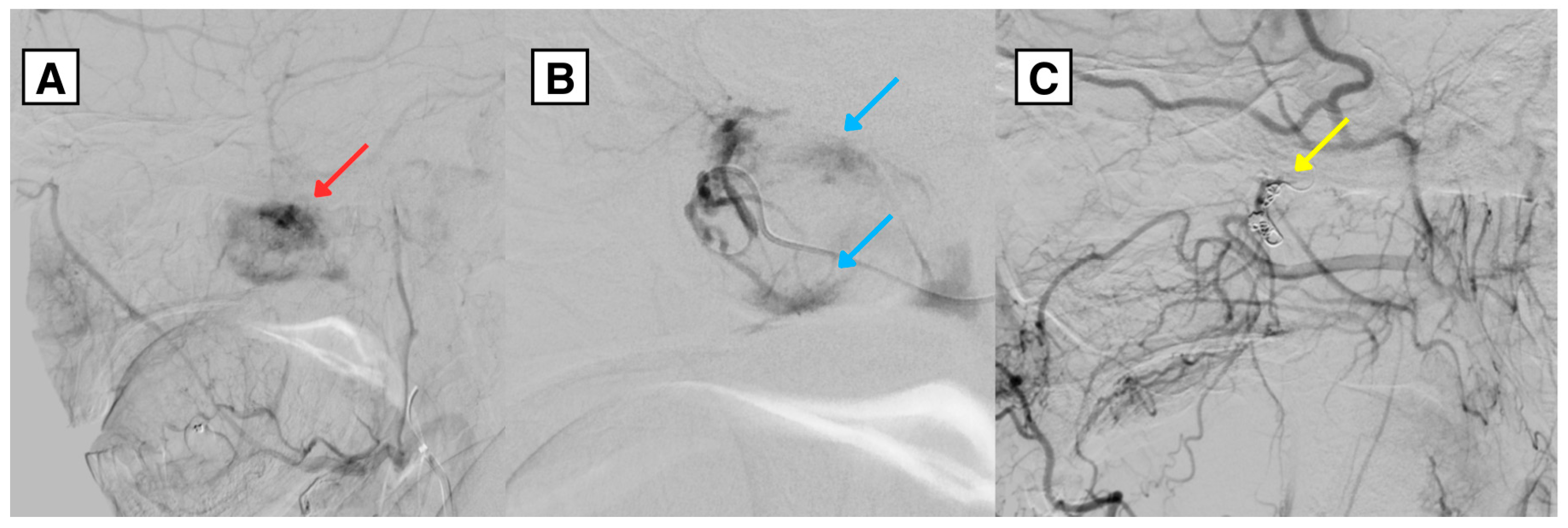
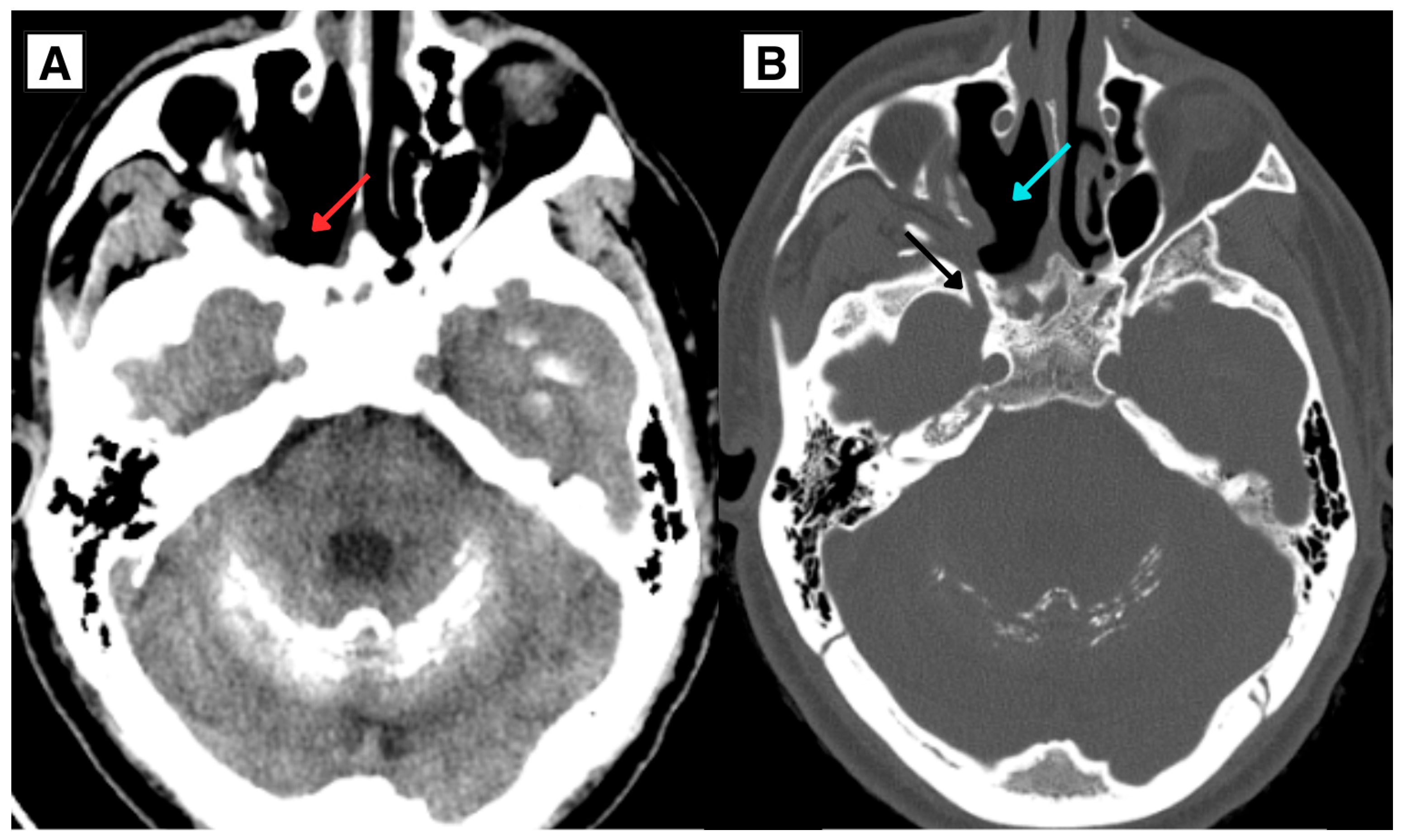
| This report (2026) | 34/M | Tracheostomy dysfunction with acute respiratory insufficiency; incidental nasopharyngeal mass; post-op hypoparathyroidism with chronic hypocalcemia and Fahr-like calcifications | MRI: 37 × 33 × 32 mm hypervascular mass with bone remodeling; right PPF/sphenopalatine region. DSA: right sphenopalatine feeders | Endovascular embolization + endoscopic resection | NR on FU imaging |
Disclaimer/Publisher’s Note: The statements, opinions and data contained in all publications are solely those of the individual author(s) and contributor(s) and not of MDPI and/or the editor(s). MDPI and/or the editor(s) disclaim responsibility for any injury to people or property resulting from any ideas, methods, instructions or products referred to in the content. |
© 2026 by the authors. Licensee MDPI, Basel, Switzerland. This article is an open access article distributed under the terms and conditions of the Creative Commons Attribution (CC BY) license.
Share and Cite
Zālīte, S.; Čudare, K.; Vērzemnieks, K.; Pavlovičs, S.; Kupčs, K.; Vilks, I.; Tone, T.; Briede, I.; Balodis, A. Juvenile Nasopharyngeal Angiofibroma in an Adult Patient: A Rare Presentation with Fahr Syndrome and Multiple Comorbidities—A Case Report and Literature Review. Diagnostics 2026, 16, 1327. https://doi.org/10.3390/diagnostics16091327
Zālīte S, Čudare K, Vērzemnieks K, Pavlovičs S, Kupčs K, Vilks I, Tone T, Briede I, Balodis A. Juvenile Nasopharyngeal Angiofibroma in an Adult Patient: A Rare Presentation with Fahr Syndrome and Multiple Comorbidities—A Case Report and Literature Review. Diagnostics. 2026; 16(9):1327. https://doi.org/10.3390/diagnostics16091327
Chicago/Turabian StyleZālīte, Sigita, Karīna Čudare, Kalvis Vērzemnieks, Sergejs Pavlovičs, Kārlis Kupčs, Ingus Vilks, Tatjana Tone, Inese Briede, and Arturs Balodis. 2026. "Juvenile Nasopharyngeal Angiofibroma in an Adult Patient: A Rare Presentation with Fahr Syndrome and Multiple Comorbidities—A Case Report and Literature Review" Diagnostics 16, no. 9: 1327. https://doi.org/10.3390/diagnostics16091327
APA StyleZālīte, S., Čudare, K., Vērzemnieks, K., Pavlovičs, S., Kupčs, K., Vilks, I., Tone, T., Briede, I., & Balodis, A. (2026). Juvenile Nasopharyngeal Angiofibroma in an Adult Patient: A Rare Presentation with Fahr Syndrome and Multiple Comorbidities—A Case Report and Literature Review. Diagnostics, 16(9), 1327. https://doi.org/10.3390/diagnostics16091327