Optical Coherence Tomography-Guided Versus Angiography-Guided PCI in Moderate-to-Severe Calcified Coronary Lesions: A Systematic Review and Meta-Analysis of Randomized Trials

 , , , , , , and
Abstract
1. Introduction
2. Materials and Methods
2.1. Registration
2.2. Search Strategies
2.3. Screening and Selection of Studies
2.4. Data Collection and Extraction
2.5. Risk-of-Bias Assessment
2.6. Certainty of Evidence Assessment
2.7. Statistical Analysis
3. Results
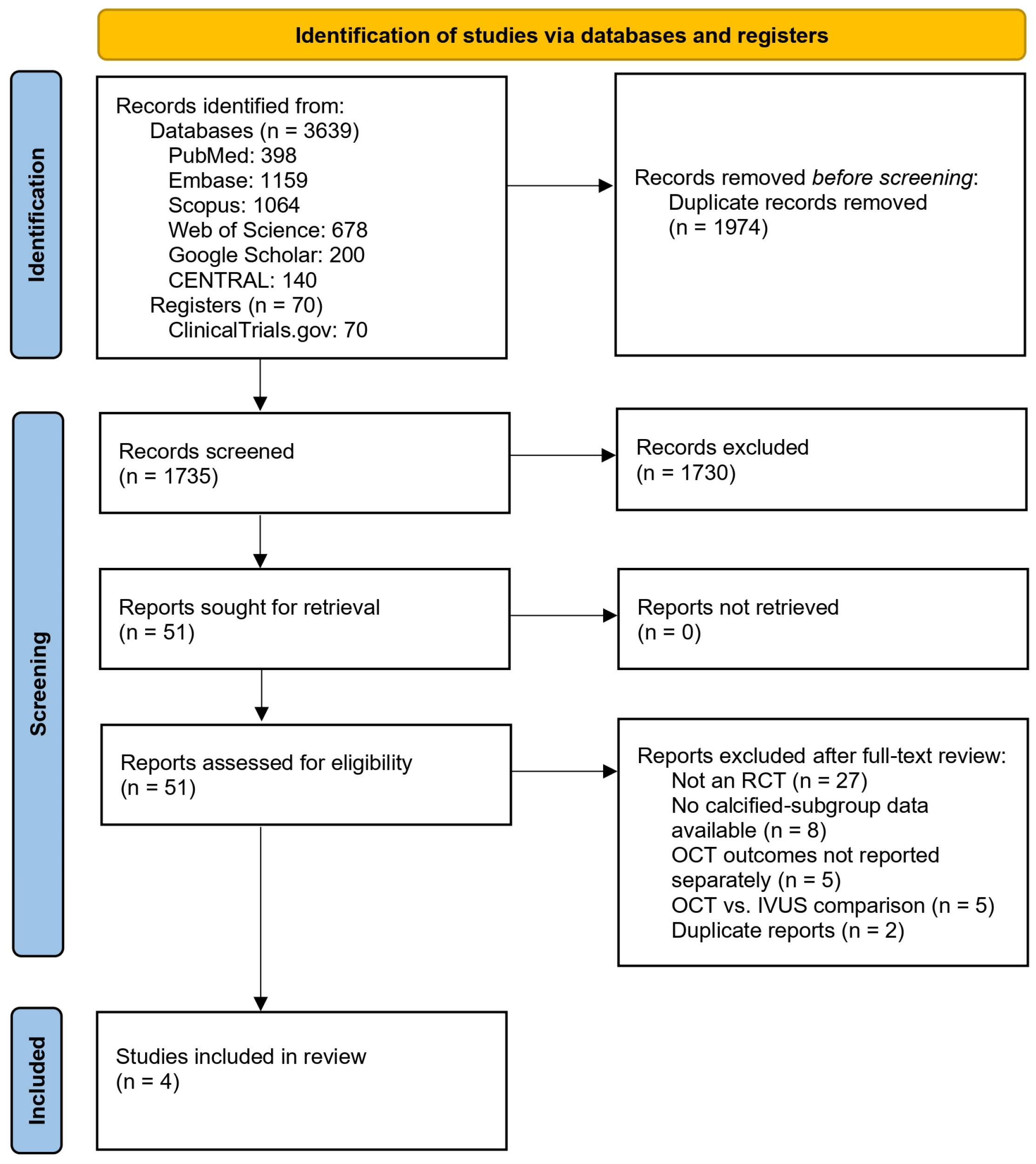
3.1. Study Selection
3.2. Characteristics of Included Studies
3.3. Risk-of-Bias Assessment
3.4. Clinical Outcomes
3.5. Imaging Outcomes
3.6. Procedural Outcomes
3.7. Certainty of Evidence Assessment
4. Discussion
5. Conclusions
Supplementary Materials
Author Contributions
Funding
Institutional Review Board Statement
Informed Consent Statement
Data Availability Statement
Acknowledgments
Conflicts of Interest
Abbreviations
| PCI | Percutaneous coronary intervention |
| MACE | Major adverse cardiovascular event |
| IVUS | Intravascular ultrasound |
| OCT | Optical coherence tomography |
| TVF | Target vessel failure |
| MSA | Minimal stent area |
| TV-MI | Target vessel myocardial infarction |
| ID-TVR | Ischemia-driven target vessel revascularization |
References
- Virani, S.S.; Alonso, A.; Benjamin, E.J.; Bittencourt, M.S.; Callaway, C.W.; Carson, A.P.; Chamberlain, A.M.; Chang, A.R.; Cheng, S.; Delling, F.N.; et al. Heart Disease and Stroke Statistics—2020 Update: A Report From the American Heart Association. Circulation 2020, 141, e139–e596. [Google Scholar] [CrossRef] [Scilit]
- Stark, B.A.; DeCleene, N.K.; Desai, E.C.; Hsu, J.M.; Johnson, C.O.; Lara-Castor, L.; LeGrand, K.E.; A, P.B.; Aalipour, M.A.; Aalruz, H.; et al. Global, Regional, and National Burden of Cardiovascular Diseases and Risk Factors in 204 Countries and Territories, 1990–2023. JACC 2025, 86, 2167–2243. [Google Scholar] [CrossRef] [Scilit] [PubMed]
- McInerney, A.; Hynes, S.O.; Gonzalo, N. Calcified Coronary Artery Disease: Pathology, Prevalence, Predictors and Impact on Outcomes. Interv. Cardiol. Rev. Res. Resour. 2025, 20, e02. [Google Scholar] [CrossRef] [Scilit]
- Sun, Z.; Xu, L. Coronary CT Angiography in the Quantitative Assessment of Coronary Plaques. Biomed Res. Int. 2014, 2014, 346380. [Google Scholar] [CrossRef] [Scilit]
- Rahman, M.; Nasir, A.; Ullah, I.; Adnan, G.; Farhad, A.; Khan, M. Comparison of Clinical Outcomes of Calcified and Non-Calcified Coronary Artery Lesion Intervention Under IVUS Guidance. J. Coll. Physicians Surg. Pak. 2023, 33, 1355–1360. [Google Scholar] [CrossRef] [Scilit] [PubMed]
- Golub, I.S.; Misic, A.; Schroeder, L.P.; Aldana-Bitar, J.; Krishnan, S.; Kianoush, S.; Benzing, T.; Ichikawa, K.; Budoff, M.J. Calcific Coronary Lesions: Management, Challenges, and a Comprehensive Review. AIMS Med. Sci. 2024, 11, 292–317. [Google Scholar] [CrossRef] [Scilit]
- Bourantas, C.V.; Zhang, Y.-J.; Garg, S.; Iqbal, J.; Valgimigli, M.; Windecker, S.; Mohr, F.W.; Silber, S.; de Vries, T.; Onuma, Y.; et al. Prognostic Implications of Coronary Calcification in Patients with Obstructive Coronary Artery Disease Treated by Percutaneous Coronary Intervention: A Patient-Level Pooled Analysis of 7 Contemporary Stent Trials. Heart 2014, 100, 1158–1164. [Google Scholar] [CrossRef] [Scilit] [PubMed]
- Généreux, P.; Madhavan, M.V.; Mintz, G.S.; Maehara, A.; Palmerini, T.; LaSalle, L.; Xu, K.; McAndrew, T.; Kirtane, A.; Lansky, A.J.; et al. Ischemic Outcomes After Coronary Intervention of Calcified Vessels in Acute Coronary Syndromes. J. Am. Coll. Cardiol. 2014, 63, 1845–1854. [Google Scholar] [CrossRef] [Scilit]
- Barbato, E.; Gallinoro, E.; Abdel-Wahab, M.; Andreini, D.; Carrié, D.; Di Mario, C.; Dudek, D.; Escaned, J.; Fajadet, J.; Guagliumi, G.; et al. Management Strategies for Heavily Calcified Coronary Stenoses: An EAPCI Clinical Consensus Statement in Collaboration with the EURO4C-PCR Group. Eur. Heart J. 2023, 44, 4340–4356. [Google Scholar] [CrossRef] [Scilit]
- Généreux, P.; Redfors, B.; Witzenbichler, B.; Arsenault, M.-P.; Weisz, G.; Stuckey, T.D.; Rinaldi, M.J.; Neumann, F.-J.; Christopher Metzger, D.; Henry, T.D.; et al. Two-Year Outcomes after Percutaneous Coronary Intervention of Calcified Lesions with Drug-Eluting Stents. Int. J. Cardiol. 2017, 231, 61–67. [Google Scholar] [CrossRef] [Scilit] [PubMed]
- Kostantinis, S.; Rempakos, A.; Simsek, B.; Karacsonyi, J.; Allana, S.S.; Alexandrou, M.; Gorgulu, S.; Alaswad, K.; Basir, M.B.; Davies, R.E.; et al. Impact of Calcium on the Procedural Techniques and Outcomes of Chronic Total Occlusion Percutaneous Coronary Intervention. Int. J. Cardiol. 2023, 390, 131254. [Google Scholar] [CrossRef] [Scilit] [PubMed]
- Tearney, G.J.; Regar, E.; Akasaka, T.; Adriaenssens, T.; Barlis, P.; Bezerra, H.G.; Bouma, B.; Bruining, N.; Cho, J.; Chowdhary, S.; et al. Consensus Standards for Acquisition, Measurement, and Reporting of Intravascular Optical Coherence Tomography Studies. J. Am. Coll. Cardiol. 2012, 59, 1058–1072. [Google Scholar] [CrossRef] [Scilit]
- Araki, M.; Park, S.-J.; Dauerman, H.L.; Uemura, S.; Kim, J.-S.; Di Mario, C.; Johnson, T.W.; Guagliumi, G.; Kastrati, A.; Joner, M.; et al. Optical Coherence Tomography in Coronary Atherosclerosis Assessment and Intervention. Nat. Rev. Cardiol. 2022, 19, 684–703. [Google Scholar] [CrossRef] [Scilit]
- Varghese, M.; Varghese, S.; Preethi, S. Revolutionizing Medical Imaging: A Comprehensive Review of Optical Coherence Tomography (OCT). J. Opt. 2025, 54, 1178–1195. [Google Scholar] [CrossRef] [Scilit]
- Mintz, G.S.; Popma, J.J.; Pichard, A.D.; Kent, K.M.; Satler, L.F.; Chien Chuang, Y.; DeFalco, R.A.; Leon, M.B. Limitations of Angiography in the Assessment of Plaque Distribution in Coronary Artery Disease. Circulation 1996, 93, 924–931. [Google Scholar] [CrossRef] [Scilit] [PubMed]
- Ali, Z.A.; Karimi Galougahi, K.; Mintz, G.S.; Maehara, A.; Shlofmitz, R.A.; Mattesini, A. Intracoronary Optical Coherence Tomography: State of the Art and Future Directions. EuroIntervention 2021, 17, e105–e123. [Google Scholar] [CrossRef] [Scilit]
- Mintz, G.S.; Popma, J.J.; Pichard, A.D.; Kent, K.M.; Satler, L.F.; Chuang, Y.C.; Ditrano, C.J.; Leon, M.B. Patterns of Calcification in Coronary Artery Disease. Circulation 1995, 91, 1959–1965. [Google Scholar] [CrossRef] [Scilit]
- Burhan, M.; Saeed, H.; Usama, M.; Tariq, A.; Shafiq, S.; Hurjkaliani, S.; Iqbal, M.; Shahid, S.; Khalid, S.; Tahirkheli, N.K. Optical Coherence Tomography-Guided Percutaneous Coronary Intervention Compared to Angiography-Guided Percutaneous Coronary Intervention for Complex Lesions. World J. Cardiol. 2025, 17, 110403. [Google Scholar] [CrossRef] [Scilit] [PubMed]
- Shuja, M.H.; Ahmed, M.; Hannat, R.; Khurram, L.; Hasnain Sheikh, H.A.; Shuja, S.H.; Raja, A.; Ahmed, J.; Soni, K.; Wani, S.A.; et al. Optical Coherence Tomography-Guided vs. Angiography-Guided Percutaneous Coronary Intervention for Complex Coronary Lesions: A Systematic Review and Meta-Analysis. Diagnostics 2025, 15, 1907. [Google Scholar] [CrossRef] [Scilit]
- Rao, S.V.; O’Donoghue, M.L.; Ruel, M.; Rab, T.; Tamis-Holland, J.E.; Alexander, J.H.; Baber, U.; Baker, H.; Cohen, M.G.; Cruz-Ruiz, M.; et al. 2025 ACC/AHA/ACEP/NAEMSP/SCAI Guideline for the Management of Patients with Acute Coronary Syndromes. JACC 2025, 85, 2135–2237. [Google Scholar] [CrossRef] [Scilit]
- Vrints, C.; Andreotti, F.; Koskinas, K.C.; Rossello, X.; Adamo, M.; Ainslie, J.; Banning, A.P.; Budaj, A.; Buechel, R.R.; Chiariello, G.A.; et al. 2024 ESC Guidelines for the Management of Chronic Coronary Syndromes. Eur. Heart J. 2024, 45, 3415–3537. [Google Scholar] [CrossRef] [Scilit]
- Carvalho, P.E.P.; Antunes, V.L.J.; Bittar de Pontes, V.; Gomes, W.F.; Polachini Assunes Goncalves, B.; Caixeta, A.; Strepkos, D.; Alexandrou, M.; Mutlu, D.; Gibson, C.M.; et al. IVUS, OCT, or Angiography as Guidance for PCI in Complex Coronary Artery Lesions. JACC Cardiovasc. Interv. 2026, 19, 31–43. [Google Scholar] [CrossRef] [Scilit]
- Stone, G.W.; Genereux, P.; Maehara, A.; Lewis, B.E.; Shlofmitz, R.A.; Dohad, S.; Choudary, J.; Dahle, T.; Pineda, A.M.; Shunk, K.; et al. Intravascular Imaging vs Angiography Guidance for PCI of Severely Calcified Lesions. JACC Cardiovasc. Interv. 2025, 18, 2338–2351. [Google Scholar] [CrossRef] [Scilit]
- Amabile, N.; Rangé, G.; Landolff, Q.; Bressollette, E.; Meneveau, N.; Lattuca, B.; Levesque, S.; Boueri, Z.; Adjedj, J.; Casassus, F.; et al. OCT vs Angiography for Guidance of Percutaneous Coronary Intervention of Calcified Lesions. JAMA Cardiol. 2025, 10, 666. [Google Scholar] [CrossRef] [Scilit]
- Ali, Z.A.; Shin, D.; Vijayvergiya, R.; Gawalkar, A.A.; Shlofmitz, R.A.; Alfonso, F.; Calligaris, G.; Canova, P.; Sakai, K.; Price, M.J.; et al. Optical Coherence Tomography-vs Angiography-Guided Coronary Stent Implantation in Calcified Lesions: The ILUMIEN IV Trial. Eur. Heart J. 2025, 46, 3201–3210. [Google Scholar] [CrossRef] [Scilit]
- Page, M.J.; McKenzie, J.E.; Bossuyt, P.M.; Boutron, I.; Hoffmann, T.C.; Mulrow, C.D.; Shamseer, L.; Tetzlaff, J.M.; Akl, E.A.; Brennan, S.E.; et al. The PRISMA 2020 Statement: An Updated Guideline for Reporting Systematic Reviews. BMJ 2021, 372, n71. [Google Scholar] [CrossRef] [Scilit] [PubMed]
- Higgins, J.P.T.; Thomas, J.; Chandler, J.; Cumpston, M.; Li, T.; Page, M.J.; Welch, V.A. (Eds.) Cochrane Handbook for Systematic Reviews of Interventions; Wiley: New York, NY, USA, 2019; ISBN 9781119536628. [Google Scholar]
- Sterne, J.A.C.; Savović, J.; Page, M.J.; Elbers, R.G.; Blencowe, N.S.; Boutron, I.; Cates, C.J.; Cheng, H.-Y.; Corbett, M.S.; Eldridge, S.M.; et al. RoB 2: A Revised Tool for Assessing Risk of Bias in Randomised Trials. BMJ 2019, 366, l4898. [Google Scholar] [CrossRef] [Scilit]
- McGuinness, L.A.; Higgins, J.P.T. Risk-of-bias VISualization (Robvis): An R Package and Shiny Web App for Visualizing Risk-of-bias Assessments. Res. Synth. Methods 2021, 12, 55–61. [Google Scholar] [CrossRef] [Scilit]
- Schünemann, H.; Brożek, J.; Guyatt, G.; Oxman, A. (Eds.) GRADE Handbook; Updated October 2013; The GRADE Working Group: Hamilton, ON, Canada, 2013; Available online: https://gdt.gradepro.org/app/handbook/handbook.html (accessed on 20 December 2025).
- McMaster University; Evidence Prime. GRADEpro GDT; Evidence Prime: Hamilton, ON, Canada, 2025; Available online: https://www.gradepro.org/ (accessed on 20 December 2025).
- Wan, X.; Wang, W.; Liu, J.; Tong, T. Estimating the Sample Mean and Standard Deviation from the Sample Size, Median, Range and/or Interquartile Range. BMC Med. Res. Methodol. 2014, 14, 135. [Google Scholar] [CrossRef] [Scilit]
- Abbas, A.; Hefnawy, M.T.; Negida, A. Meta-Analysis Accelerator: A Comprehensive Tool for Statistical Data Conversion in Systematic Reviews with Meta-Analysis. BMC Med. Res. Methodol. 2024, 24, 243. [Google Scholar] [CrossRef] [Scilit]
- Holm, N.R.; Andreasen, L.D.; Neghabat, O.; Laanmets, P.; Kumsars, I.; Bennett, J.; Olsen, N.T.; Odenstedt, J.; Hoffmann, P.; Dens, J.; et al. OCT or Angiography Guidance for PCI in Complex Bifurcation Lesions. N. Engl. J. Med. 2023, 389, 1477–1487. [Google Scholar] [CrossRef] [Scilit]
- Shin, D.; Hong, D.; Singh, M.; Lee, S.H.; Sakai, K.; Dakroub, A.; Malik, S.; Maehara, A.; Shlofmitz, E.; Stone, G.W.; et al. Intravascular Imaging-Guided Percutaneous Coronary Intervention for Heavily Calcified Coronary Lesions: A Systematic Review and Meta-Analysis. Int. J. Cardiovasc. Imaging 2024, 40, 1653–1659. [Google Scholar] [CrossRef] [Scilit]
- Fujii, K.; Carlier, S.G.; Mintz, G.S.; Yang, Y.; Moussa, I.; Weisz, G.; Dangas, G.; Mehran, R.; Lansky, A.J.; Kreps, E.M.; et al. Stent Underexpansion and Residual Reference Segment Stenosis Are Related to Stent Thrombosis after Sirolimus-Eluting Stent Implantation. J. Am. Coll. Cardiol. 2005, 45, 995–998. [Google Scholar] [CrossRef] [Scilit]
- Kim, J.H.; Kang, D.; Ahn, J.; Kweon, J.; Chae, J.; Wee, S.-B.; An, S.Y.; Park, H.; Kang, S.-J.; Park, D.; et al. Optimal Minimal Stent Area after Crossover Stenting in Patients with Unprotected Left Main Coronary Artery Disease. EuroIntervention 2025, 21, e1069–e1080. [Google Scholar] [CrossRef] [Scilit] [PubMed]
- Fujino, A.; Mintz, G.S.; Matsumura, M.; Lee, T.; Kim, S.-Y.; Hoshino, M.; Usui, E.; Yonetsu, T.; Haag, E.S.; Shlofmitz, R.A.; et al. A New Optical Coherence Tomography-Based Calcium Scoring System to Predict Stent Underexpansion. EuroIntervention 2018, 13, 2182–2189. [Google Scholar] [CrossRef] [Scilit] [PubMed]
- Fujino, A.; Mintz, G.S.; Lee, T.; Hoshino, M.; Usui, E.; Kanaji, Y.; Murai, T.; Yonetsu, T.; Matsumura, M.; Ali, Z.A.; et al. Predictors of Calcium Fracture Derived from Balloon Angioplasty and Its Effect on Stent Expansion Assessed by Optical Coherence Tomography. JACC Cardiovasc. Interv. 2018, 11, 1015–1017. [Google Scholar] [CrossRef] [Scilit]
- Wulandari, A.P.; Pintaningrum, Y.; Adheriyani, R. Outcomes of Optical Coherence Tomography Guided Percutaneous Coronary Intervention against Angiography Guided in Patients with Coronary Artery Disease: A Systematic Reviews and Meta-Analyses. Indian Heart J. 2023, 75, 108–114. [Google Scholar] [CrossRef] [Scilit]
- Hong, S.-J.; Lee, S.-J.; Lee, S.-H.; Lee, J.-Y.; Cho, D.-K.; Kim, J.W.; Kim, S.M.; Hur, S.-H.; Heo, J.H.; Jang, J.-Y.; et al. Optical Coherence Tomography-Guided versus Angiography-Guided Percutaneous Coronary Intervention for Patients with Complex Lesions (OCCUPI): An Investigator-Initiated, Multicentre, Randomised, Open-Label, Superiority Trial in South Korea. Lancet 2024, 404, 1029–1039. [Google Scholar] [CrossRef] [Scilit] [PubMed]
- Ali, Z.A.; Landmesser, U.; Maehara, A.; Matsumura, M.; Shlofmitz, R.A.; Guagliumi, G.; Price, M.J.; Hill, J.M.; Akasaka, T.; Prati, F.; et al. Optical Coherence Tomography–Guided versus Angiography-Guided PCI. N. Engl. J. Med. 2023, 389, 1466–1476. [Google Scholar] [CrossRef] [Scilit]
- Ali, Z.A.; Karimi Galougahi, K.; Maehara, A.; Shlofmitz, R.A.; Fabbiocchi, F.; Guagliumi, G.; Alfonso, F.; Akasaka, T.; Matsumura, M.; Mintz, G.S.; et al. Outcomes of Optical Coherence Tomography Compared with Intravascular Ultrasound and with Angiography to Guide Coronary Stent Implantation: One-Year Results from the ILUMIEN III: OPTIMIZE PCI Trial. EuroIntervention 2021, 16, 1085–1091. [Google Scholar] [CrossRef] [Scilit]
- Chandrasekhar, J.; Baber, U.; Sartori, S.; Stefanini, G.G.; Sarin, M.; Vogel, B.; Farhan, S.; Camenzind, E.; Leon, M.B.; Stone, G.W.; et al. Effect of Increasing Stent Length on 3-Year Clinical Outcomes in Women Undergoing Percutaneous Coronary Intervention with New-Generation Drug-Eluting Stents. JACC Cardiovasc. Interv. 2018, 11, 53–65. [Google Scholar] [CrossRef] [Scilit] [PubMed]
- Li, M.; Hou, J.; Gu, X.; Weng, R.; Zhong, Z.; Liu, S. Incidence and Risk Factors of In-Stent Restenosis after Percutaneous Coronary Intervention in Patients from Southern China. Eur. J. Med. Res. 2022, 27, 12. [Google Scholar] [CrossRef] [Scilit] [PubMed]
- Costa, H.; Silva, M.; Santo, M.E.; Mota, T.; Fernandes, R.; Palmeiro, H.; Marto, S.; Franco, P.; Carvalho, D.; Bispo, J.; et al. Contrast-Induced Nephropathy Following PCI: Can We Calculate a Safe Contrast Volume? Eur. Heart J. 2022, 43, ehac544.1403. [Google Scholar] [CrossRef] [Scilit]





| Trial (Year) | Geography | Inclusion | Calcified Lesion Population | Total Patients, n | Moderate-to-Severe Calcified Lesions, n | Primary Endpoints | Definition of TVF/MACE | |
|---|---|---|---|---|---|---|---|---|
| OCT Group | Angiography Group | |||||||
| OCTOBER (2023) [34] | Europe | True bifurcation lesions | Subgroup only | 1201 | 198 | 194 | MACE | MACE *: cardiac death, target-lesion myocardial infarction, or ischemia-driven target lesion revascularization |
| CALIPSO (2025) [24] | France | Moderate-to-severe calcified lesions | All patients | 134 | 65 | 69 | MSA | MACE: cardiovascular death, any myocardial infarction, or need for clinically driven reintervention on the target lesion |
| ECLIPSE (2025) [23] | US | Severe calcified lesions | All patients | 2005 | 819 | 759 | MSA, TVF | TVF: cardiac death, TV-MI, or ID-TVR |
| ILUMIEN IV (2025) [25] | US, Canada, Europe, Asia | Diabetic patients or complex lesions | Subgroup only | 2487 | 544 | 538 | MSA, TVF | MACE: cardiac death, TV-MI, or definite or probable stent thrombosis TVF: cardiac death, TV-MI, or ID-TVR |
| Trial (Year) | Definition of Moderate-to-Severe Calcified Lesions | Operator Experience | OCT Protocol (Pre-PCI) | OCT Protocol (Post-PCI) | Follow-Up Period | |
|---|---|---|---|---|---|---|
| Angiographic Severity | Angiographic Definition | |||||
| OCTOBER (2023) [34] | Moderate or severe | Radiopacity with contrast (moderate) or without contrast (severe) in the cardiac cycle before contrast injection | The majority of sites that enrolled patients were university centers, and most investigators had at least moderate experience with OCT guidance for PCI. | The protocol specified the timing of OCT scans, the checks and measurements to be obtained at each time point, the recommended optimizations, and the treatment goals. | The main treatment goals were optimal coverage of the coronary artery lesion with a stent, optimal stent expansion, and absence of stent malapposition or accidentally crushed or distorted stents. | 2 years |
| CALIPSO (2025) [24] | Moderate or severe | Type B or C by Mintz classification | Not reported. | An initial OCT run was acquired, and the plaque preparation was guided by a predefined algorithm based on the maximal calcium arc extension for choice of the plaque. | Results were assessed by control OCT, and potential optimization steps were proposed according to predefined success criteria in terms of stent expansion, strut apposition, and presence of dissection based on the EAPCI consensus document. | 30 days |
| ECLIPSE (2025) [23] | Severe | Radiopacities noted without cardiac motion involving both sides of the arterial wall, with total calcium length ≥ 15 mm extending into the target lesion | Not reported. | The protocol did not favor a specific IVI strategy or specify a target imaging goal; sites were asked to follow their usual, standard procedures for IVI use and stent optimization. | Selected sites were invited to participate in a voluntary OCT substudy to assess stent expansion in approximately 500 randomized patients. | 1 year |
| ILUMIEN IV (2025) [25] | Moderate or severe | Radio-opacities seen only during the cardiac cycle (moderate) or without cardiac motion generally on both sides of the arterial lumen (severe) | Operators were experienced in OCT guided PCI and underwent mandatory training. | OCT was used to assess plaque morphology, stent size, and proximal and distal reference segments. Stent size was determined based on the mean external elastic lamina or the mean lumen-based vessel diameter. | OCT was performed, and post dilatation was conducted to ensure adequate stent expansion. Additional stenting was recommended in cases of major edge dissection or residual focal inflow/outflow disease in the reference segments. | 30 days and 2 years |
| Outcomes | No. of Studies | Participants | Effect Estimate (95% CI) | p-Value | Heterogeneity (I2) |
|---|---|---|---|---|---|
| Clinical outcomes | |||||
| TVF | 3 | 2966 | RR 0.66 (0.52 to 0.82) | <0.001 | 0% |
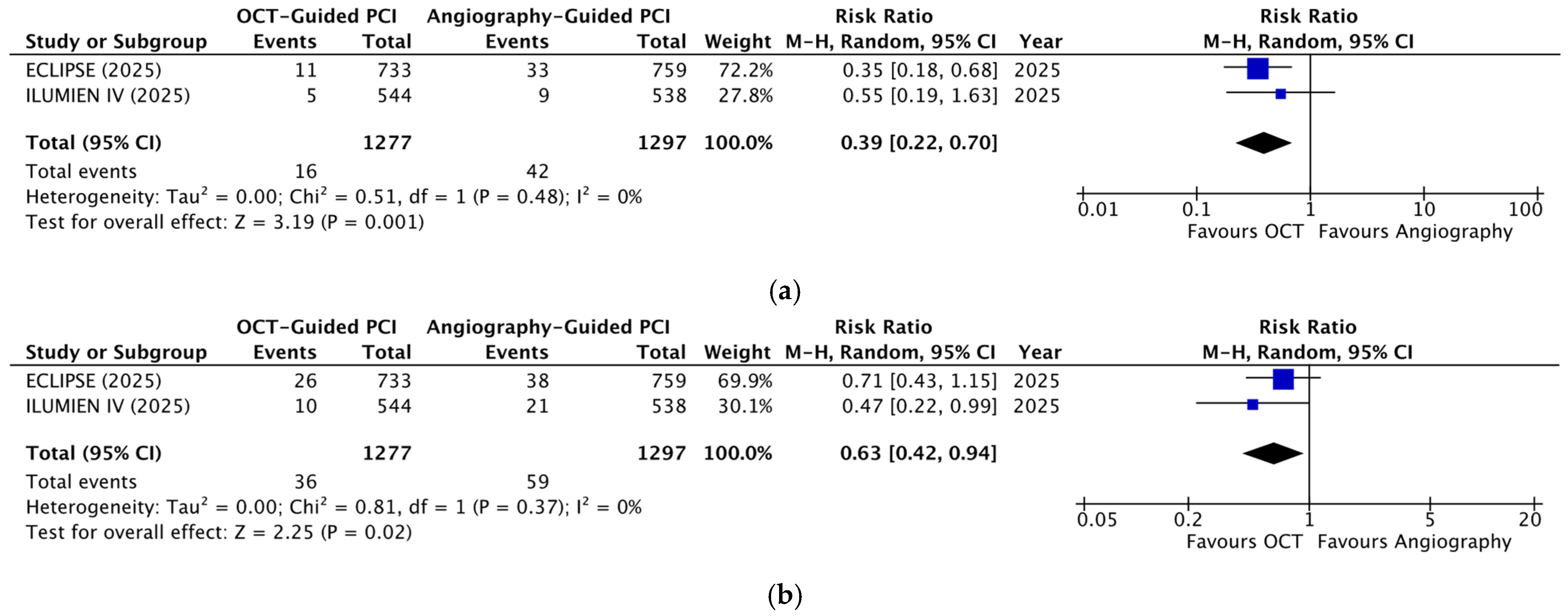
| Cardiac death | 2 | 2574 | RR 0.39 (0.22 to 0.70) | 0.001 | 0% |
| ID-TVR | 2 | 2574 | RR 0.77 (0.55 to 1.08) | 0.12 | 0% |
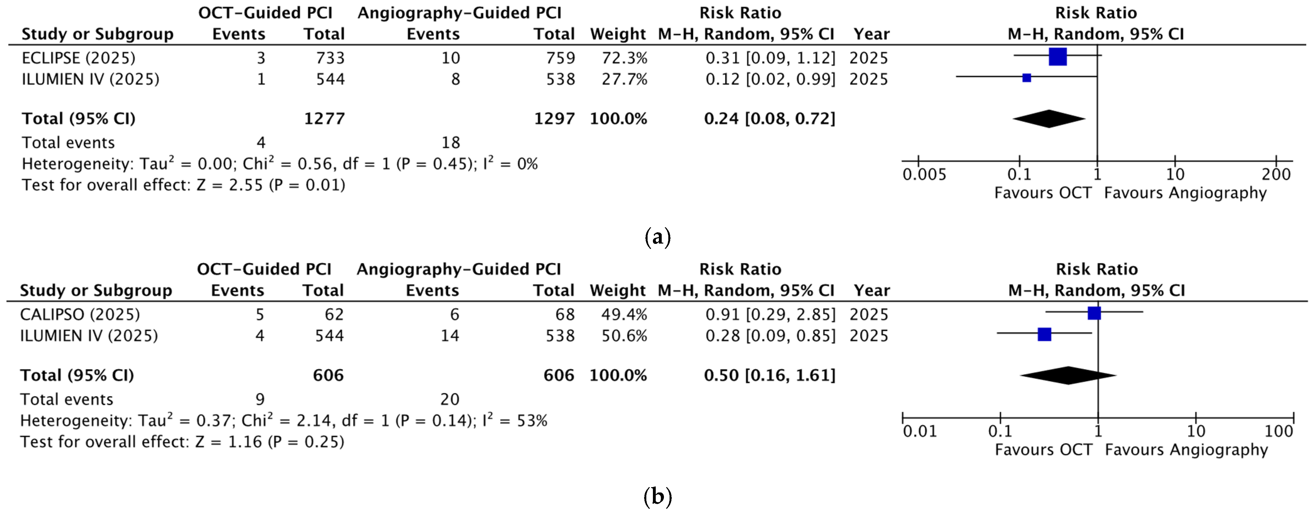
| 30-day MACEs | 2 | 1212 | RR 0.50 (0.16 to 1.61) | 0.25 | 53% |
| Stent thrombosis | 2 | 2574 | RR 0.24 (0.08 to 0.72) | 0.01 | 0% |
| TV-MI | 2 | 2574 | RR 0.63 (0.42 to 0.94) | 0.02 | 0% |
| Imaging outcomes | |||||
| Edge Dissections | 2 | 1214 | RR 0.76 (0.65 to 0.89) | <0.001 | 0% |
| Major malapposition | 2 | 1216 | RR 0.57 (0.40 to 0.81) | 0.002 | 61% |
| MSA (mm2) | 2 | 1216 | MD 0.91 (−0.45 to 2.27) | 0.19 | 95% |
| Procedural outcomes | |||||
| Contrast volume (mL) | 3 | 2708 | MD 25.68 (11.46 to 39.89) | <0.001 | 77% |
| Fluoroscopy duration (min) | 3 | 2708 | MD 1.69 (−0.54 to 3.92) | 0.14 | 79% |
| Maximum inflation pressure (atm) | 3 | 2856 | MD 0.00 (−1.32 to 1.33) | 1.00 | 95% |
| No. of balloons | 2 | 1216 | MD 0.17 (−0.12 to 0.46) | 0.26 | 74% |
| Procedure duration (min) | 3 | 2708 | MD 11.93 (6.70 to 17.15) | <0.001 | 70% |
| Radiation dose | 2 | 2574 | SMD 0.10 (−0.14 to 0.35) | 0.42 | 90% |
| Total stent length (mm) | 3 | 2856 | MD 3.79 (1.67 to 5.92) | <0.001 | 47% |
Disclaimer/Publisher’s Note: The statements, opinions and data contained in all publications are solely those of the individual author(s) and contributor(s) and not of MDPI and/or the editor(s). MDPI and/or the editor(s) disclaim responsibility for any injury to people or property resulting from any ideas, methods, instructions or products referred to in the content. |
© 2026 by the authors. Licensee MDPI, Basel, Switzerland. This article is an open access article distributed under the terms and conditions of the Creative Commons Attribution (CC BY) license.
Share and Cite
Mawar, H.E.; Baamer, M.; Althagafi, A.A.; Alghamdi, A.G.; Aleidi, M.; Alzahrani, R.S.; Alnamlah, A.; Bokhari, M.F.; Batawi, A.; Gholam, M.F.; et al. Optical Coherence Tomography-Guided Versus Angiography-Guided PCI in Moderate-to-Severe Calcified Coronary Lesions: A Systematic Review and Meta-Analysis of Randomized Trials. Diagnostics 2026, 16, 1317. https://doi.org/10.3390/diagnostics16091317
Mawar HE, Baamer M, Althagafi AA, Alghamdi AG, Aleidi M, Alzahrani RS, Alnamlah A, Bokhari MF, Batawi A, Gholam MF, et al. Optical Coherence Tomography-Guided Versus Angiography-Guided PCI in Moderate-to-Severe Calcified Coronary Lesions: A Systematic Review and Meta-Analysis of Randomized Trials. Diagnostics. 2026; 16(9):1317. https://doi.org/10.3390/diagnostics16091317
Chicago/Turabian StyleMawar, Hesham E., Maryam Baamer, Azzam A. Althagafi, Ahmad G. Alghamdi, Moudi Aleidi, Reem S. Alzahrani, Abdulrahman Alnamlah, Maya F. Bokhari, Amjaad Batawi, Mohammed F. Gholam, and et al. 2026. "Optical Coherence Tomography-Guided Versus Angiography-Guided PCI in Moderate-to-Severe Calcified Coronary Lesions: A Systematic Review and Meta-Analysis of Randomized Trials" Diagnostics 16, no. 9: 1317. https://doi.org/10.3390/diagnostics16091317
APA StyleMawar, H. E., Baamer, M., Althagafi, A. A., Alghamdi, A. G., Aleidi, M., Alzahrani, R. S., Alnamlah, A., Bokhari, M. F., Batawi, A., Gholam, M. F., & Al Bugami, S. (2026). Optical Coherence Tomography-Guided Versus Angiography-Guided PCI in Moderate-to-Severe Calcified Coronary Lesions: A Systematic Review and Meta-Analysis of Randomized Trials. Diagnostics, 16(9), 1317. https://doi.org/10.3390/diagnostics16091317