Review

Beyond One-Size-Fits-All Active Surveillance for Low-Risk Prostate Cancer: Risk-Adapted Follow-Up, De-Escalation Pathways, and Focal Therapy as Tailored Strategy

by
Fabio Zattoni
1,*,†,
Andrea Mari
2,†,
Ugo Giovanni Falagario
3,4,†,
Riccardo Giuseppe Bertolo
5,†,
Simone Albisinni
6,†,
Daniele Amparore
7,8,†,
Lorenzo Bianchi
9,†,
Riccardo Campi
10,11,†,
Roberto Contieri
12,†,
Elisa De Lorenzis
13,14,†,
Paolo Dell’Oglio
15,†,
Michele Marchioni
16,17,†,
Veronica Mollica
18,†,
Marco Moschini
19,†,
Francesco Soria
20,†,
Michele Talso
21,†,
Filippo Turri
22,† and
Savio Domenico Pandolfo
23,24,† the Uronauti Group
1
Urology Clinic, Department of Surgery, Oncology, and Gastroenterology, University of Padua, 35128 Padua, Italy
2
Department of Experimental and Clinical Medicine, University of Florence—Unit of Oncologic Minimally-Invasive Urology and Andrology, Careggi Hospital, 50134 Florence, Italy
3
Department of Molecular Medicine and Surgery, Karolinska Institute, SE-171 76 Stockholm, Sweden
4
Department of Urology, University of Foggia, 71122 Foggia, Italy
5
Department of Urology, University of Verona, A.O.U.I., 37126 Verona, Italy
6
Department of Urology, University of Tor Vergata, 00133 Rome, Italy
7
Department of Oncology, University of Turin, Orbassano, 10043 Turin, Italy
8
Division of Urology, Department of Surgery, Candiolo Cancer Institute, FPO-IRCCS, 10060 Candiolo (TO), Italy
9
Division of Urology, IRCCS Azienda Ospedaliero-Universitaria di Bologna, 40138 Bologna, Italy
10
Unit of Urology and Renal Transplantation, Department of Oncology, Careggi University Hospital, 50134 Florence, Italy
11
Department of Experimental and Clinical Medicine, University of Florence, 50134 Florence, Italy
12
Department of Urology, Istituto Nazionale Tumori “Fondazione Pascale”, 80131 Naples, Italy
13
Department of Urology, IRCCS Foundation Cà Granda—Ospedale Maggiore Policlinico, 20122 Milan, Italy
14
Department of Clinical Sciences and Community Health, University of Milan, 20122 Milan, Italy
15
Department of Urology, ASST Grande Ospedale Metropolitano Niguarda, 20162 Milan, Italy
16
UniCamillus—Saint Camillus International University of Health Sciences, 00131 Rome, Italy
17
Department of Urology, Leonardo Foundation, 35037 Abano Terme, Italy
18
Medical Oncology, IRCCS Azienda Ospedaliero-Universitaria di Bologna, 40138 Bologna, Italy
19
Division of Experimental Oncology, Department of Urology, Urological Research Institute, Vita-Salute San Raffaele University, 20132 Milan, Italy
20
Division of Urology, Department of Surgical Sciences—San Giovanni Battista Hospital, University of Studies of Torino, 10126 Turin, Italy
21
ASST Fatebenefratelli—Sacco, Ospedale Fatebenefratelli, 20121 Milan, Italy
22
Department of Urology, Fondazione Policlinico Universitario Agostino Gemelli IRCCS, Università Cattolica del Sacro Cuore, 00168 Rome, Italy
23
Department of Life, Health and Environmental Sciences, University of L’Aquila, 67100 L’Aquila, Italy
24
Department of Neurosciences, Reproductive Sciences and Odontostomatology, University of Naples “Federico II”, 80131 Naples, Italy
*
Author to whom correspondence should be addressed.
Membership of the Uronauti Group is provided in the Supplementary Materials.
Diagnostics 2026, 16(9), 1310; https://doi.org/10.3390/diagnostics16091310
Submission received: 22 February 2026 / Revised: 15 April 2026 / Accepted: 23 April 2026 / Published: 27 April 2026
(This article belongs to the Special Issue Clinical Diagnosis and Management in Urology)

Abstract

Low-risk prostate cancer (PCa) has historically been overtreated, exposing men to unnecessary morbidity. Emerging evidence supports conservative management of low-risk PCa without immediate radical intervention. Contemporary data show a marked decline in surgical overtreatment, with the proportion of radical prostatectomies yielding only Grade Group 1 cancers falling from 32.4% in 2010 to 7.8% in 2020 in the US SEER registry. Long-term studies confirm that deferring treatment is safe for low-risk disease, with PCa-specific survival exceeding 95% at 15–25 years for cohorts managed with surveillance. Major guidelines now endorse active surveillance (AS) as the preferred management for low-risk PCa. An alternative risk stratification system that expands the low-risk category was shown to reclassify 45–83% more men as low risk without increasing 15-year PCa mortality. Focal therapy has emerged as a potential middle-ground strategy, though evidence is still limited. The paradigm for managing low-risk PCa has shifted toward conservatism, with AS firmly established as the standard of care. Continued efforts to refine risk stratification and evaluate focal therapy are needed to further optimize individualized care, minimize harm, and maintain excellent cancer-specific outcomes for low-risk PCa. This comprehensive review aims to create a practical, risk-adapted framework for managing patients on AS. We will: (i) summarize inclusion criteria and outcomes, (i) compare AS follow-up schedules across major institutions and guidelines, (iii) provide evidence-based criteria to de-intensify surveillance in men with sustained stability and (iv) clarify the role of focal therapy as an intermediate treatment option within the AS continuum.

1. Introduction

Prostate cancer (PCa) remains a major cause of morbidity and can lead to premature death, particularly when clinically significant disease is diagnosed late. At the same time, the global burden is expected to increase substantially over the coming decades, largely driven by population ageing and growth, with forecasts suggesting a marked rise in new cases by 2040 [1].
Long-term randomized evidence indicates that organized PSA-based screening can reduce prostate cancer mortality, although at the cost of overdiagnosis and overtreatment when applied indiscriminately [2].
Low-risk PCa—typically defined as Gleason Grade Group 1 (GG1) (Gleason 3 + 3), PSA <10 ng/mL, and clinical stage T1–T2a—often follows an indolent course. Yet in past decades, many men with low-risk tumours received immediate radical prostatectomy or radiation. Such interventions provide minimal survival benefit in low-grade disease but can lead to substantial long-term side effects. Radical prostatectomy may cause urinary incontinence and erectile dysfunction [3,4,5], while radiation therapy (RT) can result in urinary and gastrointestinal complications, including radiation cystitis, chronic radiation proctitis, and sexual dysfunction. The recognition that many PCa are clinically insignificant drove a movement to reduce unnecessary treatment. Active surveillance (AS)—deferring definitive therapy while monitoring for signs of progression—has emerged as a strategy to preserve quality of life without compromising survival for low-risk patients [6].
Across contemporary international guidance, AS remains the preferred first-line strategy for men with GG1 PCa who have an appropriate life expectancy. The AUA/ASTRO guideline for clinically localized disease explicitly frames AS as the preferred management option in low-risk PCa, reinforcing its role as the standard of care rather than an “alternative” to treatment.
The EAU 2025 guideline similarly positions AS as a recommended option for ISUP/GG1 when other low-risk clinical features are present, aligning European practice with risk-adapted de-escalation.
Importantly, all major frameworks acknowledge that AS can be appropriate beyond GG1 in carefully selected favourable-intermediate presentations, provided adverse biological and pathological features are absent and disease burden is low.
Consistent with this, both EAU 2025 and NCCN v1.2025 support consideration of AS in selected favourable-intermediate cases—typically low-volume GG2/pattern 4 and otherwise favourable clinical parameters—while emphasizing rigorous selection and close follow-up to mitigate the higher progression risk relative to GG1.
With the present comprehensive review, we will summarize inclusion criteria and outcomes, biopsy follow-up schedules across major institutions and guidelines, discuss evidence-based criteria to de-intensify surveillance in men with sustained stability, and clarify the role of focal therapy as an intermediate treatment option within the AS continuum.

2. Materials and Methods

A comprehensive review was conducted by searching major databases, including PubMed/MEDLINE, Embase, Scopus, and the Cochrane Library, encompassing publications from January 2014 to September 2025. The aim of this approach was to provide a clinically oriented overview of the evolving management of low-risk prostate cancer.

2.1. Search Strategy and Study Selection

The search was structured around key terms pertinent to the objectives of the review. These included “low-risk prostate cancer”, “active surveillance”, “risk stratification”, “MRI”, “biomarkers”, “focal therapy”, and “overtreatment”. These keywords were selected to ensure a broad and comprehensive retrieval of relevant literature covering patient selection, diagnostic pathways, surveillance strategies, and emerging treatment approaches. Eligible items addressed localized low-risk prostate cancer (e.g., Grade Group 1, PSA ≤ 10 ng/mL, cT1–T2a or equivalent definitions) and included clinical trials, cohort studies, registry analyses, diagnostic studies, major guideline documents, and high-quality reviews. Non-English reports, animal studies, and small case series (<10 patients) were excluded.
Our review process aimed to include studies providing clinically meaningful insights into the management of localized low-risk prostate cancer, including randomized trials, large cohort studies, registry-based analyses, and major guideline documents.
To ensure a thorough and balanced selection, additional articles were identified through manual screening of reference lists from key studies, landmark trials, and relevant review articles.
Data extraction was performed independently by two authors, with a predefined strategy to resolve any discrepancies. In cases of disagreement, a third author was consulted to reach consensus. This collaborative approach ensured consistency and reliability in the interpretation of the evidence included in this review.

2.2. Data Synthesis

This review summarizes emerging trends and prospects in low-risk PCa. We searched MEDLINE/PubMed, Embase, Scopus, and the Cochrane Library from January 2014 to September 2025, supplemented by hand-searching references of key papers and guidelines. Eligible items addressed localized low-risk disease (e.g., GG1, PSA ≤ 10 ng/mL, cT1–T2a or near-equivalents) and included trials, cohort studies/registries, diagnostic studies, major guidelines, and high-quality reviews; non-English reports, animal studies, and case series (<10 patients) were excluded. The retrieved evidence was narratively synthesized to provide a clinically grounded overview of the current landscape of low-risk prostate cancer management, highlighting inclusion criteria for active surveillance, follow-up strategies, de-escalation pathways, and the emerging role of focal therapy. Evidence summaries were critically discussed and refined within a multidisciplinary expert panel (Uronauti group) to ensure a practice-oriented interpretation of the available data. Formal quantitative analysis was not performed. A detailed search strategy is reported in Table 1.
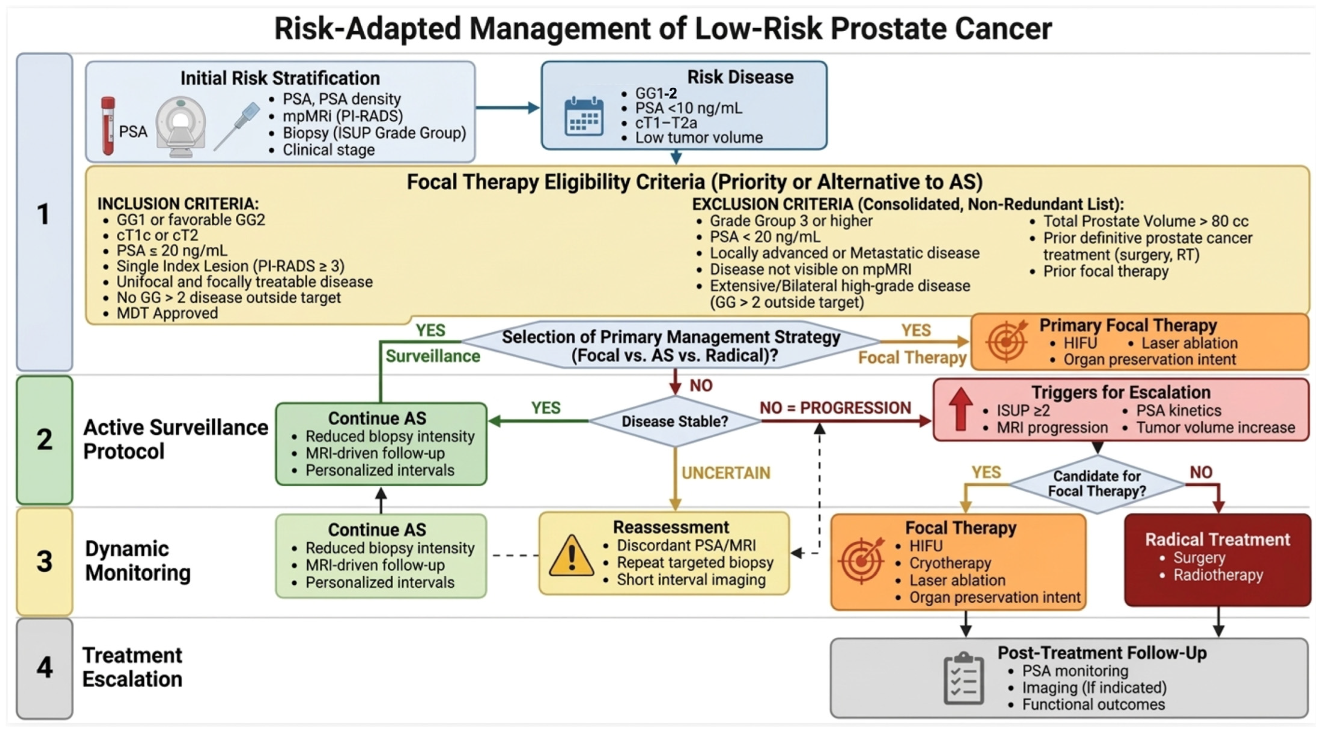
To facilitate clinical translation of the reviewed evidence, a risk-adapted management algorithm was developed and is presented. The flowchart integrates initial risk stratification, eligibility criteria for active surveillance and focal therapy, de-escalation pathways, and triggers for treatment escalation, providing a practical decision-making framework for clinicians managing low-risk Pca.

3. Discussion

3.1. Registry-Based Trends in Overtreatment and Active Surveillance

Recent large-scale cohort studies show significant declines in surgeries for indolent tumours, reflecting more judicious patient selection. Monda et al. (2025) [7] analysed pathology reports from two parallel registries, the U.S. SEER database and the Michigan Urological Surgery Improvement Collaborative (MUSIC), as a surrogate measure of overtreatment. They found a reduction in the fraction of prostatectomies that harboured only Gleason 6 cancer (pathologic GG 1) on final histology. Prostatectomies for low-grade tumours fell from 32.4% in 2010 to less than 8% by 2020. Similarly, statewide Michigan data revealed a reduction in pGG1 rates from 20.7% in 2012 to just 2.7% in 2024. However, these datasets include only men treated with prostatectomy and therefore cannot be interpreted as AS outcomes.
In a recent study including data on 26,999 men (2000–2016) on AS from the GAP3 registry [8], 10-year metastasis-free survival and overall survival were 99.4% and 84.1%, with lower rates of treatment conversion and longer treatment-free survival in more recent cohorts [8]. Such datasets are prone to substantial selection bias. Temporal changes in referral patterns, patient preferences, adherence to guidelines, and access to care may masquerade as improvement, without indicating that comparable populations were evaluated across time.
Definitions of “low-risk” disease and grading practice have evolved, producing grade migration across eras that can reclassify patients independently of true biology.
Moreover, the diffusion of mpMRI and targeted biopsy improved csPCa detection, reducing under-sampling and the risk of misclassification in the PRECISION study [9], which can make later cohorts appear safer and reduce low-grade diagnoses.
GAP3 additionally pools heterogeneous international cohorts with variable inclusion criteria, surveillance intensity, and triggers for intervention, limiting causal inference from time-period comparisons.
Incomplete capture of cause-specific outcomes (e.g., cause of death) and variable metastasis ascertainment across centres further constrain interpretation of “safety” endpoints in registry-based AS analyses.
In SEER/MUSIC, pGG1 prostatectomy is an informative system-level proxy for overtreatment, but it cannot adjudicate appropriateness in individual cases nor capture overtreatment driven by comorbidity or life expectancy.
Registry studies are also sensitive to missing data and coding changes over time; excluding incomplete cases can bias trends if missingness correlates with year or risk. Despite all these limitations, these prostatectomy-based and surveillance-based registries provide convergent evidence of improved risk stratification and increased acceptance of conservative management in low-risk disease.

3.2. Could Modernizing Risk Stratification Further Reduce Overtreatment?

The substantial decline in surgeries for low-grade PCa in favour of conservative management is strengthened by enhanced risk stratification methods, including advanced imaging and molecular biomarkers [10], which help differentiate between indolent and aggressive disease. While the “less is more” approach for low-risk PCa is gaining traction, current guidelines still rely on traditional classification parameters, suggesting a need for updated criteria that incorporate these modern diagnostic tools. Risk stratification models are evolving to better identify which patients truly need aggressive therapy and which do not.
A recent analysis of the ERSPC (European Randomized Study of Screening for Prostate Cancer) Rotterdam cohort examined whether modernizing risk stratification could further cut overtreatment [11]. The standard European Association of Urology (EAU) risk classification defines “low-risk” as any GG1 with PSA < 10 ng/mL and ≤cT2a; the study tested an alternative system that includes as low-risk any GG1 with PSA < 20 ng/mL and reclassifies certain favourable-intermediate risk patients as low-risk if they meet contemporary AS criteria (e.g., GG2 with PSA < 10 ng/mL and PSA density < 0.2 and ≤cT2a). Strikingly, this broadened definition increased the proportion of men labelled low-risk by 45% in the screening-detected cohort and 83% in the clinically detected cohort, compared to the traditional EAU criteria. Importantly, low-risk men according to the proposed criteria did not experience worse cancer-specific mortality compared to EAU low-risk men (subhazard ratio: 1.33 [95% CI: 0.66–2.68] in the screened group; 0.99 [95% CI: 0.29–3.38] in the unscreened group). In other words, many men currently classified as “intermediate-risk” by older criteria could safely be managed as low-risk, which would expand the pool of candidates for AS and further reduce overtreatment without compromising survival.

3.3. Role of MRI in Risk-Adapted Diagnostic Pathways

PCa screening began as a largely PSA-threshold–driven, one-size-fits-all pathway in which a raised PSA commonly led directly to systematic (non-targeted) biopsy. In ERSPC, repeated PSA screening reduced PCa mortality on long-term follow-up, but at the cost of substantial downstream testing and detection of cancers that would never have become clinically relevant. Interpretation of the Prostate, Lung, Colorectal and Ovarian Cancer Screening Trial (PLCO) has been complicated by opportunistic PSA testing in the control arm (“contamination”), so it compared organized screening versus a high background level of screening rather than screening versus no screening [12]. This early era therefore selected a broad population for biopsy, many men with benign disease, many with indolent GG1 cancers, and fewer with potentially lethal disease, producing the mortality benefit versus overdiagnosis/overtreatment trade-off (often estimated around 40–50% among screen-detected cases in ERSPC analyses) [13].
Risk stratification then evolved by inserting prediction models or biomarkers after PSA and before biopsy, shifting selection from a simple PSA cut-off toward an individualized probability of clinically significant PCa. Examples include ERSPC-derived risk calculators and biomarker-based models such as Stockholm3 and kallikrein panels [14]. These approaches preferentially triage men with higher predicted csPCa risk to biopsy while deferring biopsy in low-risk men despite modest PSA elevation, but when systematic biopsy remains the reference test, they remain vulnerable to undersampling and continued detection of low-grade disease, albeit at reduced rates versus PSA alone.
A major inflection point came with MRI as a triage test before biopsy, enabling targeted sampling. PROMIS established MRI as a useful triage test, and PRECISION showed that an MRI pathway with targeted biopsy increases detection of clinically significant PCa and reduces detection of clinically insignificant cancer versus standard systematic biopsy [9,15,16]. MRI-first strategies enrich for MRI-visible cancers and de-emphasize many men with PSA elevation but no MRI target, but can generate substantial numbers of MRIs in men who will not benefit and concentrate uncertainty in PI-RADS 3.
Pretreatment mpMRI risk strongly correlated with worse RP outcomes: higher PI-RADS independently predicted biochemical recurrence (BCR) in most studies, with reported HRs from about 1.75 to 6.19 depending on cutoff. After RP, men with PI-RADS 5 lesions had higher recurrence risk than those with lower-suspicion lesions; in one MRI-targeted biopsy cohort, PI-RADS 5 vs. 3 carried an HR of 2.86 for BCR [17]. In patients diagnosed with MRI-targeted biopsy (TBx) and treated by RP, adding PI-RADS score plus mpMRI features such as seminal vesicle invasion and lesion diameter improved 36-month BCR prediction versus clinical/TBx variables alone (73% vs. 66%).
Modern screening trials are increasingly using risk stratification to decide whether to perform an MRI. This involves a stepwise approach: starting with PSA-based entry and then enriching the risk for csPCa using biomarkers or models. Finally, MRI with targeted biopsy is reserved for men above a specific risk threshold. In ProScreen, a multi-step strategy (PSA → kallikrein panel risk score → MRI) detected additional high-grade cancers while minimizing additional low-grade detections [14]. Similarly, MRI-based extensions of Stockholm3 (STHLM3-MRI) were designed to reduce unnecessary biopsies and overdiagnosis by concentrating MRI and biopsy among men with a higher pretest probability of csPCa, based on integrated risk (clinical variables + biomarkers) [18].
Risk stratification can also be applied after MRI, especially for MRI-negative and PI-RADS 3 cases, redefining “at risk” as those with meaningful residual csPCa probability. PSA density has been proposed as a post-MRI modifier, and evidence syntheses suggest that combining MRI findings with PSA density thresholds may reduce unnecessary biopsy while attempting to preserve sensitivity for csPCa; however, this approach should be interpreted cautiously given heterogeneity in the evidence base and limited validation across populations [19]. “Double risk stratification” formalizes layered pre-MRI enrichment and post-MRI refinement to reduce harms while maintaining early detection of lethal cancers, reflected in Europe’s stepwise policy approach (EU Council Recommendation, 9 December 2022) and initiatives such as PRAISE-U [20]. In Germany, PROBASE extends “risk stratification” to risk-adapted retesting intervals based on baseline PSA [21]. Figure 1 describes possible risk stratification strategies in the diagnostic pathway of PCa.
These shifts affect what is labelled “low-risk PCa” and how often it is treated: MRI-targeted and layered tools reduce random undersampling, making “low risk” more biologically consistent, while reducing detection of small indolent tumours can shrink the pool diagnosed with truly low-risk disease. In addition to imaging, molecular and liquid biomarkers are increasingly integrated into risk stratification pathways and are discussed in the following section.

3.4. Evolving Diagnostic Pathways: Biomarkers

In addition to these MRI pathways, biomarkers from blood, urine, and tissue (e.g., PCA3, Oncotype DX, Decipher) are being investigated to refine stratification and management, helping identify indolent disease versus risk of progression [16,22,23]. Table 2 summarizes possible applications and roles of these biomarkers in advancing the diagnostic and management pathway of Low-Risk PCa.
A meta-analysis of five studies showed that Decipher® GC is an independent predictor of metastasis after radical prostatectomy (HR 1.30; 95% CI: 1.14–1.47; p < 0.001 per 0.1-unit increase), improving prognostic stratification across most clinicopathologic subgroups [24]. An RCT of 215 intermediate-risk PCa patients confirmed its prognostic value for disease progression (HR 1.12), biochemical failure (HR 1.22), distant metastasis (HR 1.28), and cancer-specific mortality (HR 1.45), though these were secondary endpoints [25].
Overall, while Decipher® and Oncotype DX improve risk stratification, no definitive evidence confirms improved long-term oncologic outcomes. Selective use is reasonable when results may guide management, but routine use lacks prospective outcome data support. Their use may be considered selectively when results are likely to inform management, but routine use is not yet supported by prospective outcome data. The magnitude of their clinical benefit remains modest and heterogeneous across studies, and a definitive improvement in long-term oncologic outcomes has not yet been proven in prospective trials.
High-sensitivity liquid biopsy approaches, particularly circulating tumor DNA (ctDNA), are biologically appealing because they may capture occult molecular aggressiveness not reflected by conventional clinical variables. However, in localized low-volume disease, ctDNA remains constrained by low tumor shedding and limited analytical sensitivity; accordingly, current evidence does not support its routine use in low-risk active surveillance [26], and its role should presently be regarded as investigational, potentially within multimodal models or minimal residual disease-oriented settings rather than as a stand-alone decision tool. Beyond ctDNA, a broader range of diagnostic tools may help refine the timing of transition from surveillance to active treatment. These include blood-based markers, kallikrein-based panels, urine assays, tissue genomic classifiers, and imaging-derived risk stratification approaches, which may improve the identification of biologically significant disease among patients otherwise classified as low risk. In this context, the ProScreen randomized trial [14] supports the value of a multistep strategy integrating PSA, the kallikrein panel, and MRI, reinforcing the concept that future risk-adapted pathways will likely rely on combinations of complementary biomarkers rather than on any single parameter.

3.5. Active Surveillance and Risk-Adapted Follow-Up

AS is now the cornerstone of low-risk PCa management. It aligns treatment intensity with disease aggressiveness, achieving both cancer control and quality-of-life preservation. Multiple studies have established the long-term safety of AS, and with proper patient selection and monitoring protocols (including PSA kinetics, MRI, and periodic biopsies as needed), the risk of missing the “window of curability” is exceedingly low.
The ProtecT randomized trial in the UK [3], which followed mostly low-risk (and some intermediate-risk) patients for 15 years, found no difference in PCa-specific mortality between men randomized to active monitoring versus immediate surgery or radiation. At 15 years, PCa death rates remained very low (<3%) in all arms, confirming that surveillance does not compromise survival in low-risk disease.
A recent report from the Göteborg randomized screening trial provided up to 25-year outcomes for men managed with surveillance [27]. Among those with very-low-risk or low-risk PCa, the 20-year PCa-specific survival exceeded 90–99%. In the entire AS cohort (including some intermediate-risk patients), only 14 men (out of 488) died from PCa over a median 18-years follow-up. These data demonstrate that with vigilant monitoring, the likelihood of a low-risk patient dying of PCa is extremely low—typically 1–5% over two decades, often lower than competing health risks. Furthermore, about half of men on AS in such cohorts never require active treatment even after 15–20 years.
Long-term population-based data [28] support the use of risk-adapted management and deferred treatment strategies in men with low-risk disease. The 15-year PCa-specific mortality was only 5.5%, rising to 12% at 30 years. In contrast, the risk of death from other causes in this group reached 77% over the same 30-year period, indicating that most patients are far more likely to die with the disease than from it. Among men with a life expectancy greater than 15 years, 30-year PCa mortality remained low at 11%, reinforcing the safety of conservative approaches like AS.
Across contemporary AS cohorts and guideline-based pathways, follow-up intensity is typically front-loaded (to confirm eligibility) and then individualized over time. An example is the Johns Hopkins approach, which includes confirmatory biopsy within 6–24 months, PSA every 3–6 months, annual digital rectal examination (DRE), mpMRI every 2–3 years and repeat biopsies roughly every 1–5 years depending on interim findings [29].
In contrast, an older UCSF protocol emphasizes close PSA monitoring (every 3 months), interval prostate imaging with ultrasound (every 6–12 months), and biopsy every 1–2 years, reflecting how some long-running programs pre-dated today’s widespread mpMRI integration [30]. The Sunnybrook/University of Toronto schedule is often described as PSA every 3 months for 2 years, then every 6 months, with biopsy at 12 months and then every 3–4 years. Protocol-directed multicentre AS can be even more prespecified, as in Canary PASS (PSA every 3 months, DRE every 6 months, and repeat biopsies at 6–12, 24, 48, and 72 months) [31]. Guideline frameworks generally set upper bounds rather than mandating a single “best” cadence; for example, NCCN describes PSA no more often than every 6 months, DRE no more often than every 12 months, and repeat biopsy and mpMRI no more often than every 12 months (unless clinically indicated), with an early repeat biopsy within 6 months when the diagnostic sampling is limited or discordant. ASCO’s endorsement of Cancer Care Ontario similarly suggests confirmatory biopsy at 6–12 months and then serial biopsy every 2–5 years, alongside PSA every 3–6 months and at least annual DRE [32].
However, the implementation of these risk-adapted surveillance pathways is not uniform across healthcare systems. Access to high-quality mpMRI, expert radiological interpretation, and repeat biopsy strategies may be limited in resource-constrained settings, potentially leading to disparities in patient selection and monitoring intensity. In addition, the increasing incorporation of advanced tools such as genomic classifiers may further widen inequities due to cost and availability constraints. These factors may result in underuse of AS in some populations or, conversely, suboptimal monitoring where recommended protocols cannot be fully implemented.
De-escalation strategies are increasingly framed as risk-adapted “response-to-stability.” Long-term prospective data underscore why this matters: in Canary PASS, 49% of men remained free of progression or treatment at 10 years (with <2% metastatic disease and <1% PCa mortality), highlighting a sizable subgroup that may not need perpetual high-intensity testing [33]. A particularly actionable trigger for de-intensification is a negative follow-up biopsy, which in large multicenter data is associated with substantially lower risk of conversion to treatment (adjusted hazard ratio: 0.45; 95% CI: 0.42–0.49), and subsequent upgrading (adjusted HR for conversion ~0.45; adjusted OR 0.52; 95%CI: 0.45–0.62), supporting less invasive schedules in this subset [34]. In parallel, MRI can be used to rationalize intensity rather than replace tissue confirmation outright: the EAU notes that serial DRE may be omitted when MRI is stable, and that patients with low-risk disease, stable MRI, and low PSA density (<0.15) may be exempted from repeat biopsy when a repeat MRI is performed before the planned re-biopsy. Nevertheless, such MRI-driven de-escalation strategies presuppose consistent access to high-quality imaging, which may not be universally available, reinforcing the need to adapt protocols to local resources. Finally, de-escalation should also include “time horizon” considerations—NCCN explicitly states that repeat biopsies are not indicated when life expectancy is <10 years, supporting transition from AS to observation/watchful waiting in appropriately selected older or comorbid patients.
A comparative overview of active surveillance follow-up protocols across major institutions and guideline frameworks is provided in Table 3.

3.6. Beyond Surveillance: The Role of Focal Therapy

Focal therapy (FT) represents an evolving approach in PCa management, strategically positioned between AS and radical treatment [35,36]. Its definitive role remains to be established as higher-level evidence continues to emerge. FT is not intended to replace AS in genuine low-risk cases, which typically require no immediate intervention, nor should it substitute radical treatment in high-risk scenarios. Rather, FT serves as an intermediate solution for patients uncomfortable with surveillance, yet who do not necessitate immediate whole-gland therapy.
Patient selection is crucial, encompassing specific tumor paramenters (PSA, PSAD, ISUP at biopsy, tumor size, tumor location, prostate volume, and multifocality), patient characteristics (life expectancy, individual treatment goals, pelvic and prostate anatomy, acceptance of potential recurrence, and openness to retreatment or return to AS), and hospital organization (availability of advanced FT modalities, surgeon experience, quality of MRI imaging, and biopsy accuracy). However, selection criteria remain inconsistently defined across studies, contributing to variability in reported outcomes.
The ideal candidate for FT typically falls within the intermediate-risk category, balancing between sufficient disease progression risk to justify intervention and low likelihood of immediate metastatic progression or mortality. Such patients commonly have MRI-visible, biopsy-confirmed, clinically significant tumors spatially aligned for targeted ablation. However, this paradigm is challenged by the intrinsic multifocality of PCa and the limitations of imaging and biopsy in fully characterizing disease extent. Tumors located anteriorly or adequately distanced from critical structures (e.g., urethra, distal apex) represent optimal targets, while posterior lesions should preferably be small and safely situated. Nevertheless, the risk of undetected clinically significant disease outside the treatment field remains a key concern. Caution is warranted in younger men due to the uncertainty regarding FT’s long-term oncologic durability and its potential impact on subsequent salvage treatments, which remain insufficiently studied. Transition from AS to FT should be based on biopsy-confirmed progression, supported by imaging, rather than on any single parameter alone. The main triggers are grade upgrading from ISUP Grade Group 1 to csPCa, especially ISUP Grade Group ≥ 3, detection of cribriform or intraductal carcinoma, increasing tumor burden on repeat systematic biopsy, and MRI progression requiring confirmatory biopsy before treatment change. PSA kinetics alone should not trigger intervention. However, there is currently no standardized threshold for intervention, and decision-making remains partly subjective. When systematic and MRI-targeted biopsy grades are discordant, the associated risk is intermediate, not simply that of the highest grade alone: the rate of advanced pathological stage was 23% in concordant Grade Group 3 disease and 8.8% in concordant Grade Group 2 disease, compared with 18% for systematic Grade Group 3/targeted Grade Group 2 and 15% for the reverse combination [37]. This supports a more nuanced and individualized approach to treatment escalation. FT appears most appropriate in carefully selected patients with localized, MRI-visible, biopsy-confirmed csPCa but anatomically limited disease. However, available studies remain heterogeneous in patient selection, treatment platform, follow-up, and endpoint definition. FT for clinically localized PCa demonstrates meaningful short-term oncological control, with biopsy-proven csPCa recurrence-free survival (csPCa RFS) rates of 89%, 86%, and 81% at 6, 12, and 24 months, respectively, across 50 prospective studies including 4615 patients. However, comparative effectiveness data against AS remains sparse.
In the intermediate-risk subgroup—the most clinically relevant population—the 12-month csPCa RFS was 79%, confirming moderate cancer control even in more aggressive disease [38]. Five-year failure-free survival (radical and systemic treatment-free) reached 82%, suggesting that most patients can avoid whole-gland treatments at intermediate-term follow-up. However, these findings should be interpreted with caution, as most data derive from heterogeneous, single-arm studies with limited randomized comparisons, variable outcome definitions, and inconsistent follow-up protocols.
In-field and out-of-field csPCa recurrence rates were comparable (~9% and ~8%, respectively), highlighting the importance of careful patient selection given the multifocal nature of PCa [38]. Interestingly, no significant differences in oncological outcomes were observed across the seven energy sources investigated (HIFU, cryotherapy, IRE, FLA, PDT, focal brachytherapy, RFA). This apparent equivalence may reflect insufficient comparative power rather than true clinical similarity, and no modality has demonstrated clear superiority.
Treatment failure after FT should therefore be defined using composite criteria, including biopsy-confirmed persistent or recurrent csPCa, radiologic progression on follow-up MRI, and the need for salvage focal, whole-gland, or systemic treatment. At present, outcomes are strongly influenced by patient selection, operator experience, and institutional expertise, limiting generalizability. Though long-term data—including metastasis-free and overall survival—remain limited and are critically awaited [24,25], while AS remains the gold standard for low-risk (GG1) disease, focal therapy is increasingly positioned as a “middle ground” for favourable intermediate-risk (GG2) patients. It offers a higher chance of oncologic clearance than AS while significantly reducing the morbidity associated with radical prostatectomy or whole-gland radiation (Table 4).
To facilitate clinical implementation, a management algorithm integrating risk stratification, AS eligibility, monitoring, and escalation pathways is proposed (Figure 2). This framework reflects contemporary trends toward intensification and de-intensificaion while maintaining oncologic safety.

3.7. Current Limitations of the Diagnostic Pathway in Low-Risk Prostate Cancer

Despite significant strides in risk stratification and surveillance strategies, the diagnostic journey of low-risk PCa remains fraught with limitations that may affect optimal patient selection and long-term outcomes. These challenges encompass imaging and biopsy accuracy, risk of pathological upgrading, terminological controversies around GG1, and evolving genomic and histopathological markers.
MRI sensitivity remains variable, especially for small or low-grade lesions. Studies indicate that MRI may miss tumours <0.5 mL or those lacking aggressive histologic features, with sensitivity for index lesion detection ranging from 71% to 94% depending on technique and reader experience [39]. Additionally, interpretation inconsistencies across centers impact diagnostic yield. For example, biopsies informed by MRI from low-volume or less experienced centres show significantly reduced clinically significant PCa detection compared to high-volume centers [40]. Other imaging modalities like PET/CT are still considered experimental due to their limited value in intermediate and high-risk cases. While a positive correlation between SUV and pathological prostate features exists, PET/CT sensitivity remains a key limitation [41].
Biopsy strategies themselves present diagnostic gaps. A randomized trial comparing targeted biopsy (TB) alone versus TB plus systematic biopsy (SB) in MRI-positive, biopsy-naïve patients revealed that although TB alone suffices in detecting most csPCa, the addition of SB can improve overall detection by 7.3% [42]. These limitations underscore the persistence of sampling errors and false negatives even in the MRI era.
Despite initial classification as low risk, many patients experience histological upgrading upon repeat biopsy or post-prostatectomy analysis. In a contemporary cohort, 67.3% of low-risk patients demonstrated upgrading, with PSA levels and proportion of biopsy-positive cores emerging as independent predictors [43]. Similarly, long-term AS cohorts report a cumulative 10-year upgrading rate of 62%, with 19% showing major upgrading to GG ≥3 [44]. This risk raises concerns about the initial diagnostic accuracy and justifies the need for confirmation biopsies, especially for those not pre-stratified by MRI.
The introduction of MRI-guided targeted biopsies has contributed to a “stage shift” phenomenon. Lesions previously undetected with systematic biopsy are now identified, but this comes at the cost of reclassifying many patients into higher risk categories based on upgraded biopsy results. Consequently, the average risk profile of “low-risk” patients has evolved, complicating comparisons with historical cohorts and casting doubt on the current risk stratification thresholds. Indeed, MRI-targeted biopsies are more sensitive in detecting high-grade cancers but may also lead to a better prognosis within the same GG compared to systematic biopsies. As a result, the 2019 ISUP consensus conference recommended using an aggregated GG for MRI-targeted biopsy cores from the same lesion, rather than relying solely on the highest-grade core [45].
Whether GG1 represents true cancer remains controversial. Some propose relabelling it as a benign or “indolent neoplasm” to reduce anxiety and overtreatment [46]. However, a pathology society survey showed that 81.7% of experts oppose removing the “cancer” label, citing diagnostic reproducibility and potential confusion in management [47]. Critics argue that GG1 tumours share morphologic and molecular features with higher-grade cancers, and that removing the label could undermine proper monitoring.
Genomic classifiers such as Decipher and OncotypeDx have been integrated into modern risk stratification algorithms, offering potential additional prognostic value [10,22]. Yet, evidence from large prospective cohorts suggests that high-risk genomic profiles do not reliably predict upgrading on biopsy [44]. In multivariable models, traditional factors like PSA density and age retain stronger predictive value. The role of genomics remains adjunctive rather than definitive in the current diagnostic pathway.
The health-economic and implementation implications of genomic classifiers warrant discussion, as their cost-effectiveness and real-world utility remain debated. While these assays can add prognostic information beyond clinicopathologic factors, evidence that they consistently and meaningfully change routine management—and thereby improve patient-important outcomes—remains heterogeneous across settings and pathways. Cost-effectiveness is highly context-dependent and typically assumes that testing produces net de-escalation of unnecessary treatment or better-targeted escalation in selected subgroups; if instead it adds expense while only modestly shifting decisions (or increases downstream surveillance/utilization), economic value may be attenuated. Moreover, variable payer coverage, access, and workflow constraints shape uptake and may limit generalizability and equity [16].
Histologic findings such as Atypical Intraductal Proliferation (AIP) are emerging as critical yet underutilized predictors of unsampled high-grade disease [48]. AIP is frequently associated with adverse pathology and a higher likelihood of upgrading on repeat prostate biopsy. Despite this, AIP is not consistently reported in pathology or incorporated into surveillance protocols. Its routine documentation could enhance risk stratification, guiding more tailored monitoring strategies.

4. Conclusions

The management of low-risk PCa is a success story of risk adaptation in oncology. AS has established itself as the standard of care for low-risk ISUP I PCa, supported by long-term cohort data and randomized trial evidence demonstrating oncologic safety without compromising functional outcomes. Refined risk stratification tools—including mpMRI, biomarkers, and genomic classifiers—have further reduced overtreatment, though their optimal integration into clinical pathways remains to be standardized. FT represents a promising minimally invasive alternative, yet critical evidence gaps persist: long-term cancer control durability, out-of-field progression risk, implications for salvage treatments, and absence of comparative effectiveness data against AS or radical therapy. To bridge the gap between evidence and clinical practice, a risk-adapted management approach should integrate initial risk stratification, AS eligibility and follow-up protocols, de-escalation pathways, and escalation triggers, providing a structured decision-making framework applicable across different healthcare settings.
From a clinical standpoint, key priorities include: (i) adherence to guideline-recommended AS protocols for low-risk disease; (ii) structured use of mpMRI for dynamic monitoring; and (iii) restriction of FT to clinical trials or prospective registries until robust long-term data are available.
Future research should focus on head-to-head prospective comparisons of AS and FT, standardization of oncologic and patient-reported outcome measures, and validation of integrated risk models combining clinical, imaging, and molecular data. Real-world registry-based evidence will be essential to define the evolving role of minimally invasive approaches and to ensure equitable, resource-adapted implementation across diverse healthcare systems.

Supplementary Materials

The following supporting information can be downloaded at: https://www.mdpi.com/article/10.3390/diagnostics16091310/s1.

Author Contributions

Conceptualization: F.Z. and S.D.P.; methodology: F.Z. and S.D.P.; investigation: all authors; data curation: F.Z. and S.D.P.; writing—original draft preparation: F.Z. and S.D.P.; writing—review and editing: all authors; supervision: S.D.P. All authors have read and agreed to the published version of the manuscript.

Funding

This research received no external funding.

Institutional Review Board Statement

Not applicable.

Informed Consent Statement

Not applicable.

Data Availability Statement

No new data were created or analyzed in this study. Data sharing is not applicable to this article.

Acknowledgments

All authors are members of the Uronauti Group, a collaborative Italian academic network in urology. The group is acknowledged as the academic framework within which this work was developed, but all contributions were made directly by the listed authors.

Conflicts of Interest

The authors declare no conflicts of interest.

References

  1. James, N.D.; Tannock, I.; N’Dow, J.; Feng, F.; Gillessen, S.; Ali, S.A.; Trujillo, B.; Al-Lazikani, B.; Attard, G.; Bray, F.; et al. The Lancet Commission on prostate cancer: Planning for the surge in cases. Lancet 2024, 403, 1683–1722. [Google Scholar] [CrossRef]
  2. Roobol, M.J.; de Vos, I.I.; Månsson, M.; Godtman, R.A.; Talala, K.M.; den Hond, E.; Nelen, V.; Villers, A.; Poinas, G.; Kwiatkowski, M.; et al. European Study of Prostate Cancer Screening—23-Year Follow-up. N. Engl. J. Med. 2025, 393, 1669–1680. [Google Scholar] [CrossRef] [PubMed]
  3. Hamdy, F.C.; Donovan, J.L.; Lane, J.A.; Metcalfe, C.; Davis, M.; Turner, E.L.; Martin, R.M.; Young, G.J.; Walsh, E.I.; Bryant, R.J.; et al. Fifteen-Year Outcomes after Monitoring, Surgery, or Radiotherapy for Prostate Cancer. N. Engl. J. Med. 2023, 388, 1547–1558. [Google Scholar] [CrossRef]
  4. Biasatti, A.; Soputro, N.A.; Porpiglia, F.; Perdonà, S.; Abdollah, F.; Nelson, R.; Rogers, C.; Zhao, L.C.; Ghazi, A.; Challacombe, B.; et al. The current landscape of single-port robotic surgery in urology. Nat. Rev. Urol. 2025, 23, 156–173. [Google Scholar] [CrossRef] [PubMed]
  5. Bignante, G.; Orsini, A.; Lasorsa, F.; Lambertini, L.; Pacini, M.; Amparore, D.; Pandolfo, S.D.; Del Giudice, F.; Zaytoun, O.; Buscarini, M.; et al. Robotic-assisted surgery for the treatment of urologic cancers: Recent advances. Expert Rev. Med. Devices 2024, 21, 1165–1177. [Google Scholar] [CrossRef] [PubMed]
  6. Lam, T.B.L.; MacLennan, S.; Willemse, P.P.M.; Mason, M.D.; Plass, K.; Shepherd, R.; Baanders, R.; Bangma, C.H.; Bjartell, A.; Bossi, A.; et al. EAU-EANM-ESTRO-ESUR-SIOG Prostate Cancer Guideline Panel Consensus Statements for Deferred Treatment with Curative Intent for Localised Prostate Cancer from an International Collaborative Study (DETECTIVE Study). Eur. Urol. 2019, 76, 790–813. [Google Scholar] [CrossRef] [PubMed]
  7. Monda, S.M.; Demus, T.; Jaime-Casas, S.; Meah, S.; Srivastava, A.; Sarle, R.; Labardee, C.; Ghani, K.R.; Ginsburg, K.M.; Morgan, T.M.; et al. Trends in Surgical Overtreatment of Prostate Cancer. JAMA Oncol. 2025, 11, 700–706. [Google Scholar] [CrossRef] [PubMed]
  8. Bangma, C.; Doan, P.; Zhu, L.; Remmers, S.; Nieboer, D.; Helleman, J.; Roobol, M.J.; Sugimoto, M.; Chung, B.H.; Lee, L.S.; et al. Has Active Surveillance for Prostate Cancer Become Safer? Lessons Learned from a Global Clinical Registry. Eur. Urol. Oncol. 2025, 8, 324–337. [Google Scholar] [CrossRef]
  9. Kasivisvanathan, V.; Rannikko, A.S.; Borghi, M.; Panebianco, V.; Mynderse, L.A.; Vaarala, M.H.; Briganti, A.; Budäus, L.; Hellawell, G.; Hindley, R.G.; et al. MRI-Targeted or Standard Biopsy for Prostate-Cancer Diagnosis. N. Engl. J. Med. 2018, 378, 1767–1777. [Google Scholar] [CrossRef] [PubMed]
  10. Tabriz, A.A.; Boyer, M.J.; Gordon, A.M.; Carpenter, D.J.; Gingrich, J.R.; Raman, S.R.; Sirohi, D.; Rompre-Brodeur, A.; Lunyera, J.; Basher, F.; et al. Impact of Genomic Classifiers on Risk Stratification and Treatment Intensity in Patients with Localized Prostate Cancer. Ann. Intern. Med. 2025, 178, 218–228. [Google Scholar] [CrossRef]
  11. de Vos, I.I.; Rosenstand, C.; Hogenhout, R.; van den Bergh, R.C.N.; Remmers, S.; Roobol, M.J. Reducing Overtreatment of Prostate Cancer Patients: Revisiting the European Association of Urology Pretreatment Risk Group Classification Using Long-term Follow-up Data from the European Randomized Study of Screening for Prostate Cancer Rotterdam. Eur. Urol. Oncol. 2025, 8, 747–754. [Google Scholar] [CrossRef] [PubMed]
  12. Palmstedt, E.; Månsson, M.; Hugosson, J.; Arnsrud Godtman, R. Active Surveillance for Screen-detected Low- and Intermediate-risk Prostate Cancer: Extended Follow-up up to 25 Years in the GÖTEBORG-1 Trial. Eur. Urol. 2025, 88, 373–380. [Google Scholar] [CrossRef]
  13. Scilipoti, P.; Bratt, O.; Garmo, H.; Orrason, A.W.; Gedeborg, R.; Stattin, P.; Westerberg, M. Long-Term Outcomes After Guideline-Recommended Treatment of Men with Prostate Cancer. J. Natl. Compr. Cancer Netw. 2025, 23, e257022. [Google Scholar] [CrossRef]
  14. Tosoian, J.J.; Mamawala, M.; Epstein, J.I.; Landis, P.; Macura, K.J.; Simopoulos, D.N.; Carter, H.B.; Gorin, M.A. Active Surveillance of Grade Group 1 Prostate Cancer: Long-term Outcomes from a Large Prospective Cohort. Eur. Urol. 2020, 77, 675–682. [Google Scholar] [CrossRef]
  15. Dall’Era, M.A.; Konety, B.R.; Cowan, J.E.; Shinohara, K.; Stauf, F.; Cooperberg, M.R.; Meng, M.V.; Kane, C.J.; Perez, N.; Master, V.A.; et al. Active surveillance for the management of prostate cancer in a contemporary cohort. Cancer 2008, 112, 2664–2670. [Google Scholar] [CrossRef]
  16. Newcomb, L.F.; Thompson, I.M.; Boyer, H.D.; Brooks, J.D.; Carroll, P.R.; Cooperberg, M.R.; Dash, A.; Ellis, W.J.; Fazli, L.; Feng, Z.; et al. Outcomes of Active Surveillance for Clinically Localized Prostate Cancer in the Prospective, Multi-Institutional Canary PASS Cohort. J. Urol. 2016, 195, 313–320. [Google Scholar] [CrossRef]
  17. Chen, R.C.; Rumble, R.B.; Jain, S. Active Surveillance for the Management of Localized Prostate Cancer (Cancer Care Ontario guideline): American Society of Clinical Oncology Clinical Practice Guideline Endorsement Summary. J. Oncol. Pract. 2016, 12, 267–269. [Google Scholar] [CrossRef]
  18. Tanne, J.H. Low risk prostate cancer: Active surveillance does not increase unfavourable outcomes, study finds. BMJ 2024, 385, q1213. [Google Scholar] [CrossRef] [PubMed]
  19. Beckmann, K.; Santaolalla, A.; Sugimoto, M.; Carroll, P.; Rubio, J.; Villers, A.; Bjartell, A.; Morgan, T.; Dasgupta, P.; Van Hemelrijck, M.; et al. Risk of progression following a negative biopsy in prostate cancer active surveillance. Prostate Cancer Prostatic Dis. 2023, 26, 403–409. [Google Scholar] [CrossRef] [PubMed]
  20. de Luca, S.; Checcucci, E.; Piramide, F.; Russo, F.; Alessio, P.; Garrou, D.; Peretti, D.; Sica, M.; Volpi, G.; Piana, A.; et al. MRI/real-time ultrasound image fusion guided high-intensity focused ultrasound: A prospective comparative and functional analysis of different ablative techniques. Minerva Urol. Nephrol. 2023, 75, 172–179. [Google Scholar] [CrossRef] [PubMed]
  21. Lasorsa, F.; Biasatti, A.; Orsini, A.; Bignante, G.; Farah, G.M.; Pandolfo, S.D.; Lambertini, L.; Reddy, D.; Damiano, R.; Ditonno, P.; et al. Focal Therapy for Prostate Cancer: Recent Advances and Insights. Curr. Oncol. 2025, 32, 15. [Google Scholar] [CrossRef]
  22. Scuderi, S.; Pellegrino, F.; Tin, A.; Beech, B.B.; Gandaglia, G.; Stabile, A.; Eastham, J.A.; Montorsi, F.; Briganti, A.; Vickers, A.J. The Highest Grade Group Does Not Drive the Risk of Recurrence when Systematic and Multiparametric Magnetic Resonance Imaging (MRI)-targeted Biopsies are Discordant: Preliminary Findings Using Radical Prostatectomy Pathology as a Surrogate for MRI-targeted Biopsy Grade. Eur. Urol. Focus 2024, 10, 486–488. [Google Scholar] [CrossRef] [PubMed]
  23. Ślusarczyk, A.; Gurwin, A.; Barnaś, A.; Ismail, H.; Miszczyk, M.; Zapała, P.; Przydacz, M.; Krajewski, W.; Antczak, A.; Życzkowski, M.; et al. Outcomes of Focal Therapy for Localized Prostate Cancer: A Systematic Review and Meta-analysis of Prospective Studies. Eur. Urol. Oncol. 2025, 8, 1653–1672. [Google Scholar] [CrossRef] [PubMed]
  24. Hocking, W.G.; Hu, P.; Oken, M.M.; Winslow, S.D.; Kvale, P.A.; Prorok, P.C.; Ragard, L.R.; Commins, J.; Lynch, D.A.; Andriole, G.L.; et al. Lung cancer screening in the randomized prostate, lung, colorectal, and ovarian (PLCO) cancer screening trial. J. Natl. Cancer Inst. 2010, 102, 722–731. [Google Scholar] [CrossRef] [PubMed]
  25. Schröder, F.H.; Hugosson, J.; Roobol, M.J.; Tammela, T.L.J.; Zappa, M.; Nelen, V.; Kwiatkowski, M.; Lujan, M.; Määttänen, L.; Lilja, H.; et al. Screening and prostate cancer mortality: Results of the European Randomised Study of Screening for Prostate Cancer (ERSPC) at 13 years of follow-up. Lancet 2014, 384, 2027–2035. [Google Scholar] [CrossRef]
  26. Auvinen, A.; Tammela, T.L.J.; Mirtti, T.; Lilja, H.; Tolonen, T.; Kenttämies, A.; Rinta-Kiikka, I.; Lehtimäki, T.; Natunen, K.; Nevalainen, J.; et al. Prostate Cancer Screening with PSA, Kallikrein Panel, and MRI. JAMA 2024, 331, 1452. [Google Scholar] [CrossRef]
  27. Ahmed, H.U.; El-Shater Bosaily, A.; Brown, L.C.; Gabe, R.; Kaplan, R.; Parmar, M.K.; Collaco-Moraes, Y.; Ward, K.; Hindley, R.G.; Freeman, A.; et al. Diagnostic accuracy of multi-parametric MRI and TRUS biopsy in prostate cancer (PROMIS): A paired validating confirmatory study. Lancet 2017, 389, 815–822. [Google Scholar] [CrossRef] [PubMed]
  28. Janopaul-Naylor, J.R.; Patil, D.; Joshi, S.; Sanda, M.G.; Sebastian, N.; Salari, K.; Dodge, J.; Dhere, V.R.; Hershatter, B.W.; Patel, P.R.; et al. Sociodemographic Features, Health Care Costs, and Treatment Implications of Genomic Classifier Testing for Localized Prostate Cancer in the United States. JCO Precis. Oncol. 2025, 9, e2500406. [Google Scholar] [CrossRef]
  29. Stabile, A.; Mazzone, E.; Cirulli, G.O.; De Cobelli, F.; Grummet, J.; Thoeny, H.C.; Emberton, M.; Pokorny, M.; Pinto, P.A.; Taneja, S.S. Association Between Multiparametric Magnetic Resonance Imaging of the Prostate and Oncological Outcomes after Primary Treatment for Prostate Cancer: A Systematic Review and Meta-analysis. Eur. Urol. Oncol. 2021, 4, 519–528. [Google Scholar] [CrossRef] [PubMed]
  30. Nordström, T.; Discacciati, A.; Bergman, M.; Clements, M.; Aly, M.; Annerstedt, M.; Glaessgen, A.; Carlsson, S.; Jäderling, F.; Eklund, M.; et al. Prostate cancer screening using a combination of risk-prediction, MRI, and targeted prostate biopsies (STHLM3-MRI): A prospective, population-based, randomised, open-label, non-inferiority trial. Lancet Oncol. 2021, 22, 1240–1249. [Google Scholar] [CrossRef] [PubMed]
  31. Haj-Mirzaian, A.; Burk, K.S.; Lacson, R.; Glazer, D.I.; Saini, S.; Kibel, A.S.; Khorasani, R. Magnetic Resonance Imaging, Clinical, and Biopsy Findings in Suspected Prostate Cancer. JAMA Netw. Open 2024, 7, e244258. [Google Scholar] [CrossRef]
  32. Chandran, A.; van Harten, M.; Singh, D.; Vilaseca, J.; Patasius, A.; Tupikowski, K.; Amorín, Á.G.; Galvin, D.; López, H.; Salazar, J.P.; et al. Risk-stratified Approach to Implementing Population-based Prostate Cancer Screening in Five Pilot Sites in the European Union: A Protocol for the PRAISE-U Project. Eur. Urol. Open Sci. 2024, 70, 8–17. [Google Scholar] [CrossRef] [PubMed]
  33. Krilaviciute, A.; Kaaks, R.; Seibold, P.; de Vrieze, M.; Lakes, J.; Radtke, J.P.; Kuczyk, M.; Harke, N.N.; Debus, J.; Fink, C.A.; et al. Risk-adjusted Screening for Prostate Cancer—Defining the Low-risk Group by Data from the PROBASE Trial. Eur. Urol. 2024, 86, 493–500. [Google Scholar] [CrossRef]
  34. Bologna, E.; Ditonno, F.; Licari, L.C.; Franco, A.; Manfredi, C.; Mossack, S.; Pandolfo, S.D.; De Nunzio, C.; Simone, G.; Leonardo, C.; et al. Tissue-Based Genomic Testing in Prostate Cancer: 10-Year Analysis of National Trends on the Use of Prolaris, Decipher, ProMark, and Oncotype DX. Clin. Pract. 2024, 14, 508–520. [Google Scholar] [CrossRef] [PubMed]
  35. Zhu, A.; Proudfoot, J.A.; Davicioni, E.; Ross, A.E.; Petkov, V.I.; Bonds, S.; Schussler, N.; Zaorsky, N.G.; Jia, A.Y.; Spratt, D.E.; et al. Use of Decipher Prostate Biopsy Test in Patients with Favorable-risk Disease Undergoing Conservative Management or Radical Prostatectomy in the Surveillance, Epidemiology, and End Results Registry. Eur. Urol. Oncol. 2024, 7, 1504–1512. [Google Scholar] [CrossRef] [PubMed]
  36. Spratt, D.E.; Yousefi, K.; Deheshi, S.; Ross, A.E.; Den, R.B.; Schaeffer, E.M.; Trock, B.J.; Zhang, J.; Glass, A.G.; Dicker, A.P.; et al. Individual Patient-Level Meta-Analysis of the Performance of the Decipher Genomic Classifier in High-Risk Men After Prostatectomy to Predict Development of Metastatic Disease. J. Clin. Oncol. 2017, 35, 1991–1998. [Google Scholar] [CrossRef] [PubMed]
  37. Spratt, D.E.; Liu, V.Y.T.; Michalski, J.; Davicioni, E.; Berlin, A.; Simko, J.P.; Efstathiou, J.A.; Tran, P.T.; Sandler, H.M.; Hall, W.A.; et al. Genomic Classifier Performance in Intermediate-Risk Prostate Cancer: Results from NRG Oncology/RTOG 0126 Randomized Phase 3 Trial. Int. J. Radiat. Oncol. Biol. Phys. 2023, 117, 370–377. [Google Scholar] [CrossRef] [PubMed]
  38. Sighinolfi, M.C.; Pallotta, G.; Del Re, M.; Moosavi, K.; Schubert, O.; Rossi, F.; Gavi, F.; Assumma, S.; Panio, E.; Totaro, A.; et al. Liquid Biopsy in Non-Metastatic Prostate Cancer: Clinical Evidence and Future Directions. Cancers 2026, 18, 800. [Google Scholar] [CrossRef] [PubMed]
  39. Lorusso, V.; Talso, M.; Palmisano, F.; Branger, N.; Granata, A.M.; Fiori, C.; Gregori, A.; Pignot, G.; Walz, J. Is imaging accurate enough to detect index lesion in prostate cancer? Analysis of the performance of MRI and other imaging modalities. Minerva Urol. Nephrol. 2024, 76, 22–30. [Google Scholar] [CrossRef] [PubMed]
  40. Droghetti, M.; Bianchi, L.; Gaudiano, C.; Corcioni, B.; Rustici, A.; Piazza, P.; Beretta, C.; Balestrazzi, E.; Costa, F.; Feruzzi, A.; et al. Comparison of prostate cancer detection rate at targeted biopsy of hub and spoke centers mpMRI: Experience matters. Minerva Urol. Nephrol. 2023, 75, 42–49. [Google Scholar] [CrossRef] [PubMed]
  41. Spena, G.; Moretti, T.B.; Dávila, F.S.; Dos Anjos, G.; Khan, I.; Calace, F.P.; Aveta, A.; Pandolfo, S.D.; Tufano, A.; Izzo, A.; et al. Ga68-PSMA PET for lymph node staging in intermediate and high-risk prostate cancer patients undergoing robotic assisted radical prostatectomy. Minerva Urol. Nephrol. 2024, 76, 467–473. [Google Scholar] [CrossRef] [PubMed]
  42. Porpiglia, F.; Checcucci, E.; de Cillis, S.; Piramide, F.; Amparore, D.; Piana, A.; Volpi, G.; Granato, S.; Zamengo, D.; Stura, I.; et al. A prospective randomized controlled trial comparing target prostate biopsy alone approach vs. target plus standard in naïve patients with positive mpMRI. Minerva Urol. Nephrol. 2023, 75, 31–41. [Google Scholar] [CrossRef] [PubMed]
  43. Porcaro, A.B.; Inverardi, D.; Corsi, P.; Sebben, M.; Cacciamani, G.; Tafuri, A.; Processali, T.; Pirozzi, M.; Mattevi, D.; De Marchi, D.; et al. Prostate-specific antigen levels and proportion of biopsy positive cores are independent predictors of upgrading patterns in low-risk prostate cancer. Minerva Urol. Nefrol. 2020, 72, 66–71. [Google Scholar] [CrossRef] [PubMed]
  44. Shee, K.; Song, J.J.; Cowan, J.E.; Wang, L.; Nie, J.; Chappidi, M.; Washington, S.L.; Nguyen, H.G.; Cooperberg, M.R.; Shinohara, K.; et al. The Natural History of Confirmed Grade Group 1 Prostate Cancer Managed with Active Surveillance in the Modern Era. Eur. Urol. Oncol. 2025, 8, 1094–1100. [Google Scholar] [CrossRef] [PubMed]
  45. Van Leenders, G.J.L.H.; Van Der Kwast, T.H.; Grignon, D.J.; Evans, A.J.; Kristiansen, G.; Kweldam, C.F.; Litjens, G.; McKenney, J.K.; Melamed, J.; Mottet, N.; et al. The 2019 International Society of Urological Pathology (ISUP) Consensus Conference on Grading of Prostatic Carcinoma. Am. J. Surg. Pathol. 2020, 44, e87–e99. [Google Scholar] [CrossRef] [PubMed]
  46. Epstein, J.I. Is Grade Group 1 (Gleason score 3 + 3 = 6) adenocarcinoma of the prostate really cancer? Curr. Opin. Urol. 2022, 32, 91–95. [Google Scholar] [CrossRef] [PubMed]
  47. Zhou, M.; Amin, A.; Fine, S.W.; Rao, P.; Siadat, F.; Shah, R.B. Should grade group 1 prostate cancer be reclassified as “non-cancer”? A pathology community perspective. Urol. Oncol. 2023, 41, 62–64. [Google Scholar] [CrossRef] [PubMed]
  48. Bhattarai, R.; McKenney, J.K.; Alaghehbandan, R.; Liu, X.; Cox, R.M.; Myles, J.L.; Przybycin, C.G.; Williamson, S.R.; Weight, C.J.; Schwen, Z.; et al. Atypical Intraductal Proliferation in Prostate Needle Core Biopsy. Am. J. Surg. Pathol. 2025, 49, 515–522. [Google Scholar] [CrossRef] [PubMed]
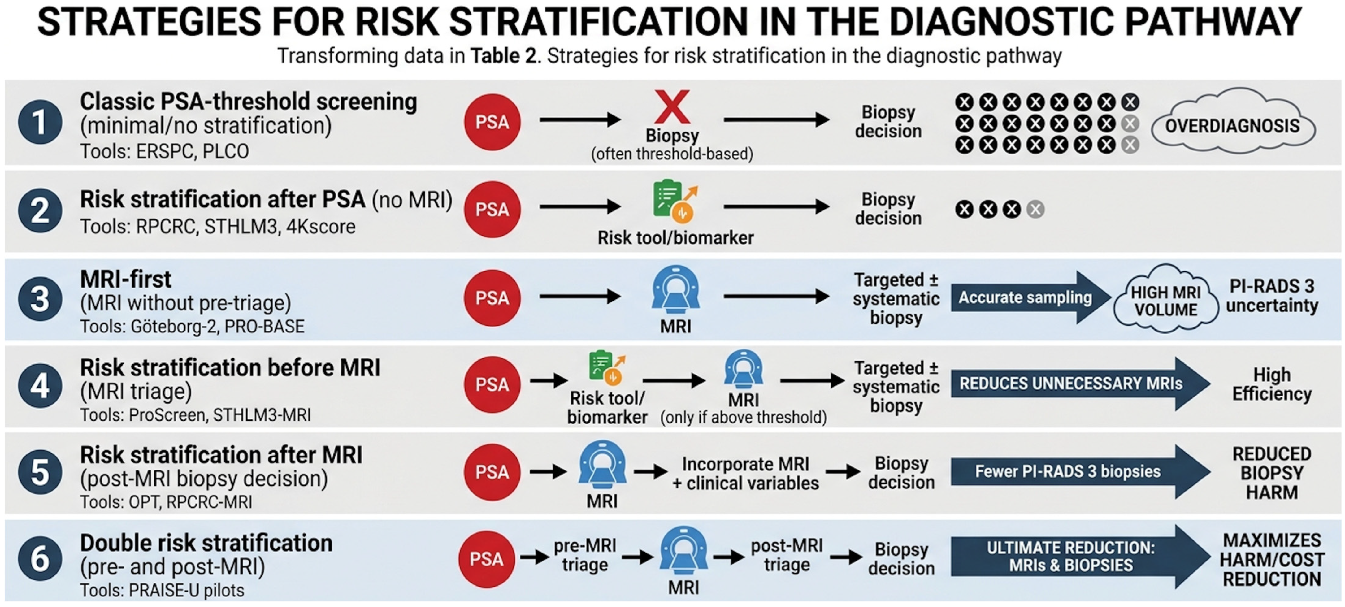
Figure 1. Strategies for risk stratification in the diagnostic pathway. PSA = Prostate-Specific Antigen; PCa = prostate cancer; csPCa = clinically significant prostate cancer; MRI = Magnetic Resonance Imaging;; RPCRC = Rotterdam Prostate Cancer Risk Calculator; STHLM3 = Stockholm3 model; ERSPC = European Randomized Study of Screening for Prostate Cancer; PLCO = Prostate, Lung, Colorectal, and Ovarian (Cancer Screening Trial); OPT = “optimal”/optimized post-test strategy (as used in some pathway descriptions; often referencing concepts like PSA density); PROBASE = Prostate Cancer Screening in Young Men (PROBASE trial); PRAISE-U = PRAISE-U pilots/trials (double risk-stratification pathway).
Diagnostics 16 01310 g001
Figure 2. Risk-Adapted Management Algorithm for active surveillance and focal therapy.
Diagnostics 16 01310 g002
Table 1. Search strategy summary.
ItemsSpecifications
Databases and other sources searchedPubMed/MEDLINE, Embase, Scopus, Cochrane Library + manual reference screening
Search terms usedlow-risk prostate cancer, active surveillance, MRI, biomarkers, focal therapy, overtreatment
Timeframe2014–2025
Inclusion criteriaStudies on localized low-risk PCa (GG1, PSA ≤ 10, cT1–T2a), including trials, cohorts, registries, and guidelines
Selection processTwo authors independently performed data collection; discrepancies were resolved with a third author; final synthesis validated by expert panel
Table 2. Comparative tools for translational evaluation of “low-risk” prostate cancer.
Tool CategorySpecific
Tests
Pathway
Integration
Clinical UtilityStrategic
Advantage
Critical Limitations
Liquid Biopsy (Serum/Urine)PHI, 4Kscore, S3M, SelectMDx, ExoDx (EPI)Pre-Biopsy/Pre-MRI TriageRefines the “gray zone” PSA signal; provides a probability of clinically significant PCa (csPCa, GG ≥ 2).Reduces unnecessary MRI and biopsy burden; non-invasive.Significant cost; potential for geographic/population calibration drift.
Morphometric and Advanced ImagingPSAD, mpMRI (PI-RADS 2.1), Micro-ultrasound (MicroUS)Pre-Biopsy/Diagnostic LocalizationPSAD: PSA/V prostate. Imaging: Localizes lesions to transition from systematic to targeted sampling.Improves detection of csPCa while minimizing GG1 (overdiagnosis).High inter-reader variability; MRI access and infrastructure constraints.
Molecular
Imaging
PSMA PET/CT (intraprostatic)Adjunct to MRI/Equivocal TriageProvides biologic signal (PSMA expression) where anatomy (MRI) is uncertain.High specificity for high-grade disease; helps resolve MRI-negative suspicion.High cost; radiation exposure; evolving role in primary diagnosis.
Epigenetic Tissue AnalysisConfirmMDxPost-Negative Biopsy TriageIdentifies DNA methylation “field effects” to detect cancer missed by the needle.Addresses sampling error/undersampling; reduces unnecessary repeat biopsies.Requires existing tissue; not a primary grading or prognostic tool.
Genomic Classifiers (Tissue-based)GPS, CCP, DecipherPost-Diagnosis/Treatment SelectionAnalyzes RNA expression to distinguish “true low-risk” from “aggressive” biology.Supports AS vs. intervention; predicts metastatic risk.Not recommended for routine use; selective use based on clinical uncertainty.
PSA, prostate-specific antigen; PSAD, PSA density; PHI, Prostate Health Index; S3M, Stockholm3 model; EPI, Exosomal RNA-based Prostate Intelliscore; GPS, Genomic Prostate Score; CCP, Cell Cycle Progression; mpMRI, multiparametric magnetic resonance imaging; PI-RADS v2.1, Prostate Imaging Reporting and Data System version 2.1; MicroUS, micro-ultrasound; PSMA, prostate-specific membrane antigen; PET/CT, positron emission tomography/computed tomography; PCa, prostate cancer; csPCa, clinically significant prostate cancer; GG, Grade Group; AS, active surveillance.
Table 3. Comparison of Active Surveillance Follow-up Protocols across major institutions and guidelines.
Institution/GuidelinePSADREMRIBiopsyTrigger for InterventionNotes
Johns HopkinsEvery 3–6 monthsYearlyEvery 2–3 years1–5 years (risk-adapted)Upgrade to ≥GG2, PSA kinetics, MRI progressionEarly confirmatory biopsy; highly structured protocol
UCSFEvery 3 monthsNot standardizedVariableEvery 1–2 yearsPathologic upgrade, PSA riseOlder protocol, pre-MRI era
Sunnybrook (Toronto)q3 months (2 yrs), then q6 monthsVariableSelective12 months, then q3–4 yearsUpgrade, volume progression, PSA kineticsWidely adopted risk-adapted model
Canary PASSEvery 3 monthsEvery 6 monthsNot mandatory6–12, 24, 48, 72 monthsProtocol-defined progression criteriaProspective multicenter protocol
NCCN≤every 6 months≤yearlyAs indicated≤yearly (early confirmatory)Upgrade, clinical progressionUpper limits rather than fixed schedule
EAURisk-adaptedRisk-adaptedIncreasing roleRisk-adaptedUpgrade, MRI progression, PSA densityMRI-driven de-escalation strategies
PSA = Prostate-Specific Antigen; DRE = digital rectal examination; MRI = Magnetic Resonance Imaging; GG = Grade Group; AS = active surveillance; NCCN = National Comprehensive Cancer Network; EAU = European Association of Urology; UCSF = University of California, San Francisco; PASS = Prostate Active Surveillance Study.
Table 4. Comparison of focal therapies, their selected patient indications and therapeutic outcomes.
ModalityTypical Patient SelectionMechanismReported Oncologic Outcomes *Functional OutcomesKey Limitations
HIFUIntermediate-risk (GG2–3), MRI-visible, localized; apical lesions preferred.Thermal ablation via high-intensity focused ultrasound.csPCa-free survival ~75–85% at 2 yrs; FFS ~70% at 5 yrs.High continence (~95%); Moderate ED risk (20–30%).Difficulty treating large glands or calcifications; operator-dependent.
CryotherapyIntermediate-risk; localized; better for posterior/peripheral lesions.Freeze–thaw cycles (Argon/Helium) causing ice ball necrosis.Similar to HIFU; ~75% 5-yr biochemical recurrence-free survival.Good continence; Higher ED risk than HIFU due to ice ball spread.Risk of rectal fistula (though rare with warming probes); heterogeneous data.
IRELesions also near critical structures (e.g., urethra, neurovascular bundle).Non-thermal permanent cell membrane pore formation (electroporation).Early series: 80% control at 2 yrs; limited long-term data.Superior preservation of nerves and collagenous structures.Requires general anesthesia and muscle paralysis; high cost.
FLASmall, MRI-visible, low–intermediate risk (GG1–2) lesions.Laser-induced thermotherapy causing coagulative necrosis.~70–80% short-term (1 yr) biopsy-free rate.Excellent; minimal impact on urinary or sexual function.Small treatment zones; high rate of “in-field” recurrence in some series.
VTP (PDT)Low-risk (GG1) or favorable intermediate-risk.Vascular-targeted light-activated cytotoxic reaction (e.g., Tookad).RCT shows ~50% reduction in progression to radical RX vs. AS.Very high functional preservation; mild transient urinary symptoms.Requires specialized laser fibers and light-shielding post-op.
Focal BrachyIntermediate-risk (GG2); localized and well-defined.Localized radiation (LDR seeds or HDR temporary needles).High local control; comparable to whole-gland brachytherapy.Moderate urinary irritative symptoms; gradual decline in ED over time.Late radiation toxicity; “focal” boundaries are less distinct than ablation.
RFASmall, localized lesions; primarily in research settings.Thermal ablation via high-frequency alternating current.Sparse; early data suggests ~70% short-term success.Likely favorable; similar to FLA or HIFU.Very limited data; largely superseded by HIFU and Cryo in the US/EU.
* csPCa: Clinically significant Prostate Cancer (typically defined as Grade Group 2); FFS: Failure-Free Survival (avoidance of salvage radical treatment or systemic therapy); ED: Erectile Dysfunction; AS (Active Surveillance); IRE—Irreversible Electroporation; FLA—Focal Laser Ablation; VTP (PDT)—Vascular-Targeted Photodynamic Therapy (Photodynamic Therapy); Focal Brachy—Focal Brachytherapy; RFA—Radiofrequency Ablation.
Disclaimer/Publisher’s Note: The statements, opinions and data contained in all publications are solely those of the individual author(s) and contributor(s) and not of MDPI and/or the editor(s). MDPI and/or the editor(s) disclaim responsibility for any injury to people or property resulting from any ideas, methods, instructions or products referred to in the content.