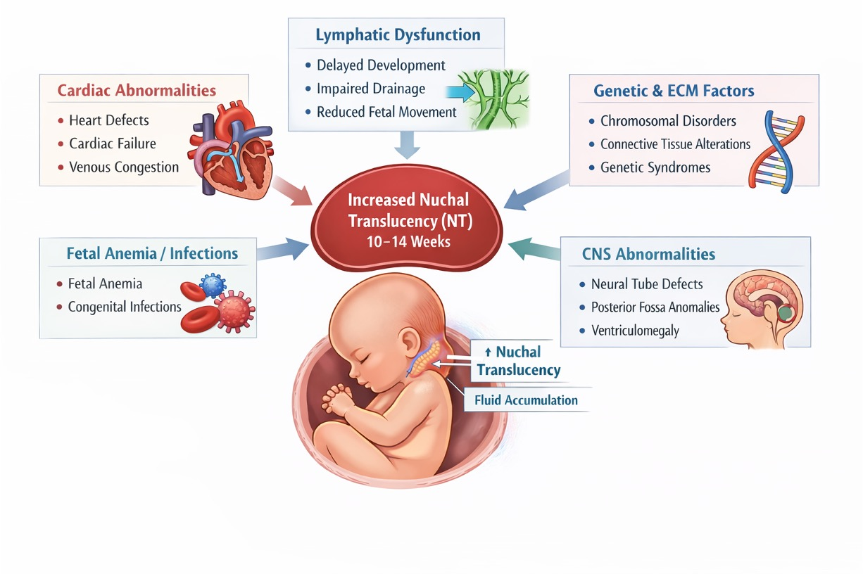
Association Between Increased Nuchal Translucency and Foetal CNS Abnormalities in Euploid Foetuses: Systematic Review and Meta-Analysis


 , ,
, ,
Abstract
1. Introduction
2. Methods
2.1. Literature Search and Study Selection
2.2. Data Extraction and Quality Assessment
2.3. Statistical Analysis
3. Results
Quality of Studies
4. Discussion
4.1. Main Findings and Interpretation of Results
4.2. Clinical Implications
4.3. Strengths and Limitations
5. Conclusions
Supplementary Materials
Author Contributions
Funding
Institutional Review Board Statement
Informed Consent Statement
Data Availability Statement
Acknowledgments
Conflicts of Interest
Appendix A
Appendix A.1. Search Strategies
- ((“pregnancy trimester, first”[MeSH Terms]) OR (“foetus”[MeSH Terms])) OR (First trimester[Title/Abstract] OR first-trimester[Title/Abstract] OR 1st trimester[Title/Abstract] OR Gestational week[Title/Abstract] OR gestation*[Title/Abstract] OR foetus[Title/Abstract] OR foetal[Title/Abstract] OR foetal[Title/Abstract] OR foetus[Title/Abstract])
- (((“nuchal translucency measurement”[MeSH Terms]) OR (Nuchal translucency[Title/Abstract] OR NT[Title/Abstract]))) OR ((nuchal[Title/Abstract] AND (ultrasound[Title/Abstract] OR ultrasonography[Title/Abstract] OR scan*[Title/Abstract])))
- (((((“brain”[MeSH Terms]) OR (“brain diseases”[MeSH Terms])) OR (“central nervous system diseases”[MeSH Terms])) OR (“nervous system malformations”[MeSH Terms])) OR (“chromosome aberrations”[MeSH Terms])) OR (Brain*[Title/Abstract] OR (brain[Title/Abstract] AND (pathology*[Title/Abstract] OR defect*[Title/Abstract] OR anomal*[Title/Abstract] OR malformation*[Title/Abstract] OR adverse[Title/Abstract])) OR CNS[Title/Abstract] OR “central nervous system”[Title/Abstract] OR “neurological complication*”[Title/Abstract])
- 1, 2, and 3
- ‘first trimester pregnancy’/exp OR ‘foetus’/exp OR ‘first trimester’:ab,ti OR ‘first-trimester 1st trimester’:ab,ti OR ‘gestational week’:ab,ti OR gestation*:ab,ti OR foetus:ab,ti OR foetal:ab,ti OR foetal:ab,ti OR foetus:ab,ti
- ‘nuchal translucency measurement’/exp OR ‘nuchal translucency’:ab,ti OR nt:ab,ti OR (nuchal:ab,ti AND (ultrasound:ab,ti OR ultrasonography:ab,ti OR scan*:ab,ti))
- ‘brain disease’/exp OR ‘brain’/exp OR ‘central nervous system disease’/exp OR ‘nervous system malformation’/exp OR ‘chromosome aberration’/exp OR brain*:ab,ti OR (brain:ab,ti AND (pathology*:ab,ti OR defect*:ab,ti OR anomal*:ab,ti OR malformation*:ab,ti OR adverse:ab,ti)) OR cns:ab,ti OR ‘central nervous system’:ab,ti OR ‘neurological complication*’:ab,ti
- 1, 2, and 3
- 4 and [embase]/lim not ([embase]/lim and [medline]/lim)
- 5 and (‘article’/it or ‘review’/it)
- S1.
- (MH “Pregnancy Trimester, First”)
- S2.
- (MH “Foetus+”)
- S3.
- TI (First trimester OR first-trimester 1st trimester OR Gestational week OR gestation* OR foetus OR foetal OR foetal OR foetus) OR AB (First trimester OR first-trimester 1st trimester OR Gestational week OR gestation* OR foetus OR foetal OR foetal OR foetus)
- S4.
- S1 OR S2 OR S3
- S5.
- (MH “Nuchal Translucency Measurement”)
- S6.
- TI (Nuchal translucency OR NT) OR AB (Nuchal translucency OR NT) OR TI ((nuchal AND (ultrasound OR ultrasonography OR scan*)) OR AB ((nuchal AND (ultrasound OR ultrasonography OR scan*))
- S7.
- S5 OR S6
- S8.
- (MH “Brain+”)
- S9.
- (MH “Brain Diseases+”)
- S10.
- (MH “Central Nervous System Diseases+”)
- S11.
- (MH “Nervous System Abnormalities+”)
- S12.
- (MH “Chromosome Aberrations+”)
- S13.
- TI (Brain* OR (brain AND (pathology* OR defect* OR anomal* OR malformation* OR adverse)) OR CNS OR “central nervous system” OR “neurological complication*”) OR AB (Brain* OR (brain AND (pathology* OR defect* OR anomal* OR malformation* OR adverse)) OR CNS OR “central nervous system” OR “neurological complication*”)
- S14.
- S8 OR S9 OR S10 OR S11 OR S12 OR S13
- S15.
- S4 AND S7 AND S14
Appendix A.2. PRISMA Checklist

| Section and Topic | Item # | Checklist Item | Location Where Item Is Reported |
|---|---|---|---|
| Title | |||
| Title | 1 | Identify the report as a systematic review. | Page 1 (Title) |
| Abstract | |||
| Abstract | 2 | See the PRISMA 2020 for Abstracts checklist. | Page 1 (Abstract) |
| Introduction | |||
| Rationale | 3 | Describe the rationale for the review in the context of existing knowledge. | Page 1 (Introduction, paragraphs 1–2) |
| Objectives | 4 | Provide an explicit statement of the objective(s) or question(s) the review addresses. | Page 2 (Introduction, final paragraph) |
| Methods | |||
| Eligibility criteria | 5 | Specify the inclusion and exclusion criteria for the review and how studies were grouped for the syntheses. | Page 2 (Methods–Literature search and study selection) |
| Information sources | 6 | Specify all databases, registers, websites, organisations, reference lists, and other sources searched or consulted to identify studies. Specify the date when each source was last searched or consulted. | Page 2 (Methods–Literature search and study selection) |
| Search strategy | 7 | Present the full search strategies for all databases, registers, and websites, including any filters and limits used. | Appendix A.2 |
| Selection process | 8 | Specify the methods used to decide whether a study met the inclusion criteria of the review, including how many reviewers screened each record and each report retrieved, whether they worked independently, and, if applicable, details of automation tools used in the process. | Page 2 (Methods–Literature search and study selection) |
| Data collection process | 9 | Specify the methods used to collect data from reports, including how many reviewers collected data from each report, whether they worked independently, any processes for obtaining or confirming data from study investigators, and, if applicable, details of automation tools used in the process. | Page 2 (Methods–Data extraction and quality assessment) |
| Data items | 10a | List and define all outcomes for which data were sought. Specify whether all results that were compatible with each outcome domain in each study were sought (e.g., for all measures, time points, analyses), and if not, the methods used to decide which results to collect. | Page 2 (Methods–Data extraction) |
| 10b | List and define all other variables for which data were sought (e.g., participant and intervention characteristics, funding sources). Describe any assumptions made about any missing or unclear information. | Page 2 (Methods–Data extraction) | |
| Study risk of bias assessment | 11 | Specify the methods used to assess risk of bias in the included studies, including details of the tool(s) used, how many reviewers assessed each study, and whether they worked independently, and if applicable, details of automation tools used in the process. | Page 2 (Methods–Data extraction and quality assessment) |
| Effect measures | 12 | Specify for each outcome the effect measure(s) (e.g., risk ratio, mean difference) used in the synthesis or presentation of results. | Page 3–4 (Methods–Statistical analysis) |
| Synthesis methods | 13a | Describe the processes used to decide which studies were eligible for each synthesis (e.g., tabulating the study intervention characteristics and comparing against the planned groups for each synthesis (item #5)). | Page 3–4 (Methods–Study selection and data extraction) |
| 13b | Describe any methods required to prepare the data for presentation or synthesis, such as handling of missing summary statistics or data conversions. | Page 3–4 (Methods–Statistical analysis) | |
| 13c | Describe any methods used to tabulate or visually display the results of individual studies and syntheses. | Page 4 (Methods–Statistical analysis; Forest plots) | |
| 13d | Describe any methods used to synthesise results and provide a rationale for the choice(s). If meta-analysis was performed, describe the model(s), method(s) to identify the presence and extent of statistical heterogeneity, and software package(s) used. | Page 3–4 (Methods–Statistical analysis) | |
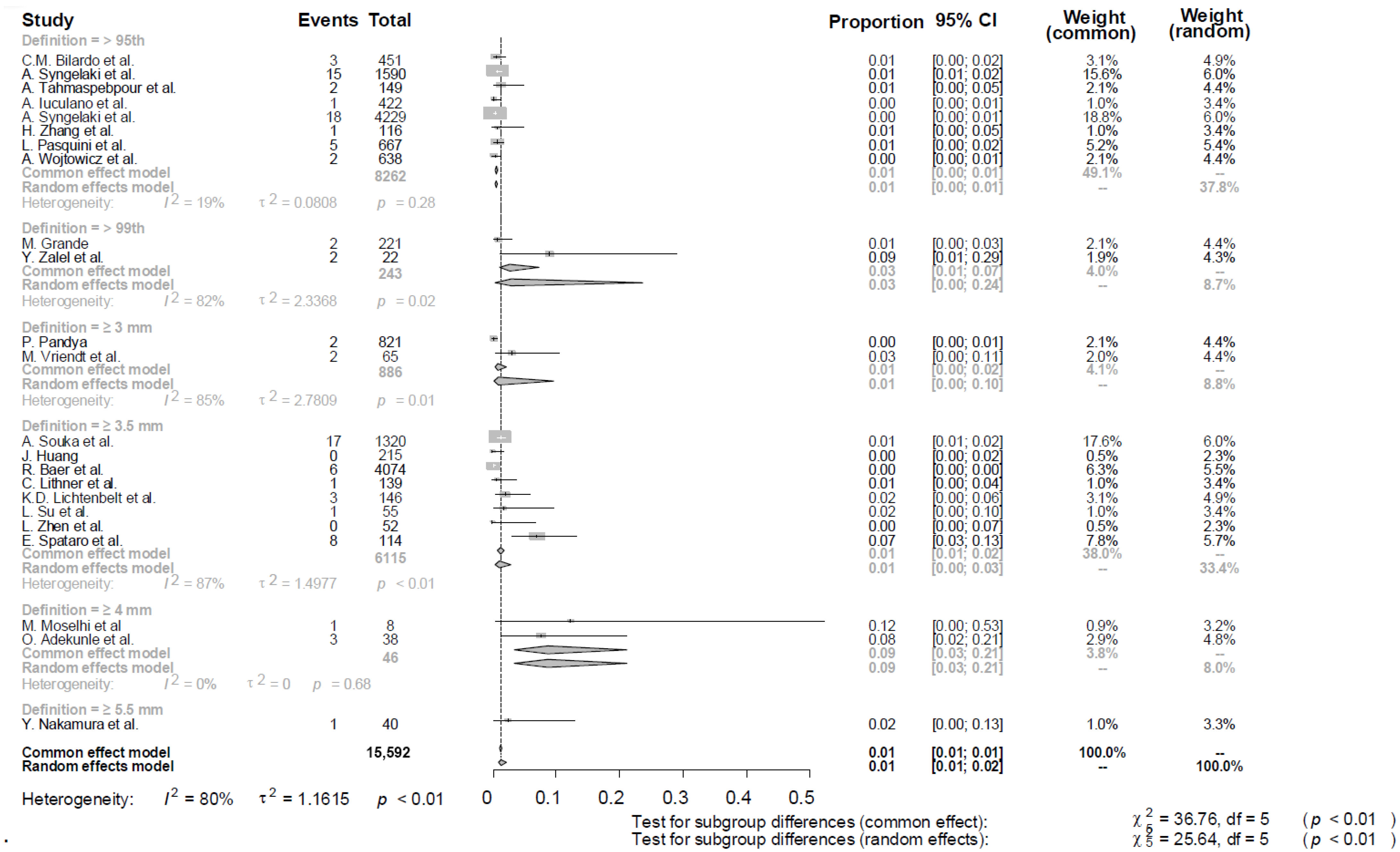
| 13e | Describe any methods used to explore possible causes of heterogeneity among study results (e.g., subgroup analysis, meta-regression). | Page 4 (Methods–Statistical analysis; subgroup analyses) | |
| 13f | Describe any sensitivity analyses conducted to assess the robustness of the synthesised results. | Page 4 (Methods–Statistical analysis) | |
| Reporting bias assessment | 14 | Describe any methods used to assess the risk of bias due to missing results in a synthesis (arising from reporting biases). | Page 2 (Methods–quality assessment) |
| Certainty assessment | 15 | Describe any methods used to assess certainty (or confidence) in the body of evidence for an outcome. | Page 2 (Methods–quality assessment) |
| Results | |||
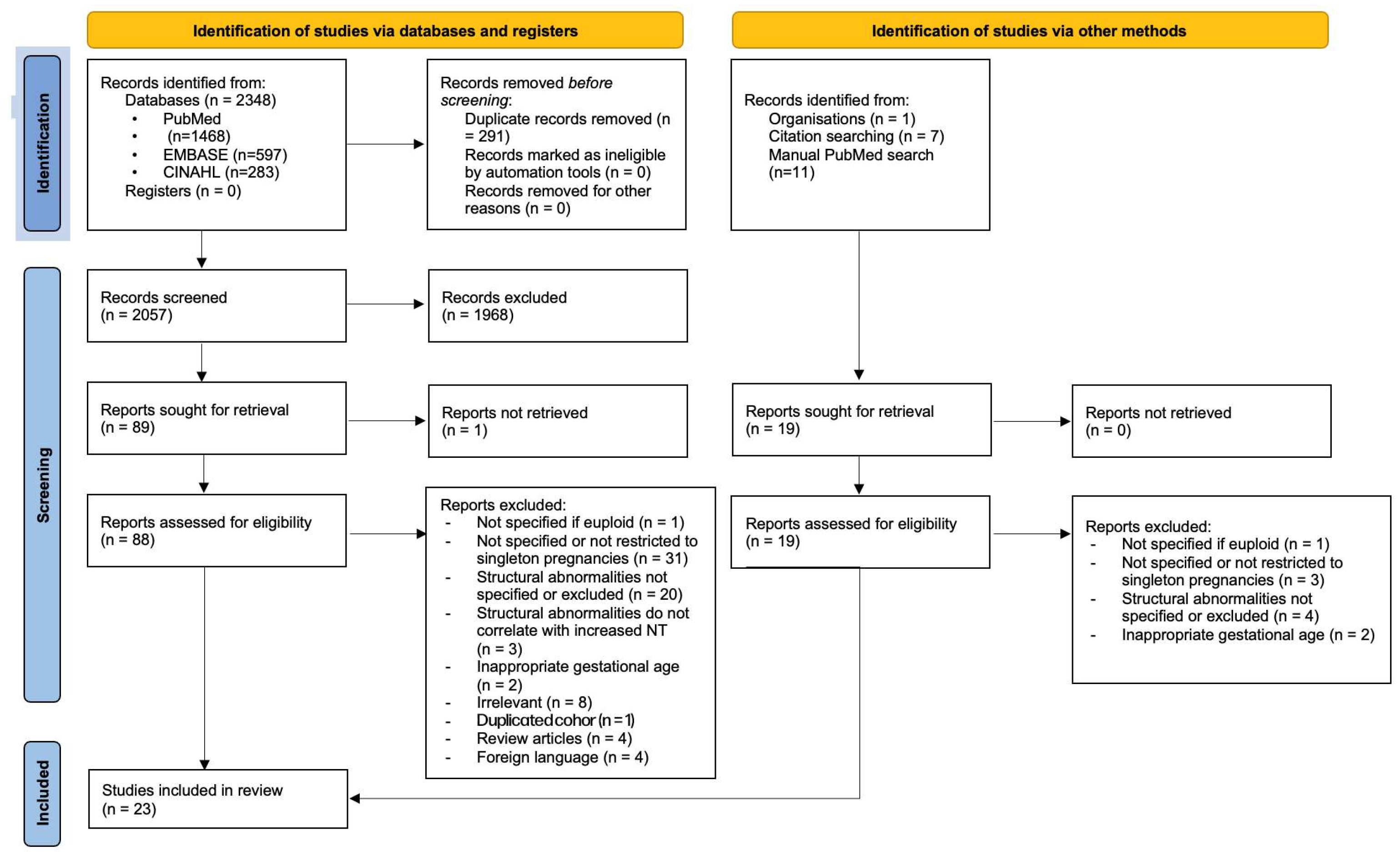
| Study selection | 16a | Describe the results of the search and selection process, from the number of records identified in the search to the number of studies included in the review, ideally using a flow diagram. | Page 4, 10 (Results–Study selection; Figure 1) |
| 16b | Cite studies that might appear to meet the inclusion criteria, but which were excluded, and explain why they were excluded. | Page 4, 10 (Results–Study selection) | |
| Study characteristics | 17 | Cite each included study and present its characteristics. | Page 4 (Results; Table 1) |
| Risk of bias in studies | 18 | Present assessments of risk of bias for each included study. | Page 6, 14 (Results–Quality of studies; Appendix A.3) |
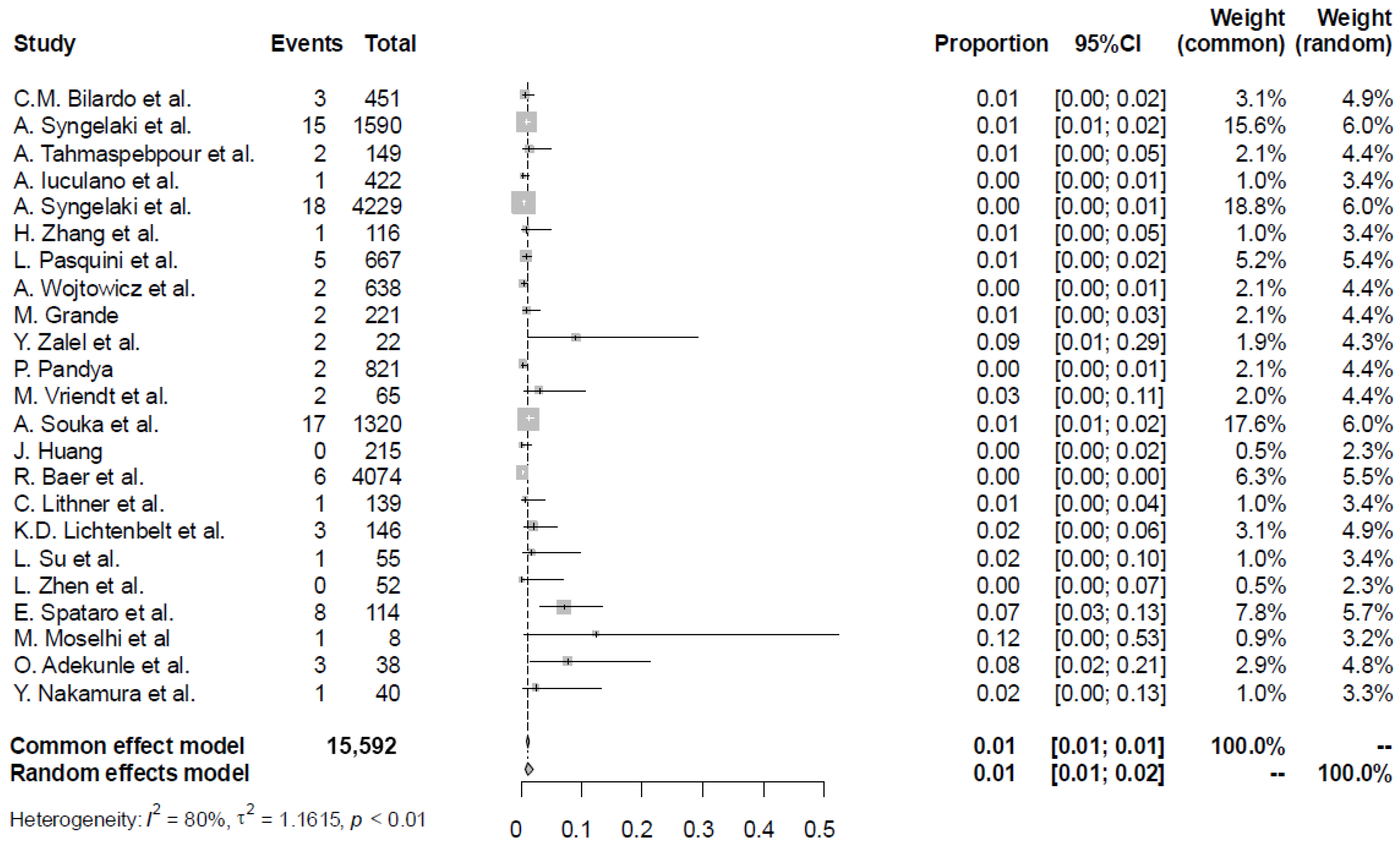
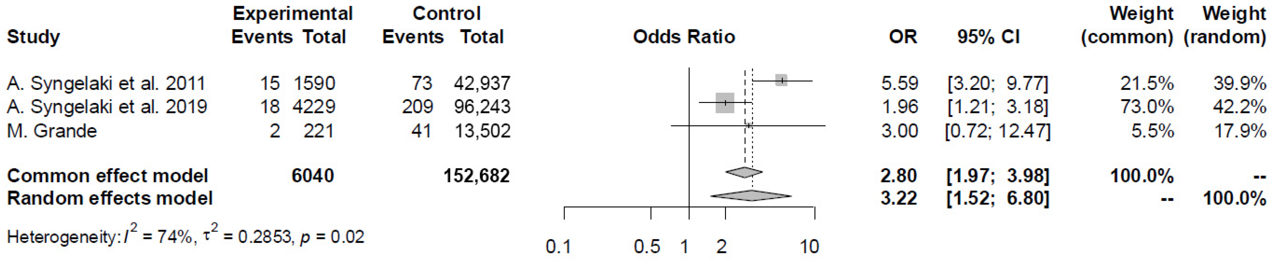
| Results of individual studies | 19 | For all outcomes, present, for each study: (a) summary statistics for each group (where appropriate) and (b) an effect estimate and its precision (e.g., confidence/credible interval), ideally using structured tables or plots. | Page 5, 6 (Results; Table 1, Figure 2, Figure 3 and Figure 4) |
| Results of syntheses | 20a | For each synthesis, briefly summarise the characteristics and risk of bias among contributing studies. | Page 6 (Results–Quality of studies) |
| 20b | Present the results of all statistical syntheses conducted. If meta-analysis was done, present for each the summary estimate and its precision (e.g., confidence/credible interval) and measures of statistical heterogeneity. If comparing groups, describe the direction of the effect. | Page 5, 6 (Results; Figure 2, Figure 3 and Figure 4) | |
| 20c | Present the results of all investigations of possible causes of heterogeneity among study results. | Page 5, 6 (Results; subgroup analyses) | |
| 20d | Present results of all sensitivity analyses conducted to assess the robustness of the synthesised results. | Not applicable | |
| Reporting biases | 21 | Present assessments of risk of bias due to missing results (arising from reporting biases) for each synthesis assessed. | Page 6 (Results quality of studies) |
| Certainty of evidence | 22 | Present assessments of certainty (or confidence) in the body of evidence for each outcome assessed. | Page 5, 6 (Results) |
| Discussion | |||
| Discussion | 23a | Provide a general interpretation of the results in the context of other evidence. | Page 7–9 (Discussion–Main findings) |
| 23b | Discuss any limitations of the evidence included in the review. | Page 8 (Discussion–Strengths and limitations) | |
| 23c | Discuss any limitations of the review processes used. | Page 8 (Discussion–Strengths and limitations) | |
| 23d | Discuss implications of the results for practice, policy, and future research. | Page 8 (Discussion–Clinical implications and future research) | |
| Other information | |||
| Registration and protocol | 24a | Provide registration information for the review, including the register name and registration number, or state that the review was not registered. | Page 2 (Methods–PROSPERO registration) |
| 24b | Indicate where the review protocol can be accessed, or state that a protocol was not prepared. | Page 2 (Methods) | |
| 24c | Describe and explain any amendments to information provided at registration or in the protocol. | No amendments | |
| Support | 25 | Describe sources of financial or non-financial support for the review, and the role of the funders or sponsors in the review. | Page 1 (Abstract), Page 9 (Funding) |
| Competing interests | 26 | Declare any competing interests of review authors. | Page 9 (Competing interests) |
| Availability of data, code, and other materials | 27 | Report which of the following are publicly available and where they can be found: template data collection forms; data extracted from included studies; data used for all analyses; analytic code; any other materials used in the review. | Data extracted from included studies |
Appendix A.3. Newcastle Ottawa Scale Quality Assessment
| Author | Selection | Comparability | Outcome | Final Score | Final Score |
|---|---|---|---|---|---|
| Pandya, 1995 [11] | 2 | 1 | 1 | 4/9 | Poor |
| Moselhi, 1996 [12] | 2 | 1 | 1 | 4/9 | Poor |
| Adekunle, 1999 [13] | 2 | 1 | 1 | 4/9 | Poor |
| Souka, 2001 [14] | 2 | 1 | 3 | 6/9 | Fair |
| Bilardo, 2007 [15] | 2 | 1 | 3 | 6/9 | Fair |
| Syngelaki, 2011 [16] | 4 | 2 | 3 | 9/9 | Good |
| Grande, 2012 [17] | 4 | 1 | 3 | 8/9 | Good |
| Tahmasebpour, 2012 [18] | 3 | 1 | 2 | 6/9 | Good |
| Huang et al., 2014 [19] | 2 | 1 | 2 | 5/9 | Fair |
| Baer et al., 2014 [20] | 4 | 2 | 3 | 9/9 | Good |
| Lithner et al., 2015 [21] | 2 | 1 | 2 | 5/9 | Fair |
| Lichtenbelt et al., 2015 [22] | 3 | 1 | 1 | 5/9 | Poor |
| Iuculano et al., 2016 [23] | 2 | 1 | 1 | 4/9 | Poor |
| Zalel et al., 2017 [24] | 2 | 1 | 2 | 5/9 | Fair |
| Syngelaki et al., 2019 [25] | 4 | 2 | 3 | 9/9 | Good |
| Su et al., 2019 [26] | 3 | 1 | 1 | 5/9 | Poor |
| Nakamura et al., 2020 [27] | 2 | 1 | 1 | 4/9 | Poor |
| Zhen et al., 2022 [28] | 2 | 1 | 2 | 5/9 | Fair |
| Zhang et al., 2023 [29] | 3 | 1 | 2 | 6/9 | Fair |
| Spataro et al., 2023 [30] | 3 | 1 | 3 | 7/9 | Good |
| Pasquini et al., 2023 [31] | 4 | 1 | 1 | 6/9 | Poor |
| Wojtowicz et al., 2024 [32] | 3 | 1 | 1 | 5/9 | Poor |
| Vriendt et al., 2025 [33] | 3 | 1 | 3 | 8/9 | Good |
References
- Nicolaides, K.H.; Azar, G.; Byrne, D.; Mansur, C.; Marks, K. Fetal nuchal translucency: Ultrasound screening for chromosomal defects in first trimester of pregnancy. BMJ 1992, 304, 867–869. [Google Scholar] [CrossRef]
- Nicolaides, K.H. First-trimester screening for chromosomal abnormalities. Semin. Perinatol. 2005, 29, 190–194. [Google Scholar] [CrossRef] [PubMed]
- Westin, M.; Saltvedt, S.; Almström, H.; Grunewald, C.; Valentin, L. By how much does increased nuchal translucency increase the risk of adverse pregnancy outcome in chromosomally normal fetuses? A study of 16,260 fetuses derived from an unselected pregnant population. Ultrasound Obstet. Gynecol. 2007, 29, 150–158. [Google Scholar] [CrossRef]
- Nicolaides, K.H.; Heath, V.; Cicero, S. Increased fetal nuchal translucency at 11–14 weeks. Prenat. Diagn. 2002, 22, 308–315. [Google Scholar] [CrossRef] [PubMed]
- Ghi, T.; Huggon, I.; Zosmer, N.; Nicolaides, K. Incidence of major structural cardiac defects associated with increased nuchal translucency but normal karyotype. Ultrasound Obstet. Gynecol. 2001, 18, 610–614. [Google Scholar] [CrossRef] [PubMed]
- Wright, D.; Kagan, K.O.; Molina, F.S.; Gazzoni, A.; Nicolaides, K.H. A mixture model of nuchal translucency thickness in screening for chromosomal defects. Ultrasound Obstet. Gynecol. 2008, 31, 376–383. [Google Scholar] [CrossRef]
- Page, M.J.; McKenzie, J.E.; Bossuyt, P.M.; Boutron, I.; Hoffmann, T.C.; Mulrow, C.D.; Shamseer, L.; Tetzlaff, J.M.; Akl, E.A.; Brennan, S.E.; et al. The PRISMA 2020 statement: An updated guideline for reporting systematic reviews. BMJ 2021, 372, n71. [Google Scholar] [CrossRef]
- Gualdi-Russo, E.; Zaccagni, L. The Newcastle–Ottawa Scale for Assessing the Quality of Studies in Systematic Reviews. Publications 2026, 14, 4. [Google Scholar] [CrossRef]
- Friedrich, J.O.; Adhikari, N.K.; Beyene, J. Inclusion of zero total event trials in meta-analyses maintains analytic consistency and incorporates all available data. BMC Med. Res. Methodol. 2007, 7, 5. [Google Scholar] [CrossRef]
- Riley, R.D.; Abrams, K.R.; Sutton, A.J.; Lambert, P.C.; Thompson, J.R. Bivariate random-effects meta-analysis and the estimation of between-study correlation. BMC Med. Res. Methodol. 2007, 7, 3. [Google Scholar] [CrossRef]
- Pandya, P.P.; Santiago, C.; Snijders, R.J.; Nicolaides, K.H. First trimester fetal nuchal translucency. Curr. Opin. Obstet. Gynecol. 1995, 7, 95–102. [Google Scholar] [CrossRef]
- Moselhi, M.; Thilaganathan, B. Nuchal translucency: A marker for the antenatal diagnosis of aortic coarctation. Br. J. Obstet. Gynaecol. 1996, 103, 1044–1045. [Google Scholar] [CrossRef] [PubMed]
- Adekunle, O.; Gopee, A.; el-Sayed, M.; Thilaganathan, B. Increased first trimester nuchal translucency: Pregnancy and infant outcomes after routine screening for Down’s syndrome in an unselected antenatal population. Br. J. Radiol. 1999, 72, 457–460. [Google Scholar] [CrossRef] [PubMed]
- Souka, A.P.; Krampl, E.; Bakalis, S.; Heath, V.; Nicolaides, K.H. Outcome of pregnancy in chromosomally normal fetuses with increased nuchal translucency in the first trimester. Ultrasound Obstet. Gynecol. 2001, 18, 9–17. [Google Scholar] [CrossRef] [PubMed]
- Bilardo, C.M.; Müller, M.A.; Pajkrt, E.; Clur, S.A.; van Zalen, M.M.; Bijlsma, E.K. Increased nuchal translucency thickness and normal karyotype: Time for parental reassurance. Ultrasound Obstet. Gynecol. 2007, 30, 11–18. [Google Scholar] [CrossRef]
- Syngelaki, A.; Chelemen, T.; Dagklis, T.; Allan, L.; Nicolaides, K.H. Challenges in the diagnosis of fetal non-chromosomal abnormalities at 11–13 weeks. Prenat. Diagn. 2011, 31, 90–102. [Google Scholar] [CrossRef]
- Grande, M.; Arigita, M.; Borobio, V.; Jimenez, J.M.; Fernandez, S.; Borrell, A. First-trimester detection of structural abnormalities and the role of aneuploidy markers. Ultrasound Obstet. Gynecol. 2012, 39, 157–163. [Google Scholar] [CrossRef]
- Tahmasebpour, A.; Rafiee, N.B.; Ghaffari, S.; Jamal, A. Increased nuchal translucency and pregnancy outcome. Iran. J. Public. Health 2012, 41, 92–97. [Google Scholar]
- Huang, J.; Poon, L.C.; Akolekar, R.; Choy, K.W.; Leung, T.Y.; Nicolaides, K.H. Is high fetal nuchal translucency associated with submicroscopic chromosomal abnormalities on array CGH? Ultrasound Obstet. Gynecol. 2014, 43, 620–624. [Google Scholar] [CrossRef]
- Baer, R.J.; Norton, M.E.; Shaw, G.M.; Flessel, M.C.; Goldman, S.; Currier, R.J.; Jelliffe-Pawlowski, L.L. Risk of selected structural abnormalities in infants after increased nuchal translucency measurement. Am. J. Obstet. Gynecol. 2014, 211, 675.e1–675.e19. [Google Scholar] [CrossRef]
- Lithner, C.U.; Kublickas, M.; Ek, S. Pregnancy outcome for fetuses with increased nuchal translucency but normal karyotype. J. Med. Screen. 2016, 23, 1–6. [Google Scholar] [CrossRef] [PubMed]
- Lichtenbelt, K.D.; Diemel, B.D.; Koster, M.P.; Manten, G.T.; Siljee, J.; Schuring-Blom, G.H.; Page-Christiaens, G.C. Detection of fetal chromosomal anomalies: Does nuchal translucency measurement have added value in the era of non-invasive prenatal testing? Prenat. Diagn. 2015, 35, 663–668. [Google Scholar] [CrossRef] [PubMed]
- Iuculano, A.; Pagani, G.; Stagnati, V.; Floris, M.; Ibba, R.M.; Monni, G. Pregnancy outcome and long-term follow-up of fetuses with isolated increased NT: A retrospective cohort study. J. Perinat. Med. 2016, 44, 237–242. [Google Scholar] [CrossRef] [PubMed]
- Zalel, Y.; Zemet, R.; Kivilevitch, Z. The added value of detailed early anomaly scan in fetuses with increased nuchal translucency. Prenat. Diagn. 2017, 37, 235–243. [Google Scholar] [CrossRef]
- Syngelaki, A.; Hammami, A.; Bower, S.; Zidere, V.; Akolekar, R.; Nicolaides, K.H. Diagnosis of fetal non-chromosomal abnormalities on routine ultrasound examination at 11–13 weeks’ gestation. Ultrasound Obstet. Gynecol. 2019, 54, 468–476. [Google Scholar] [CrossRef]
- Su, L.; Huang, H.; An, G.; Cai, M.; Wu, X.; Li, Y.; Xie, X.; Lin, Y.; Wang, M.; Xu, L. Clinical application of chromosomal microarray analysis in fetuses with increased nuchal translucency and normal karyotype. Mol. Genet. Genom. Med. 2019, 7, e811. [Google Scholar] [CrossRef]
- Nakamura, Y.; Fujita, S.; Yamada, K.; Song, M.; Tajima, A.; Matsumoto, J.; Kihira, C.; Kurata, Y.; Arakawa, H.; Tamura, C. Outcome of the fetuses with severely increased nuchal translucency thickness in the first trimester. J. Obstet. Gynaecol. Res. 2020, 46, 1977–1981. [Google Scholar] [CrossRef]
- Zhen, L.; Pan, M.; Li, Y.T.; Cao, Q.; Xu, L.L.; Li, D.Z. The 16-week sonographic findings in fetuses with increased nuchal translucency and a normal array. J. Matern. Fetal Neonatal Med. 2022, 35, 9435–9439. [Google Scholar] [CrossRef]
- Zhang, H.; Wang, S.; Feng, C.; Zhao, H.; Zhang, W.; Sun, Y.; Yang, H. Chromosomal abnormalities and structural defects in fetuses with increased nuchal translucency at a Chinese tertiary medical center. Front. Med. 2023, 10, 1158554. [Google Scholar] [CrossRef]
- Spataro, E.; Cordisco, A.; Luchi, C.; Filardi, G.R.; Masini, G.; Pasquini, L. Increased nuchal translucency with normal karyotype and genomic microarray analysis: A multicenter observational study. Int. J. Gynaecol. Obstet. 2023, 161, 1040–1045. [Google Scholar] [CrossRef]
- Pasquini, L.; Ponziani, I.; Spataro, E.; Masini, G.; Biancareddu, E.; Cordisco, A.; Petraglia, F. Elevated nuchal translucency, is it time to discuss the cut off? Int. J. Gynaecol. Obstet. 2023, 163, 540–546. [Google Scholar] [CrossRef]
- Wójtowicz, A.; Kowalczyk, K.; Szewczyk, K.; Madetko-Talowska, A.; Wójtowicz, W.; Huras, H.; Bik-Multanowski, M.; Beata, N. Array Comparative Genomic Hybridization (aCGH) Results among Patients Referred to Invasive Prenatal Testing after First-Trimester Screening: A Comprehensive Cohort Study. Diagnostics 2024, 14, 2186. [Google Scholar] [CrossRef] [PubMed]
- Vriendt, M.D.E.; Rooryck, C.; Madar, H.; Coatleven, F.; Vincienne, M.; Prier, P.; Naudion, S.; Sentilhes, L.; Bouchghoul, H. Outcomes associated with fetal nuchal translucency between 3.0 and 3.4 mm in the first trimester. Acta Obstet. Gynecol. Scand. 2025, 104, 629–636. [Google Scholar] [CrossRef] [PubMed]
- EUROCAT Prevalence Tables: Nervous System Anomalies. European Surveillance of Congenital Anomalies. Available online: https://eu-rd-platform.jrc.ec.europa.eu/eurocat/eurocat-data/prevalence_en (accessed on 1 February 2026).
- Malinger, G.; Birnbam, R.; Haratz, K.K. Dedicated neurosonography for recognition of pathology associated with mild-to-moderate ventriculomegaly. Ultrasound Obstet. Gynecol. 2020, 56, 319–323. [Google Scholar] [CrossRef] [PubMed]
- Malinger, G.; Paladini, D.; Haratz, K.K.; Monteagudo, A.; Pilu, G.L.; Timor-Tritsch, I.E. ISUOG Practice Guidelines (updated): Sonographic examination of the fetal central nervous system. Part 1: Performance of screening examination and indications for targeted neurosonography. Ultrasound Obstet. Gynecol. 2020, 56, 476–484, Erratum in Ultrasound Obstet. Gynecol. 2022, 60, 591. [Google Scholar] [CrossRef]
- Paladini, D.; Malinger, G.; Birnbaum, R.; Monteagudo, A.; Pilu, G.; Salomon, L.J.; Timor-Tritsch, I.E. ISUOG Practice Guidelines (updated): Sonographic examination of the fetal central nervous system. Part 2: Performance of targeted neurosonography. Ultrasound Obstet. Gynecol. 2021, 57, 661–671, Erratum in Ultrasound Obstet. Gynecol. 2022, 60, 591. [Google Scholar] [CrossRef]
- Weichert, J.; Scharf, J.L. Advancements in Artificial Intelligence for Fetal Neurosonography: A Comprehensive Review. J. Clin. Med. 2024, 13, 5626. [Google Scholar] [CrossRef]
- Tarui, T.; Gimovsky, A.C.; Madan, N. Fetal neuroimaging applications for diagnosis and counseling of brain anomalies: Current practice and future diagnostic strategies. Semin. Fetal Neonatal Med. 2024, 29, 101525. [Google Scholar] [CrossRef]
- Volpe, N.; Dall’Asta, A.; Di Pasquo, E.; Frusca, T.; Ghi, T. First-trimester fetal neurosonography: Technique and diagnostic potential. Ultrasound Obstet. Gynecol. 2021, 57, 204–214. [Google Scholar] [CrossRef]
- Altmann, R.; Schertler, C.; Scharnreitner, I.; Arzt, W.; Dertinger, S.; Scheier, M. Diagnosis of Fetal Posterior Fossa Malformations in High-Risk Pregnancies at 12–14 Gestational Weeks by Transvaginal Ultrasound Examination. Fetal Diagn. Ther. 2020, 47, 182–187. [Google Scholar] [CrossRef]
- Syngelaki, A.; Mitsigiorgi, R.; Goadsby, J.; Hamed, K.; Akolekar, R.; Nicolaides, K.H. Routine 36-week scan: Diagnosis of fetal abnormalities. Ultrasound Obstet. Gynecol. 2025, 65, 427–435. [Google Scholar] [CrossRef]
- Vargas-Rodríguez, C.; Chimenea, A.; Antiñolo, G.; García-Díaz, L. Insights into neurosonographic indicators for prenatal diagnosis of fetal neurological anomalies and cortical development: A systematic review of the literature. Early Hum. Dev. 2024, 197, 106–109. [Google Scholar] [CrossRef]


| First Author, Year | NT Cut-Off | Total | Defects | Specified Defect |
|---|---|---|---|---|
| Pandya, 1995 [11] | ≥3 mm | 821 | 2 | Anencephaly, holoprosencephaly |
| Moselhi, 1996 [12] | ≥4 mm | 8 | 1 | Spina bifida |
| Adekunle, 1999 [13] | ≥4 mm | 38 | 3 | Anencephaly, encephalocele, macrocephaly |
| Souka, 2001 [14] | ≥3.5 mm | 1320 | 17 | Anencephaly, spina bifida, encephalocele, holoprosencephaly, ventriculomegaly, Dandy Walker malformation |
| Bilardo, 2007 [15] | >95th | 451 | 3 | Anencephaly, spina bifida, Dandy-Walker malformation |
| Syngelaki, 2011 [16] | >95th | 1590 | 15 | Acrania, spina bifida, holoprosencephaly, agenesis of corpus callosum, vermian agenesis |
| Grande, 2012 [17] | >99th | 221 | 2 | Acrania, encephalocele |
| Tahmasebpour, 2012 [18] | >95th | 149 | 2 | Ventriculomegaly, Dandy-Walker malformation |
| Huang et al., 2014 [19] | ≥3.5 mm | 215 | 0 | None |
| Baer et al., 2014 [20] | ≥3.5 mm | 4074 | 6 | Hydrocephalus, encephalocele, brain deformity |
| Lithner et al., 2015 [21] | ≥3.5 mm | 139 | 1 | CNS defect (not specified) |
| Lichtenbelt et al., 2015 [22] | ≥3.5 mm | 146 | 3 | Ventriculomegaly, holoprosencephaly, complex brain |
| Iuculano et al., 2016 [23] | >95th | 422 | 1 | Arachnoid cyst |
| Zalel et al., 2017 [24] | >99th | 22 | 2 | Holoprosencephaly, vermian agenesis |
| Syngelaki et al., 2019 [25] | >95th | 4229 | 18 | Holoprosencephaly, encephalocele, spina bifida, agenesis of corpus callosum, arachnoid cyst, craniosynostosis, occipital dermoid cyst |
| Su et al., 2019 [26] | ≥3.5 mm | 55 | 1 | Ventriculomegaly |
| Nakamura et al., 2020 [27] | ≥5.5 mm | 40 | 1 | Encephalocele |
| Zhen et al., 2022 [28] | ≥3.5 mm | 52 | 0 | None |
| Zhang et al., 2023 [29] | >95th | 116 | 1 | Ventriculomegaly |
| Spataro et al., 2023 [30] | ≥3.5 mm | 114 | 8 | Ventriculomegaly, agenesis of corpus callosum, vermis hypoplasia |
| Pasquini et al., 2023 [31] | >95th | 667 | 5 | Holoprosencephaly, agenesis of corpus callosum, polymicrogyria, complex brain |
| Wojtowicz et al., 2024 [32] | >95th | 638 | 2 | Microcephaly, cerebellar hypoplasia |
| Vriendt et al., 2025 [33] | ≥3 mm | 65 | 2 | Agenesis of the corpus callosum, posterior fossa abnormality |
| Total | 15,592 | 96 |
Disclaimer/Publisher’s Note: The statements, opinions and data contained in all publications are solely those of the individual author(s) and contributor(s) and not of MDPI and/or the editor(s). MDPI and/or the editor(s) disclaim responsibility for any injury to people or property resulting from any ideas, methods, instructions or products referred to in the content. |
© 2026 by the authors. Licensee MDPI, Basel, Switzerland. This article is an open access article distributed under the terms and conditions of the Creative Commons Attribution (CC BY) license.
Share and Cite
Mackina, G.; Ricci, B.M.; Moser, M.; Chatzakis, C.; Nicolaides, K.H.; Arechvo, A. Association Between Increased Nuchal Translucency and Foetal CNS Abnormalities in Euploid Foetuses: Systematic Review and Meta-Analysis. Diagnostics 2026, 16, 1250. https://doi.org/10.3390/diagnostics16091250
Mackina G, Ricci BM, Moser M, Chatzakis C, Nicolaides KH, Arechvo A. Association Between Increased Nuchal Translucency and Foetal CNS Abnormalities in Euploid Foetuses: Systematic Review and Meta-Analysis. Diagnostics. 2026; 16(9):1250. https://doi.org/10.3390/diagnostics16091250
Chicago/Turabian StyleMackina, Giula, Belen M. Ricci, Mirjam Moser, Christos Chatzakis, Kypros H. Nicolaides, and Anastasija Arechvo. 2026. "Association Between Increased Nuchal Translucency and Foetal CNS Abnormalities in Euploid Foetuses: Systematic Review and Meta-Analysis" Diagnostics 16, no. 9: 1250. https://doi.org/10.3390/diagnostics16091250
APA StyleMackina, G., Ricci, B. M., Moser, M., Chatzakis, C., Nicolaides, K. H., & Arechvo, A. (2026). Association Between Increased Nuchal Translucency and Foetal CNS Abnormalities in Euploid Foetuses: Systematic Review and Meta-Analysis. Diagnostics, 16(9), 1250. https://doi.org/10.3390/diagnostics16091250