Association of Angiography-Derived Coronary Microvascular Parameters with Periprocedural Myocardial Injury in Uncomplicated Elective Percutaneous Coronary Interventions

 , , ,
, , ,
Abstract
1. Introduction
2. Materials and Methods
2.1. Study Design
2.2. Angiographic Analysis–μFR, CFVR and AMR
2.3. Clinical Outcome Measures
2.4. Data Analysis
2.5. Use of Artificial Intelligence
3. Results
3.1. Study Population Baseline Characteristics
3.2. Baseline Angiography and Procedural Characteristics
3.3. Coronary Physiology in Overall Population
3.4. Periprocedural MI and Coronary Physiologic Indexes
4. Discussion
5. Limitations
6. Conclusions
7. Impact on Daily Practice–Perspectives
Author Contributions
Funding
Institutional Review Board Statement
Informed Consent Statement
Data Availability Statement
Acknowledgments
Conflicts of Interest
References
- Sandoval, Y.; Jaffe, A.S. Type 2 Myocardial Infarction and Myocardial Injury. Clin. Chem. 2017, 63, 101–107. [Google Scholar] [CrossRef] [Scilit] [PubMed]
- Thygesen, K.; Alpert, J.S.; Jaffe, A.S.; Chaitman, B.R.; Bax, J.J.; Morrow, D.A.; White, H.D. Executive Group on behalf of the Joint European Society of Cardiology (ESC)/American College of Cardiology (ACC)/American Heart Association (AHA)/World Heart Federation (WHF) Task Force for the Universal Definition of Myocardial Infarction. Fourth Universal Definition of Myocardial Infarction. J. Am. Coll. Cardiol. 2018, 72, 2231–2264. [Google Scholar] [PubMed]
- Ueki, Y.; Kuwahara, K. Periprocedural myocardial infarction in patients undergoing percutaneous coronary intervention. J. Cardiol. 2023, 81, 364–372. [Google Scholar] [CrossRef] [Scilit] [PubMed]
- Herrmann, J. Peri-procedural myocardial injury: 2005 update. Eur. Heart J. 2005, 26, 2493–2519. [Google Scholar] [CrossRef] [Scilit]
- Fearon, W.F.; Kobayashi, Y. Invasive Assessment of the Coronary Microvasculature: The Index of Microcirculatory Resistance. Circ. Cardiovasc. Interv. 2017, 10, e005361. [Google Scholar] [CrossRef] [Scilit]
- El Farissi, M.; Zimmermann, F.M.; De Maria, G.L.; van Royen, N.; van Leeuwen, M.A.; Carrick, D.; Carberry, J.; Wijnbergen, I.F.; Konijnenberg, L.S.; Hoole, S.P.; et al. The Index of Microcirculatory Resistance After Primary PCI: A Pooled Analysis of Individual Patient Data. JACC Cardiovasc. Interv. 2023, 16, 2383–2392. [Google Scholar] [CrossRef] [Scilit]
- Lee, J.M.; Layland, J.; Jung, J.-H.; Lee, H.-J.; Echavarria-Pinto, M.; Watkins, S.; Yong, A.S.; Doh, J.-H.; Nam, C.-W.; Shin, E.-S.; et al. Integrated physiologic assessment of ischemic heart disease in real-world practice using index of microcirculatory resistance and fractional flow reserve: Insights from the International Index of Microcirculatory Resistance Registry. Circ. Cardiovasc. Interv. 2015, 8, e002857. [Google Scholar] [CrossRef] [Scilit]
- Oikonomou, E.; Theofilis, P.; Lampsas, S.; Katsarou, O.; Kalogeras, K.; Marinos, G.; Tsatsaragkou, A.; Anastasiou, A.; Lysandrou, A.; Gounaridi, M.-I.; et al. Current Concepts and Future Applications of Non-Invasive Functional and Anatomical Evaluation of Coronary Artery Disease. Life 2022, 12, 1803. [Google Scholar] [CrossRef] [Scilit]
- Campisi, R. Noninvasive assessment of coronary microvascular function in women at risk for ischaemic heart disease. Int. J. Clin. Pract. 2008, 62, 300–307. [Google Scholar] [CrossRef] [Scilit]
- Neumann, F.-J.; Sousa-Uva, M.; Ahlsson, A.; Alfonso, F.; Banning, A.P.; Benedetto, U.; A Byrne, R.; Collet, J.-P.; Falk, V.; Head, S.J.; et al. 2018 ESC/EACTS Guidelines on myocardial revascularization. Eur. Heart J. 2019, 40, 87–165. [Google Scholar] [CrossRef] [Scilit]
- Tu, S.; Ding, D.; Chang, Y.; Li, C.; Wijns, W.; Xu, B. Diagnostic accuracy of quantitative flow ratio for assessment of coronary stenosis significance from a single angiographic view: A novel method based on bifurcation fractal law. Catheter. Cardiovasc. Interv. 2021, 97, 1040–1047. [Google Scholar] [CrossRef] [Scilit] [PubMed]
- Ding, D.; Tu, S.; Li, Y.; Li, C.; Yu, W.; Liu, X.; Leone, A.M.; Aurigemma, C.; Romagnoli, E.; Vergallo, R.; et al. Quantitative flow ratio modulated by intracoronary optical coherence tomography for predicting physiological efficacy of percutaneous coronary intervention. Catheter. Cardiovasc. Interv. 2023, 102, 36–45. [Google Scholar] [CrossRef] [Scilit] [PubMed]
- Tu, S.; Echavarria-Pinto, M.; von Birgelen, C.; Holm, N.R.; Pyxaras, S.A.; Kumsars, I.; Lam, M.K.; Valkenburg, I.; Toth, G.G.; Li, Y.; et al. Fractional Flow Reserve and Coronary Bifurcation Anatomy: A Novel Quantitative Model to Assess and Report the Stenosis Severity of Bifurcation Lesions. JACC Cardiovasc. Interv. 2015, 8, 564–574. [Google Scholar] [CrossRef] [Scilit] [PubMed]
- Chamuleau, S.A.; Meuwissen, M.; van Eck-Smit, B.L.; Koch, K.T.; de Jong, A.; de Winter, R.J.; Schotborgh, C.E.; Bax, M.; Verberne, H.J.; Tijssen, J.G.; et al. Fractional flow reserve, absolute and relative coronary blood flow velocity reserve in relation to the results of technetium-99m sestamibi single-photon emission computed tomography in patients with two-vessel coronary artery disease. J. Am. Coll. Cardiol. 2001, 37, 1316–1322. [Google Scholar] [CrossRef] [Scilit]
- Tu, S.; Westra, J.; Yang, J.; Von Birgelen, C.; Ferrara, A.; Pellicano, M.; Nef, H.; Tebaldi, M.; Murasato, Y.; Lansky, A.; et al. Diagnostic Accuracy of Fast Computational Approaches to Derive Fractional Flow Reserve From Diagnostic Coronary Angiography: The International Multicenter FAVOR Pilot Study. JACC Cardiovasc. Interv. 2016, 9, 2024–2035. [Google Scholar] [CrossRef] [Scilit]
- Wang, H.; Wu, Q.; Yang, L.; Chen, L.; Liu, W.; Guo, J.; Xu, J. Application of AMR in evaluating microvascular dysfunction after ST-elevation myocardial infarction. Clin. Cardiol. 2024, 47, e24196. [Google Scholar] [CrossRef] [Scilit]
- Ellis, S.G.; Vandormael, M.G.; Cowley, M.J.; DiSciascio, G.; Deligonul, U.; Topol, E.J.; Bulle, T.M. Coronary morphologic and clinical determinants of procedural outcome with angioplasty for multivessel coronary disease. Implications for patient selection. Multivessel angioplasty prognosis study group. Circulation 1990, 82, 1193–1202. [Google Scholar] [CrossRef] [Scilit]
- Gaetano Antonio Lanza Morrone, D.; Pizzi, C.; Tritto, I.; Bergamaschi, L.; De Vita, A.; Villano, A.; Crea, F. Diagnostic approach for coronary microvascular dysfunction in patients with chest pain and no obstructive coronary artery disease. Trends Cardiovasc. Med. 2022, 32, 128–134. [Google Scholar] [CrossRef] [Scilit]
- Gulati, M.; Cooper-DeHoff, R.M.; McClure, C.; Johnson, B.D.; Shaw, L.J.; Handberg, E.M. Adverse cardiovascular outcomes in women with nonobstructive coronary artery disease: A report from the Women’s Ischemia Syndrome Evaluation Study and the St James Women Take Heart Project. Arch. Intern. Med. 2009, 169, 843–850. [Google Scholar] [CrossRef] [Scilit]
- Jespersen, L.; Hvelplund, A.; Abildstrøm, S.Z.; Pedersen, F.; Galatius, S.; Madsen, J.K.; Jørgensen, E.; Kelbæk, H.; Prescott, E. Stable angina pectoris with no obstructive coronary artery disease is associated with increased risks of major adverse cardiovascular events. Eur. Heart J. 2012, 33, 734–744. [Google Scholar] [CrossRef] [Scilit]
- De Maria, G.L.; Cuculi, F.; Patel, N.; Dawkins, S.; Fahrni, G.; Kassimis, G.; Choudhury, R.P.; Forfar, J.C.; Prendergast, B.D.; Channon, K.M.; et al. How does coronary stent implantation impact on the status of the microcirculation during primary percutaneous coronary intervention in patients with ST-elevation myocardial infarction? Eur. Heart J. 2015, 36, 3165–3177. [Google Scholar] [CrossRef] [Scilit] [PubMed]
- Scarsini, R.; Shanmuganathan, M.; De Maria, G.L.; Borlotti, A.; Kotronias, R.A.; Burrage, M.K.; Terentes-Printzios, D.; Langrish, J.; Lucking, A.; Fahrni, G.; et al. Coronary Microvascular Dysfunction Assessed by Pressure Wire and CMR After STEMI Predicts Long-Term Outcomes. JACC Cardiovasc. Imaging 2021, 14, 1948–1959. [Google Scholar] [CrossRef] [Scilit] [PubMed]
- Kwon, W.; Choi, K.H.; Lee, S.H.; Hong, D.; Shin, D.; Kim, H.K.; Park, K.H.; Choo, E.H.; Kim, C.J.; Kim, M.C.; et al. Clinical Value of Single-Projection Angiography-Derived FFR in Noninfarct-Related Artery. Circ. Cardiovasc. Interv. 2024, 17, E013844. [Google Scholar] [CrossRef] [Scilit] [PubMed]
- Fan, Y.; Fezzi, S.; Sun, P.; Ding, N.; Li, X.; Hu, X.; Wang, S.; Wijns, W.; Lu, Z.; Tu, S. In Vivo Validation of a Novel Computational Approach to Assess Microcirculatory Resistance Based on a Single Angiographic View. J. Pers. Med. 2022, 12, 1798. [Google Scholar] [CrossRef] [Scilit]
- Pim, A.L.; Tonino, P.A.; De Bruyne, B.; Pijls, N.H.; Siebert, U.; Ikeno, F.; vant Veer, M.; Klauss, V.; Manoharan, G.; Engstrøm, T.; et al. Fractional flow reserve versus angiography for guiding percutaneous coronary intervention. Rev. Port. Cardiol. 2009, 28, 229–230. [Google Scholar]
- Fearon, W.F.; Low, A.F.; Yong, A.S.; McGeoch, R.; Berry, C.; Shah, M.G.; Ho, M.Y.; Kim, H.-S.; Loh, J.P.; Oldroyd, K.G. Prognostic value of the Index of Microcirculatory Resistance measured after primary percutaneous coronary intervention. Circulation 2013, 127, 2436–2441. [Google Scholar] [CrossRef] [Scilit]
- Cortigiani, L.; Gaibazzi, N.; Ciampi, Q.; Rigo, F.; Tuttolomondo, D.; Bovenzi, F.; Gregori, D.; Carerj, S.; Pepi, M.; Pellikka, P.A.; et al. Reduction of Coronary Flow Velocity Reserve as the Main Driver of Prognostically Beneficial Coronary Revascularization. J. Am. Soc. Echocardiogr. 2025, 38, 24–32. [Google Scholar] [CrossRef] [Scilit]
- Ueki, Y.; Otsuka, T.; Bär, S.; Koskinas, K.C.; Heg, D.; Häner, J.; Siontis, G.C.M.; Praz, F.; Hunziker, L.; Lanz, J.; et al. Frequency and Outcomes of Periprocedural MI in Patients with Chronic Coronary Syndromes Undergoing PCI. J. Am. Coll. Cardiol. 2022, 79, 513–526. [Google Scholar] [CrossRef] [Scilit] [PubMed]
- Sarkisian, L.; Saaby, L.; Poulsen, T.S.; Gerke, O.; Jangaard, N.; Hosbond, S.; Diederichsen, A.C.P.; Thygesen, K.; Mickley, H. Clinical Characteristics and Outcomes of Patients with Myocardial Infarction, Myocardial Injury, and Nonelevated Troponins. Am. J. Med. 2016, 129, 446.e5–446.e21. [Google Scholar] [CrossRef] [Scilit]
- Zeitouni, M.; Silvain, J.; Guedeney, P.; Kerneis, M.; Yan, Y.; Overtchouk, P.; Barthelemy, O.; Hauguel-Moreau, M.; Choussat, R.; Helft, G.; et al. ACTION Study Group. Periprocedural myocardial infarction and injury in elective coronary stenting. Eur. Heart J. 2018, 39, 1100–1109. [Google Scholar] [CrossRef] [Scilit] [PubMed]
- Armillotta, M.; Bergamaschi, L.; Paolisso, P.; Belmonte, M.; Angeli, F.; Sansonetti, A.; Stefanizzi, A.; Bertolini, D.; Bodega, F.; Amicone, S.; et al. Prognostic Relevance of Type 4a Myocardial Infarction and Periprocedural Myocardial Injury in Patients with Non-ST-Segment-Elevation Myocardial Infarction. Circulation 2025, 151, 760–772. [Google Scholar] [CrossRef] [Scilit] [PubMed] [PubMed Central]
- de Lemos, J.A.; Newby, L.K.; Mills, N.L. A proposal for modest revision of the definition of type 1 and type 2 myocardial infarction. Circulation 2019, 140, 1773–1775. [Google Scholar] [CrossRef] [Scilit]
- Nishi, T.; Murai, T.; Ciccarelli, G.; Shah, S.V.; Kobayashi, Y.; Derimay, F.; Waseda, K.; Moonen, A.; Hoshino, M.; Hirohata, A.; et al. Prognostic Value of Coronary Microvascular Function Measured Immediately after Percutaneous Coronary Intervention in Stable Coronary Artery Disease: An International Multicenter Study. Circ. Cardiovasc. Interv. 2019, 12, e007889. [Google Scholar] [CrossRef] [Scilit] [PubMed]
- Mangiacapra, F.; Bressi, E.; Di Gioia, G.; Pellicano, M.; Di Serafino, L.; Peace, A.J.; Bartunek, J.; Morisco, C.; Wijns, W.; De Bruyne, B.; et al. Coronary microcirculation and peri-procedural myocardial injury during elective percutaneous coronary intervention. Int. J. Cardiol. 2020, 306, 42–46. [Google Scholar] [CrossRef] [Scilit] [PubMed]
- Teira Calderón, A.; Sans-Roselló, J.; Fernández-Peregrina, E.; Vilchez-Tschischke, J.P.; Martinez-Rubio, A.; Garcia-Garcia, H.M. Impact of plaque modification techniques on coronary microcirculation using an angiography-derived index of microcirculatory resistance. Int. J. Cardiovasc. Imaging 2024, 40, 1671–1682. [Google Scholar] [CrossRef] [Scilit] [PubMed]
- Wu, Z.; Ye, F.; You, W.; Zhang, J.; Xie, D.; Chen, S. Microcirculatory significance of periprocedural myocardial necrosis after percutaneous coronary intervention assessed by the index of microcirculatory resistance. Int. J. Cardiovasc. Imaging 2014, 30, 995–1002. [Google Scholar] [CrossRef] [Scilit]
- Kapoor, J.R.; Kapoor, R. Stent overexpansion and myocardial no-reflow. J. Am. Coll. Cardiol. 2009, 54, 2340–2341. [Google Scholar] [CrossRef] [Scilit]
- Ganesha Babu, G.; Malcolm Walker, J.; Yellon, D.M.; Hausenloy, D.J. Peri-procedural myocardial injury during percutaneous coronary intervention: An important target for cardioprotection. Eur. Heart J. 2011, 32, 23–31. [Google Scholar] [CrossRef] [Scilit]
- Bulluck, H.; Paradies, V.; Barbato, E.; Baumbach, A.; Bøtker, H.E.; Capodanno, D.; De Caterina, R.; Cavallini, C.; Davidson, S.M.; Feldman, D.N.; et al. Prognostically relevant periprocedural myocardial injury and infarction associated with percutaneous coronary interventions: A Consensus Document of the ESC Working Group on Cellular Biology of the Heart and European Association of Percutaneous Cardiovascular Interventions (EAPCI). Eur. Heart J. 2021, 42, 2630–2642. [Google Scholar]
- Khattak, S.; Townend, J.N.; Thomas, M.R. Impact of antiplatelet therapy on microvascular thrombosis during ST-elevation myocardial infarction. Front. Mol. Biosci. 2024, 11, 1287553. [Google Scholar] [CrossRef] [Scilit]
- Fang, J.; Zhang, Y.; Zheng, Y.; Chen, D.; Yidilisi, A.; Ji, R.; Xiang, J.; Zhang, X.; Jiang, J. Comparison of Ticagrelor with Clopidogrel on Coronary Microvascular Dysfunction Following Acute Myocardial Infarction Using Angiography-Derived Index of Microcirculatory Resistance. Cardiovasc. Drugs Ther. 2024, 39, 1097–1111. [Google Scholar] [CrossRef] [Scilit]
- Izumi, C.; Kondo, H.; Tamura, T.; Inoko, M.; Kitaguchi, S.; Hirozane, T.; Himura, Y.; Iga, K.; Gen, H.; Konishi, T. Coronary flow after successful angioplasty in patients with acute myocardial infarction: Comparison between coronary flow reserve by nicorandil and papaverine. J. Cardiol. 2000, 35, 175–180. (In Japanese) [Google Scholar] [PubMed]
- De Luca, G.; Verburg, A.; Hof, A.V.; Berg, J.T.; Kereiakes, D.J.; Coller, B.S.; Gibson, C.M. Current and Future Roles of Glycoprotein IIb-IIIa Inhibitors in Primary Angioplasty for ST-Segment Elevation Myocardial Infarction. Biomedicines 2024, 12, 2023. [Google Scholar] [CrossRef] [Scilit]
- Eitel, I.; Saraei, R.; Jurczyk, D.; Fach, A.; Hambrecht, R.; Wienbergen, H.; Frerker, C.; Schmidt, T.; Allali, A.; Joost, A.; et al. Glycoprotein IIb/IIIa inhibitors in acute myocardial infarction and angiographic microvascular obstruction: The REVERSE-FLOW trial. Eur. Heart J. 2024, 45, 5058–5067. [Google Scholar] [CrossRef] [Scilit] [PubMed]
- Spagnolo, M.; Occhipinti, G.; Laudani, C.; Greco, A.; Capodanno, D. Periprocedural myocardial infarction and injury. Eur. Heart J. Acute Cardiovasc. Care 2024, 13, 433–445. [Google Scholar] [CrossRef] [Scilit] [PubMed]
- Zhou, Y.; Chen, Z.; Ma, J.; Chen, A.; Lu, D.; Wu, Y.; Ren, D.; Zhang, C.; Dai, C.; Zhang, Y.; et al. Incidence, predictors, and clinical significance of periprocedural myocardial injury in patients undergoing elective percutaneous coronary intervention. J. Cardiol. 2020, 76, 309–316. [Google Scholar] [CrossRef] [Scilit] [PubMed]
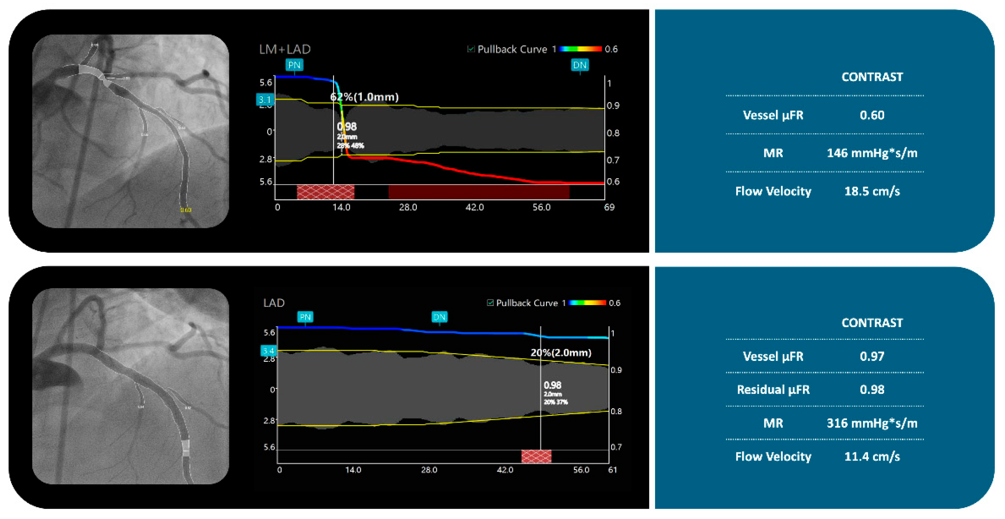



| Overall N = 330 | No PMI N = 146 | PMI N = 184 | p | |
|---|---|---|---|---|
| Age, Years (25th–75th percentil) | 65.0 (58.1–73.2) | 64.0 (61.2–69.3) | 65.0 (60.0–72.2) | 0.64 |
| Male sex, n (%) | 179.0 (54.2) | 82.0 (56.1) | 97.0 (52.7) | 0.81 |
| Hypertension, n (%) | 287.0 (86.9) | 135.0 (92.4) | 152.0 (82.6) | 0.74 |
| Dyslipidemia, n (%) | 241.0 (73.1) | 112.0 (76.7) | 129.0 (70.1) | 0.67 |
| DM, n (%) | 173.0 (52.4) | 81.0 (55.4) | 92.0 (50.0) | 0.68 |
| Obesity, n (%) | 102.0 (30.9) | 46.0 (31.5) | 56.0 (30.4) | 0.75 |
| Prior MI, n (%) | 129.0 (39.0) | 49.0 (33.5) | 70.0 (38.0) | 0.45 |
| Angina CCS Class III/IV, n (%) | 239.0 (72.4) | 107 (73.3) | 132 (71.7) | 0.38 |
| COPD, n (%) | 65.0 (19.7) | 30 (20.5) | 35 (19.0) | 0.61 |
| History of cancer (%) | 22 (6.6) | 8 (5.4) | 14 (7.6) | 0.08 |
| PAD (%) | 38 (11.5) | 12 (8.2) | 26 (14.1) | 0.04 |
| Prior coronary angioplasty, n (%) | 98.0 (29.7) | 42.0 (28.7) | 56.0 (30.4) | 0.88 |
| LVEF (%) ± SD | 55.5 ± 10.1 | 56.2 ± 10.0 | 54.7 ± 10.8 | 0.92 |
| CrCl (ml/min) ± SD | 64.6 ± 18.8 | 65.6 ± 18.5 | 65.6± 16.7 | 0.79 |
| Troponin/URL (25th–75th) | 6.1 (1.9–20.9) | 1.7 (0.8–3.0) | 18.1 (8.2–37.9) | <0.01 |
| Baseline (330) | No pMI (146) | pMI (184) | p | |
|---|---|---|---|---|
| Target vessel | ||||
| Left Main PCI, n (%) | 20.0 (6.1) | 9.0 (6.1) | 11.0 (5.9) | 0.46 |
| LAD, n (%) | 117.0 (35.4) | 52.0 (35.6) | 65.0 (35.3) | 0.29 |
| Circumflex, n (%) | 81.0 (24.5) | 38.0 (26.0) | 43.0 (23.3) | 0.37 |
| Right coronary PCI, n (%) | 112.0 (33.9) | 47.0 (32.2) | 65.0 (35.3) | 0.55 |
| Mean stent diameter (mm) ± SD | 3.1 ± 0.4 | 3.0 ± 0.4 | 3.2 ± 0.3 | 0.48 |
| Minimun Lumen Diameter (mm) ± SD | 1.1 ± 0.3 | 1.1 ± 0.1 | 1.1 ± 0.4 | 0.19 |
| Maximum Stenosis Diameter (%) ± SD | 78.1 ± 12.2 | 76.2 ± 12.0 | 77.7 ± 12.6 | 0.63 |
| Proximal diameter (mm) ± SD | 2.7 ± 0.6 | 2.7 ± 0.4 | 2.6 ± 0.5 | 0.58 |
| Distal diameter (mm) ± SD | 2.3 ± 0.4 | 2.40 ± 0.4 | 2.3 ± 0.4 | 0.49 |
| Pre dilatation balloon diameter (mm) ± SD | 3.6 ± 0.1 | 3.5 ± 0.3 | 3.6 ± 0.2 | 0.81 |
| Post dilatation balloon diameter (mm) ± SD | 3.3 ± 0.4 | 2.3 ± 0.3 | 2.4 ± 0.2 | 0.74 |
| Radial Access, n (%) | 267.0 (80.9) | 117.0 (80.1) | 150.0 (81.5) | 0.43 |
| Femoral Access, n (%) | 63.0 (19.1) | 29.0 (19.9) | 34.0 (18.5) | 0.41 |
| Overall N = 330 | No pMI N = 146 | pMI N = 184 | p | |
|---|---|---|---|---|
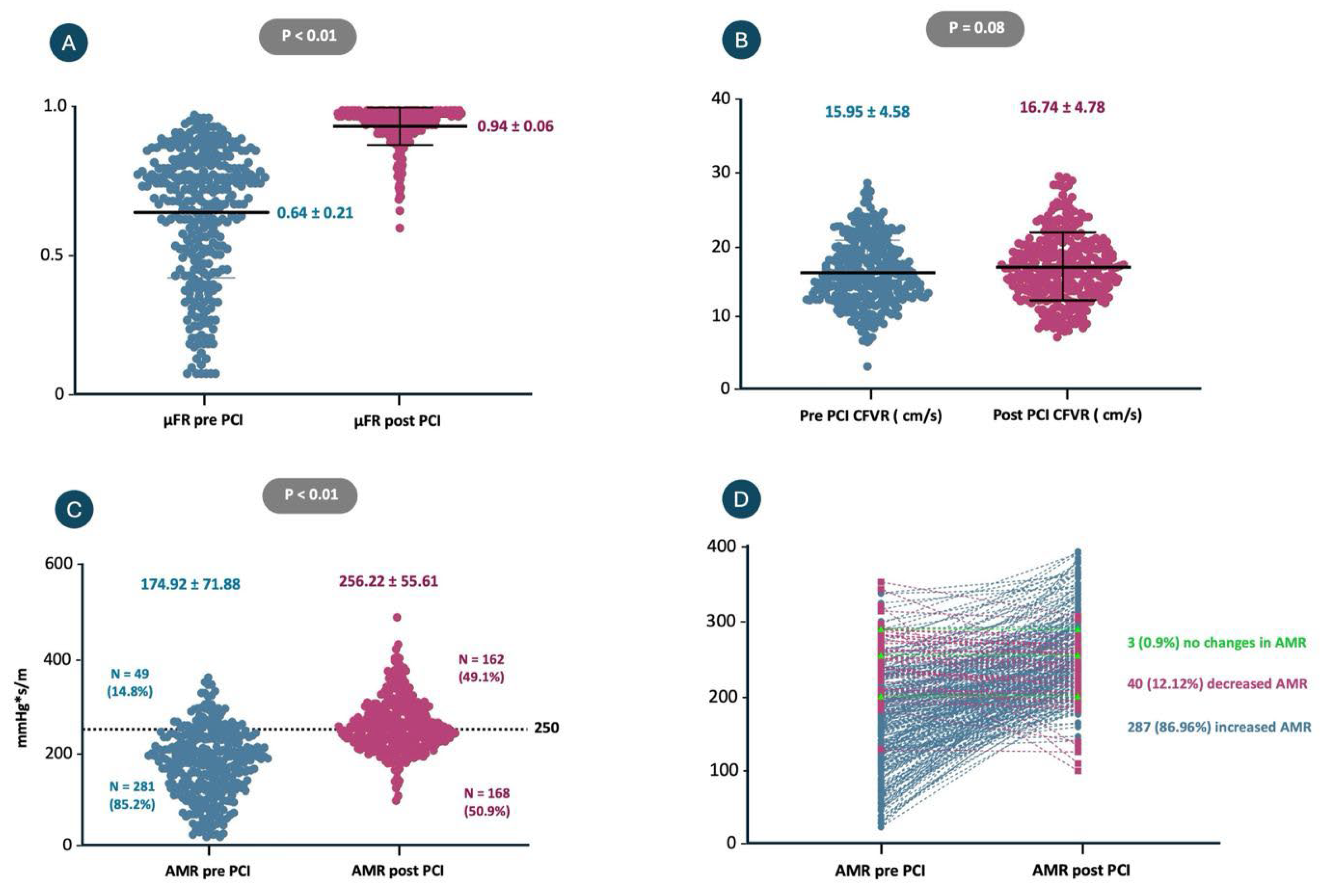
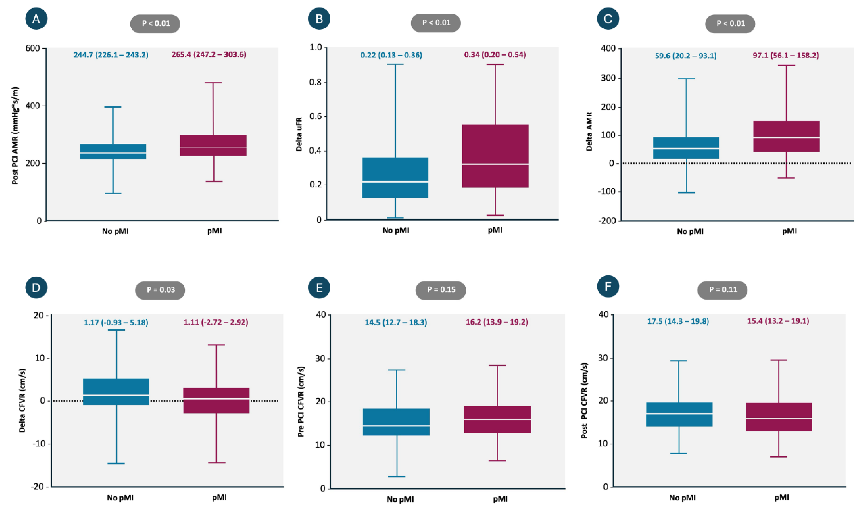
| Pre PCI CFVR ± SD | 15.95 ± 4.42 | 15.58 ± 4.84 | 16.25 ± 4.35 | 0.15 |
| Post PCI CFVR ± SD | 16.74 ± 4.72 | 17.12 ± 4.57 | 16.45 ± 4.93 | 0.11 |
| Delta CFVR ± SD | 0.78 ± 5.22 | 0.26 ± 4.95 | −1.53 ± 5.38 | 0.03 |
| Pre PCI μFR ± SD | 0.64 ± 0.21 | 0.68 ± 0.20 | 0.60 ± 0.22 | 0.05 |
| Post PCI μFR ± SD | 0.96 ± 0.02 | 0.96 ± 0.02 | 0.95 ± 0.03 | 0.94 |
| Delta μFR ± SD | 0.32 ± 0.22 | 0.26 ± 0.19 | 0.36 ± 0.22 | <0.01 |
| Pre PCI AMR (mmHg*m/s) ± SD | 174.9 ± 71.88 | 191.4 ± 66.86 | 161.7 ± 73.17 | <0.01 |
| Post PCI AMR (mmHg*m/s) ± SD | 256.2 ± 55.61 | 244.71 ± 52.47 | 265.42 ± 56.44 | <0.01 |
| Delta AMR ± SD | 82.42 ± 74.27 | 61.67 ± 69.87 | 97.14 ± 76.31 | <0.01 |
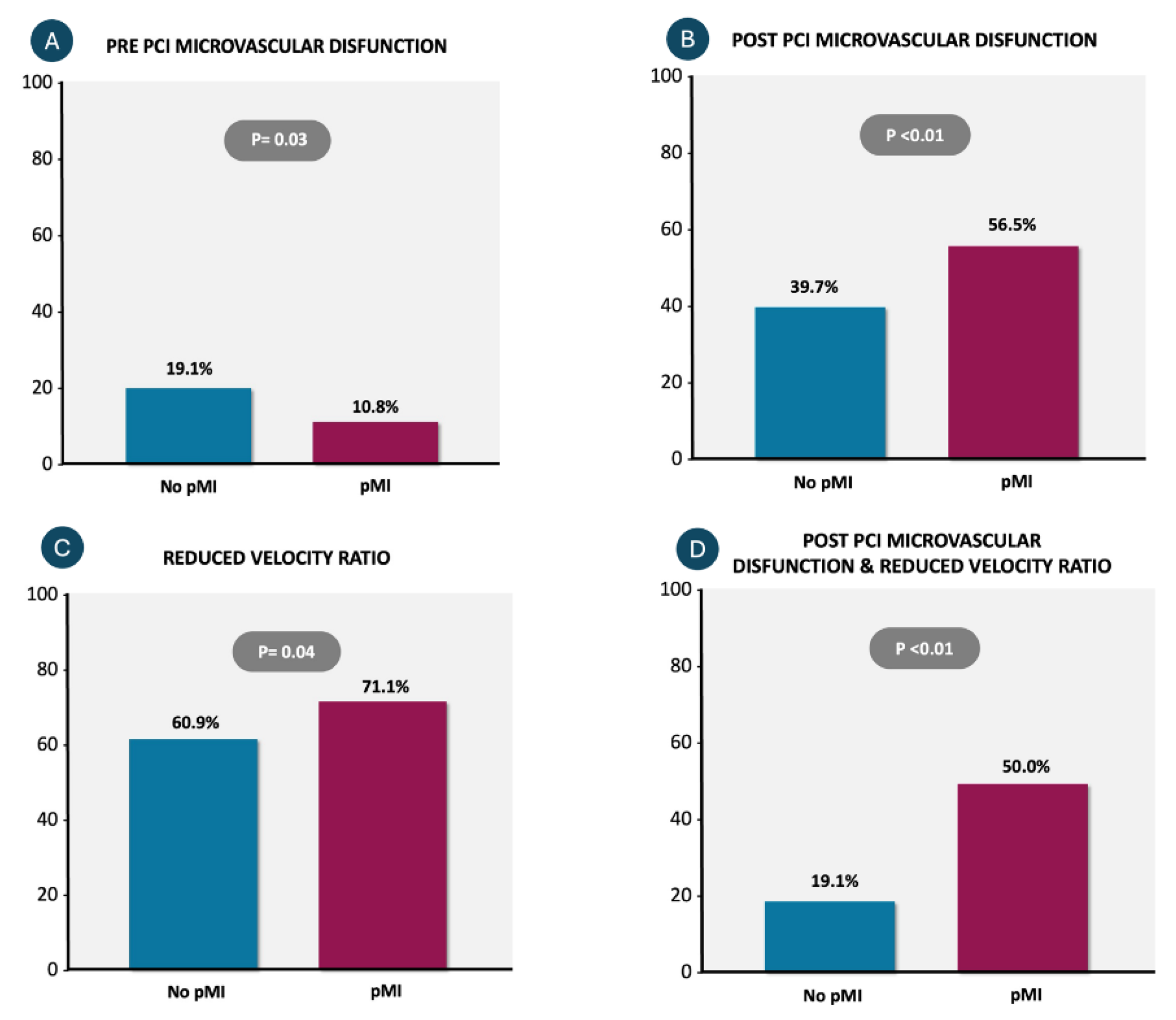
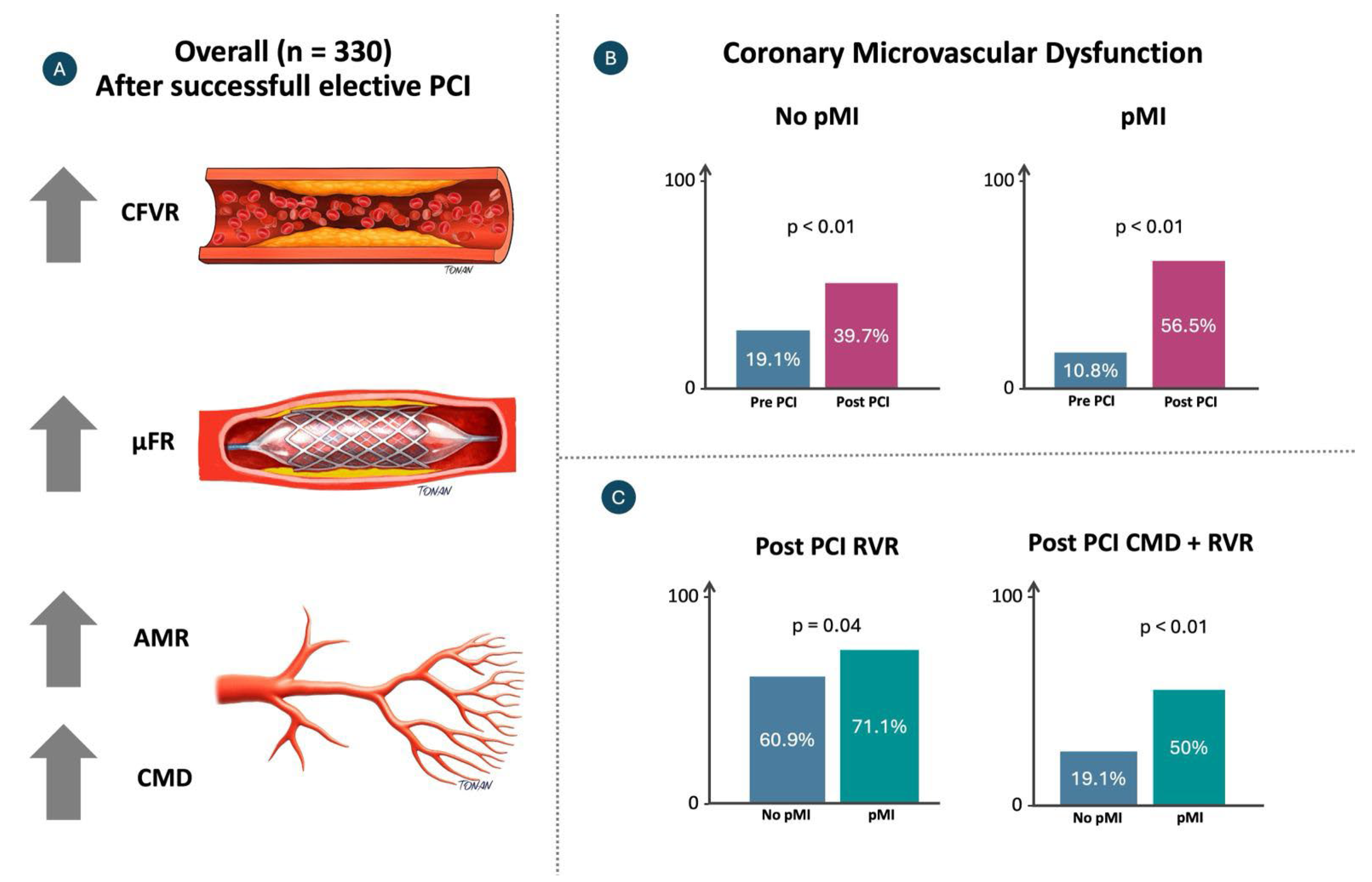
| Pre PCI Microvascular Disfunction n (%) | 48 (14.5) | 28 (19.1) | 20 (10.8) | 0.03 |
| Post PCI Microvascular Disfunction, n (%) | 162 (49.0) | 58 (39.7) | 104 (56.5) | <0.01 |
| Reduced velocity ratio (RVR) (%) | 220 (66.6) | 89 (60.9) | 131 (71.1) | 0.04 |
| Post PCI microvascular disfunction & Reduced flow velocity ratio (RVR) (%) | 120 (36.3) | 28 (19.1) | 92 (50.0) | <0.01 |
| Univariable Analysis | Multivariable Analysis | |||||
|---|---|---|---|---|---|---|
| OR | 95% CI | p | OR | 95% CI | p | |
| Post PCI AMR (mmHg*s/m) | 1.08 | 1.06–1.10 | <0.01 | 1.02 | 1.01–1.03 | <0.01 |
| Delta CFVR (cm/s) | 0.90 | 0.88–0.92 | <0.01 | 0.92 | 0.91–0.93 | <0.01 |
Disclaimer/Publisher’s Note: The statements, opinions and data contained in all publications are solely those of the individual author(s) and contributor(s) and not of MDPI and/or the editor(s). MDPI and/or the editor(s) disclaim responsibility for any injury to people or property resulting from any ideas, methods, instructions or products referred to in the content. |
© 2026 by the authors. Licensee MDPI, Basel, Switzerland. This article is an open access article distributed under the terms and conditions of the Creative Commons Attribution (CC BY) license.
Share and Cite
Jallad, P.; Maksud, D.; Garcia-Garcia, H.M.; da Mota Rocha, B.A.; Calderon, J.C.M.; Godinho, R.R.; Sampaio, B.P.A.; Lopes, N.H.M.; Gowdak, L.H.W.; Melo, P.; et al. Association of Angiography-Derived Coronary Microvascular Parameters with Periprocedural Myocardial Injury in Uncomplicated Elective Percutaneous Coronary Interventions. Diagnostics 2026, 16, 997. https://doi.org/10.3390/diagnostics16070997
Jallad P, Maksud D, Garcia-Garcia HM, da Mota Rocha BA, Calderon JCM, Godinho RR, Sampaio BPA, Lopes NHM, Gowdak LHW, Melo P, et al. Association of Angiography-Derived Coronary Microvascular Parameters with Periprocedural Myocardial Injury in Uncomplicated Elective Percutaneous Coronary Interventions. Diagnostics. 2026; 16(7):997. https://doi.org/10.3390/diagnostics16070997
Chicago/Turabian StyleJallad, Pedro, Danilo Maksud, Hector M. Garcia-Garcia, Bruno Alves da Mota Rocha, Jean Carlo Mayta Calderon, Roger Renault Godinho, Brunna Pileggi Azevedo Sampaio, Neuza Helena Moreira Lopes, Luis Henrique Wolff Gowdak, Pedro Melo, and et al. 2026. "Association of Angiography-Derived Coronary Microvascular Parameters with Periprocedural Myocardial Injury in Uncomplicated Elective Percutaneous Coronary Interventions" Diagnostics 16, no. 7: 997. https://doi.org/10.3390/diagnostics16070997
APA StyleJallad, P., Maksud, D., Garcia-Garcia, H. M., da Mota Rocha, B. A., Calderon, J. C. M., Godinho, R. R., Sampaio, B. P. A., Lopes, N. H. M., Gowdak, L. H. W., Melo, P., Freire, A. F. D., de Matos Soeiro, A., Rizk, S. I., Kalil Filho, R., Hajjar, L., Abizaid, A., & Campos, C. M. (2026). Association of Angiography-Derived Coronary Microvascular Parameters with Periprocedural Myocardial Injury in Uncomplicated Elective Percutaneous Coronary Interventions. Diagnostics, 16(7), 997. https://doi.org/10.3390/diagnostics16070997