Functional Magnetic Resonance Imaging for Investigating the Role of the Hippocampus in Migraine with Aura



Abstract
1. Introduction
2. Materials and Methods
2.1. Participants
2.2. MRI Data Acquisition and Post-Processing
2.3. Statistical Analyses
3. Results
3.1. Study Population
3.2. Seed-Based FC Results
4. Discussion
5. Conclusions
Author Contributions
Funding
Institutional Review Board Statement
Informed Consent Statement
Data Availability Statement
Acknowledgments
Conflicts of Interest
Abbreviations
| FC | functional connectivity |
| HCs | healthy controls |
| MRI | magnetic resonance imaging |
| MwA | Migraine with aura |
| MwAv | MwA patients with pure visual aura |
| MwAvsd | MwA patients with visual, somatosensory, and dysphasic aura |
References
- Headache Classification Committee of the International Headache Society (IHS). The International classification of Headache disorders. Cephalalgia 2018, 38, 1–211. [Google Scholar] [CrossRef]
- Goadsby, P.J.; Holland, P.R.; Martins-Oliveira, M.; Hoffmann, J.; Schankin, C.; Akerman, S. Pathophysiology of Migraine: A Disorder of Sensory Processing. Physiol. Rev. 2017, 97, 553–622. [Google Scholar] [CrossRef]
- Petrusic, I.; Viana, M.; Dakovic, M.; Goadsby, P.J.; Zidverc-Trajkovic, J. Proposal for a migraine aura complexity score. Cephalalgia 2018, 39, 732–741. [Google Scholar] [CrossRef]
- Viana, M.; Sances, G.; Linde, M.; Ghiotto, N.; Guaschino, E.; Allena, M.; Terrazzino, S.; Nappi, G.; Goadsby, P.J.; Tassorelli, C. Clinical features of migraine aura: Results from a prospective diary-aided study. Cephalalgia 2017, 37, 979–989. [Google Scholar] [CrossRef]
- Coppola, G.; Renzo, A.D.; Tinelli, E.; Petolicchio, B.; Parisi, V.; Serrao, M.; Porcaro, C.; Fiorelli, M.; Caramia, F.; Schoenen, J.; et al. Thalamo-cortical networks in subtypes of migraine with aura patients. J. Headache Pain 2021, 22, 58. [Google Scholar] [CrossRef] [PubMed]
- Mitrovic, K.; Petrusic, I.; Dakovic, M.; Savić, A. Migraine with aura detection and subtype classification using machine learning algorithms and morphometric magnetic resonance imaging data. Front. Neurol. 2023, 14, 1106612. [Google Scholar] [CrossRef] [PubMed]
- Messina, R.; Rocca, M.A.; Goadsby, P.J.; Filippi, M. Insights into migraine attacks from neuroimaging. Lancet Neurol. 2023, 22, 834–846. [Google Scholar] [CrossRef] [PubMed]
- Petrusic, I.; Viana, M.; Dakovic, M.; Zidverc-Trajkovic, J. Application of the Migraine Aura Complexity score (MACS): Clinical and Neuroimaging Study. Front. Neurol. 2019, 10, 1112. [Google Scholar] [CrossRef]
- Petrusic, I.; Jovanovic, V.; Kovic, V.; Savic, A.M. P3 latency as a biomarker for the complexity of migraine with aura: Event-related potential study. Cephalalgia 2022, 42, 1022–1030. [Google Scholar] [CrossRef]
- Coppola, G.; Bracaglia, M.; Di Lenola, S.; Di Lorenzo, C.; Serrao, M.; Parisi, V.; Di Renzo, A.; Martelli, F.; Fadda, A.; Schoenen, J.; et al. Visual evoked potentials in subgroups of migraine with aura patients. J. Headache Pain 2015, 16, 92. [Google Scholar] [CrossRef]
- Silvestro, M.; Tessitore, A.; Di Nardo, F.; Scotto di Clemente, F.; Trojsi, F.; Cirillo, M.; Esposito, F.; Tedeschi, G.; Russo, A. Functional connectivity changes in complex migraine aura: Beyond the visual network. Eur. J. Neurol. 2022, 29, 295–304. [Google Scholar] [PubMed]
- Abagnale, C.; Di Renzo, A.; Sebastianelli, G.; Casillo, F.; Tinelli, E.; Giuliani, G.; Tullo, M.G.; Serrao, M.; Parisi, V.; Fiorelli, M.; et al. Whole brain surface-based morphometry and tract-based spatial statistics in migraine with aura patients: Difference between pure visual and complex auras. Front. Hum. Neurosci. 2023, 17, 1146302. [Google Scholar] [CrossRef] [PubMed]
- Petrušić, I.; Savić, A.; Mitrović, K.; Bačanin, N.; Sebastianelli, G.; Secci, D.; Coppola, G. Machine learning classification meets migraine: Recommendations for study evaluation. J. Headache Pain 2024, 25, 215. [Google Scholar] [CrossRef] [PubMed]
- Cankaya, S.; Ayyildiz, B.; Sayman, D.; Duran, U.; Ucak, D.; Karaca, R.; Ayyildiz, S.; Oktem, E.O.; Lakadamyalı, H.; Sayman, C.; et al. Hippocampal connectivity dynamics and volumetric alterations predict cognitive status in migraine: A resting-state fMRI study. Neuroimage 2025, 305, 120961. [Google Scholar]
- Petrušić, I.; Radović, M.; Daković, M.; Radojičić, A.; Coppola, G. Subsegmentation of the hippocampus in subgroups of migraine with aura patients: Advanced structural neuroimaging study. J. Headache Pain 2024, 25, 182. [Google Scholar] [CrossRef]
- He, M.; Kis-Jakab, G.; Komaromy, H.; Perlaki, G.; Orsi, G.; Bosnyák, E.; Rozgonyi, R.; John, F.; Trauninger, A.; Eklics, K.; et al. The volume of the thalamus and hippocampus in a right-handed female episodic migraine group. Front. Neurol. 2023, 14, 1254628. [Google Scholar] [CrossRef]
- Feng, Q.; Chen, W.; Ke, J.; Geng, D.; Xiong, X.; Dai, L.; Zhao, H.; Hu, C. Volumetric analysis of hippocampal subregions in migraine without aura: An exploratory study on mechanisms underlying migraine chronification. J. Headache Pain 2025, 26, 236. [Google Scholar] [CrossRef]
- Dalton, M.A.; D’Souza, A.; Lv, J.; Calamante, F. New insights into anatomical connectivity along the anterior-posterior axis of the human hippocampus using in vivo quantitative fibre tracking. eLife 2022, 11, e76143. [Google Scholar]
- Chan, R.W.; Leong, A.T.L.; Ho, L.C.; Gao, P.P.; Wong, E.C.; Dong, C.M.; Wang, X.; He, J.; Chan, Y.-S.; Lim, L.W.; et al. Low-frequency hippocampal-cortical activity drives brain-wide resting-state functional MRI connectivity. Proc. Natl. Acad. Sci. USA 2017, 114, E6972–E6981. [Google Scholar]
- Piai, V.; Anderson, K.L.; Lin, J.J.; Dewar, C.; Parvizi, J.; Dronkers, N.F.; Knight, R.T. Direct brain recordings reveal hippocampal rhythm underpinnings of language processing. Proc. Natl. Acad. Sci. USA 2016, 113, 11366–11371. [Google Scholar]
- Maleki, N.; Becerra, L.; Brawn, J.; McEwen, B.; Burstein, R.; Borsook, D. Common hippocampal structural and functional changes in migraine. Brain Struct. Funct. 2013, 218, 903–912. [Google Scholar] [CrossRef] [PubMed]
- Chong, C.D.; Dumkrieger, G.; Schwedt, T.J. Structural co-variance patterns in migraine: A cross-sectional study exploring the role of the Hippocampus. Headache 2017, 57, 1522–1531. [Google Scholar] [CrossRef] [PubMed]
- Tsai, C.L.; Chou, K.H.; Lee, P.L.; Liang, C.-S.; Kuo, C.-Y.; Lin, G.-Y.; Lin, Y.-K.; Hsu, Y.-C.; Ko, C.-A.; Yang, F.-C.; et al. Shared alterations in hippocampal structural covariance in subjective cognitive decline and migraine. Front. Aging Neurosci. 2023, 15, 1191991. [Google Scholar] [CrossRef] [PubMed]
- Mitrović, K.; Savić, A.; Radojičić, A.; Daković, M.; Petrušić, I. Machine learning approach for Migraine Aura Complexity Score prediction based on magnetic resonance imaging data. J. Headache Pain 2023, 24, 169. [Google Scholar] [CrossRef]
- Smitha, K.A.; Akhil Raja, K.; Arun, K.M.; Rajesh, P.G.; Thomas, B.; Kapilamoorthy, T.R.; Kesavadas, C. Resting state fMRI: A review on methods in resting state connectivity analysis and resting state networks. Neuroradiol. J. 2017, 30, 305–317. [Google Scholar] [CrossRef]
- Colombo, B.; Rocca, M.A.; Messina, R.; Guerrieri, S.; Filippi, M. Resting-state fMRI functional connectivity: A new perspective to evaluate pain modulation in migraine? Neurol. Sci. 2015, 36, 41–45. [Google Scholar] [CrossRef]
- Skorobogatykh, K.; van Hoogstraten, W.S.; Degan, D.; Prischepa, A.; Savitskaya, A.; Ileen, B.M.; Bentivegna, E.; Skiba, I.; D’Acunto, L.; Ferri, L.; et al. Functional connectivity studies in migraine: What have we learned? J. Headache Pain 2019, 20, 108. [Google Scholar] [CrossRef]
- Worsley, K.J. Statistical analysis of activation images. In Book Functional MRI: An Introduction to Methods; Jezzard, P., Matthews, P.M., Smith, S.M., Eds.; Oxford University Press: Oxford, UK, 2001; pp. 251–270. [Google Scholar]
- Wei, H.L.; Chen, J.; Chen, Y.C.; Yu, Y.-S.; Zhou, G.-P.; Qu, L.-J.; Yin, X.; Li, J.; Zhang, H. Impaired functional connectivity of limbic system in migraine without aura. Brain Imaging Behav. 2020, 14, 1805–1814. [Google Scholar] [CrossRef]
- Zhu, Y.; Dai, L.; Zhao, H.; Ji, B.; Yu, Y.; Dai, H.; Hu, C.; Wang, X.; Ke, J. Alterations in Effective Connectivity of the Hippocampus in Migraine without Aura. J. Pain Res. 2021, 14, 3333–3343. [Google Scholar] [CrossRef]
- Karsan, N.; Silva, E.; Goadsby, P.J. Evaluating migraine with typical aura with neuroimaging. Front. Hum. Neurosci. 2023, 17, 1112790. [Google Scholar] [CrossRef]
- Ayata, C.; Lauritzen, M. Spreading Depression, Spreading Depolarizations, and the Cerebral Vasculature. Physiol. Rev. 2015, 95, 953–993. [Google Scholar] [CrossRef]
- Arren Hill, A.; Amendolara, A.B.; Small, C.; Guzman, S.C.; Pfister, D.; McFarland, K.; Settelmayer, M.; Baker, S.; Donnelly, S.; Payne, A.; et al. Metabolic Pathophysiology of Cortical Spreading Depression: A Review. Brain Sci. 2024, 14, 1026. [Google Scholar] [CrossRef] [PubMed]
- Florez, C.; Lukankin, V.; Sugumar, S.; McGinn, R.; Zhang, Z.; Zhang, L.; Carlen, P. Hypoglycemia-induced alterations in hippocampal intrinsic rhythms: Decreased inhibition, increased excitation, seizures and spreading depression. Neurobiol. Dis. 2015, 82, 213–225. [Google Scholar] [CrossRef] [PubMed]
- Sereno, M.I.; Huang, R.-S. Multisensory maps in parietal cortex. Curr. Opin. Neurobiol. 2014, 24, 39–46. [Google Scholar] [CrossRef] [PubMed]
- Freud, E.; Behrmann, M.; Snow, J.C. What Does Dorsal Cortex Contribute to Perception? Open Mind 2020, 4, 40–56. [Google Scholar] [CrossRef]
- Orban, G.A.; Sepe, A.; Bonini, L. Parietal maps of visual signals for bodily action planning. Brain Struct. Funct. 2021, 226, 2967–2988. [Google Scholar] [CrossRef]
- Wiener, S.I.; Berthoz, A.; Zugaro, M.B. Multisensory processing in the elaboration of place and head direction responses by limbic system neurons. Brain Res. Cogn. Brain Res. 2002, 14, 75–90. [Google Scholar] [CrossRef]
- Zhou, L.; Xu, T.; Feng, T. The hippocampus-IPL connectivity links to ADHD traits through sensory processing sensitivity. Cereb. Cortex 2025, 35, bhaf063. [Google Scholar] [CrossRef]
- Friedman, N.P.; Robbins, T.W. The role of prefrontal cortex in cognitive control and executive function. Neuropsychopharmacology 2021, 47, 72–89. [Google Scholar] [CrossRef]
- Seminowicz, D.A.; Moayedi, M. The dorsolateral prefrontal cortex in acute and chronic pain. J. Pain 2018, 18, 1027–1035. [Google Scholar] [CrossRef]
- Clemente, L.; Paparella, G.; Scannicchio, S.; Abbatantuono, C.; Tancredi, G.; Ladisa, E.; Delussi, M.D.; Ammendola, E.; Prudenzano, A.M.P.; de Tommaso, M. Dorso lateral prefrontal cortex stimulation with TMS in chronic migraine individuals refractory to anti-CGRP monoclonal antibodies: Clinical, neuropsychological and neurophysiological effects. Cephalalgia 2025, 45, 3331024251364843. [Google Scholar] [CrossRef]
- Mungoven, T.J.; Marciszewski, K.K.; Macefiel, V.G.; Macey, P.M.; Henderson, L.A.; Meylakh, N. Alterations in pain processing circuitries in episodic migraine. J. Headache Pain 2022, 23, 9. [Google Scholar] [CrossRef]
- Preston, A.R.; Eichenbaum, H. Interplay of hippocampus and prefrontal cortex in memory. Curr. Biol. 2013, 23, R764–R773. [Google Scholar] [CrossRef]
- Nobre, A.C.; Gresch, D. How the brain shifts between external and internal attention. Neuron 2025, 113, 2382–2398. [Google Scholar] [CrossRef]
- Kuhnke, P.; Chapman, C.A.; Cheung, V.K.M.; Turker, S.; Graessner, A.; Martin, S.; Williams, K.A.; Hartwigsen, G. The role of the angular gyrus in semantic cognition: A synthesis of five functional neuroimaging studies. Brain Struct. Funct. 2022, 228, 273–291. [Google Scholar] [CrossRef]
- Kim, H.; Wang, K.; Cutting, L.E.; Willcutt, E.G.; Petrill, S.A.; Leopold, D.R.; Reineberg, A.E.; Thompson, L.A.; Banich, M.T. The Angular Gyrus as a Hub for Modulation of Language-related Cortex by Distinct Prefrontal Executive Control Regions. J. Cogn. Neurosci. 2022, 34, 2275–2296. [Google Scholar] [CrossRef]



| Variables | MwAvsd (n = 27) | MwAv (n = 18) | p-Value |
|---|---|---|---|
| Disease duration in years, mean ± sd | 18.2 ± 10.9 | 16.3 ± 9.9 | 0.538 |
| MwA frequency per year, mean ± sd | 8.3 ± 9.1 | 3.9 ± 2.5 | 0.244 |
| Photophobia, number of patients (%) | 26 (96) | 15 (83) | 0.286 |
| Phonophobia, number of patients (%) | 16 (59) | 6 (33) | 0.130 |
| Nausea, number of patients (%) | 17 (63) | 8 (44) | 0.241 |
| Headache intensity, mean ± sd | 6.7 ± 2.2 | 6.4 ± 2.5 | 0.712 |
| MwoA, number of patients (%) | 14 (52) | 6 (33) | 0.359 |
| Cluster Voxels | p Value | Z-Max | Peak MNI Coordinates | Brain Region | ||
|---|---|---|---|---|---|---|
| X | Y | Z | ||||
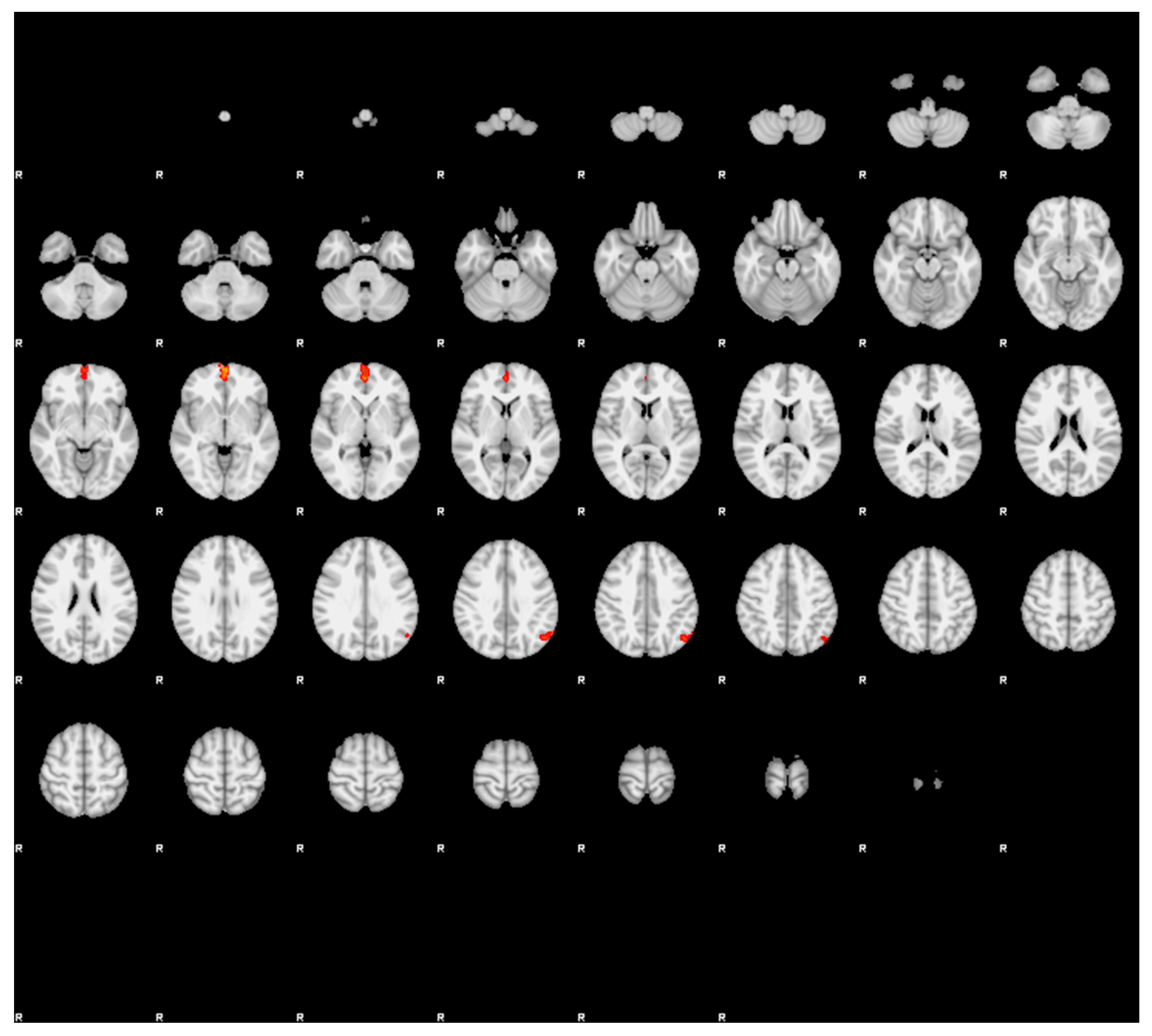
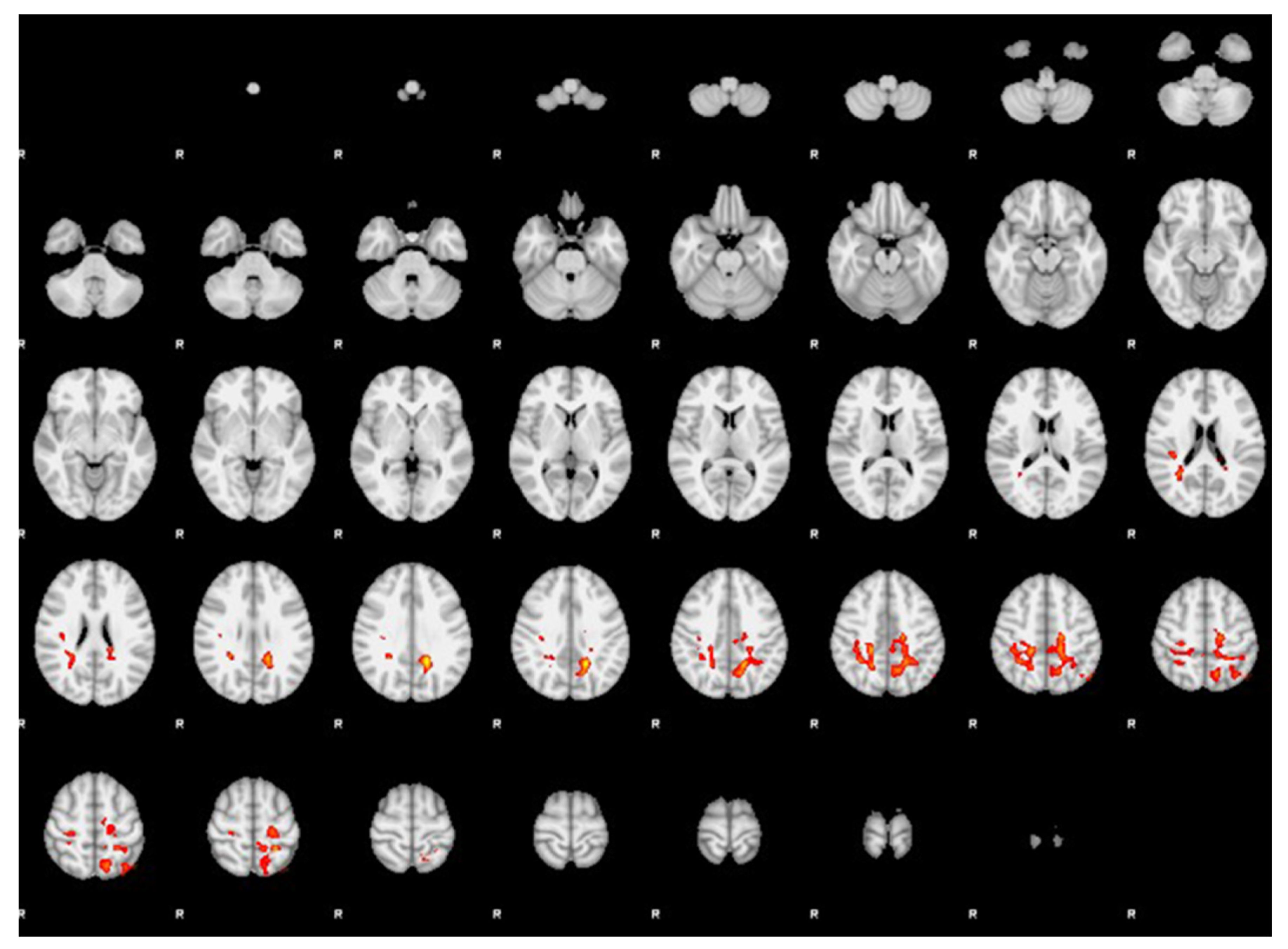
| FC of the right hippocampus in MwAvsd > MwAv | ||||||
| 2227 | <0.001 | 4.05 | −18 | −50 | 34 | Left dorsal parietal cortex |
| 1055 | 0.032 | 3.62 | 20 | −36 | 46 | Right sensory association cortex |
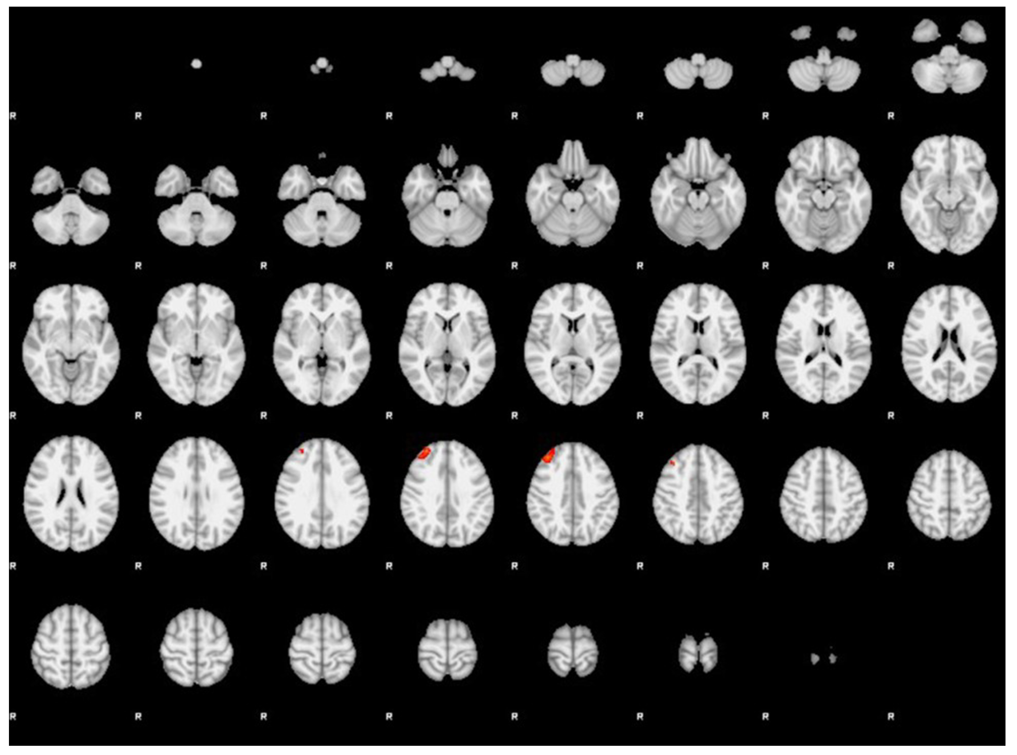
| FC of the left hippocampus in MwAv > MwAvsd | ||||||
| 212 | 0.031 | 3.46 | 36 | 38 | 40 | Right dorsolateral prefrontal cortex |
| FC of the left hippocampus in MwAv > HCs | ||||||
| 321 | 0.002 | 3.84 | 0 | 62 | −4 | Bilateral anterior prefrontal cortices |
| 206 | 0.037 | 3.56 | −60 | −60 | 38 | Left angular cortex |
| Left Dorsal Parietal Cortex | Right Sensory Association Cortex | Right Dorsolateral Prefrontal Cortex | |
|---|---|---|---|
| MwA attack frequency per year | rho = −0.112, p = 0.476 | rho = −0.120, p = 0.443 | rho = −0.009, p = 0.955 |
| Disease duration in years | rho = −0.125, p = 0.424 | rho = −0.251, p = 0.105 | rho = −0.043, p = 0.784 |
| Headache intensity | rho = −0.164, p = 0.293 | rho = 0.069, p = 0.658 | rho = −0.244, p = 0.115 |
| Volume of the whole left hippocampus | rho = 0.018, p = 0.909 | rho = 0.127, p = 0.418 | rho = 0.108, p = 0.490 |
| Volume of the whole right hippocampus | rho = 0.001, p = 0.994 | rho = −0.165, p = 0.291 | rho = 0.090, p = 0.566 |
Disclaimer/Publisher’s Note: The statements, opinions and data contained in all publications are solely those of the individual author(s) and contributor(s) and not of MDPI and/or the editor(s). MDPI and/or the editor(s) disclaim responsibility for any injury to people or property resulting from any ideas, methods, instructions or products referred to in the content. |
© 2026 by the authors. Licensee MDPI, Basel, Switzerland. This article is an open access article distributed under the terms and conditions of the Creative Commons Attribution (CC BY) license.
Share and Cite
Radović, M.; Daković, M.; Radojičić, A.; Petrušić, I. Functional Magnetic Resonance Imaging for Investigating the Role of the Hippocampus in Migraine with Aura. Diagnostics 2026, 16, 1111. https://doi.org/10.3390/diagnostics16071111
Radović M, Daković M, Radojičić A, Petrušić I. Functional Magnetic Resonance Imaging for Investigating the Role of the Hippocampus in Migraine with Aura. Diagnostics. 2026; 16(7):1111. https://doi.org/10.3390/diagnostics16071111
Chicago/Turabian StyleRadović, Mojsije, Marko Daković, Aleksandra Radojičić, and Igor Petrušić. 2026. "Functional Magnetic Resonance Imaging for Investigating the Role of the Hippocampus in Migraine with Aura" Diagnostics 16, no. 7: 1111. https://doi.org/10.3390/diagnostics16071111
APA StyleRadović, M., Daković, M., Radojičić, A., & Petrušić, I. (2026). Functional Magnetic Resonance Imaging for Investigating the Role of the Hippocampus in Migraine with Aura. Diagnostics, 16(7), 1111. https://doi.org/10.3390/diagnostics16071111