Biomarkers of Treatment Response in Paediatric Medulloblastoma
 , ,
, ,
Abstract
1. Introduction
2. Molecular Classification and Predictive Biomarkers

| Molecular Subgroup | Frequency | Key Molecular Features/Genetic Alterations | Predictive Biomarkers | Clinical Characteristics/Prognosis | Therapeutic Implications | Metastatic Potential | Ref. |
|---|---|---|---|---|---|---|---|
| WNT | ~10% | Activation of WNT/β-catenin pathway; CTNNB1 mutations; monosomy 6; specific DNA methylation signatures | Nuclear β-catenin accumulation; subgroup-specific methylation patterns | Excellent prognosis; low metastatic potential | High sensitivity to standard therapy; potential for therapy de-escalation to reduce craniospinal irradiation and long-term neurotoxicity | Low (<10% metastatic at diagnosis) | [26,27] |
| SHH | ~25–33% | Dysregulation of Hedgehog pathway; mutations in PTCH1, SMO, SUFU; GLI amplification | TP53 status (mutant vs. wild-type); age and Tumour genetics influence biomarker relevance | Heterogeneous outcomes; TP53-mutant Tumours show poor prognosis; TP53–wild-type Tumours (especially infants) respond better | Chemotherapy-based regimens preferred for some patients; targeted SMO inhibitors show activity but resistance common; biomarkers guide individualized therapy | Moderate (15–20% metastatic) | [28,29] |
| Group 3 | ~15–20% (approx.) | MYC amplification/overexpression; extensive chromosomal instability; high metastasis at diagnosis | MYC status; transcriptional signatures linked to stemness, hypoxia, immune evasion | Most aggressive subtype; poorest prognosis; early relapse common | High-risk patients may need intensified therapy, novel targeted agents, or immunotherapies | High (40–45% metastatic) | [30,31] |
| Group 4 | ~35–40% | Isochromosome 17q; SNCAIP duplication; enhancer hijacking of GFI1, PRDM6; heterogeneous genomic profiles | DNA methylation patterns; copy number variation profiles emerging | Largest and most heterogeneous subgroup; intermediate, variable outcomes | Biomarker-driven risk stratification emerging; may guide individualized therapy | Moderate (30–35% metastatic; lower in subtypes 4/7) | [32,33] |
3. Epigenetic and Transcriptomic Biomarkers
| Biomarker Type | Key Mechanism/Features | Examples/Specific Signatures | Association with Tumour Biology | Predictive Value/Clinical Relevance | Notes/Emerging Applications | Ref. |
|---|---|---|---|---|---|---|
| DNA Methylation | Epigenetic modification of CpG islands; stable and heritable patterns reflect cellular origin and differentiation state | Subgroup-specific methylation profiles (WNT, SHH, Group 3, Group 4); methylation of promoters for cell cycle genes (CDKN2A, RB1), DNA repair genes (MGMT), and neuronal differentiation markers | Distinguishes molecular subgroups and substructures; reflects differentiation state—more differentiated patterns (neuronal gene hypomethylation) correlate with favourable biology | Aberrant hypermethylation of tumour suppressor genes → chemotherapy/radiotherapy resistance; hypomethylation of oncogenic pathways → treatment sensitivity; methylation-based subtypes 4 and 7 within Group 3/4 predict favourable response to reduced-intensity therapy | Incorporated into diagnostic workflows (EPIC arrays, methylation classifiers); guides risk stratification and clinical trial eligibility | [46,47] |
| Histone Modifications & Chromatin Remodelling | Epigenetic regulation of chromatin structure affects gene accessibility | Alterations in chromatin regulators | Influences Tumour plasticity and survival under cytotoxic stress | May predict response in high-risk or therapy-resistant Tumours | Under investigation; potential target for epigenetic therapies | [48,49] |
| Transcriptomic Signatures | Gene expression profiling capturing functional programs | Proliferation, stemness, hypoxia, immune modulation, metabolic adaptation; MYC-driven networks in Group 3 | Reflects intrinsic Tumour biology and microenvironment interactions | High MYC/stemness expression → poor response and early relapse; differentiation/apoptosis signatures → favourable response | RNA-seq increasingly used to identify predictive signatures | [50,51] |
| Non-coding RNAs (microRNAs, lncRNAs) | Post-transcriptional gene regulation | Dysregulated miRNAs/lncRNAs affecting DNA repair, drug resistance, and cell survival | Modulates pathways governing therapy response | Altered expression associated with chemotherapy/radiotherapy sensitivity; potential predictive biomarkers | Potential therapeutic targets; clinical validation ongoing | [52,53] |
| Liquid Biopsy (CSF-based) | Detection of circulating Tumour nucleic acids (DNA/RNA) | Methylated DNA fragments, Tumour-derived RNA transcripts | Reflects Tumour burden and molecular state | Enables longitudinal monitoring of treatment response and early relapse | Minimally invasive; promising for dynamic biomarker assessment | [54,55] |
4. Protein and Imaging Biomarkers
| Biomarker Type | Examples/Specific Markers | Biological Basis/Mechanism | Predictive/Prognostic Value | Clinical/Practical Applications | Notes/Emerging Approaches | Ref. |
|---|---|---|---|---|---|---|
| Protein Biomarkers | MYC, p53, survivin, BCL-2, neuronal differentiation markers | Reflect downstream activity of oncogenic pathways, apoptosis regulation, and differentiation | MYC overexpression → poor response and early relapse (esp. Group 3); p53 alterations → impaired DNA repair, therapy resistance; neuronal differentiation markers → improved response | Detection in tumour tissue or CSF; guide risk stratification and therapy adaptation | CSF-derived proteins provide minimally invasive monitoring; serial assessment allows real-time treatment evaluation | [68] |
| Liquid Biopsy/CSF Protein Markers | Tumour-derived proteins, cytokines, growth factors, ctDNA | Reflect tumour burden and biological activity | Early detection of minimal residual disease, monitoring of treatment response, and prediction of relapse | Minimally invasive; can detect molecular relapse before radiographic progression | Integration with molecular and protein biomarkers enhances longitudinal monitoring | [69] |
| Diffusion MRI (DWI/ADC) | ADC values from diffusion-weighted imaging | Surrogate of tumour cellularity | Early increases in ADC indicate an effective cytotoxic response | Non-invasive assessment of early treatment response | Functional imaging complements anatomical MRI | [70] |
| Perfusion MRI | Dynamic susceptibility contrast, arterial spin labelling | Measures tumour vascularity and blood flow | Changes correlate with treatment sensitivity/resistance | Evaluate tumour physiology in real-time | Provides insight into microenvironmental adaptations to therapy | [71] |
| Magnetic Resonance Spectroscopy (MRS) | Choline, N-acetylaspartate, lactate | Metabolic profiling of tumours | Metabolite shifts indicate therapy response or progression | Complementary to MRI for metabolic assessment | Useful for distinguishing viable tumour vs. post-therapy changes | [72] |
| PET Imaging | Glucose metabolism tracers, amino acid transport tracers | Functional metabolic imaging | Distinguishes viable tumour tissue from treatment-related changes | Post-therapy evaluation; treatment monitoring | Enhances detection of residual or recurrent disease | [73] |
| Radiomics & AI-driven Imaging | Quantitative imaging features: shape, texture, heterogeneity, spatial complexity | High-dimensional feature extraction from imaging | Predict molecular subgroup, treatment response, risk of relapse | Non-invasive, longitudinal disease monitoring; supports precision medicine | Integrates with molecular and clinical data for individualised risk prediction | [74] |
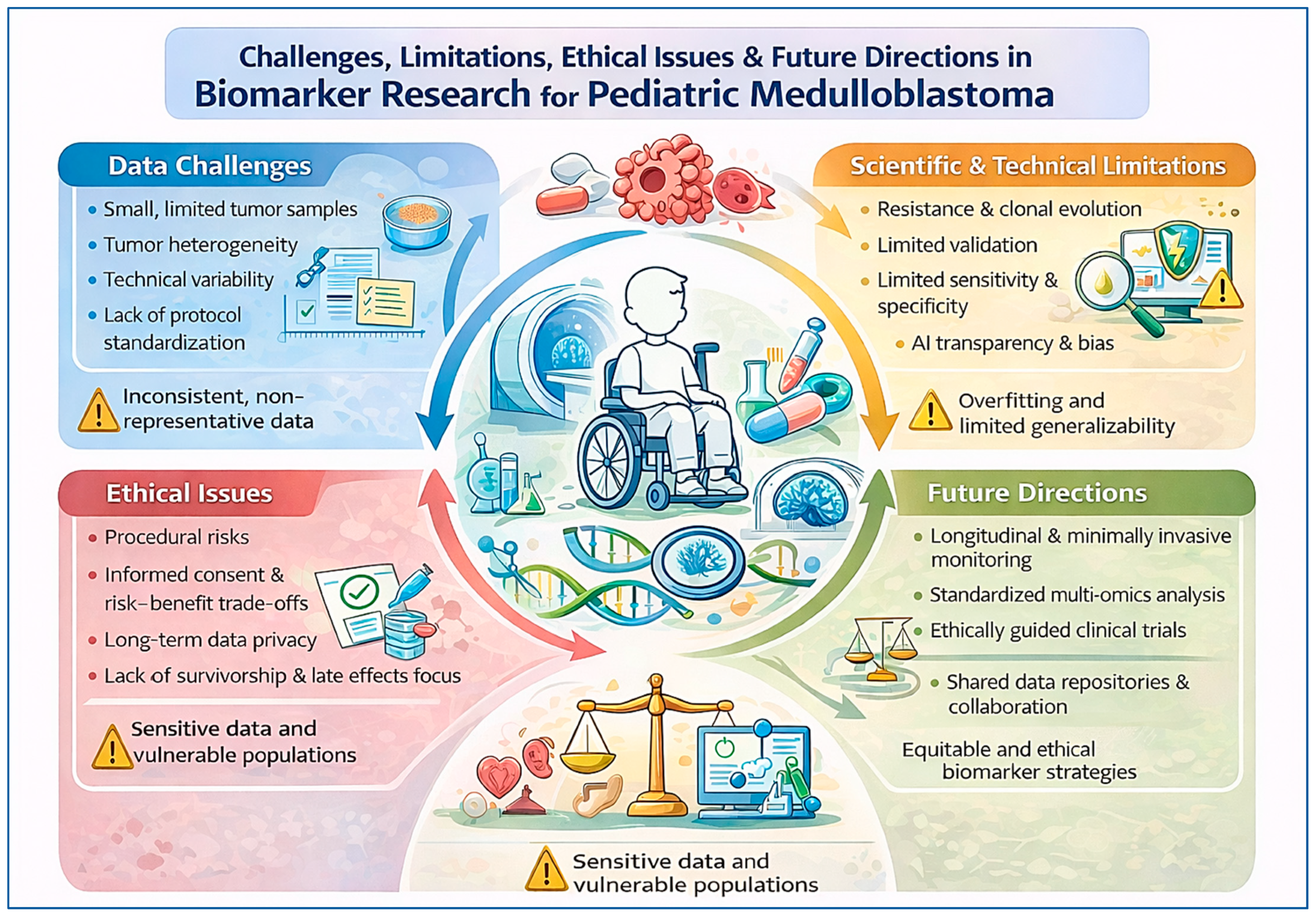
5. Challenges, Limitations, Ethical Issues and Future Directions

6. Conclusions
Author Contributions
Funding
Institutional Review Board Statement
Informed Consent Statement
Data Availability Statement
Acknowledgments
Conflicts of Interest
References
- Bailey, S.; Jacobs, S.; Kourti, M.; Massimino, M.; Andre, N.; Doz, F.; Dufour, C.; Vennarini, S.; Padovani, L.; Aquilina, K.; et al. Medulloblastoma therapy: Consensus treatment recommendations from SIOP-Europe and the European Reference Network. EJC Paediatr. Oncol. 2025, 5, 100205. [Google Scholar] [CrossRef] [Scilit]
- Gupta, T.; Chatterjee, A. Existing and Evolving Landscape of Medulloblastoma: Towards Optimization and Personalization. Diagnostics 2024, 14, 598. [Google Scholar] [CrossRef] [Scilit]
- Pan, Z.; Bao, J.; Wei, S. Advancing medulloblastoma therapy: Strategies and survival insights. Clin. Exp. Med. 2025, 25, 119. [Google Scholar] [CrossRef] [Scilit]
- Kim, D.T.; Uloho-Okundaye, M.; Frederico, S.C.; Guru, S.; Kim, M.J.; Chang, S.D. Advancing Medulloblastoma Therapy in Pediatrics: Integrative Molecular Classification and Emerging Treatments. Brain Sci. 2025, 15, 896. [Google Scholar] [CrossRef] [Scilit]
- Jermakowicz, A.M.; Ruiz, L.; Chu, J.; Jangde, N.; Suter, R.K.; Kadan-Lottick, N.S.; Hanson, D.; Ayad, N.G. In silico drug sensitivity predicts subgroup-specific therapeutics in medulloblastoma patients. Sci. Rep. 2025, 15, 42373. [Google Scholar] [CrossRef] [Scilit]
- Rosenberg, T.; Cooney, T. Current Open Trials and Molecular Update for Pediatric Embryonal Tumors. Pediatr. Neurosurg. 2023, 58, 299–306. [Google Scholar] [CrossRef] [Scilit]
- Thompson, E.M.; Ashley, D.; Landi, D. Current medulloblastoma subgroup specific clinical trials. Transl. Pediatr. 2020, 9, 157–162. [Google Scholar] [CrossRef] [Scilit]
- Delaidelli, A.; Burwag, F.; Ben-Neriah, S.; Suk, Y.; Shyp, T.; Kosteniuk, S.; Dunham, C.; Cheng, S.; Okonechnikov, K.; Schrimpf, D.; et al. High-resolution proteomic analysis of medulloblastoma clinical samples identifies therapy-resistant subgroups and MYC immunohistochemistry as a powerful outcome predictor. Neuro-Oncology 2025, 27, 2431–2444. [Google Scholar] [CrossRef] [Scilit] [PubMed]
- van Bree, N.; Wilhelm, M. The Tumor Microenvironment of Medulloblastoma: An Intricate Multicellular Network with Therapeutic Potential. Cancers 2022, 14, 5009. [Google Scholar] [CrossRef] [Scilit] [PubMed]
- Slika, H.; Alimonti, P.; Raj, D.; Caraway, C.; Alomari, S.; Jackson, E.M.; Tyler, B. The Neurodevelopmental and Molecular Landscape of Medulloblastoma Subgroups: Current Targets and the Potential for Combined Therapies. Cancers 2023, 15, 3889. [Google Scholar] [CrossRef] [Scilit] [PubMed]
- Jiang, Z.; Zhang, H.; Gao, Y.; Sun, Y. Multi-omics strategies for biomarker discovery and application in personalized oncology. Mol. Biomed. 2025, 6, 115. [Google Scholar] [CrossRef] [Scilit] [PubMed]
- Ramírez-Chiquito, J.C.; Juárez-Méndez, S. Strategies for the Molecular Classification of Medulloblastoma. Biomedicines 2025, 13, 2845. [Google Scholar] [CrossRef] [Scilit]
- Sheng, H.; Li, H.; Zeng, H.; Zhang, B.; Lu, Y.; Liu, X.; Xu, Z.; Zhang, J.; Zhang, L. Heterogeneity and tumoral origin of medulloblastoma in the single-cell era. Oncogene 2024, 43, 839–850. [Google Scholar] [CrossRef] [Scilit]
- Ki, M.-R.; Kim, D.H.; Abdelhamid, M.A.A.; Pack, S.P. Cancer and Aging Biomarkers: Classification, Early Detection Technologies and Emerging Research Trends. Biosensors 2025, 15, 737. [Google Scholar] [CrossRef] [Scilit]
- Ranade, M.; Epari, S.; Gurav, M.; Sahay, A.; Shetty, P.; Prasad, M.; Gupta, T.; Chinnaswamy, G. Lymphoid enhancer-binding factor 1 (LEF1): A reliable immunohistochemical predictive marker for WNT-activated medulloblastoma. Child’s Nerv. Syst. 2025, 41, 213. [Google Scholar] [CrossRef] [Scilit]
- Alaña, L.; Nunes-Xavier, C.E.; Zaldumbide, L.; Martin-Guerrero, I.; Mosteiro, L.; Alba-Pavón, P.; Villate, O.; García-Obregón, S.; González-García, H.; Herraiz, R.; et al. Identification and Functional Analysis of a Novel CTNNB1 Mutation in Pediatric Medulloblastoma. Cancers 2022, 14, 421. [Google Scholar] [CrossRef] [Scilit]
- Moreno, D.A.; Bonatelli, M.; Antoniazzi, A.P.; de Paula, F.E.; Leal, L.F.; Garcia, F.A.d.O.; de Paula, A.E.; Teixeira, G.R.; Santana, I.V.V.; Saggioro, F.; et al. High frequency of WNT-activated medulloblastomas with CTNNB1 wild type suggests a higher proportion of hereditary cases in a Latin-Iberian population. Front. Oncol. 2023, 13, 1237170. [Google Scholar] [CrossRef] [Scilit] [PubMed]
- Boycho, M.E.; Colli, S.L.; Christiansen, S.B.; Mangone, F.M.; Bonatelli, M.; de Paula, F.E.; Lombardi, M.G.; Attardo, A.; De Matteo, E.N.; Rivello, H.J.G.; et al. An integrated approach to molecular profiling supports precision diagnosis of pediatric medulloblastoma in Argentina amid the resource-constrained setting. Sci. Rep. 2025, 16, 2113. [Google Scholar] [CrossRef] [Scilit]
- Marques, R.F.; Moreno, D.A.; da Silva, L.; Leal, L.F.; de Paula, F.E.; Santana, I.; Teixeira, G.; Saggioro, F.; Neder, L.; Junior, C.A.; et al. Digital expression profile of immune checkpoint genes in medulloblastomas identifies CD24 and CD276 as putative immunotherapy targets. Front. Immunol. 2023, 14, 1062856. [Google Scholar] [CrossRef] [Scilit]
- Gold, M.P.; Ong, W.; Masteller, A.M.; Ghasemi, D.R.; Galindo, J.A.; Park, N.R.; Huynh, N.C.; Donde, A.; Pister, V.; Saurez, R.A.; et al. Developmental basis of SHH medulloblastoma heterogeneity. Nat. Commun. 2024, 15, 270. [Google Scholar] [CrossRef] [Scilit]
- Suchors, C.; Kim, J. Canonical Hedgehog Pathway and Noncanonical GLI Transcription Factor Activation in Cancer. Cells 2022, 11, 2523. [Google Scholar] [CrossRef] [Scilit]
- Mirgayazova, R.; Khadiullina, R.; Gilyazova, E.; Davletshin, D.; Ganeeva, I.; Zmievskaya, E.; Chasov, V.; Valiullina, A.; Bulatov, E. The importance of TP53 status in cancer therapy: The example of chronic lymphocytic leukemia. Mol. Biol. Res. Commun. 2025, 14, 179–198. [Google Scholar] [CrossRef] [Scilit]
- Kuchařová, Z.; Glasow, A.; Kortmann, R.-D.; Patties, I. Antitumor Activity of Radiation Therapy Combined with Checkpoint Kinase Inhibition in SHH/p53-Mutated Human Medulloblastoma. Int. J. Mol. Sci. 2025, 26, 2577. [Google Scholar] [CrossRef] [Scilit]
- Abdel-Rasol, M.A.; El-Sayed, W.M. Advancing Medulloblastoma Treatment: Molecular Mechanisms, Drug Repurposing, and Precision Therapies. Mol. Diagn. Ther. 2025, 30, 75–90. [Google Scholar] [CrossRef] [Scilit] [PubMed]
- Collins, R.R.J.; Gee, R.R.F.; Tozandehjani, S.; Bayat, T.; Sanchez, M.C.H.; Gutierrez, J.S.S.; Breznik, B.; Lee, A.K.; Peters, S.T.; Connelly, J.P.; et al. Melanoma antigens in pediatric medulloblastoma contribute to tumor heterogeneity and species-specificity of group 3 tumors. Acta Neuropathol. Commun. 2025, 13, 164. [Google Scholar] [CrossRef] [Scilit]
- Crosier, S.; Hicks, D.; Schwalbe, E.C.; Williamson, D.; Nicholson, S.L.; Smith, A.; Lindsey, J.C.; Michalski, A.; Pizer, B.; Bailey, S.; et al. Advanced molecular pathology for rare tumours: A national feasibility study and model for centralised medulloblastoma diagnostics. Neuropathol. Appl. Neurobiol. 2021, 47, 736–747. [Google Scholar] [CrossRef] [Scilit]
- Chiang, J.; Moreira, D.C.; Pytel, N.J.; Liu, Y.; Blackburn, P.R.; Shi, Z.; Cardenas, M.; Wheeler, D.A.; Furtado, L.V. A CTNNB1-altered medulloblastoma shows the immunophenotypic, DNA methylation and transcriptomic profiles of SHH-activated, and not WNT-activated, medulloblastoma. Neuropathol. Appl. Neurobiol. 2022, 48, e12815. [Google Scholar] [CrossRef] [Scilit]
- Talbot, J.; Fombonne, J.; Torrejon, J.; Babcock, B.R.; McSwain, L.F.; Rama, N.; Severini, L.L.; Bonerandi, E.; Marsaud, V.; Bernardi, F.; et al. Sonic hedgehog medulloblastomas are dependent on Netrin-1 for survival. Nat. Commun. 2025, 16, 5137. [Google Scholar] [CrossRef] [Scilit]
- Yan, Z.; Cheng, M.; Hu, G.; Wang, Y.; Zeng, S.; Huang, A.; Xu, L.; Liu, Y.; Shi, C.; Deng, L.; et al. Positive feedback of SuFu negating protein 1 on Hedgehog signaling promotes colorectal tumor growth. Cell Death Dis. 2021, 12, 199. [Google Scholar] [CrossRef] [Scilit]
- Hylton-McComas, H.M.; Cordes, A.; Floros, K.V.; Faber, A.C.; Drapkin, B.J.; Miles, W.O. Myc family proteins: Molecular drivers of tumorigenesis and resistance in neuroendocrine tumors. Biochim. Biophys. Acta (BBA)—Rev. Cancer 2025, 1880, 189332. [Google Scholar] [CrossRef] [Scilit]
- Dhanasekaran, R.; Hansen, A.S.; Park, J.; Lemaitre, L.; Lai, I.; Adeniji, N.; Kuruvilla, S.; Suresh, A.; Zhang, J.; Swamy, V.; et al. MYC Overexpression Drives Immune Evasion in Hepatocellular Carcinoma That Is Reversible through Restoration of Proinflammatory Macrophages. Cancer Res. 2023, 83, 626–640. [Google Scholar] [CrossRef] [Scilit]
- Sursal, T.; Ronecker, J.S.; Dicpinigaitis, A.J.; Mohan, A.L.; Tobias, M.E.; Gandhi, C.D.; Jhanwar-Uniyal, M. Molecular Stratification of Medulloblastoma: Clinical Outcomes and Therapeutic Interventions. Anticancer. Res. 2022, 42, 2225–2239. [Google Scholar] [CrossRef] [Scilit]
- Lee, J.J.; Johnston, M.J.; Farooq, H.; Chen, H.-M.; Younes, S.T.; Suarez, R.; Zwaig, M.; Juretic, N.; Weiss, W.A.; Ragoussis, J.; et al. 3D genome topology distinguishes molecular subgroups of medulloblastoma. Am. J. Hum. Genet. 2024, 111, 2720–2734. [Google Scholar] [CrossRef] [Scilit]
- Harry, J.L.; Shezi, N.B.; Mwazha, A. Molecular classification of medulloblastoma using immunohistochemistry: A single centre study. Ann. Diagn. Pathol. 2025, 76, 152463. [Google Scholar] [CrossRef] [Scilit] [PubMed]
- Williamson, D.; Schwalbe, E.C.; Hicks, D.; Aldinger, K.A.; Lindsey, J.C.; Crosier, S.; Richardson, S.; Goddard, J.; Hill, R.M.; Castle, J.; et al. Medulloblastoma group 3 and 4 tumors comprise a clinically and biologically significant expression continuum reflecting human cerebellar development. Cell Rep. 2022, 40, 111162. [Google Scholar] [CrossRef] [Scilit] [PubMed]
- Kubon, N.; Bălan, M.; Koppstein, D.; Praeger, S.; Wolter, M.; Ebert, P.; Pauck, D.; Felsberg, J.; Beez, T.; Picard, D.; et al. Detection of structural DNA variants in medulloblastomas using optical genome mapping. Acta Neuropathol. Commun. 2026, 14, 50. [Google Scholar] [CrossRef] [Scilit]
- Bonifacio-Mundaca, J.; Casavilca-Zambrano, S.; Desterke, C.; Casafont, Í.; Mata-Garrido, J. Deciphering Medulloblastoma: Epigenetic and Metabolic Changes Driving Tumorigenesis and Treatment Outcomes. Biomedicines 2025, 13, 1898. [Google Scholar] [CrossRef] [Scilit]
- Ng, C.X.; Lee, S.Y.; Yap, X.Y.; Wong, Y.H.; Loh, J.S.; Ang, K.P.; Lee, W.H.; Xu, W.; Yong, P.V.C.; Lee, S.H. Epigenetic reprogramming as the nexus of cancer stemness and therapy resistance: Implications for biomarker discovery. Discov. Oncol. 2025, 16, 2220. [Google Scholar] [CrossRef] [Scilit]
- Kakodkar, P.; Conway, K.; Santana-Santos, L.; McCord, M.; Sukhanova, M.; Castellani, R.; Jamshidi, P. DNA Methylation-Based Classification of the Central Nervous System Tumors, Achievements, and Challenges. In Advanced Concepts and Strategies in Central Nervous System Tumors; IntechOpen: London, UK, 2025. [Google Scholar]
- Geissler, F.; Nesic, K.; Kondrashova, O.; Dobrovic, A.; Swisher, E.M.; Scott, C.L.; Wakefield, M.J. The role of aberrant DNA methylation in cancer initiation and clinical impacts. Ther. Adv. Med Oncol. 2024, 16, 1–23. [Google Scholar] [CrossRef] [Scilit] [PubMed]
- Tonn, S.; Korshunov, A.; Obrecht, D.; Sill, M.; Spohn, M.; von Hoff, K.; Milde, T.; Pietsch, T.; Goschzik, T.; Bison, B.; et al. Risk prediction in early childhood sonic hedgehog medulloblastoma treated with radiation-avoiding chemotherapy: Evidence for more than 2 subgroups. Neuro-Oncology 2023, 25, 1518–1529. [Google Scholar] [CrossRef] [Scilit]
- Irikura, T.; Fukuoka, K.; Nakazawa, A.; Ichimura, K.; Kurihara, J.; Koh, K. Molecular characterization of long-term survivors of metastatic medulloblastoma treated with reduced-dose craniospinal irradiation. Child’s Nerv. Syst. 2023, 39, 2505–2507. [Google Scholar] [CrossRef] [Scilit]
- Zhou, W.-T.; Wu, T.; Lu, Y.-F.; Du, S.-X.; Zhao, H.-G.; Ren, S.-K.; Zhao, C.; Tian, Y.-J.; Zhao, F. Development of an integrated risk stratification model for metastatic medulloblastoma (M2/3) using clinical, radiologic, and molecular variables. Neuro-Oncology Adv. 2026, 8, vdaf265. [Google Scholar] [CrossRef] [Scilit] [PubMed]
- Dai, W.; Qiao, X.; Fang, Y.; Guo, R.; Bai, P.; Liu, S.; Li, T.; Jiang, Y.; Wei, S.; Na, Z.; et al. Epigenetics-targeted drugs: Current paradigms and future challenges. Signal Transduct. Target. Ther. 2024, 9, 332. [Google Scholar] [CrossRef] [Scilit] [PubMed]
- Rembiałkowska, N.; Rekiel, K.; Urbanowicz, P.; Mamala, M.; Marczuk, K.; Wojtaszek, M.; Żywica, M.; Radzevičiūtė-Valčiukė, E.; Novickij, V.; Kulbacka, J. Epigenetic Dysregulation in Cancer: Implications for Gene Expression and DNA Repair-Associated Pathways. Int. J. Mol. Sci. 2025, 26, 6531. [Google Scholar] [CrossRef] [Scilit] [PubMed]
- Li, L.; Chen, R.; Zhang, H.; Li, J.; Huang, H.; Weng, J.; Tan, H.; Guo, T.; Wang, M.; Xie, J. The epigenetic modification of DNA methylation in neurological diseases. Front. Immunol. 2024, 15, 1401962. [Google Scholar] [CrossRef] [Scilit]
- Tekin, I.; Oskay, A.; Oskay, T.; Seyit, M.; Özen, M.; Yılmaz, A.; Berberoğlu, Y.; Elfiky, A.A.; Lengerova, G.; Bozhkova, M.; et al. State-Dependent DNA Methylation Signatures Distinguish Acute from Stable Coronary Syndromes. Int. J. Mol. Sci. 2026, 27, 2459. [Google Scholar] [CrossRef] [Scilit]
- Marei, H.E. Epigenetic regulators in cancer therapy and progression. npj Precis. Oncol. 2025, 9, 206. [Google Scholar] [CrossRef] [Scilit]
- Ladel, L.; Sailo, B.; Das, P.; Lin, E.S.; Tan, W.Y.; Chhoda, A.; Tang, H.; Ang-Olson, O.; He, L.; John, N.; et al. Epigenetic Regulation of Higher-Order Chromatin Structure (HOCS) and Its Implication in Human Diseases. Cancers 2026, 18, 483. [Google Scholar] [CrossRef] [Scilit]
- Hassan, A.; Hafiz, B.; Alsinani, T.; Bokhari, R.; Mirdad, D.; Tayyib, A.; Alkhotani, A.; Fallata, A.; Mirza, I.; Faizo, E.; et al. RNA Expression Signatures in Glioblastoma: A Systematic Review of Tumour Biology and Therapeutic Targets. Oncol. Res. Featur. Preclin. Clin. Cancer Ther. 2025, 33, 3293–3325. [Google Scholar] [CrossRef] [Scilit]
- Wang, S.; Zhang, D.; Wang, C.; Liu, Y. MYC promotes group 3 medulloblastoma cell proliferation and alleviates ROS-induced cell death by upregulating transketolase. Acta Neuropathol. Commun. 2025, 13, 139. [Google Scholar] [CrossRef] [Scilit]
- Ismail, N.H.; Mussa, A.; Al-Khreisat, M.J.; Yusoff, S.M.; Husin, A.; Al-Jamal, H.A.N.; Johan, M.F.; Islam, A. Dysregulation of Non-Coding RNAs: Roles of miRNAs and lncRNAs in the Pathogenesis of Multiple Myeloma. Non-Coding RNA 2023, 9, 68. [Google Scholar] [CrossRef] [Scilit]
- Peng, C.-Y.; Chiu, Y.-W.; Huang, C.-C.; Tang, P.-H.; Chang, F.-Y.; Liao, Y.-W.; Yu, C.-C.; Wang, S.-M. Long non-coding RNA PP7080 enhances cancer stemness and tumorigenic features in oral cancer. J. Dent. Sci. 2026, 21, 125–132. [Google Scholar] [CrossRef] [Scilit]
- Ma, L.; Guo, H.; Zhao, Y.; Liu, Z.; Wang, C.; Bu, J.; Sun, T.; Wei, J. Liquid biopsy in cancer: Current status, challenges and future prospects. Signal Transduct. Target. Ther. 2024, 9, 336. [Google Scholar] [CrossRef] [Scilit]
- Xia, S.; Dai, W.; Wu, J. Brain tumor detection based on transcription regulation features identified from public cerebrospinal fluid cell-free DNA sequencing data. Transl. Cancer Res. 2026, 15, 103. [Google Scholar] [CrossRef] [Scilit]
- Wang, D.; Ritz, C.; Luo, Y.; Suresh, A.; Pierce, A.; Veo, B.; Brunt, B.; Dahl, N.; Serkova, N.; Venkataraman, S.; et al. Transcriptional regulation of protein synthesis by mediator kinase represents a therapeutic vulnerability in MYC-driven medulloblastoma. Nat. Commun. 2025, 16, 11152. [Google Scholar] [CrossRef] [Scilit] [PubMed]
- Zheng, Z.; Pupwe, G.; Takehiro, K.; Yu, G.; Zhan, M.; Xu, B. The function and mechanisms of action of non-coding RNAs in prostatic diseases. Cell Investig. 2025, 1, 100021. [Google Scholar] [CrossRef] [Scilit]
- Attathikhun, M.; Jurj, A.; Calin, G.A. MicroRNAs as key regulators of cancer drug resistance: Insights and future directions in chemotherapy, targeted-therapy, radiotherapy, and immunotherapy. Cancer Drug Resist. 2025, 8, 64. [Google Scholar] [CrossRef] [Scilit] [PubMed]
- Zafar, S.; Hafeez, A.; Shah, H.; Mutiullah, I.; Ali, A.; Khan, K.; Figueroa-González, G.; Reyes-Hernández, O.D.; Quintas-Granados, L.I.; Peña-Corona, S.I.; et al. Emerging biomarkers for early cancer detection and diagnosis: Challenges, innovations, and clinical perspectives. Eur. J. Med Res. 2025, 30, 760. [Google Scholar] [CrossRef] [Scilit]
- Sporn, K.; Kumar, R.; Marla, K.; Ravi, P.; Vaja, S.; Paladugu, P.; Zaman, N.; Tavakkoli, A. Epigenetic Profiling of Cell-Free DNA in Cerebrospinal Fluid: A Novel Biomarker Approach for Metabolic Brain Diseases. Life 2025, 15, 1181. [Google Scholar] [CrossRef] [Scilit]
- Chiu, F.-Y.; Yen, Y. Imaging biomarkers for clinical applications in neuro-oncology: Current status and future perspectives. Biomark. Res. 2023, 11, 35. [Google Scholar] [CrossRef] [Scilit]
- Grandioso, A.; Tollis, P.; Pellegrini, F.R.; Falvo, E.; Palma, A.; Migliaccio, F.; Belvedere, A.; Rea, J.; Tisci, G.; Carissimo, A.; et al. The MYC-dependent lncRNA MB3 inhibits apoptosis in Group 3 Medulloblastoma by regulating the TGF-β pathway via HMGN5. Cell Death Dis. 2025, 16, 800. [Google Scholar] [CrossRef] [Scilit]
- Butera, A.; Amelio, I. Deciphering the significance of p53 mutant proteins. Trends Cell Biol. 2025, 35, 258–268. [Google Scholar] [CrossRef] [Scilit]
- Alharbi, F.; Almanifi, E.; Ashrafuzzaman. Targeting BCL-2 family proteins using BH3 mimetic drugs for cancer therapy: A systematic review of randomized clinical trials. Med. Drug Discov. 2024, 24, 100199. [Google Scholar] [CrossRef] [Scilit]
- Hickman, R.A.; Miller, A.M.; Arcila, M.E. Cerebrospinal fluid: A unique source of circulating tumor DNA with broad clinical applications. Transl. Oncol. 2023, 33, 101688. [Google Scholar] [CrossRef] [Scilit]
- Chen, W.-W.; Chu, T.S.M.; Xu, L.; Zhao, C.-N.; Poon, W.-S.; Leung, G.K.-K.; Kong, F.-M. Immune related biomarkers for cancer metastasis to the brain. Exp. Hematol. Oncol. 2022, 11, 105. [Google Scholar] [CrossRef] [Scilit]
- Pandey, S.; Yadav, P. Liquid biopsy in cancer management: Integrating diagnostics and clinical applications. Pract. Lab. Med. 2025, 43, e00446. [Google Scholar] [CrossRef] [Scilit] [PubMed]
- Fairlie, W.D.; Lee, E.F. Co-Operativity between MYC and BCL-2 Pro-Survival Proteins in Cancer. Int. J. Mol. Sci. 2021, 22, 2841. [Google Scholar] [CrossRef] [Scilit]
- Ge, Q.; Zhang, Z.-Y.; Li, S.-N.; Ma, J.-Q.; Zhao, Z. Liquid biopsy: Comprehensive overview of circulating tumor DNA (Review). Oncol. Lett. 2024, 28, 548. [Google Scholar] [CrossRef] [Scilit] [PubMed]
- Santucci, D.; Faiella, E.; Calabrese, A.; Zobel, B.B.; Ascione, A.; Cerbelli, B.; Iannello, G.; Soda, P.; de Felice, C. On the Additional Information Provided by 3T-MRI ADC in Predicting Tumor Cellularity and Microscopic Behavior. Cancers 2021, 13, 5167. [Google Scholar] [CrossRef] [Scilit] [PubMed]
- Qu, Y.; Ma, A.; Yan, X.; Ou, X.; Zou, X.; Rui, Q.; Wen, H.; Wang, X.; Zhu, D.; Qin, Q.; et al. Velocity-Selective arterial spin labeling (VSASL) for cerebral blood volume assessment in gliomas: Comparison with VSASL based cerebral blood flow and dynamic susceptibility contrast MRI. Neuroradiology 2025, 67, 2689–2700. [Google Scholar] [CrossRef] [Scilit]
- Saleh, S.R.; Yousif, S.A.; Ahmed, I.Q. Brain lesion MRI and co-related MRS spectroscopy dataset. Data Brief 2025, 59, 111288. [Google Scholar] [CrossRef] [Scilit] [PubMed]
- Zhang-Yin, J.T.; Girard, A.; Bertaux, M. What Does PET Imaging Bring to Neuro-Oncology in 2022? A Review. Cancers 2022, 14, 879. [Google Scholar] [CrossRef] [Scilit] [PubMed]
- Fan, H.; Luo, Y.; Gu, F.; Tian, B.; Xiong, Y.; Wu, G.; Nie, X.; Yu, J.; Tong, J.; Liao, X. Artificial intelligence-based MRI radiomics and radiogenomics in glioma. Cancer Imaging 2024, 24, 36. [Google Scholar] [CrossRef] [Scilit] [PubMed]
- Saltarelli, G.; Di Cerbo, G.; Innocenzi, A.; De Felici, C.; Splendiani, A.; Di Cesare, E. Quantitative MRI in Neuroimaging: A Review of Techniques, Biomarkers, and Emerging Clinical Applications. Brain Sci. 2025, 15, 1088. [Google Scholar] [CrossRef] [Scilit]
- Xiao, Y.; Yang, F.; Deng, Q.; Ming, Y.; Tang, L.; Yue, S.; Li, Z.; Zhang, B.; Liang, H.; Huang, J.; et al. Comparison of conventional diffusion-weighted imaging and multiplexed sensitivity-encoding combined with deep learning-based reconstruction in breast magnetic resonance imaging. Magn. Reson. Imaging 2025, 117, 110316. [Google Scholar] [CrossRef] [Scilit]
- Gamaleldin, O.; Iannella, G.; Cavalcanti, L.; Desouky, S.; Shama, S.; Gamaleldin, A.; Elwany, Y.; Magliulo, G.; Greco, A.; Pace, A.; et al. Differentiation of Benign and Malignant Neck Neoplastic Lesions Using Diffusion-Weighted Magnetic Resonance Imaging. J. Imaging 2024, 10, 257. [Google Scholar] [CrossRef] [Scilit]
- Alsulami, T.A.; Hyare, H.; Thomas, D.L.; Golay, X. The value of arterial spin labelling (ASL) perfusion MRI in the assessment of post-treatment progression in adult glioma: A systematic review and meta-analysis. Neuro-Oncology Adv. 2023, 5, vdad122. [Google Scholar] [CrossRef] [Scilit]
- Attia, N.M.; Sayed, S.A.A.; Riad, K.F.; Korany, G.M. Magnetic resonance spectroscopy in pediatric brain tumors: How to make a more confident diagnosis. Egypt. J. Radiol. Nucl. Med. 2020, 51, 14. [Google Scholar] [CrossRef] [Scilit]
- Garg, P.; Singhal, G.; Horne, D.; Kulkarni, P.; Salgia, R.; Singhal, S.S. Molecular PET imaging: Unlocking the secrets of cancer metabolism. Biochem. Pharmacol. 2025, 242, 117202. [Google Scholar] [CrossRef] [Scilit]
- Maniaci, A.; Lavalle, S.; Gagliano, C.; Lentini, M.; Masiello, E.; Parisi, F.; Iannella, G.; Cilia, N.D.; Salerno, V.; Cusumano, G.; et al. The Integration of Radiomics and Artificial Intelligence in Modern Medicine. Life 2024, 14, 1248. [Google Scholar] [CrossRef] [Scilit]
- Lin, D.-F.; Li, H.-L.; Liu, T.; Lv, X.-F.; Xie, C.-M.; Ou, X.-M.; Guan, J.; Zhang, Y.; Yan, W.-B.; He, M.-L.; et al. Radiomic signatures associated with tumor immune heterogeneity predict survival in locally recurrent nasopharyngeal carcinoma. JNCI J. Natl. Cancer Inst. 2024, 116, 1294–1302. [Google Scholar] [CrossRef] [Scilit] [PubMed]
- Manco, L.; Proietti, I.; Scribano, G.; Pirisino, R.; Bagni, O.; Potenza, C.; Pellacani, G.; Filippi, L. PET Radiomics Signatures and Artificial Intelligence for Decoding Immunotherapy Response in Advanced Cutaneous Squamous Cell Carcinoma: A Retrospective Single-Center Study. Appl. Sci. 2025, 15, 6453. [Google Scholar] [CrossRef] [Scilit]
- Zhao, Q.; Zhang, C.; Zhang, W.; Zhang, S.; Liu, Q.; Guo, Y. Applications and challenges of biomarker-based predictive models in proactive health management. Front. Public Health 2025, 13, 1633487. [Google Scholar] [CrossRef] [Scilit]
- Taylor, K.E.; Jacob, H.; Oladosu, T.A.; Nwajiugo, G.K.; Adigun, M.V.; Nzunde, M.S.; Ugo, C.H. Spatial Tumor Heterogeneity: The Next Frontier in Understanding Cancer Resistance. Oncol. Nucl. Med. Transplantology 2025, 1, onmt007. [Google Scholar] [CrossRef] [Scilit]
- Tonello, S.; Rolla, R.; Tillio, P.A.; Sainaghi, P.P.; Colangelo, D. Microenvironment and Tumor Heterogeneity as Pharmacological Targets in Precision Oncology. Pharmaceuticals 2025, 18, 915. [Google Scholar] [CrossRef] [Scilit]
- Passaro, A.; Al Bakir, M.; Hamilton, E.G.; Diehn, M.; André, F.; Roy-Chowdhuri, S.; Mountzios, G.; Wistuba, I.I.; Swanton, C.; Peters, S. Cancer biomarkers: Emerging trends and clinical implications for personalized treatment. Cell 2024, 187, 1617–1635. [Google Scholar] [CrossRef] [Scilit]
- Baker, A.; Bhat K, V.; Moreira, D.C.; Qaddoumi, I. Sub specialization and centralization of pediatric neuro-oncology services: A strategy to improve outcomes in low-resource settings. Front. Oncol. 2025, 15, 1618469. [Google Scholar] [CrossRef] [Scilit] [PubMed]
- Magro, D.; Venezia, M.; Balistreri, C.R. The omics technologies and liquid biopsies: Advantages, limitations, applications. Med. Omics 2024, 11, 100039. [Google Scholar] [CrossRef] [Scilit]
- Aljohani, M.A. Exosomes in Clinical Laboratory: From Biomarker Discovery to Diagnostic Implementation. Medicina 2025, 61, 1930. [Google Scholar] [CrossRef] [Scilit]
- Lvovs, D.; Creason, A.L.; Levine, S.S.; Noble, M.; Mahurkar, A.; White, O.; Fertig, E.J. Balancing ethical data sharing and open science for reproducible research in biomedical data science. Cell Rep. Med. 2025, 6, 102080. [Google Scholar] [CrossRef] [Scilit]
- Zubair, M.; Khan, A.H.; Bilal, S.F.; Li, J. Deep learning approaches for resolving genomic discrepancies in cancer: A systematic review and clinical perspective. Briefings Bioinform. 2025, 26, bbaf541. [Google Scholar] [CrossRef] [Scilit] [PubMed]
- Cross, J.L.; Choma, M.A.; Onofrey, J.A. Bias in medical AI: Implications for clinical decision-making. PLOS Digital Health 2024, 3, e0000651. [Google Scholar] [CrossRef] [Scilit]
- Delgado-Coka, L.A.; Roa-Peña, L.; Flescher, A.; Escobar-Hoyos, L.F.; Shroyer, K.R. Advancing Precision Medicine in PDAC: An Ethical Scoping Review and Call to Action for IHC Implementation. Cancers 2025, 17, 1899. [Google Scholar] [CrossRef] [Scilit]
- Geraci, J.; Qorri, B.; Tsay, M.; Cumbaa, C.; Leonczyk, P.; Alphs, L.; Ballard, E.D.; Zarate, C.A.; Pani, L. Explainable AI-driven precision clinical trial enrichment: Demonstration of the NetraAI platform with a phase II depression trial. npj Digit. Med. 2025, 8, 749. [Google Scholar] [CrossRef] [Scilit]
- Noor, N.M.; Zheng, H.; Robertson, D.S.; Savage, J.; Yap, C.; Jorgensen, A.L.; Wason, J.M.S. on behalf of the MRC-NIHR Trial Methodology Research Partnership (TMRP) Precision Medicine Working Group. Interpreting results from biomarker-guided clinical trials: Avoiding “negative” or “failed” terminology. BMC Med. 2025, 23, 314. [Google Scholar] [CrossRef] [Scilit]
- Khan, S.Y.; Bah, T.; Rahman, R.L. The Role of Molecular Profiling in De-Escalation of Toxic Therapy in Breast Cancer. Int. J. Mol. Sci. 2025, 26, 1332. [Google Scholar] [CrossRef] [Scilit]
- Zafar, I.; Unar, A.; Khan, N.U.; Abalkhail, A.; Jamal, A. Molecular biology in the exabyte era: Taming the data deluge for biological revelation and clinical transformation. Comput. Biol. Chem. 2025, 119, 108535. [Google Scholar] [CrossRef] [Scilit] [PubMed]
- Patrinos, D.; Knoppers, B.M.; Laplante, D.P.; Rahbari, N.; Wazana, A. Sharing and Safeguarding Pediatric Data. Front. Genet. 2022, 13, 872586. [Google Scholar] [CrossRef] [Scilit]
- Quezada-Pinedo, H.G.; Cajachagua-Torres, K.N.; Ahanchi, N.S.; Khatami, F.; Muka, T.; Huicho, L.; Kavousi, M.; Eisenga, M.F.; Trajanoska, K. Genetically predicted iron status and cardiovascular function and structure: A Mendelian randomization study. Eur. J. Prev. Cardiol. 2025, 33, 394–403. [Google Scholar] [CrossRef] [Scilit]
- Aldeeva, I.A.; Glebova, E.V.; Sarkisyan, R.A.; Romanova, E.N.; Karpova, N.M.; Shapovalova, V.G.; Karelin, A.F. Long-term neurocognitive sequelae in pediatric medulloblastoma survivors treated according to the HIT protocol. J. Neuro-Oncology 2025, 174, 401–409. [Google Scholar] [CrossRef] [Scilit] [PubMed]
Disclaimer/Publisher’s Note: The statements, opinions and data contained in all publications are solely those of the individual author(s) and contributor(s) and not of MDPI and/or the editor(s). MDPI and/or the editor(s) disclaim responsibility for any injury to people or property resulting from any ideas, methods, instructions or products referred to in the content. |
© 2026 by the authors. Licensee MDPI, Basel, Switzerland. This article is an open access article distributed under the terms and conditions of the Creative Commons Attribution (CC BY) license.
Share and Cite
Bayanova, M.; Saliev, T.; Zhakupov, A.; Abdikadirova, A.; Sapargaliyeva, M.; Ibraimov, B.; Bolatov, A. Biomarkers of Treatment Response in Paediatric Medulloblastoma. Diagnostics 2026, 16, 1089. https://doi.org/10.3390/diagnostics16071089
Bayanova M, Saliev T, Zhakupov A, Abdikadirova A, Sapargaliyeva M, Ibraimov B, Bolatov A. Biomarkers of Treatment Response in Paediatric Medulloblastoma. Diagnostics. 2026; 16(7):1089. https://doi.org/10.3390/diagnostics16071089
Chicago/Turabian StyleBayanova, Mirgul, Timur Saliev, Askhat Zhakupov, Aizhan Abdikadirova, Malika Sapargaliyeva, Bakytkali Ibraimov, and Aidos Bolatov. 2026. "Biomarkers of Treatment Response in Paediatric Medulloblastoma" Diagnostics 16, no. 7: 1089. https://doi.org/10.3390/diagnostics16071089
APA StyleBayanova, M., Saliev, T., Zhakupov, A., Abdikadirova, A., Sapargaliyeva, M., Ibraimov, B., & Bolatov, A. (2026). Biomarkers of Treatment Response in Paediatric Medulloblastoma. Diagnostics, 16(7), 1089. https://doi.org/10.3390/diagnostics16071089