Fraction Conversion Models Based on Ultrasound Attenuation Coefficient for Assessing Liver Steatosis


Abstract
1. Introduction
2. Materials and Methods
2.1. Study Participants
2.2. AC Measurement Procedures
2.3. MRI-Derived Proton Density Fat Fraction
2.4. Statistical Analysis
3. Results
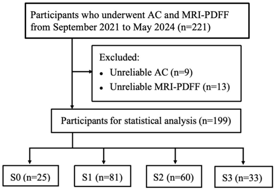
3.1. Participant Characteristics
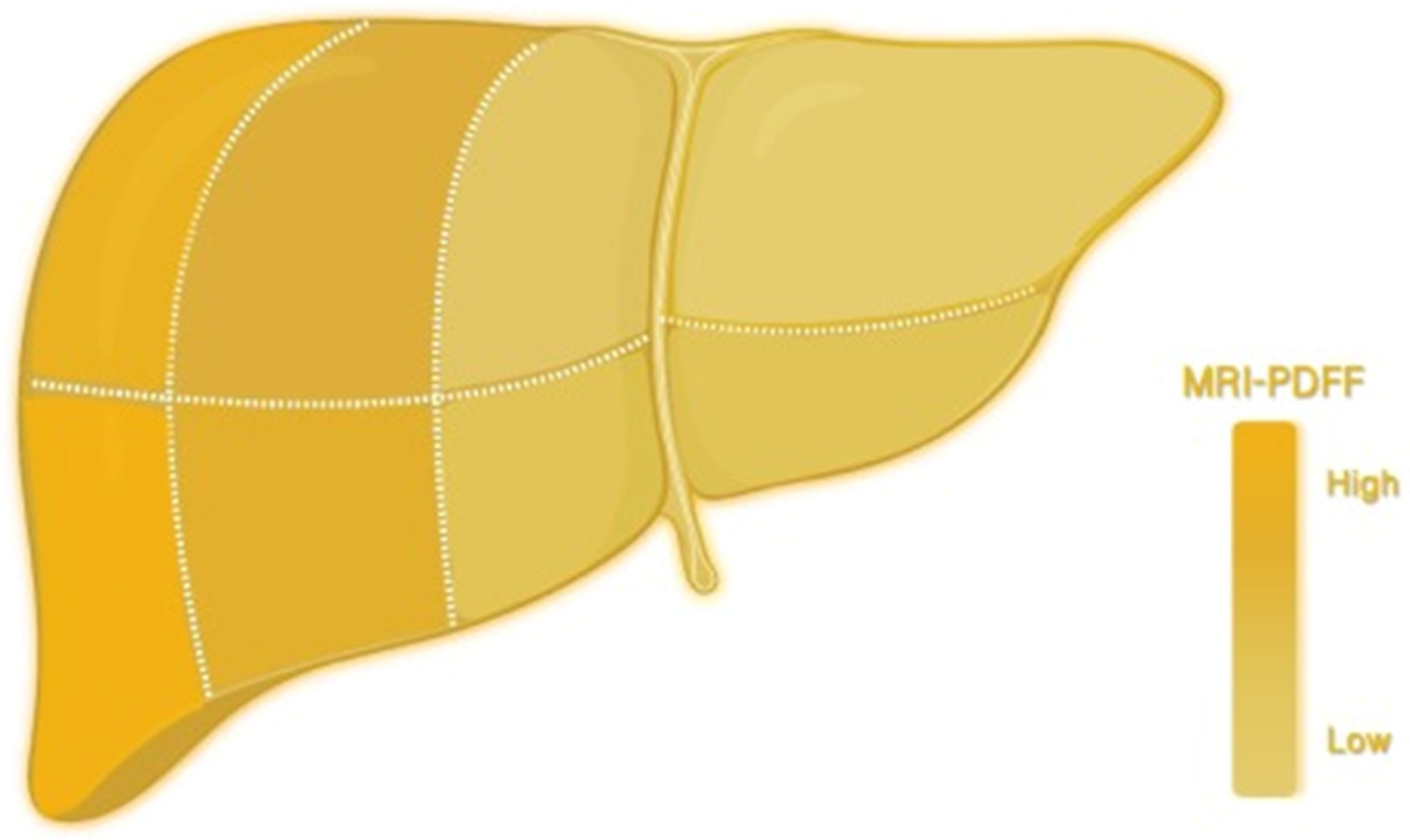
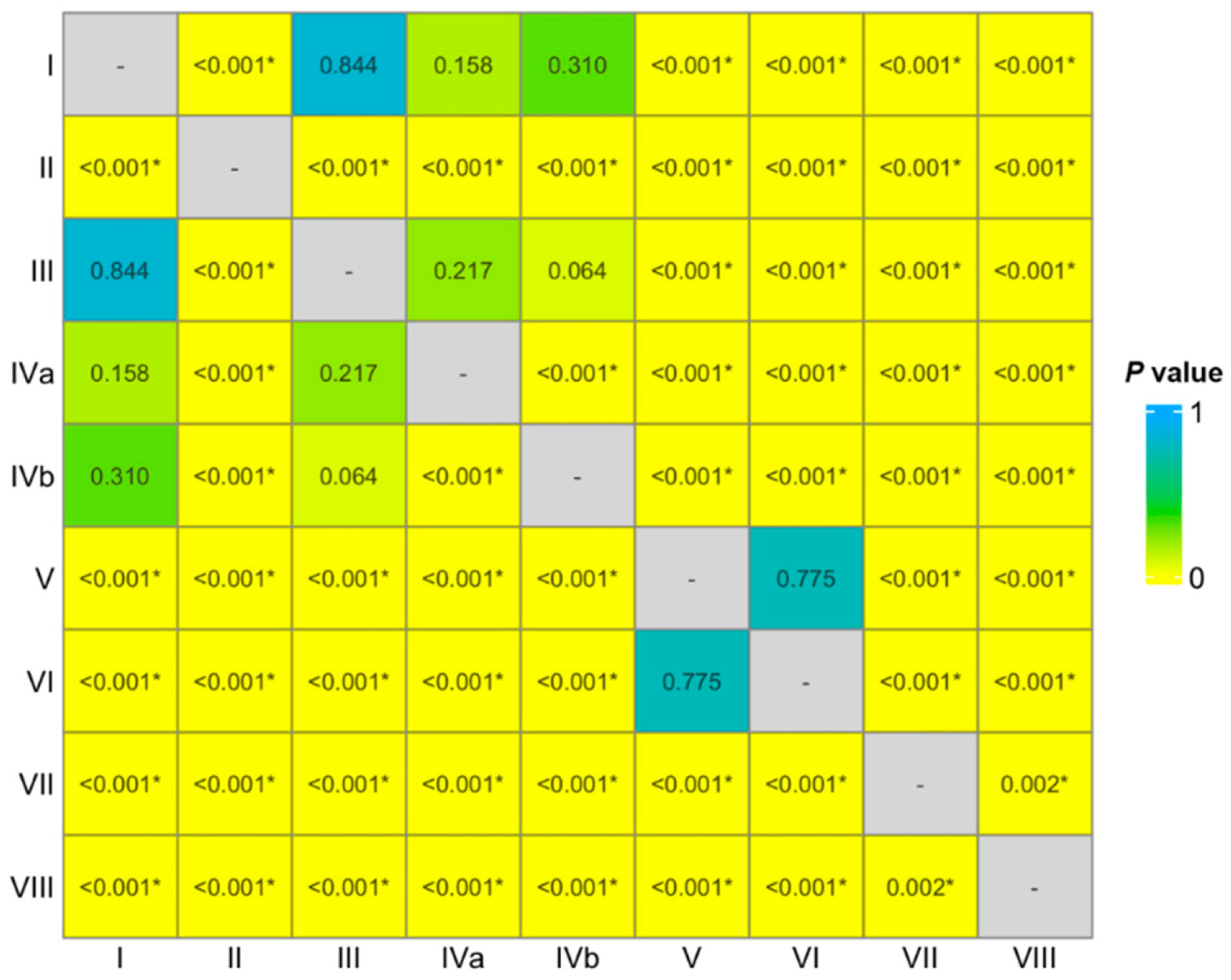
3.2. Analysis of the PDFF Values in Different Liver Segments
3.3. Analysis of AC Values Between Two Devices
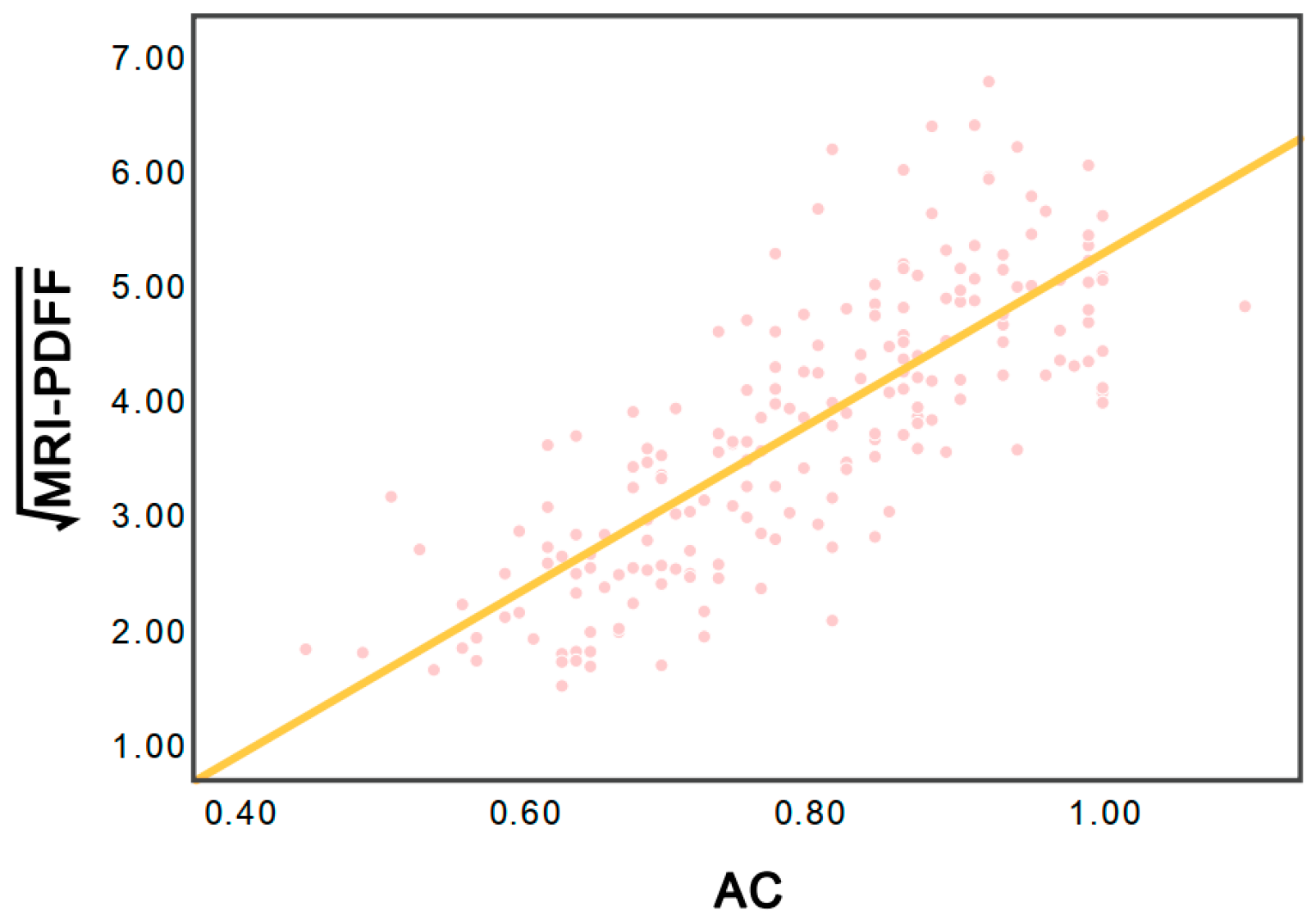
3.4. Model of the PDFF Conversion Fraction (PCF)
3.5. Model of the Attenuation Level Fraction (ALF)
3.6. Diagnostic Performances of the AC, ALF, and PCF
4. Discussion
5. Conclusions
Author Contributions
Funding
Institutional Review Board Statement
Informed Consent Statement
Data Availability Statement
Acknowledgments
Conflicts of Interest
References
- Rinella, M.E.; Lazarus, J.V.; Ratziu, V.; Francque, S.M.; Sanyal, A.J.; Kanwal, F.; Romero, D.; Abdelmalek, M.F.; Anstee, Q.M.; Arab, J.P.; et al. A multisociety Delphi consensus statement on new fatty liver disease nomenclature. Hepatology 2023, 78, 1966–1986. [Google Scholar] [CrossRef] [Scilit]
- Yip, T.C.; Vilar-Gomez, E.; Petta, S.; Yilmaz, Y.; Wong, G.L.; Adams, L.A.; de Lédinghen, V.; Sookoian, S.; Wong, V.W. Geographical similarity and differences in the burden and genetic predisposition of NAFLD. Hepatology 2023, 77, 1404–1427. [Google Scholar] [CrossRef] [Scilit] [PubMed]
- Teng, M.L.; Ng, C.H.; Huang, D.Q.; Chan, K.E.; Tan, D.J.; Lim, W.H.; Yang, J.D.; Tan, E.; Muthiah, M.D. Global incidence and prevalence of nonalcoholic fatty liver disease. Clin. Mol. Hepatol. 2023, 29, S32–S42. [Google Scholar] [CrossRef] [Scilit]
- Rinella, M.E.; Neuschwander-Tetri, B.A.; Siddiqui, M.S.; Abdelmalek, M.F.; Caldwell, S.; Barb, D.; Kleiner, D.E.; Loomba, R. AASLD Practice Guidance on the clinical assessment and management of nonalcoholic fatty liver disease. Hepatology 2023, 77, 1797–1835. [Google Scholar] [CrossRef] [Scilit] [PubMed]
- Younossi, Z.M.; Golabi, P.; Paik, J.M.; Henry, A.; Van Dongen, C.; Henry, L. The global epidemiology of nonalcoholic fatty liver disease (NAFLD) and nonalcoholic steatohepatitis (NASH): A systematic review. Hepatology 2023, 77, 1335–1347. [Google Scholar] [CrossRef] [Scilit] [PubMed]
- Kleiner, D.E.; Brunt, E.M.; Van Natta, M.; Behling, C.; Contos, M.J.; Cummings, O.W.; Ferrell, L.D.; Liu, Y.C.; Torbenson, M.S.; Unalp-Arida, A.; et al. Design and validation of a histological scoring system for nonalcoholic fatty liver disease. Hepatology 2005, 41, 1313–1321. [Google Scholar] [CrossRef] [Scilit]
- Brunt, E.M.; Janney, C.G.; Di Bisceglie, A.M.; Neuschwander-Tetri, B.A.; Bacon, B.R. Nonalcoholic steatohepatitis: A proposal for grading and staging the histological lesions. Am. J. Gastroenterol. 1999, 94, 2467–2474. [Google Scholar] [CrossRef]
- Ratziu, V.; Charlotte, F.; Heurtier, A.; Gombert, S.; Giral, P.; Bruckert, E.; Grimaldi, A.; Capron, F.; Poynard, T. Sampling variability of liver biopsy in nonalcoholic fatty liver disease. Gastroenterology 2005, 128, 1898–1906. [Google Scholar] [CrossRef] [Scilit]
- Powell, E.E.; Wong, V.W.; Rinella, M. Non-alcoholic fatty liver disease. Lancet 2021, 397, 2212–2224. [Google Scholar] [CrossRef] [Scilit]
- Goldstein, N.S.; Hastah, F.; Galan, M.V.; Gordon, S.C. Fibrosis heterogeneity in nonalcoholic steatohepatitis and hepatitis C virus needle core biopsy specimens. Am. J. Clin. Pathol. 2005, 123, 382–387. [Google Scholar] [CrossRef]
- Azizi, N.; Naghibi, H.; Shakiba, M.; Morsali, M.; Zarei, D.; Abbastabar, H.; Ghanaati, H. Evaluation of MRI proton density fat fraction in hepatic steatosis: A systematic review and meta-analysis. Eur. Radiol. 2025, 35, 1794–1807. [Google Scholar] [CrossRef] [Scilit] [PubMed]
- Caussy, C.; Reeder, S.B.; Sirlin, C.B.; Loomba, R. Noninvasive, quantitative assessment of liver fat by MRI-PDFF as an endpoint in NASH trials. Hepatology 2018, 68, 763–772. [Google Scholar] [CrossRef] [Scilit]
- QIBA Proton Density Fat Fraction Biomarker Committee. MRI-Based Proton Density Fat Fraction (PDFF) of the Liver. 2024. Available online: https://qibawiki.rsna.org/index.php/Profiles (accessed on 10 September 2024).
- Ferraioli, G.; Kumar, V.; Ozturk, A.; Nam, K.; de Korte, C.L.; Barr, R.G. US attenuation for liver fat quantification: An AIUM-RSNA QIBA pulse-echo quantitative ultrasound initiative. Radiology 2022, 302, 495–506. [Google Scholar] [CrossRef] [Scilit]
- Ferraioli, G.; Barr, R.G.; Berzigotti, A.; Sporea, I.; Wong, V.W.; Reiberger, T.; Karlas, T.; Thiele, M.; Cardoso, A.C.; Ayonrinde, O.T.; et al. WFUMB Guidelines/Guidance on Liver Multiparametric Ultrasound. Part 2: Guidance on Liver Fat Quantification. Ultrasound Med. Biol. 2024, 50, 1088–1098. [Google Scholar] [CrossRef] [Scilit]
- Ozturk, A.; Kumar, V.; Pierce, T.T.; Li, Q.; Baikpour, M.; Rosado-Mendez, I.; Wang, M.; Guo, P.; Schoen, S., Jr.; Gu, Y.; et al. The Future Is Beyond Bright: The Evolving Role of Quantitative US for Fatty Liver Disease. Radiology 2023, 309, e223146. [Google Scholar] [CrossRef] [Scilit]
- Jang, J.K.; Choi, S.H.; Lee, J.S.; Kim, S.Y.; Lee, S.S.; Kim, K.W. Accuracy of the ultrasound attenuation coefficient for the evaluation of hepatic steatosis: A systematic review and meta-analysis of prospective studies. Ultrasonography 2022, 41, 83–92. [Google Scholar] [CrossRef] [Scilit]
- Labyed, Y.; Milkowski, A. Novel Method for Ultrasound-Derived Fat Fraction Using an Integrated Phantom. J. Ultrasound Med. 2020, 39, 2427–2438. [Google Scholar] [CrossRef] [Scilit]
- Dillman, J.R.; Thapaliya, S.; Tkach, J.A.; Trout, A.T. Quantification of hepatic steatosis by ultrasound: Prospective comparison with MRI proton density fat fraction as reference standard. Am. J. Roentgenol. 2022, 219, 784–791. [Google Scholar] [CrossRef] [Scilit]
- Starekova, J.; Hernando, D.; Pickhardt, P.J.; Reeder, S.B. Quantification of liver fat content with CT and MRI: State of the art. Radiology 2021, 301, 250–262. [Google Scholar] [CrossRef] [Scilit] [PubMed]
- DeLong, E.R.; DeLong, D.M.; Clarke-Pearson, D.L. Comparing the areas under two or more correlated receiver operating characteristic curves: A nonparametric approach. Biometrics 1988, 44, 837–845. [Google Scholar] [CrossRef] [Scilit] [PubMed]
- Obuchowski, N.A. Receiver operating characteristic curves and their use in radiology. Radiology 2003, 229, 3–8. [Google Scholar] [CrossRef] [Scilit]
- Youden, W.J. Index for rating diagnostic tests. Cancer 1950, 3, 32–35. [Google Scholar] [CrossRef] [Scilit] [PubMed]
- Campo, C.A.; Hernando, D.; Schubert, T.; Bookwalter, C.A.; Pay, A.J.V.; Reeder, S.B. Standardized Approach for ROI-Based Measurements of Proton Density Fat Fraction and R2* in the Liver. Am. J. Roentgenol. 2017, 209, 592–603. [Google Scholar] [CrossRef] [Scilit] [PubMed]
- Ferraioli, G.; Roccarina, D.; Barr, R.G. Intersystem and Interoperator Agreement of US Attenuation Coefficient for Quantifying Liver Steatosis. Radiology 2024, 313, e240162. [Google Scholar] [CrossRef] [Scilit] [PubMed]
- Qi, R.; Lu, L.; He, T.; Zhang, L.; Lin, Y.; Bao, L. Comparing ultrasound-derived fat fraction and MRI-PDFF for quantifying hepatic steatosis: A real-world prospective study. Eur. Radiol. 2025, 35, 2580–2588. [Google Scholar] [CrossRef] [Scilit]
- Huang, Y.; Li, J.; Liu, C.; Song, D.; Zhu, C.; Ren, Y.; Lv, J.; Jiang, L.; Shan, R.; Wang, H.; et al. Noninvasive Quantification of Hepatic Steatosis Using Ultrasound-Derived Fat Fraction (CHESS2303): A Prospective Multicenter Study. MedComm 2025, 6, e70123. [Google Scholar] [CrossRef] [Scilit]



| Characteristics | Value | Number |
|---|---|---|
| Age (y) | 31 ± 8 | 199 |
| Sex * | 199 | |
| M | 84 (42%) | |
| F | 115 (58%) | |
| BMI (kg/m2) | 33.7 ± 4.5 | 192 |
| Waist (cm) | 105.2 ± 15.7 | 193 |
| Hypertension * | 48 (27%) | 177 |
| Fasting Blood Glucose (mmol/L) | 5.41 ± 1.62 | 185 |
| Oral Glucose Tolerance Test-2 h (mmol/L) | 8.22 ± 3.20 | 175 |
| HbA1c (%) | 5.7 ± 1.1 | 168 |
| Triglycerides (mmol/L) | 2.06 ± 1.79 | 171 |
| High-Density Lipoprotein Cholesterol (mmol/L) | 1.09 ± 0.38 | 171 |
| MRI-PDFF * | 199 | |
| S0 (<5%) | 25 (13%) | |
| S1 (5≤ to <15%) | 81 (41%) | |
| S2 (15≤ to <25%) | 60 (30%) | |
| S3 (≥25%) | 33 (17%) | |
| AC (dB/cm/MHz) | 0.79 ± 0.13 | 199 |
| Steatosis Grade | Canon | Mindray | p Value * |
|---|---|---|---|
| S0 | 0.63 ± 0.06 | 0.59 ± 0.12 | 0.33 (−0.04, 0.11) |
| S1 | 0.74 ± 0.10 | 0.71 ± 0.08 | 0.06 (0.00, 0.08) |
| S2 | 0.88 ± 0.10 | 0.87 ± 0.08 | 0.68 (−0.04, 0.06) |
| S3 | 0.92 ± 0.05 | 0.93 ± 0.07 | 0.78 (−0.04, 0.03) |
| Parameters | S0 (n = 25) | S1 (n = 81) | p * Value | S2 (n = 60) | p ** Value | S3 (n = 33) | p *** Value |
|---|---|---|---|---|---|---|---|
| MRI-PDFF | 3.5% ± 0.7% | 9.6% (7.0–12.5%) a | <0.05 | 19.4% ± 3.0% | <0.05 | 28.4% (26.3–35.1%) a | <0.05 |
| AC | 0.62 ± 0.08 | 0.73 ± 0.09 | <0.05 | 0.87 ± 0.09 | <0.05 | 0.92 ± 0.06 | <0.05 |
| AUC * | Cut-Off | Sensitivity * | Specificity * | ||
|---|---|---|---|---|---|
| AC | |||||
| ≥S1 | 0.92 (0.87, 0.96) | 0.73 | 76% (69%, 82%) | 96% (80%, 100%) | |
| ≥S2 | 0.93 (0.90, 0.96) | 0.77 | 95% (88%, 98%) | 77% (67%, 84%) | |
| ≥S3 | 0.87 (0.82, 0.92) | 0.86 | 91% (76%, 98%) | 74% (67%, 81%) | |
| ALF | |||||
| ≥S1 | 0.92 (0.87, 0.96) | 45% | 76% (69%, 82%) | 96% (80%, 100%) | |
| ≥S2 | 0.93 (0.90, 0.96) | 53% | 95% (88%, 98%) | 77% (67%, 84%) | |
| ≥S3 | 0.87 (0.82, 0.92) | 71% | 91% (76%, 98%) | 74% (67%, 81%) | |
| PCF | |||||
| ≥S1 | 0.92 (0.87, 0.96) | 33% | 76% (69%, 82%) | 96% (80%, 100%) | |
| ≥S2 | 0.93 (0.90, 0.96) | 36% | 95% (88%, 98%) | 77% (67%, 84%) | |
| ≥S3 | 0.87 (0.82, 0.92) | 42% | 91% (76%, 98%) | 74% (67%, 81%) |
Disclaimer/Publisher’s Note: The statements, opinions and data contained in all publications are solely those of the individual author(s) and contributor(s) and not of MDPI and/or the editor(s). MDPI and/or the editor(s) disclaim responsibility for any injury to people or property resulting from any ideas, methods, instructions or products referred to in the content. |
© 2026 by the authors. Licensee MDPI, Basel, Switzerland. This article is an open access article distributed under the terms and conditions of the Creative Commons Attribution (CC BY) license.
Share and Cite
Zhang, Y.; Jiang, T.; Xu, C.; He, J.; Zhang, H.; Chen, T.; Zeng, J. Fraction Conversion Models Based on Ultrasound Attenuation Coefficient for Assessing Liver Steatosis. Diagnostics 2026, 16, 1086. https://doi.org/10.3390/diagnostics16071086
Zhang Y, Jiang T, Xu C, He J, Zhang H, Chen T, Zeng J. Fraction Conversion Models Based on Ultrasound Attenuation Coefficient for Assessing Liver Steatosis. Diagnostics. 2026; 16(7):1086. https://doi.org/10.3390/diagnostics16071086
Chicago/Turabian StyleZhang, Yin, Ting Jiang, Chuli Xu, Jiajun He, Hongjun Zhang, Tufeng Chen, and Jie Zeng. 2026. "Fraction Conversion Models Based on Ultrasound Attenuation Coefficient for Assessing Liver Steatosis" Diagnostics 16, no. 7: 1086. https://doi.org/10.3390/diagnostics16071086
APA StyleZhang, Y., Jiang, T., Xu, C., He, J., Zhang, H., Chen, T., & Zeng, J. (2026). Fraction Conversion Models Based on Ultrasound Attenuation Coefficient for Assessing Liver Steatosis. Diagnostics, 16(7), 1086. https://doi.org/10.3390/diagnostics16071086