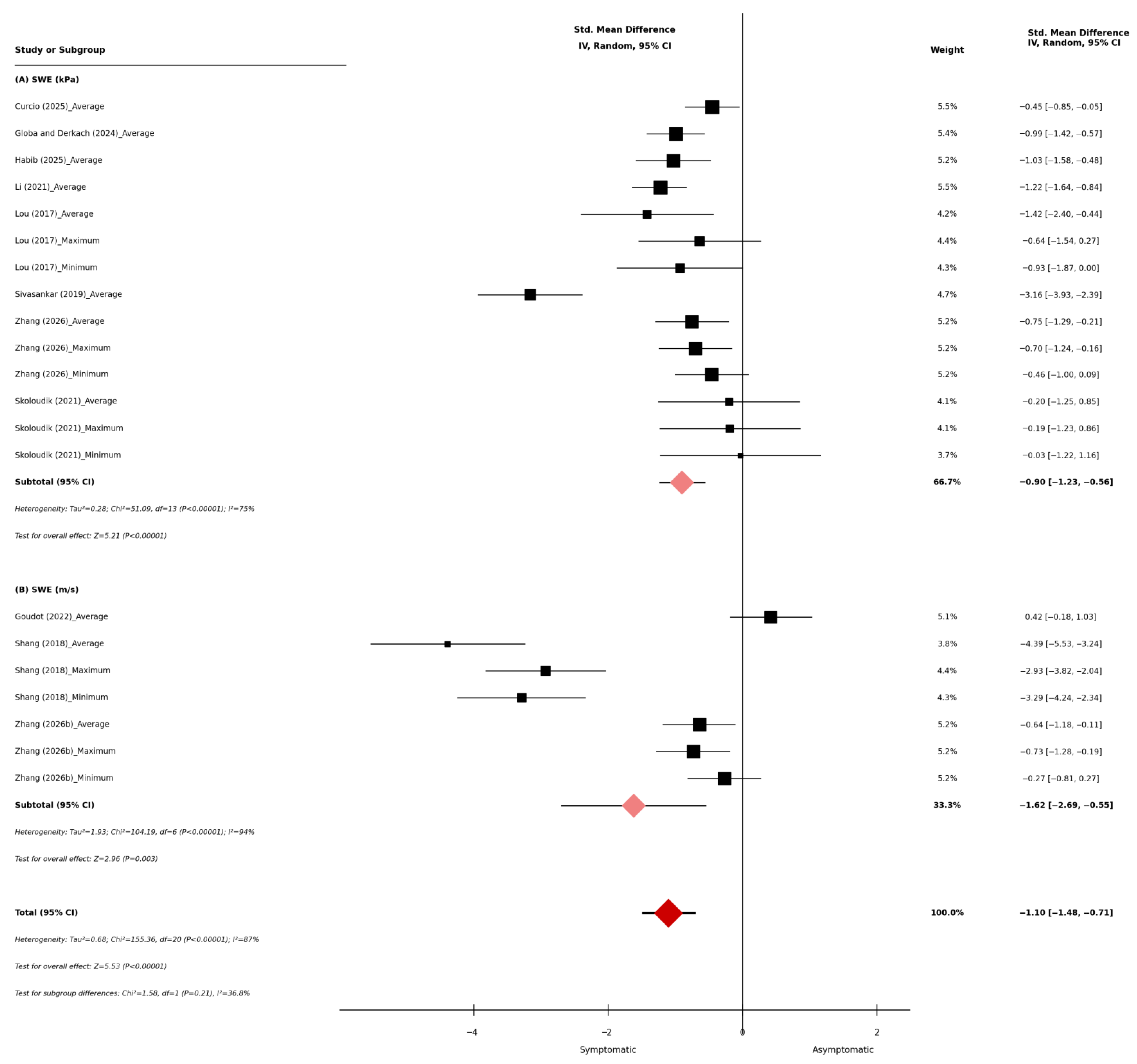
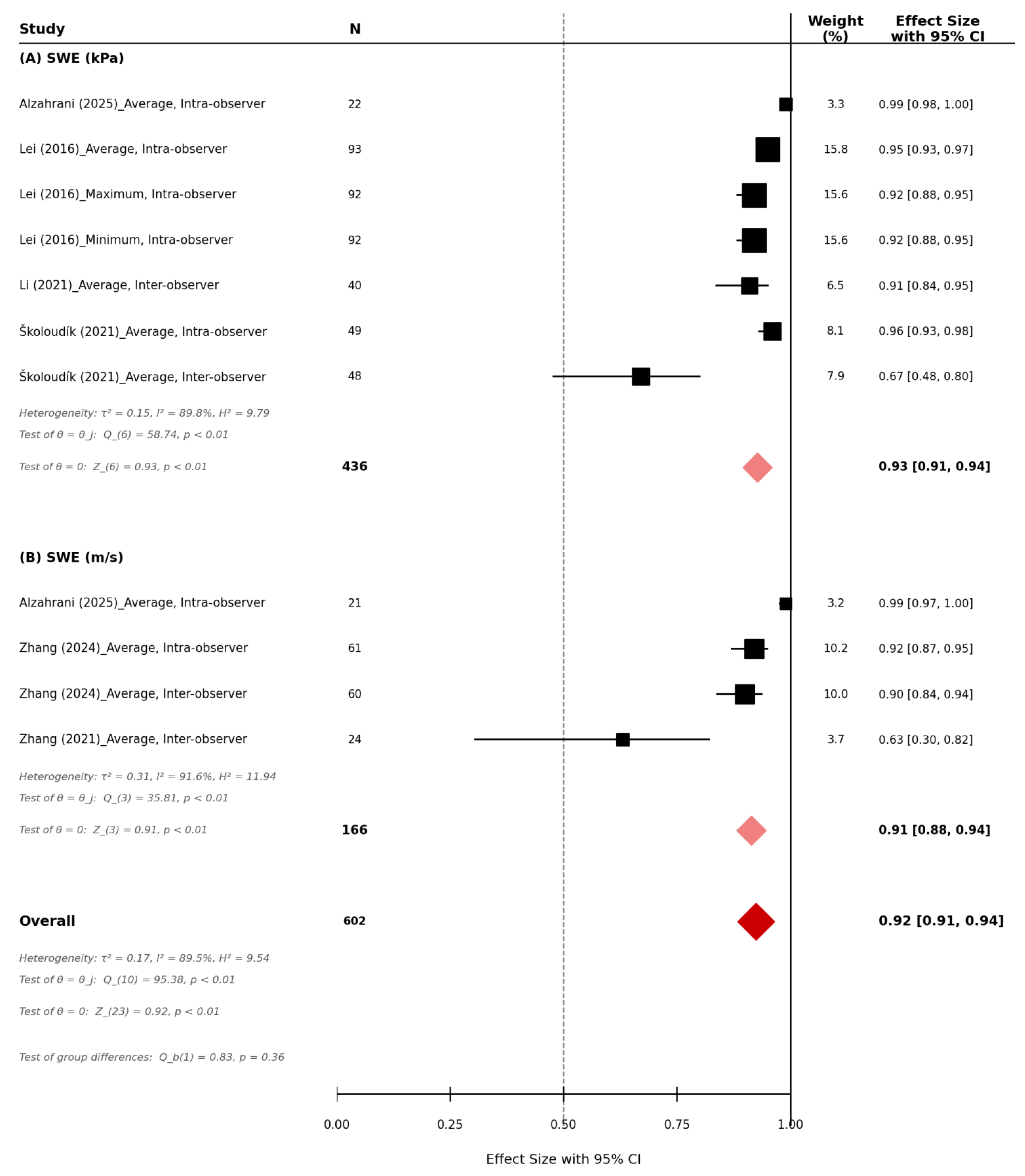
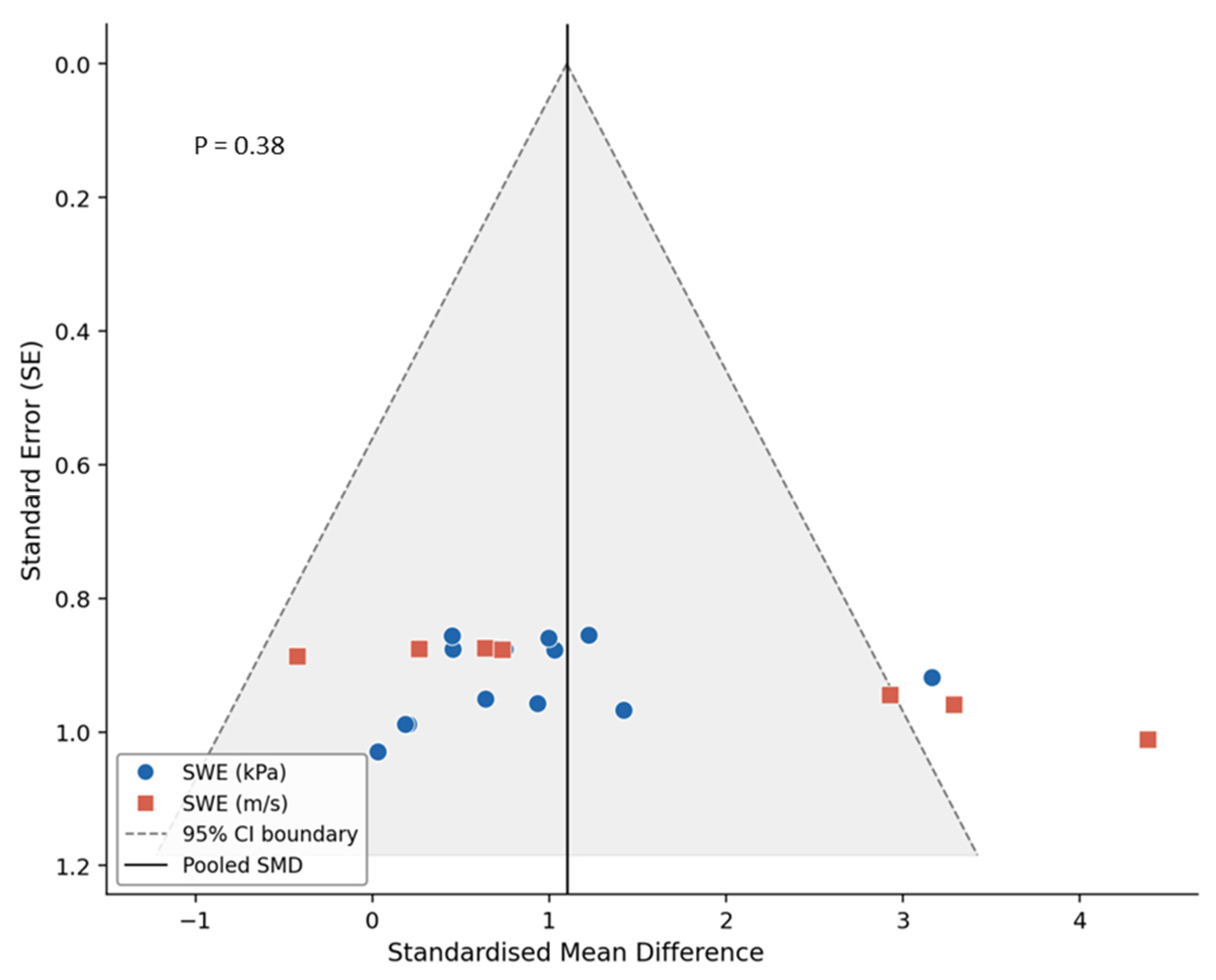
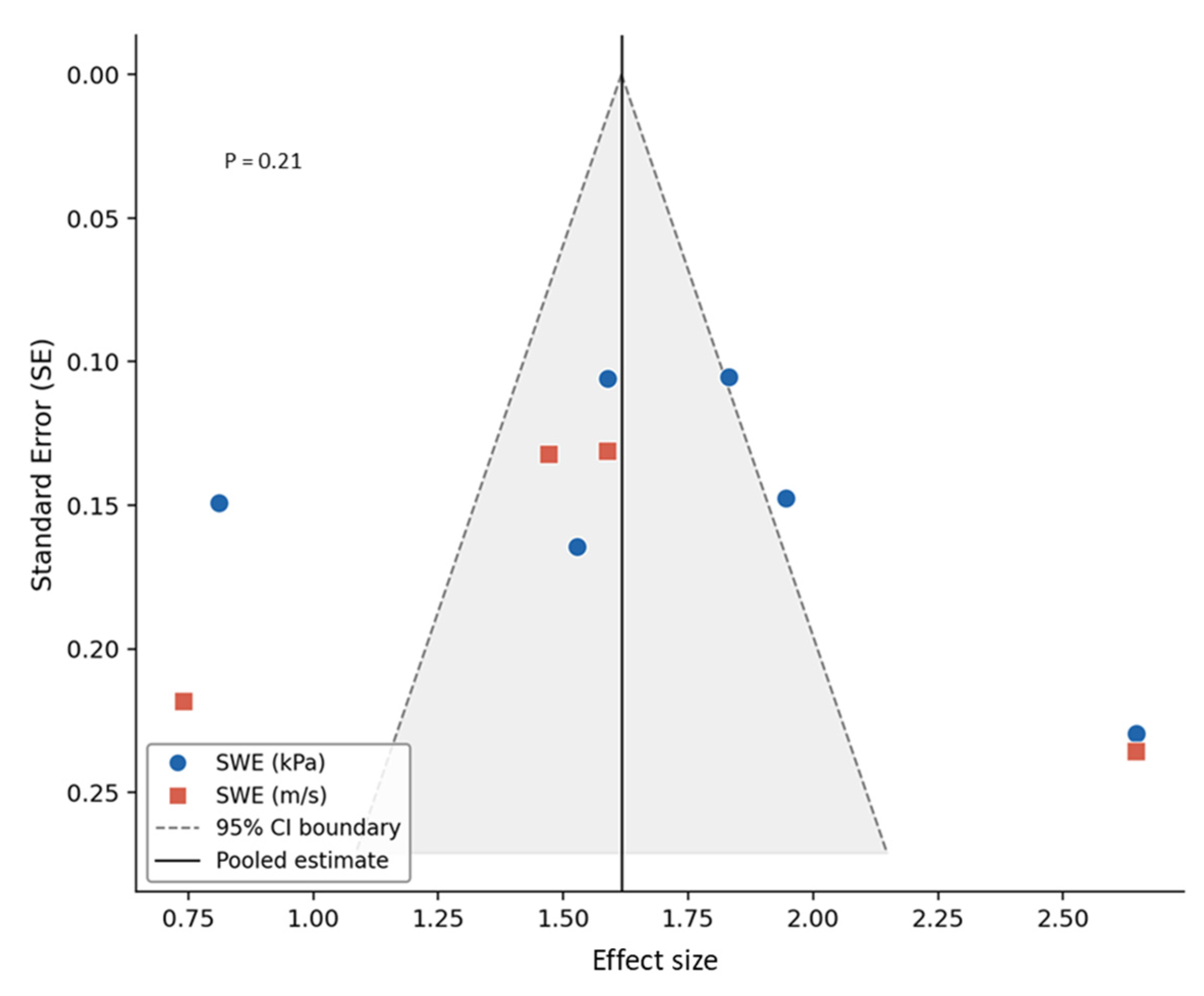
Quantitative Ultrasound SWE of Carotid Plaque in Symptomatic and Asymptomatic Patients: A Systematic Review and Meta-Analysis

 , , , , , , , , , and
Abstract
1. Introduction
2. Methods
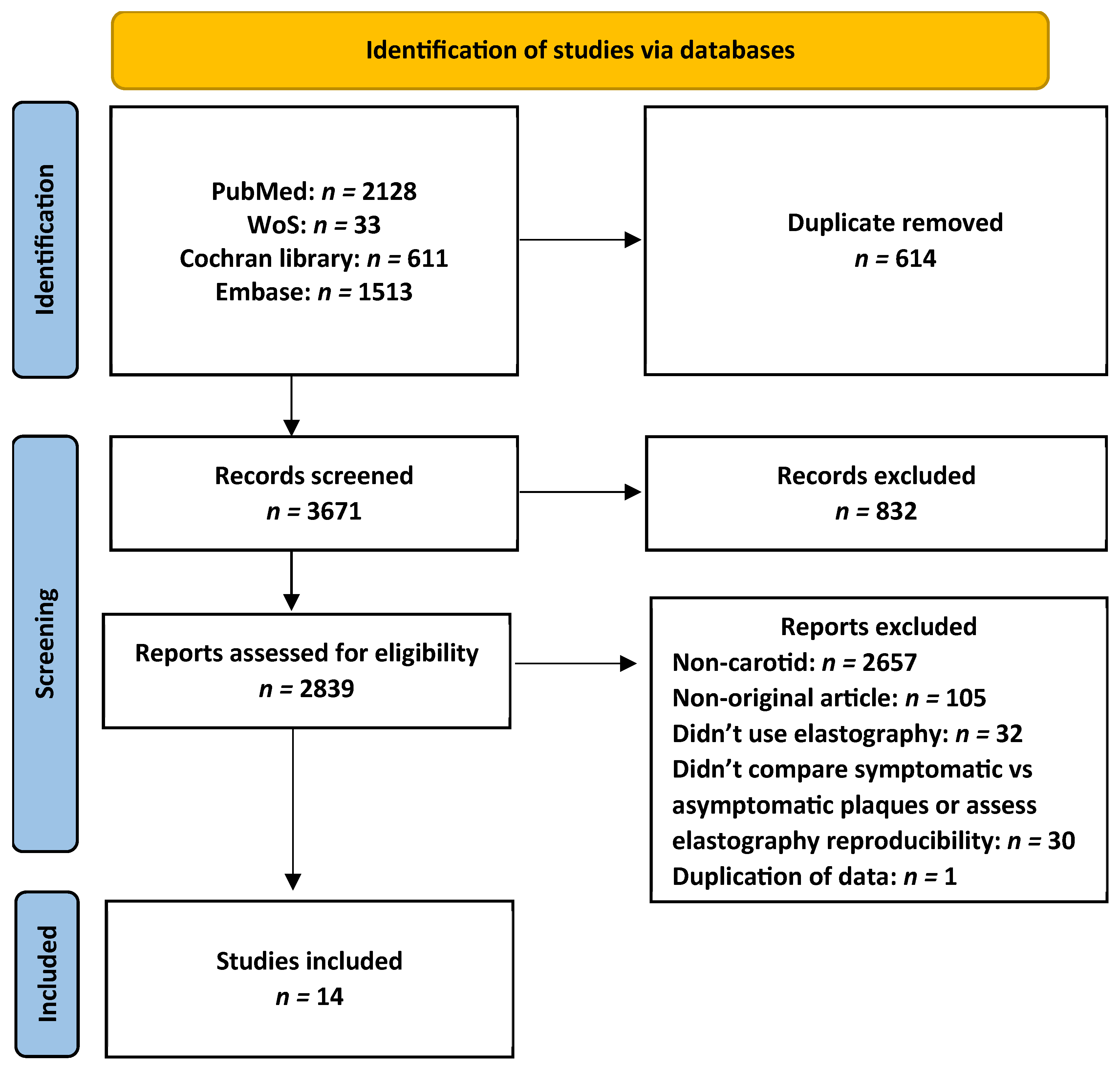
2.1. Search Strategy and Study Selection
2.2. Data Acquisition
2.3. Quality
2.4. Statistical Analysis
3. Results
3.1. SWE in Symptomatic and Asymptomatic Carotid Plaques
3.2. Reproducibility of SWE in the Evaluation of Carotid Plaque Stiffness
3.3. Quality and Publication Bias
4. Discussion
5. Conclusions
Supplementary Materials
Author Contributions
Funding
Institutional Review Board Statement
Informed Consent Statement
Data Availability Statement
Conflicts of Interest
References
- Grønholdt, M.L.M.; Nordestgaard, B.G.; Schroeder, T.V.; Vorstrup, S.; Sillesen, H. Ultrasonic echolucent carotid plaques predict future strokes. Circulation 2001, 104, 68–73. [Google Scholar] [CrossRef] [Scilit] [PubMed]
- Spanos, K.; Tzorbatzoglou, I.; Lazari, P.; Maras, D.; Giannoukas, A.D. Carotid artery plaque echomorphology and its association with histopathologic characteristics. J. Vasc. Surg. 2018, 68, 1772–1780. [Google Scholar] [CrossRef] [Scilit] [PubMed]
- Sultan, S.R. Ultrasound diagnostic potential of carotid plaques in symptomatic and asymptomatic patients: Insights from quantitative gray-scale analysis. J. Radiat. Res. Appl. Sci. 2024, 17, 100776. [Google Scholar] [CrossRef] [Scilit]
- David, E.; Grazhdani, H.; Aliotta, L.; Gavazzi, L.M.; Foti, P.V.; Palmucci, S.; Inì, C.; Tiralongo, F.; Castiglione, D.; Renda, M.; et al. Imaging of Carotid Stenosis: Where Are We Standing? Comparison of Multiparametric Ultrasound, CT Angiography, and MRI Angiography, with Recent Developments. Diagnostics 2024, 14, 1708. [Google Scholar] [CrossRef] [Scilit]
- Coli, S.; Magnoni, M.; Sangiorgi, G.; Marrocco-Trischitta, M.M.; Melisurgo, G.; Mauriello, A.; Spagnoli, L.; Chiesa, R.; Cianflone, D.; Maseri, A. Contrast-enhanced ultrasound imaging of intraplaque neovascularization in carotid arteries: Correlation with histology and plaque echogenicity. J. Am. Coll. Cardiol. 2008, 52, 223–230. [Google Scholar] [CrossRef] [Scilit]
- Varetto, G.; Gibello, L.; Castagno, C.; Quaglino, S.; Ripepi, M.; Benintende, E.; Gattuso, A.; Garneri, P.; Zan, S.; Capaldi, G.; et al. Use of Contrast-Enhanced Ultrasound in Carotid Atherosclerotic Disease: Limits and Perspectives. BioMed Res. Int. 2015, 2015, 293163. [Google Scholar] [CrossRef] [Scilit]
- Sultan, S.R.; Bashmail, F.T.; Alzahrani, N.A.; Alharbi, S.I.; Anbar, R.; Alkharaiji, M. Contrast-enhanced ultrasound for the evaluation of symptomatic and asymptomatic carotid plaques: A systematic review and meta-analysis. Echocardiography 2022, 39, 1032–1043. [Google Scholar]
- Amzar, D.; Stoian, D.; Oglat, A.A.; Abukhalil, T. Ultrasound Elastography: Methods, Clinical Applications, and Limitations: A Review Article. Appl. Sci. 2024, 14, 4308. [Google Scholar] [CrossRef] [Scilit]
- Cui, X.W.; Li, K.N.; Yi, A.J.; Wang, B.; Wei, Q.; Wu, G.G.; Dietrich, C.F. Ultrasound elastography. Endosc. Ultrasound 2022, 11, 252–274. [Google Scholar] [CrossRef] [Scilit]
- Bamber, J.; Cosgrove, D.; Dietrich, C.F.; Fromageau, J.; Bojunga, J.; Calliada, F.; Cantisani, V.; Correas, J.-M.; D’Onofrio, M.; Drakonaki, E.E.; et al. EFSUMB guidelines and recommendations on the clinical use of ultrasound elastography. Part 1: Basic principles and technology. Ultraschall Med. 2013, 34, 169–184. [Google Scholar] [CrossRef] [Scilit]
- Ferraioli, G.; Barr, R.G.; Farrokh, A.; Radzina, M.; Cui, X.W.; Dong, Y.; Rocher, L.; Cantisani, V.; Polito, E.; D’Onofrio, M.; et al. How to perform shear wave elastography. Part I. Med. Ultrason. 2022, 24, 95–106. [Google Scholar] [CrossRef] [Scilit]
- Sigrist, R.M.S.; Liau, J.; Kaffas, A.E.; Chammas, M.C.; Willmann, J.K. Ultrasound elastography: Review of techniques and clinical applications. Theranostics 2017, 7, 1303–1329. [Google Scholar] [CrossRef] [Scilit] [PubMed]
- Zhang, J.; Wu, X.; Gao, Y.; Wang, L. Morphological and biomechanical characteristics of carotid plaques with mild stenosis using ultrafast and high-definition ultrasonography. Eur. Radiol. 2026, 36, 437–449. [Google Scholar] [PubMed]
- Garrard, J.W.; Ummur, P.; Nduwayo, S.; Kanber, B.; Hartshorne, T.C.; West, K.P.; Moore, D.; Robinson, T.G.; Ramnarine, K.V. Shear Wave Elastography May Be Superior to Greyscale Median for the Identification of Carotid Plaque Vulnerability: A Comparison with Histology. Ultraschall Med. 2015, 36, 386–390. [Google Scholar] [CrossRef] [Scilit]
- Di Leo, N.; Venturini, L.; de Soccio, V.; Forte, V.; Lucchetti, P.; Cerone, G.; Alagna, G.; Caratozzolo, M.; Messineo, D.; Di Gioia, C.; et al. Multiparametric ultrasound evaluation with CEUS and shear wave elastography for carotid plaque risk stratification. J. Ultrasound 2018, 21, 293–300. [Google Scholar] [CrossRef] [Scilit]
- Lei, Z.; Qiang, Y.; Pu, T.; Jie, L. Quantitative assessment of carotid atherosclerotic plaque: Initial clinical results using ShearWave Elastography. Int. J. Clin. Exp. Med. 2016, 9, 9347–9355. [Google Scholar]
- Shang, J.; Wang, W.; Feng, J.; Luo, G.-G.; Dang, Y.; Sun, J.; Yang, Y.-Q.; Ruan, L.-T. Carotid Plaque Stiffness Measured with Supersonic Shear Imaging and Its Correlation with Serum Homocysteine Level in Ischemic Stroke Patients. Korean J. Radiol. 2018, 19, 15–22. [Google Scholar] [CrossRef] [Scilit] [PubMed]
- Alzahrani, A.; Alharbi, A.A.; Alharbi, A.K.; Alkhaldi, A.; Filimban, A.Z.; Alfatni, A.; Kaifi, R.; Albngali, A.; Alkharaiji, M.; Alserihy, O.; et al. Evaluating Carotid Plaque Stiffness with Ultrasound 2D Shear-Wave Elastography in Patients Undergoing Coronary Artery Bypass Grafting. Diagnostics 2025, 15, 338. [Google Scholar] [CrossRef] [Scilit]
- Page, M.J.; McKenzie, J.E.; Bossuyt, P.M.; Boutron, I.; Hoffmann, T.C.; Mulrow, C.D.; Shamseer, L.; Tetzlaff, J.M.; Akl, E.A.; Brennan, S.E.; et al. The PRISMA 2020 statement: An updated guideline for reporting systematic reviews. BMJ 2021, 372, n71. [Google Scholar] [CrossRef] [Scilit]
- Urbak, L.; Sandholt, B.V.; Graebe, M.; Bang, L.E.; Bundgaard, H.; Sillesen, H. Patients with Unstable Atherosclerosis Have More Echolucent Carotid Plaques Compared with Stable Atherosclerotic Patients: A 3-D Ultrasound Study. Ultrasound Med. Biol. 2020, 46, 2164–2172. [Google Scholar] [CrossRef] [Scilit]
- Whiting, P.; Rutjes, A.W.; Reitsma, J.B.; Bossuyt, P.M.; Kleijnen, J. The development of QUADAS: A tool for the quality assessment of studies of diagnostic accuracy included in systematic reviews. BMC Med. Res. Methodol. 2003, 3, 25. [Google Scholar]
- Wan, X.; Wang, W.; Liu, J.; Tong, T. Estimating the sample mean and standard deviation from the sample size, median, range and/or interquartile range. BMC Med. Res. Methodol. 2014, 14, 135. [Google Scholar]
- Hozo, S.P.; Djulbegovic, B.; Hozo, I. Estimating the mean and variance from the median, range, and the size of a sample. BMC Med. Res. Methodol. 2005, 5, 13. [Google Scholar] [CrossRef] [Scilit]
- Koo, T.K.; Li, M.Y. A Guideline of Selecting and Reporting Intraclass Correlation Coefficients for Reliability Research. J. Chiropr. Med. 2016, 15, 155–163. [Google Scholar] [CrossRef] [Scilit] [PubMed]
- Egger, M.; Smith, G.D.; Schneider, M.; Minder, C. Bias in meta-analysis detected by a simple, graphical test. Br. Med. J. 1997, 315, 629–634. [Google Scholar]
- Habib, A.H.; Baz, A.A.; Mohamed, S.F.; Al-Azayem, S.A. Diagnostic value of ultrasound and shear wave elastography in carotid plaque risk stratification. J. Ultrasound 2025, 28, 691–700. [Google Scholar] [CrossRef] [Scilit]
- Curcio, N.; Conti, M.; Cardani, R.; Renna, L.V.; Dell’Antonio, G.; Bari, V.; Nano, G.; Matrone, G.; Mazzaccaro, D. Biomechanical assessment of vulnerable plaque: From histological evidence to ultrasound elastography and image-based computational patient-specific modelling. Front. Bioeng. Biotechnol. 2025, 13, 1478408. [Google Scholar] [CrossRef] [Scilit]
- Zhang, L.; Jia, C.; Gu, S.; Chen, J.; Wu, R. Intraplaque neovascularization combined with plaque elasticity for predicting ipsilateral stroke in patients with asymptomatic mild carotid stenosis. Quant. Imaging Med. Surg. 2024, 14, 4815–4824. [Google Scholar] [CrossRef] [Scilit] [PubMed]
- Globa, M.V.; Derkach, B.V. Modern approaches to ultrasonographic assessment of carotid plaque in terms of its potential instability. Wiad. Lek. 2024, 77, 919–925. [Google Scholar] [CrossRef] [Scilit]
- Goudot, G.; Sitruk, J.; Jimenez, A.; Julia, P.; Khider, L.; Alsac, J.-M.; El Batti, S.; Bruneval, P.; Amemyia, K.; Pedreira, O.; et al. Carotid Plaque Vulnerability Assessed by Combined Shear Wave Elastography and Ultrafast Doppler Compared to Histology. Transl. Stroke Res. 2022, 13, 100–111. [Google Scholar] [CrossRef] [Scilit]
- Li, Y.; Zheng, S.; Zhang, J.; Wang, F.; Liu, X.; He, W. Advance ultrasound techniques for the assessment of plaque vulnerability in symptomatic and asymptomatic carotid stenosis: A multimodal ultrasound study. Cardiovasc. Diagn. Ther. 2021, 11, 28–38. [Google Scholar] [CrossRef] [Scilit]
- Školoudík, D.; Kešnerová, P.; Vomáčka, J.; Hrbáč, T.; Netuka, D.; Forostyak, S.; Roubec, M.; Herzig, R.; Belšan, T. Shear-Wave Elastography Enables Identification of Unstable Carotid Plaque. Ultrasound Med. Biol. 2021, 47, 1704–1710. [Google Scholar] [CrossRef] [Scilit]
- Zhang, Y.; Cao, J.; Zhou, J.; Zhang, C.; Li, Q.; Chen, S.; Feinstein, S.; Grayburn, P.A.; Huang, P. Plaque Elasticity and Intraplaque Neovascularisation on Carotid Artery Ultrasound: A Comparative Histological Study. Eur. J. Vasc. Endovasc. Surg. 2021, 62, 358–366. [Google Scholar] [CrossRef] [Scilit]
- Sivasankar, R.; Singh, R.; Hashim, P.; Soni, B.; Patel, R.; Bajpai, A. Evaluation of carotid plaque vulnerability using shear-wave elastography: An observational comparative study. J. Mar. Med. Soc. 2019, 21, 134–137. [Google Scholar] [CrossRef] [Scilit]
- Lou, Z.; Yang, J.; Tang, L.; Jin, Y.; Zhang, J.; Liu, C.; Li, Q. Shear Wave Elastography Imaging for the Features of Symptomatic Carotid Plaques: A Feasibility Study. J. Ultrasound Med. 2017, 36, 1213–1223. [Google Scholar] [CrossRef] [Scilit]
- Jansen, I.; Cahalane, R.; Hengst, R.; Akyildiz, A.; Farrell, E.; Gijsen, F.; Aikawa, E.; van der Heiden, K.; Wissing, T. The interplay of collagen, macrophages, and microcalcification in atherosclerotic plaque cap rupture mechanics. Basic Res. Cardiol. 2024, 119, 193. [Google Scholar] [CrossRef] [Scilit] [PubMed]
- Shiina, T.; Nightingale, K.R.; Palmeri, M.L.; Hall, T.J.; Bamber, J.C.; Barr, R.G.; Castera, L.; Choi, B.I.; Chou, Y.-H.; Cosgrove, D.; et al. WFUMB guidelines and recommendations for clinical use of ultrasound elastography: Part 1: Basic principles and terminology. Ultrasound Med. Biol. 2015, 41, 1126–1147. [Google Scholar] [CrossRef] [Scilit]
- Ryu, H.; Ahn, S.J.; Yoon, J.H.; Lee, J.M. Inter-platform reproducibility of liver stiffness measured with two different point shear wave elastography techniques and 2-dimensional shear wave elastography using the comb-push technique. Ultrasonography 2019, 38, 345–354. [Google Scholar] [CrossRef] [Scilit] [PubMed]
- Sultan, S.R. Ultrasound 2D-SWE inter-system variability and reliability: Insights from an elasticity phantom model. J. Radiat. Res. Appl. Sci. 2025, 18, 102022. [Google Scholar]



| Reference (Author/Year) | Primary Aim | Sample Size (Symptomatic/Asymptomatic) | Histology as a Reference | ROI | Machine/Transducer | Findings |
|---|---|---|---|---|---|---|
| Zhang et al., 2026 [13] | To evaluate the efficiency of multimodal ultrasonography in symptomatic and asymptomatic carotid plaques as potential biomarkers of ischemic stroke | 476 (105/371) | No | Entire plaque | Mindray Resona 9/Linear array 15-3 MHz | SWE demonstrated lower stiffness values in symptomatic carotid plaques compared with asymptomatic plaques |
| Habib et al., 2025 [26] | To identify differences in atherosclerotic plaque elasticity and plaque echogenicity among symptomatic and asymptomatic groups. | 58 (29/29) | No | Entire plaque | Canon Aplio/Linear array 15-4 MHz | SWE can help to identify unstable plaque |
| Curcio et al., 2025 [27] | To study the ability of non-invasive methods, including SWE, for assessing the biomechanical features of vulnerable and stable plaques in patients undergoing CEA | 100 (43/57) | Yes | Entire plaque | Esaote MyLabX8/Linear array 15-4 MHz | SWE stiffness measurements were able to differentiate patients with vulnerable plaques from stable plaques |
| Alzahrani et al., 2025 [18] | To assess carotid plaque stiffness in patients undergoing CABG using SWE, and its reproducibility | 43 | No | Maximum plaque thickness | Siemens Redwood/Linear array 10-4 MHz | SWE is a reproducible tool for quantifying carotid plaque stiffness, with significant correlation to GSM and reduced stiffness in CABG compared to non-CABAG patients |
| Zhang et al., 2024 [28] | To evaluate IPN and plaque elasticity In patients with asymptomatic carotid stenosis, and its reproducibility | 121 | No | Entire plaque | Phillips EPIQ/Linear array 15-4 MHz | SWE is reproducible and demonstrated significant predictive value for the risk of stroke in patients with asymptomatic carotid plaque |
| Globa and Derkach, 2024 [29] | To identify characteristics of carotid plaque using SMI and SWE in symptomatic and asymptomatic patients | 96 (51/45) | No | Entire plaque | Canon Aplio/Linear array 15-4 MHz | The low stiffness of the plaque based on to SWE data can characterize the potential instability of carotid plaques |
| Goudot et al., 2022 [30] | To evaluate plaque stiffness and WSS as potential biomarkers of vulnerability | 46 (29/17) | Yes | Entire plaque | Aixplorer SuperSonic/Linear array 10-2 MHz | SWE of plaque stiffness showed no significant differences between vulnerable and stable plaques |
| Li et al., 2021 [31] | To assess vulnerable plaques using advanced ultrasound techniques | 123 (65/58) | No | Entire plaque | Canon Aplio/Linear array 15-4 MHz | SWE can identify plaque characteristics that are associated with ischemic events and potentially vulnerable plaque |
| Školoudík et al., 2021 [32] | To identify differences in atherosclerotic plaque elasticity measured using SWE among individuals with symptomatic and asymptomatic stable carotid plaques | 97 (11/86) | No | Entire plaque | Aixplorer SuperSonic/Linear array 15-4 MHz | SWE is useful for differentiating stable from unstable atherosclerotic plaques in carotid arteries. |
| Zhang et al., 2021 [33] | To assess the association between IPN and plaque elasticity and to correlate ultrasound findings with histopathological features | 94 | Yes | Multiple regions within the plaque | Siemens Acuson/Linear array 9-4 MHz | SWE can evaluate the vulnerability of the carotid plaque by assessing the elasticity of the plaque |
| Sivasankar et al., 2019 [34] | To assess carotid plaque stiffness using SWE and to explore the association between stiffness metrics and plaque vulnerability | 60 (30/30) | No | Multiple regions within the plaque | Siemens Acuson/Linear array 9-4 MHz | SWE is useful tool that can be used for the early detection of vulnerable carotid plaques |
| Shang et al., 2018 [17] | To evaluate the feasibility of SWE in assessing the stiffness of carotid plaque | 129 (78/51) | No | Entire plaque | Aixplorer SuperSonic/Linear array 15-4 MHz | SWE can be used to evaluate the stiffness of carotid plaques, and shows an association with symptomatic ischemic stroke |
| Lou et al., 2017 [35] | To evaluate the feasibility of SWE of carotid plaques in patients presenting with cerebrovascular incidents | 61 (31/30) | No | Entire plaque | Aixplorer SuperSonic/Linear array 15-4 MHz | SWE can evaluate carotid plaque stably and can detect symptomatic carotid plaques |
| Lei et al., 2016 [16] | To assess the carotid plaque stiffness using SWE and to evaluate its reproducibility | 277 | No | Maximum plaque thickness | Aixplorer SuperSonic/Linear array 10-2 MHz | SWE is a reproducible and reliable imaging method for the assessment of carotid plaque, providing information for the quantitative assessment of carotid plaque stiffness. |
| Reference (Author/Year) | SWE Value in Symptomatic (kPa) | SWE Value in Asymptomatic (kPa) | ||
|---|---|---|---|---|
| Zhang et al., 2026 [13] | Average | 18.9 | Average | 24 |
| Maximum | 31.2 | Maximum | 48.5 | |
| Minimum | 7.6 | Minimum | 9.5 | |
| Habib et al., 2025 [26] | Average | 34.2 | Average | 64.8 |
| Curcio et al., 2025 [27] | Average | 12.4 | Average | 34.7 |
| Globa and Derkach, 2024 [29] | Average | 69.6 | Average | 83.58 |
| Li et al., 2021 [31] | Average | 51.5 | Average | 78.5 |
| Školoudík et al., 2021 [32] | Average | 36.4 | Average | 41.3 |
| Maximum | 59.1 | Maximum | 66.5 | |
| Minimum | 13.3 | Minimum | 13.8 | |
| Sivasankar et al., 2019 [34] | Average | 30.5 | Average | 42.2 |
| Lou et al., 2017 [35] | Average | 81.1 | Average | 115.7 |
| Maximum | 159.6 | Maximum | 215.3 | |
| Minimum | 11.1 | Minimum | 26.7 | |
| Reference (Author/Year) | SWE Value in Symptomatic (m/s) | SWE Value in Asymptomatic (m/s) | ||
|---|---|---|---|---|
| Zhang et al., 2026 [13] | Average | 2.5 | Average | 2.8 |
| Maximum | 3.2 | Maximum | 4.1 | |
| Minimum | 1.7 | Minimum | 1.8 | |
| Goudot et al., 2022 [30] | Average | 3.6 | Average | 3 |
| Shang et al., 2018 [17] | Average | 2.09 | Average | 4.29 |
| Maximum | 2.67 | Maximum | 4.67 | |
| Minimum | 1.51 | Minimum | 3.91 | |
Disclaimer/Publisher’s Note: The statements, opinions and data contained in all publications are solely those of the individual author(s) and contributor(s) and not of MDPI and/or the editor(s). MDPI and/or the editor(s) disclaim responsibility for any injury to people or property resulting from any ideas, methods, instructions or products referred to in the content. |
© 2026 by the authors. Licensee MDPI, Basel, Switzerland. This article is an open access article distributed under the terms and conditions of the Creative Commons Attribution (CC BY) license.
Share and Cite
Sultan, S.R.; Albin Hajji, F.; Fatani, M.; Albngali, A.; Alfatni, A.; Alsalamah, A.; Alturki, R.T.; Abdullah, A.M.; Jamjoom, R.; Khalil, M.; et al. Quantitative Ultrasound SWE of Carotid Plaque in Symptomatic and Asymptomatic Patients: A Systematic Review and Meta-Analysis. Diagnostics 2026, 16, 1085. https://doi.org/10.3390/diagnostics16071085
Sultan SR, Albin Hajji F, Fatani M, Albngali A, Alfatni A, Alsalamah A, Alturki RT, Abdullah AM, Jamjoom R, Khalil M, et al. Quantitative Ultrasound SWE of Carotid Plaque in Symptomatic and Asymptomatic Patients: A Systematic Review and Meta-Analysis. Diagnostics. 2026; 16(7):1085. https://doi.org/10.3390/diagnostics16071085
Chicago/Turabian StyleSultan, Salahaden R., Faisal Albin Hajji, Muyaser Fatani, Ahmad Albngali, Abrar Alfatni, Amal Alsalamah, Reem T. Alturki, Abdullah M. Abdullah, Reda Jamjoom, Mohammad Khalil, and et al. 2026. "Quantitative Ultrasound SWE of Carotid Plaque in Symptomatic and Asymptomatic Patients: A Systematic Review and Meta-Analysis" Diagnostics 16, no. 7: 1085. https://doi.org/10.3390/diagnostics16071085
APA StyleSultan, S. R., Albin Hajji, F., Fatani, M., Albngali, A., Alfatni, A., Alsalamah, A., Alturki, R. T., Abdullah, A. M., Jamjoom, R., Khalil, M., Alkharaiji, M., & Alzahrani, A. (2026). Quantitative Ultrasound SWE of Carotid Plaque in Symptomatic and Asymptomatic Patients: A Systematic Review and Meta-Analysis. Diagnostics, 16(7), 1085. https://doi.org/10.3390/diagnostics16071085