Article

Spectrum of Copy Number Variants in Fetal Congenital Heart Disease and Their Clinical Implications: A Retrospective Study from a Tertiary Care Center


1
Medical Genetic Diagnosis and Therapy Center, Fujian Maternity and Child Health Hospital College of Clinical Medicine for Obstetrics & Gynecology and Pediatrics, Fujian Medical University, Fuzhou 350001, China
2
Fujian Key Laboratory for Prenatal Diagnosis and Birth Defect, Fuzhou 350001, China
3
National Key Obstetric Clinical Specialty Construction Institution of China, Fuzhou 350001, China
4
Department of Clinical Laboratory, Fujian Maternity and Child Health Hospital College of Clinical Medicine for Obstetrics & Gynecology and Pediatrics, Fujian Medical University, Fuzhou 350001, China
*
Authors to whom correspondence should be addressed.
These authors contributed equally to this work.
Diagnostics 2026, 16(6), 854; https://doi.org/10.3390/diagnostics16060854
Submission received: 9 December 2025 / Revised: 12 February 2026 / Accepted: 12 February 2026 / Published: 13 March 2026
(This article belongs to the Section Pathology and Molecular Diagnostics)

Abstract

Background/Objectives: This study assesses the genetic basis of fetal congenital heart disease (CHD), which exhibits a complex etiology, by using chromosomal microarray analysis (CMA); it also elucidates perinatal outcomes and postnatal development to support prenatal diagnosis and genetic counseling. Methods: Pregnant women (n = 1195) who were diagnosed with fetal CHD based on echocardiography were selected along with those having an interventional prenatal diagnosis, all of whom underwent CMA. Depending on the gestational age, amniotic fluid or umbilical cord blood samples were collected. Patients were included if they were diagnosed with fetal CHD based on echocardiography. Those who could not consent to amniocentesis or umbilical vein puncture or who had contraindications for amniocentesis or umbilical vein puncture were excluded. Patients were studied until May 2025. Results: Of the 1195 fetuses with CHD, 140 had pathogenic copy number variation (pCNV). The pCNV detection rate in cases with a single CHD was 3.17%, whereas it was 13.51% in the group with multiple CHDs. The detection rate for pCNVs in patients with extracardiac abnormalities was 28.62%. The fetal and postnatal mortality rates were highest for fetuses with multiple CHDs. The survival rate was highest for fetuses with a single CHD. Early detection of CHD and timely genetic testing can inform clinical management of CHD-affected pregnancies; however, larger prospective studies are needed to establish their impact on perinatal outcomes. Conclusions: CMA provides valuable information for genetic counselling, as it identifies pathogenic variants associated with CHD. However, prognostic predictions should consider multiple clinical factors.

1. Introduction

Congenital heart disease (CHD) is one of the most common birth defects [1,2,3]. Prenatal echocardiography can be used to detect CHD and determine the type of CHD in more than 85% of fetuses [4]. Because of advancements in surgical techniques, most children with CHD achieve a good prognosis through surgery. However, some fetuses with complex pathogenic copy number variations (pCNVs) may develop problems, such as developmental delay, special facial features, intellectual disability, and low immunity after birth, making it impossible for these children to receive effective treatment and posing a huge burden on affected families and the broader society [5]. Therefore, fetal screening for the diagnosis of CHD should not be limited to ultrasound imaging. Genetic testing should also be conducted to better assess the prognosis of patients with CHD and reduce the birth defect rate of fetuses.
The pathogenesis of CHD involves multiple interacting factors. Genetic factors are crucial in this process; however, the specific pathogenic mechanisms have not been clearly defined [6]. Genetic causes of CHD include chromosomal abnormalities, such as trisomy 13, trisomy 18, trisomy 21, and Turner syndrome, as well as structural abnormalities including 22q11 microdeletion [7]. pCNVs significantly affect the neurological development, quality of life, and life expectancy of children with CHD [8]. Therefore, pCNV detection during prenatal assessment is critical. Chromosome microarray analysis (CMA) can detect a wide range of abnormalities, from aneuploidy to small microdeletions and/or microduplications. The American College of Obstetricians and Gynecologists and the Society for Maternal-Fetal Medicine have also recommended using CMA for the prenatal diagnosis of one or more fetal structural abnormalities [9]. CMA has a higher diagnostic yield for CNVs in fetal CHD. Moreover, there are practical constraints of whole-exome sequencing (WES; e.g., cost, turnaround time) in prenatal contexts.
In this study, we aimed to retrospectively analyzed fetuses with CHD who underwent both echocardiography and CMA at our hospital to assess the genetic causes of CHD and provide a scientific basis for fetal prognosis assessment, genetic counseling of pregnant women, and fertility guidance. pCNVs were detected at a higher rate in cases with a single CHD than those with multiple CHDs. This was accompanied by higher fetal and postnatal mortality rates for fetuses with multiple CHDs.

2. Materials and Methods

2.1. Patient Data

We selected 1195 fetuses from Fujian Provincial Maternal and Child Health Hospital who underwent fetal echocardiography for CHD and received an interventional prenatal diagnosis from January 2020 to December 2024. CMA was performed for all fetuses. Pregnant women were included if fetal CHD was confirmed via prenatal ultrasound screening performed by at least two experienced maternal-fetal specialists, using standardized diagnostic criteria (e.g., International Society of Ultrasound in Obstetrics and Gynecology [ISUOG] guidelines). Cases were excluded if they (1) lacked consent for amniocentesis/umbilical vein puncture, (2) had contraindications to these procedures, or (3) showed discordant ultrasound diagnoses between observers. All included cases required a unanimous CHD diagnosis by the specialist team. The gestational age was 21 ± 3 weeks (16–34 weeks), and the maternal age was 29 ± 6 years (19–45 years). Amniocentesis was performed on pregnant women with gestational ages ranging from 16 to 24 weeks. Umbilical vein puncture was performed through the abdomen to draw umbilical blood for those with a gestational age ≥ 24 weeks. All participants signed an informed consent form for interventional prenatal diagnosis. This study was approved by the Ethics Committee of Fujian Maternal and Child Health Hospital (protocol code 2014042).

2.2. Diagnostic Criteria for Fetal CHD

Prenatal ultrasound diagnosis of CHD was made according to the screening guidelines for fetal echocardiography issued by the ISUOG [10]. Fetal echocardiography was performed in accordance with current international guidelines [11]. Voluson E8 (GE Healthcare, Mascot, Austria) and Voluson E10 (GE Healthcare, Austria) color Doppler ultrasound diagnostic devices, with a frequency range of 3–5 MHz, were used as convex array probes. Grade III ultrasound is a comprehensive fetal echocardiography procedure performed at specialized prenatal diagnostic centers. It involves systematic cardiac evaluation (including four-chamber view, outflow tracts, aortic/ductal arches, and venous connections), spectral/color Doppler studies for hemodynamic assessment, and concurrent screening for extra-cardiac anomalies. Depending on the presence of single or multiple CHDs and whether they were accompanied by extracardiac abnormalities, patients were divided into three groups: single CHD, multiple CHD, and CHD combined with extracardiac abnormalities. Single CHD refers to single congenital heart defects involving a single primary cardiac anomaly, such as simple transposition of the great arteries without additional cardiac or extracardiac defects. The multiple CHD group included patients having congenital heart defects with two or more primary cardiac anomalies such as tetralogy of Fallot (TOF), which comprises a ventricular septal defect (VSD) that overrides the aorta, right ventricular outflow tract obstruction, and right ventricular hypertrophy. CHD with extracardiac abnormalities represents the congenital heart defects that were accompanied by one or more major structural or syndromic anomalies outside the cardiovascular system, exemplified by a VSD with cleft palate.

2.3. CMA

AF samples were processed following standard operating procedures using a CytoScan 750k Chip Reagent Kit (Affymetrix, Santa Clara, CA, USA). Data were analyzed using ChAS interpretation software version 4.2. Based on data from public databases—including the DECIPHER database related to disease-causing CNVs, the OMIM database of known genetic diseases and disease-causing genes in humans, the DGV database of polymorphic variations, the UCSC (Genome Browser and PubMed) database for analyzing gene content and function in CNVs, and internal reference databases (including the CAGdb database of the Affymetrix chip platform and the ISCA database of the International Consortium for Cell Genomic Chips)—the results of the ChAS software analysis were analyzed to determine the pathogenicity of the CNVs. CNVs were classified according to the latest guidelines of the American College of Medical Genetics and Genomics (ACMG, 2019) as pathogenic, likely pathogenic, of unknown clinical significance, benign, or likely benign. All variants of unknown significance (VUS) classifications were based on the 2019 ACMG/AMP guidelines and subsequent updates. Following our internal review process, variants with conflicting evidence were reviewed by at least two clinical geneticists to reach a consensus, and unresolved cases were referred to external databases.

2.4. Pregnancy Outcomes

All enrolled pregnant women underwent systematic follow-up via outpatient visits or structured telephone interviews at predefined intervals (e.g., 1, 3, and 6 months post-delivery). Pregnancy outcomes, neonatal birth defects, and infant prognoses were prospectively recorded. To address potential loss-to-follow-up bias, baseline characteristics were compared between cases with complete follow-up and those lost to follow-up (no significant differences were observed in maternal age, gestational age at diagnosis, or CHD severity, p > 0.05). Given the limited missing data for primary outcomes (<5% of cases), formal imputation was not performed for the final analysis.

2.5. Statistical Analysis

Statistical analysis was conducted using SPSS software (version 21.0; IBM, Armonk, NY, USA). Count data were expressed as frequencies and percentages, and comparisons between groups were performed using the chi square test. Statistical significance was set at p < 0.05. The sample size for this study was based on practical considerations, as this was an exploratory study.

3. Results

3.1. Type of Fetal Heart Structure Malformation

Among the 1195 fetuses, 662 (55.40%) had a single CHD, 222 (18.58%) had multiple CHDs, and 311 (26.03%) had CHD with extracardiac abnormalities (Table 1). In the single CHD group, the most common anomaly was VSD in 444 cases (37.15%), followed by aortic arch anomaly in 140 cases (11.72%) and persistent left superior vena cava in 51 cases (4.27%). Among patients with CHD accompanied by extracardiac abnormalities, multiple system abnormalities were the most common (n = 74, 6.19%), followed by skeletal system abnormalities (n = 42, 3.51%), the absence of nasal bone (n = 27, 2.26%), and nuchal translucency thickening (n = 27, 2.26%).

3.2. Results of CMA for CHD Fetuses

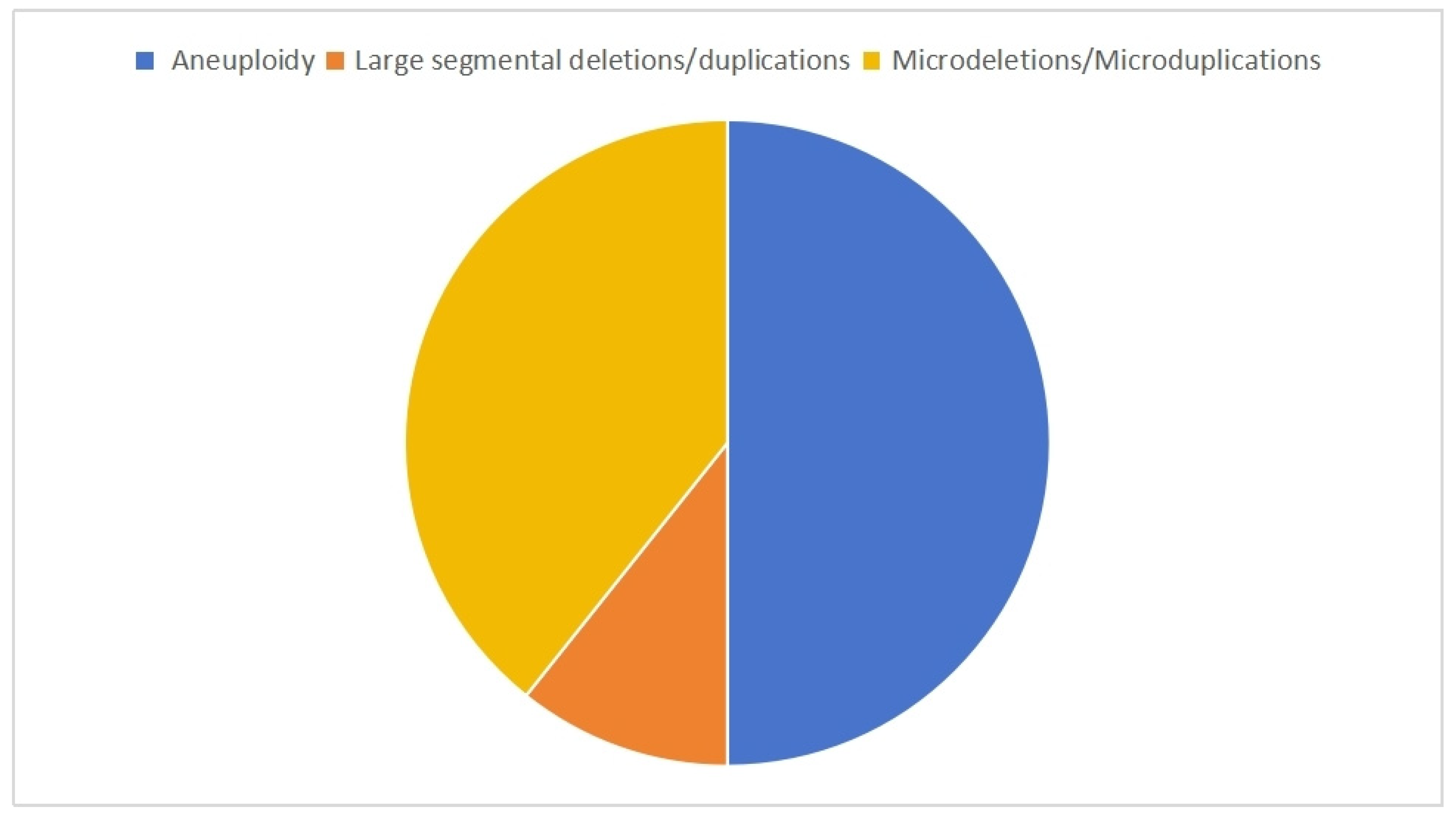
Pathogenic CNVs were detected in 11.7% (140/1195) of fetuses with CHD (Table 2), comprising three main categories: (1) 70 aneuploidies (trisomy 21 [n = 25], trisomy 18 [n = 24], and 45X [n = 10] were most frequent); (2) 15 large segmental CNVs; and (3) 55 microdeletions/duplications, predominantly at 22q11.2 (n = 17) and 15q11.2 (n = 6) (Figure 1). CMA identified both well-established CHD-associated CNVs (e.g., 22q11.2 involving TBX1/CRKL, 7q11.23 with ELN) and potential candidates (e.g., 15q11.2, 16p13.11). Additionally, 2.1% (25/1195) of cases showed VUSs, spanning 0.15–58.06 Mb across multiple chromosomes, with 72% (18/25) occurring in cases with single CHD.

3.3. Detection of pCNVs in Different Types of CHD Fetuses

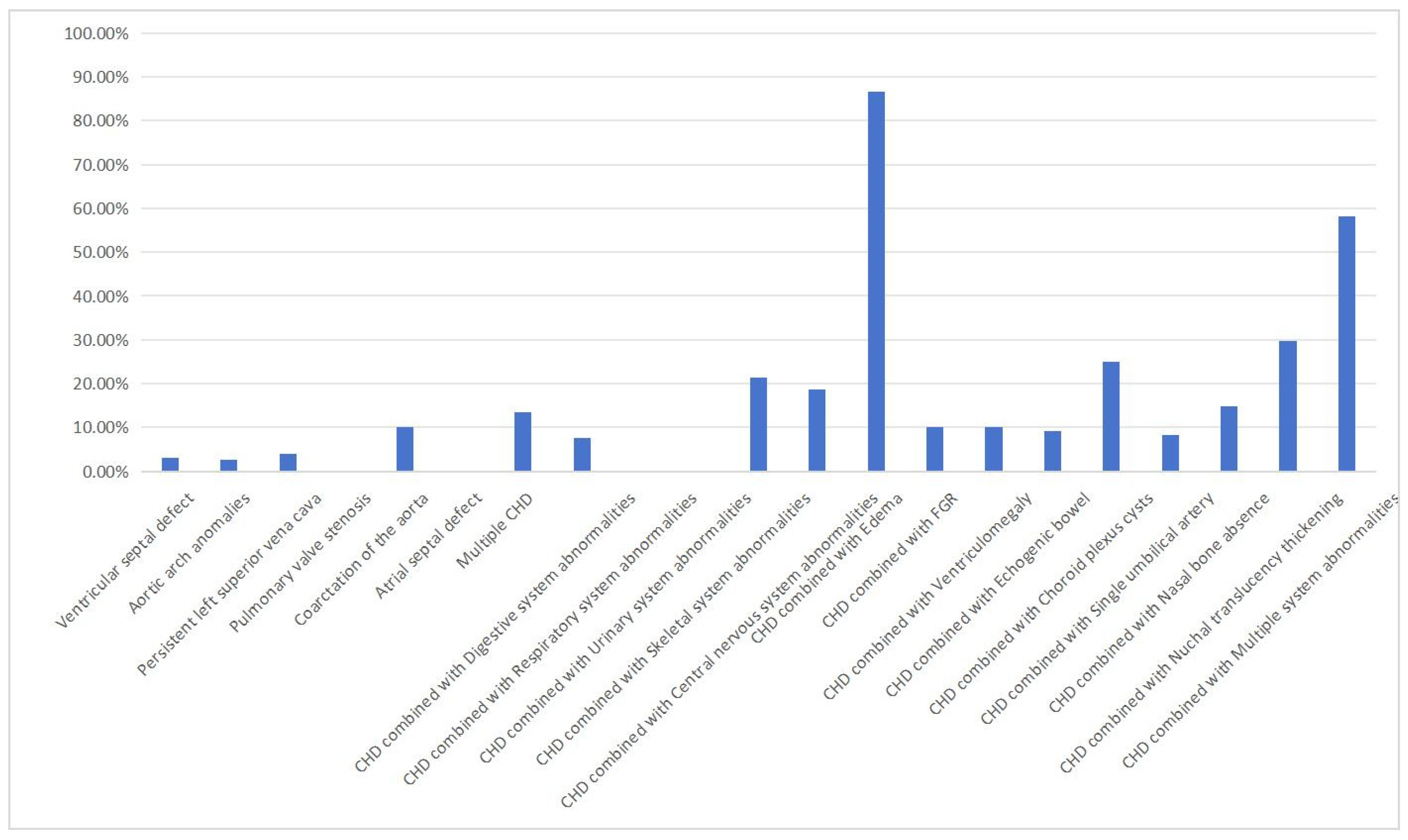
The detection rate of pCNVs was 3.17% (21/662) in patients with a single CHD, 13.51% (30/222) in the group with multiple CHDs, and 28.62% (89/311) in those with CHD and extracardiac abnormalities. The detection rates of pCNVs varied significantly among the three groups (χ2 = 133.01, p < 0.0001). Among patients with a single CHD, the detection rate of pCNVs was highest for those with aorta coarctation (10.00%, 1/10), followed by those with aortic arch anomalies (4.29%, 6/140) and persistent left superior vena cava (3.90%, 2/51). Among patients with CHD accompanied by extracardiac abnormalities, the detection rate of pCNVs was highest for those with edema (86.67%, 13/15), followed by those with multiple system abnormalities (58.11%, 43/74) and nuchal translucency thickening (29.63%, 8/27) (Figure 2).

3.4. Pregnancy Outcomes

Among the 1195 cases with CHD, 90 were lost to follow-up, giving a follow-up success rate of 92.47% (1105/1195). Among those that completed follow-up, 186 cases underwent pregnancy termination (among which 133 cases carried pCNVs), 51 represented stillbirths, 5 denoted perinatal deaths, and the remaining 863 cases were live births. Statistical analysis revealed significant outcome differences by CHD subtype (all p < 0.001). Fetuses with CHD plus extracardiac abnormalities had a significantly higher termination rate (40.00%, 116/290) than those with single CHD (8.59%, 70/815, p < 0.001). Fetuses with multiple CHDs showed elevated stillbirth/perinatal mortality (12.62%, 26/206) compared to those with an single CHD (0.49%, 3/609, p < 0.001). Pathogenic CNVs were associated with higher rates of pregnancy termination, while CNVs of VUS were more prevalent in live births (Table 3).

4. Discussion

CMA can detect genetic variations related to CHD in fetuses. CMA has the advantages of superior sensitivity and faster turnaround time compared to traditional chromosome karyotype analysis. pCNVs were detected in 140 of the 1195 cases with CHD included in this study. Of those with pCNVs, 70 had aneuploidies, 15 had large segmental deletions/duplications, and 55 had microdeletions/microduplications. The detection rate of pCNV was higher in patients with CHD and extracardiac abnormalities than in patients with only CHD [12]. However, Luo et al. [13] found no significant difference in the pCNV detection rate between patients with simple and multiple CHDs. The results of this study showed that pCNV detection rates were higher in the group with both CHD and extracardiac abnormalities and the group with multiple CHDs when compared with the single CHD group. These differences in pCNV detection rates among the three groups were statistically significant. Multiple CHD cases and CHD combined with extracardiac abnormalities were more likely to be accompanied by pCNVs, meriting attention in clinical practice and indicating that CMA testing should be promptly conducted.
Among the key genes affected by 22q11.2 deletion/duplication, TBX1 is a core regulatory factor involved in the development of the cardiac outflow tract and the aortic arch. It affects the separation of major arteries by regulating the proliferation of pharyngeal arch mesenchymal cells [14]. Among patients with 22q11.2 deletion, more than 70% had cardiac malformations (such as TOF and persistent truncus arteriosus). Tbx1 deletion reproduces these phenotypes in a mouse model. As a linker protein, CRKL mediates neural crest cell migration via the FGF8 signaling pathway. Its absence leads to abnormal development of the cardiac outflow tract. The mouse Crkl-knockout model shows conotruncal artery malformation, overlapping with the human 22q11.2 deletion phenotype [15]. ELN (elastin gene) deletion in the 7q11.21 and 11q11.23 regions reduces the number of elastic fibers in the vascular wall, resulting in stenosis of large blood vessels, aortic valve stenosis, and pulmonary artery stenosis [16]. Among the key genes affected by 1p36 deletion, SKI is a negative regulator of the TGF-β signaling pathway. Its absence leads to excessive activation of TGF-β signaling, which affects atrioventricular cushion remodeling and interventricular septal closure [17]. 11q24.2q25 deletion leads to the regulation of endothelial-mesenchymal transition by ETS1, which is crucial for the formation of heart valves and ventricular septa. An insufficient single-copy dose of ETS1 is associated with VSD and TOF [18]. Possible microdeletions/microduplications associated with CHD include: 15q11.2 microdeletion (CYFIP1), 16p13.11 microdeletion/microduplication (MYH11), 16p11.2 microdeletion/microduplication (TBX6), and 1q21.1 microduplication (GJA5). The detection rate of 15q11.2 microdeletion was relatively high in patients with CHD. However, the underlying pathogenic mechanisms remain unclear. CYFIP1 may affect cardiac development through the Rac1 signaling pathway [19]. The MYH11 gene in the 16p13.11 deletion/duplication region is mainly associated with aortic diseases, but this variant has been detected in some patients with CHD (such as those with aortic stenosis) [20]. The TBX6 gene, involved in segmental development, is present in the 16p11.2 deletion/duplication region. Its absence may result in abnormalities of the spine and heart [21]. Microduplication of the GJA5 gene (encoding connexin 40) in 1q21.1 is clearly associated with atrial arrhythmias and septal defects [22]. However, the associations of other pCNV variations (such as the 6q26q27 and 4q22.2q24 deletions) with CHD merit further verification.
Previous studies have shown that 3–14% of single CHD cases may involve pCNVs [23,24]. Among single CHDs, VSDs are the most predominant type, followed by aortic arch anomalies. Because of their high prevalence, the genetic abnormalities and clinical outcomes associated with these lesions may overshadow other less common CHDs in this category. In this study, single VSD accounted for 37.15% (n = 444) of all CHD cases, with a pCNV detection rate of 3.17%; this result is consistent with previous reports [25]. However, the predominance of VSDs and aortic arch anomalies may mask the genetic and phenotypic diversity of rarer CHD subtypes, warranting further investigation into their distinct molecular mechanisms and clinical implications. In this study, the detection rate of pCNVs in the single CHD group was highest for those with aortic coarctation (10.00%, 1/10). Coarctation of the aorta refers to a congenital heart disease characterized by local narrowing of the aortic arch, accounting for 5–7% of all fetuses with CHD [26]. Previous genetic etiological studies have shown that the detection rate for fetal coarctation of the aorta when using CMA varies from 0% to 100% [27,28,29]. A possible reason for these inconsistencies may be heterogeneity in the study population, as the distribution of genetic causes of CHD varies among different races and regions. Although the detection rate of pCNVs in single CHD is low, it significantly increases when accompanied by extracardiac abnormalities. In a study of newborns with CHD, extracardiac abnormalities, and neurodevelopmental delays, the detection rate of CMA was 17–53% [30,31]. Our detection rate of pCNVs in CHD combined with extracardiac abnormalities was 28.62% (89/311), similar to the rate reported for newborns with CHD. Similarly, Jansen et al. [32] conducted a meta-analysis and found that the incidence of pCNVs was high in fetuses with multiple CHDs. Reversed A-waves in the ductus venosus and nuchal translucency thickening are closely related to the occurrence of CHD [33]. In fetuses with CHD and edema, the detection rate of pCNVs by CMA can be as high as 40–60% [27]. In fetuses with CHD showing translucency thickening, the detection rate of pCNVs by CMA is 20–30% [27]. In this study, the detection rate of pCNV in patients with CHD combined with edema (86.67%, 13/15) was the highest, followed by those with CHD combined with multiple system abnormalities (58.11%, 43/74) and CHD combined with nuchal translucency thickening (29.63%, 8/27). This result is consistent with those of the previous studies. Therefore, when CHD is detected prenatally and is accompanied by other systemic abnormalities, especially edema or nuchal translucency thickening, patients should visit a genetic counseling clinic.
In clinical settings, the pathogenicity of CNVs is evaluated based on the gene content, size of CNVs, and gene structure [34]. Some CNVs have unclear clinical significance, and it is difficult to accurately classify them. However, through continuous research, their pathogenicity may be discovered in the future [35,36]. Twenty-five cases with VUSs were identified in this study (2.09%, 25/1195). VUSs increase the difficulty of prenatal genetic counseling, causing great anxiety for parents. These variants should be evaluated in combination with the CNV test results of the parents. The influence of incomplete penetrance and clinical phenotypic differences should also be considered. While we identified VUSs in this study, it was not possible to provide further guidance for clinical consultations because the parents of the fetus did not consent to perform the kinship CMA test. Although limited by cohort size, our data indicate a potential association between pathogenic CNVs and higher pregnancy termination rates, whereas CNVs of VUS were more frequent in live births. Long-term follow-up is warranted to assess the clinical implications of VUS.
In this retrospective study, most fetuses with a single CHD had live births with generally good postnatal conditions, especially those with VSD. For fetuses with multiple CHDs and those with both CHD and extracardiac abnormalities, the parents often choose to terminate the pregnancy after multidisciplinary prenatal consultations. The pregnancy termination rate was highest for fetuses with CHD and edema. Fetal and postnatal mortality rates were highest for fetuses with multiple CHDs. In this study, the pregnancies for 133 fetuses carrying pCNVs were terminated. During follow-up, there were five live births with pCNVs (one case of 15q11.2 microdeletion, two cases of 1q21.1 microdeletion/microduplication, and two cases of 2p16.3 microdeletion). 15q11.2 microdeletion is associated with neurodevelopmental disorders, such as autism and schizophrenia; however, the penetrance is low (approximately 10%), and most carriers are asymptomatic [37]. The phenotypic spectrum of 1q21.1 microdeletion/microduplication is extensive, ranging from normal intelligence to congenital malformations or mental disorders [38]. The 2p16.3 microdeletion is associated with autism and schizophrenia; however, its expression is incomplete [39]. The subsequent development of the abovementioned five live-born infants needs to be closely monitored over a long period to identify any abnormalities and revise the recommendations for consultation accordingly. This finding indicates the complexity of prenatal pCNV testing in clinical practice. The coexistence of high termination rates and live births underscores the need for more precise genetic counseling, a more comprehensive follow-up system, and attention to socio-psychological factors. In the future, it will be necessary to achieve a balance between technological capabilities and ethical responsibilities to prevent the unnecessary termination of pregnancies due to “technological determinism”.
This study has several limitations. First, as this was a retrospective study, pedigree verification was not conducted for all VUS cases. Such cases pose a challenge for genetic counseling. After searching databases and the relevant literature, genetic counselors should interpret and explain the test results for VUSs. Based on the full understanding of the pregnant woman and her family, unnecessary terminations of pregnancies should be minimized. Second, the average follow-up period for live-born infants in this study was only 6 months. No follow-up was performed during early childhood. However, the follow-up period could be extended to include early childhood. Third, single-gene mutations play a significant role in CHD [40,41,42]. WES was not conducted for cases with negative CMA results. This may have led to the omission of cases caused by single-gene mutations. With advancements in WES technology and improvements in genetic maps, an increasing number of pathogenic variations have been discovered. This has facilitated the prenatal diagnosis and etiological research of fetuses with CHD, allowing more detailed consultations for pregnant women and the formulation of evidence-based pregnancy and treatment plans [43]. Fourth, a notable limitation of our study is the inability to fully assess the relationship between genetic abnormalities and postnatal outcomes. Future prospective studies with long-term follow-up are needed to clarify how specific genetic abnormalities influence survival and postnatal complications in CHD-affected pregnancies. Fifth, while our statistical approach provided valid primary analyses, the study was underpowered for comprehensive multivariate adjustments (e.g., maternal age, parity) and subgroup analyses. This precluded the examination of potential effect modifiers and confounders that may refine clinical interpretations. Future studies with larger cohorts should incorporate these advanced analytical methods to strengthen causal inference. Sixth, as this study was conducted at a single institution, our findings may be influenced by local patient demographics, clinical practices, and referral patterns. Our cohort primarily comprised individuals of insert specific ethnic/geographic background. Given known variations in genetic architecture across populations, the applicability of our genotype-phenotype correlations to other ethnic groups warrants cautious interpretation and future investigation in diverse cohorts. Finally, the clinical significance of third-trimester ultrasound examinations for detecting late-emerging CHD is uncertain. Specifically, certain conditions, such as rhabdomyomas, pulmonary stenosis, ventricular septal defects, and mild aortic coarctation, may only become apparent during later gestation. Our study’s design may have hindered the detection of late-emerging anomalies because of the timing of follow-up scans [44].

5. Conclusions

In summary, the genetic factors contributing to fetal CHD are complex, and comprehensive assessments cannot rely solely on prenatal ultrasound. CMA offers valuable diagnostic utility by detecting pCNVs associated with CHD. Our findings support the consideration of CMA for the genetic evaluation of fetuses with CHD; however, its clinical application should account for the availability of regional healthcare resources. While early detection through CMA may inform pregnancy management and genetic counseling, we emphasize the need for prospective multicenter studies to validate the following factors: (1) the diagnostic yield of CMA across diverse CHD subtypes (single vs. non-single), and (2) its long-term impact on perinatal decision-making and outcomes. Prognostic interpretations should integrate CMA results with detailed phenotyping and family history.

Author Contributions

Conceptualization, L.X. and M.F.; writing—original draft preparation, M.C.; writing—review and editing, H.H.; formal analysis, M.Z., Y.Q. and N.L. All authors have read and agreed to the published version of the manuscript.

Funding

This research was funded by the Joint Funds for the Innovation of Science and Technology, Fujian Province, grant number 2025Y9610; the Fujian Provincial Natural Science Foundation, grant number 2019J01509; the Joint Funds for the Innovation of Science and Technology, Fujian Province, grant number 2020Y9159; the Innovation Platform Project of Science and Technology, Fujian Province, grant number 2021Y2012; the National Key Clinical Specialty Construction Program of China (Obstetric), Key Project on the Integration of Industry, Education and Research Collaborative Innovation of Fujian Province, grant number 2021YZ034011; and the Key Project on Science and Technology Program of Fujian Health Commission, grant number 2021ZD01002.

Institutional Review Board Statement

The study was conducted in accordance with the Declaration of Helsinki, and approved by the Ethics Committee of Fujian Maternal and Child Health Hospital (protocol code 2014042; approval date: 2 September 2021). All procedures were applied following the Declaration of Helsinki, as well as international and national laws, guidelines and regulations. Signed informed consent was obtained from all the pregnant women enrolled in this study.

Informed Consent Statement

Informed consent was obtained from all subjects involved in the study.

Data Availability Statement

The datasets analyzed during the current study are not publicly available due to institutional restrictions but are available from the corresponding author upon reasonable request. All aggregated results are included in this published article.

Acknowledgments

We sincerely appreciate all the patients who participated in this study.

Conflicts of Interest

The authors declare no conflicts of interest.

Abbreviations

The following abbreviations are used in this manuscript:
ACMGAmerican College of Medical Genetics and Genomics
CHDCongenital heart disease
CMAChromosomal microarray analysis
CNVCopy number variation
pCNVPathogenic copy number variation
VSDVentricular septal defect

References

  1. Arnaout, R.; Curran, L.; Zhao, Y.; Levine, J.C.; Moon-Grady, A.J. An ensemble of neural networks provides expert-level prenatal detection of complex congenital heart disease. Nat. Med. 2021, 27, 882–891. [Google Scholar] [CrossRef] [Scilit]
  2. Liu, Y.; Chen, S.; Zühlke, L.; Black, G.C.; Choy, M.K.; Li, N.; Keavney, B.D. Global birth prevalence of congenital heart defects 1970–2017: Updated systematic review and meta-analysis of 260 studies. Int. J. Epidemiol. Off. J. Int. Epidemiol. Assoc. 2019, 48, 455–463. [Google Scholar] [CrossRef] [Scilit]
  3. Wu, W.; He, J.; Shao, X. Incidence and mortality trend of congenital heart disease at the global, regional, and national level, 1990–2017. Medicine 2020, 99, 8. [Google Scholar] [CrossRef] [Scilit] [PubMed]
  4. Jerves, T.; Beaton, A.; Kruszka, P. The genetic workup for structural congenital heart disease. Am. J. Med. Genet. Part C Semin. Med. Genet. 2020, 184, 178–186. [Google Scholar] [PubMed]
  5. Hopkins, M.K.; Dugoff, L.; Kuller, J.A. Congenital Heart Disease: Prenatal Diagnosis and Genetic Associations. Obstet. Gynecol. Surv. 2019, 74, 497–503. [Google Scholar] [CrossRef] [Scilit] [PubMed]
  6. Diab, N.S.; Barish, S.; Dong, W.; Zhao, S.; Allington, G.; Yu, X.; Kahle, K.T.; Brueckner, M.; Jin, S.C. Molecular Genetics and Complex Inheritance of Congenital Heart Disease. Genes 2019, 12, 1020. [Google Scholar] [CrossRef] [Scilit] [PubMed]
  7. Audain, E.; Wilsdon, A.; Breckpot, J.; Izarzugaza, J.M.; Hitz, M.P. Integrative analysis of genomic variants reveals new associations of candidate haploinsufficient genes with congenital heart disease. PLoS Genet. 2021, 17, e1009679. [Google Scholar]
  8. Miller, S.P.; Mcquillen, P.S.; Hamrick, S.; Xu, D.; Glidden, D.V.; Charlton, N.; Karl, T.; Azakie, A.; Ferriero, D.M.; Barkovich, A.J. Abnormal brain development in newborns with congenital heart disease. N. Engl. J. Med. 2007, 357, 1928. [Google Scholar] [CrossRef] [Scilit] [PubMed]
  9. Society for Maternal-Fetal Medicine. Genetics CO: Committee Opinion No.682: Microarrays and Next-Generation Sequencing Technology: The Use of Advanced Genetic Diagnostic Tools in Obstetrics and Gynecology. Obstet. Gynecol. 2016, 128, e262–e268. [Google Scholar] [CrossRef] [Scilit] [PubMed]
  10. Carvalho, J.; Allan, L.; Chaoui, R.; Copel, J.; Devore, G.; Hecher, K.; Lee, W.; Munoz, H.; Paladini, D.; Tutschek, B. ISUOG Practice Guidelines (updated): Sonographic screening examination of the fetal heart. Ultrasound Obstet. Gynecol. 2013, 41, 348. [Google Scholar] [CrossRef] [Scilit]
  11. Quaresima, P.; Fesslova, V.; Farina, A.; Kagan, K.O.; Candiani, M.; Morelli, M.; Crispi, F.; Cavoretto, P.I. How to do a fetal cardiac scan. Arch. Gynecol. Obstet. 2023, 307, 1269–1276. [Google Scholar] [CrossRef] [Scilit]
  12. Wang, Y.; Cao, L.; Liang, D.; Meng, L.; Wu, Y.; Qiao, F.; Ji, X.; Luo, C.; Zhang, J.; Xu, T. Prenatal chromosomal microarray analysis in fetuses with congenital heart disease: A prospective cohort study. Am. J. Obstet. Gynecol. 2018, 218, 244.e241. [Google Scholar] [CrossRef] [Scilit]
  13. Luo, S.; Meng, D.; Li, Q.; Hu, X.; Chen, Y.; He, C.; Xie, B.; She, S.; Li, Y.; Fu, C. Genetic Testing and Pregnancy Outcome Analysis of 362 Fetuses with Congenital Heart Disease Identified by Prenatal Ultrasound. Arq. Bras. Cardiol. 2018, 111, 571–577. [Google Scholar] [CrossRef] [Scilit]
  14. Rana, M.S.; Theveniau-Ruissy, M.; De Bono, C.; Mesbah, K.; Francou, A.; Rammah, M.; Dominguez, J.N.; Roux, M.; Laforest, B.; Anderson, R.H. Tbx1 coordinates addition of posterior second heart field progenitor cells to the arterial and venous poles of the heart. Circ. Res. 2015, 115, 790. [Google Scholar] [CrossRef] [Scilit]
  15. Favicchia, I.; Flore, G.; Cioffi, S.; Lania, G.; Baldini, A.; Illingworth, E. Pharmacological Rescue of the Brain Cortex Phenotype of Tbx1 Mouse Mutants: Significance for 22q11.2 Deletion Syndrome. Front. Mol. Neurosci. 2021, 14, 12. [Google Scholar] [CrossRef] [Scilit] [PubMed]
  16. Velandia-Piedrahita, C.A.; Morel, A.; Fonseca-Mendoza, D.J.; Huertas-Quiones, V.M.; Cabrera, R. A Novel Splice-Site Mutation in the ELN Gene Suggests an Alternative Mechanism for Vascular Elastinopathies. Appl. Clin. Genet. 2020, 13, 233–240. [Google Scholar] [CrossRef] [Scilit]
  17. Jordan, V.K.; Zaveri, H.P.; Scott, D.A. 1p36 deletion syndrome: An update. Appl. Clin. Genet. 2015, 8, 189–200. [Google Scholar] [CrossRef] [Scilit]
  18. Yalcintepe, S.; Zhuri, D.; Guler, H.S.; Atli, E.; Demir, S.; Atli, E.I.; Mail, C.; Gurkan, H. First Report of Jacobsen Syndrome with Dextrocardia Diagnosed with del(11)(q24q25). Mol. Syndromol. 2022, 13, 235–239. [Google Scholar] [CrossRef] [Scilit] [PubMed]
  19. Chu, F.C.; Shaw, S.W.; Lee, C.H.; Lo, L.M.; Hsu, J.J.; Hung, T.H. Adverse Perinatal and Early Life Outcomes following 15q11.2 CNV Diagnosis. Genes 2021, 12, 1480. [Google Scholar] [CrossRef] [Scilit]
  20. Zhao, Y.; Song, L.; Zhang, S.; Hou, F.; Shan, S.; Jin, H. Prenatal diagnosis and postnatal follow-up of 15 fetuses with 16p13.11 microduplication syndrome. Front. Genet. 2024, 15, 1486974. [Google Scholar] [CrossRef] [Scilit] [PubMed]
  21. Han, J.Y.; Cho, Y.G.; Jo, D.S.; Park, J. Diversity of Clinical and Molecular Characteristics in Korean Patients with 16p11.2 Microdeletion Syndrome. Int. J. Mol. Sci. 2024, 25, 253. [Google Scholar] [CrossRef] [Scilit]
  22. Soemedi, R.; Topf, A.; Wilson, I.J.; Darlay, R.; Rahman, T.; Glen, E.; Hall, D.; Huang, N.; Bentham, J.; Bhattacharya, S. Phenotype-specific effect of chromosome 1q21.1 rearrangements and GJA5 duplications in 2436 congenital heart disease patients and 6760 controls. Hum. Mol. Genet. 2012, 21, 1513–1520. [Google Scholar] [CrossRef] [Scilit]
  23. Du, L.; Xie, H.N.; Huang, L.H.; Xie, Y.J.; Wu, L.H. Prenatal diagnosis of submicroscopic chromosomal aberrations in fetuses with ventricular septal defects by chromosomal microarray-based analysis. Prenat. Diagn. 2016, 36, 1178. [Google Scholar] [CrossRef] [Scilit]
  24. Cowan, J.R.; Ware, S.M. Genetics and Genetic Testing in Congenital Heart Disease. Clin. Perinatol. 2015, 42, 373–393. [Google Scholar] [CrossRef] [Scilit]
  25. Mastromoro, G.; Khaleghi Hashemian, N.; Guadagnolo, D.; Giuffrida, M.G.; Torres, B.; Bernardini, L.; Ventriglia, F.; Piacentini, G.; Pizzuti, A. Chromosomal Microarray Analysis in Fetuses Detected with singled Cardiovascular Malformation: A Multicenter Study, Systematic Review of the Literature and Meta-Analysis. Diagnostics 2022, 12, 1328. [Google Scholar] [CrossRef] [Scilit]
  26. He, X.; Chen, J.; Li, G. Study on the views and methods of ultrasonic screening and diagnosis for abnormal aortic arch in infants. Cardiovasc. Ultrasound 2021, 19, 8. [Google Scholar] [CrossRef] [Scilit] [PubMed]
  27. Zhang, Z.; Hu, T.; Wang, J.; Liu, S. Pregnancy outcomes of fetuses with congenital heart disease after a prenatal diagnosis with chromosome microarray. Prenat. Diagn. 2021, 42, 79–86. [Google Scholar] [CrossRef] [Scilit]
  28. Mustafa, H.J.; Jacobs, K.M.; Tessier, K.M.; Narasimhan, S.L.; Cross, S.N. Chromosomal Microarray Analysis in the Investigation of Prenatally Diagnosed Congenital Heart Disease. Am. J. Obstet. Gynecol. 2019, 2, 100078. [Google Scholar] [CrossRef] [Scilit]
  29. Sun, H.; Yi, T.; Hao, X.; Yan, H.; Wang, J.; Li, Q.; Gu, X.; Zhou, X.; Wang, S.; Wang, X. Contribution of single-gene defects to congenital cardiac left-sided lesions in the prenatal setting. Ultrasound Obstet. Gynecol. 2020, 56, 225–232. [Google Scholar] [CrossRef] [Scilit] [PubMed]
  30. Hu, H.; Zhou, B.; Shan, Q.; Lan, X. Genetic test results and one-year developmental outcomes of fetuses with congenital heart disease. Front. Pediatr. 2025, 13, 1518784. [Google Scholar] [CrossRef] [Scilit] [PubMed]
  31. Syrmou, A.; Tzetis, M.; Fryssira, H.; Kosma, K.; Oikonomakis, V.; Giannikou, K.; Makrythanasis, P.; Kitsiou-Tzeli, S.; Kanavakis, E. Array comparative genomic hybridization as a clinical diagnostic tool in syndromic and nonsyndromic congenital heart disease. Pediatr. Res. 2013, 73, 772–776. [Google Scholar] [CrossRef] [Scilit]
  32. Jansen, F.A.R.; Blumenfeld, Y.J.; Fisher, A.; Cobben, J.M.; Odibo, A.O.; Borrell, A.; Haak, M.C. Array comparative genomic hybridization and fetal congenital heart defects: A systematic review and meta-analysis. Ultrasound Obstet. Gynecol. 2015, 45, 27–35. [Google Scholar] [CrossRef] [Scilit] [PubMed]
  33. Ratnayake, C.B.B.; Escott, A.B.J.; Phillips, A.R.J.; Windsor, J.A. The anatomy and physiology of the terminal thoracic duct and ostial valve in health and disease: Potential implications for intervention. J. Anat. 2018, 233, 1–14. [Google Scholar] [CrossRef] [Scilit] [PubMed]
  34. Liu, J.; Zhang, X.; Zhao, Y.; Wang, Y. The association between allergic rhinitis and sleep: A systematic review and meta-analysis of observational studies. PLoS ONE 2020, 15, e0228533. [Google Scholar] [CrossRef] [Scilit] [PubMed]
  35. Stull, D.E.; Roberts, L.; Frank, L.; Heithoff, K. Relationship of nasal congestion with sleep, mood, and productivity. Curr. Med. Res. Opin. 2007, 23, 811–819. [Google Scholar] [CrossRef] [Scilit]
  36. Burns, C.; Yeates, L.; Spinks, C.; Semsarian, C.; Ingles, J. Attitudes, knowledge and consequences of uncertain genetic findings in hypertrophic cardiomyopathy. Eur. J. Hum. Genet. 2017, 25, 809. [Google Scholar] [CrossRef] [Scilit]
  37. Butler, M.G. Magnesium Supplement and the 15q11.2 BP1–BP2 Microdeletion (Burnside–Butler) Syndrome: A Potential Treatment? Int. J. Mol. Sci. 2019, 20, 2914. [Google Scholar] [CrossRef] [Scilit]
  38. Upadhyai, P.; Amiri, E.F.; Guleria, V.S.; Bielas, S.L.; Shukla, A. Recurrent 1q21.1 deletion syndrome: Report on variable expression, nonpenetrance and review of literature. Clin. Dysmorphol. 2020, 29, 127–131. [Google Scholar] [CrossRef] [Scilit] [PubMed]
  39. Bermudez-Wagner, K.; Jeng, L.J.B.; Slavotinek, A.M.; Sanford, E.F. 2p16.3 microdeletion with partial deletion of the neurexin-1 gene in a female with developmental delays, short stature, and a congenital diaphragmatic hernia. Clin. Dysmorphol. 2013, 22, 22–24. [Google Scholar] [CrossRef] [Scilit]
  40. Zheng, S.Q.; Chen, H.X.; Liu, X.C.; Yang, Q.; He, G.W. Identification of Variants of ISL1 Gene Promoter and Cellular Functions in singled Ventricular Septal Defects. Am. J. Physiol. Cell Physiol. 2021, 321, C443–C452. [Google Scholar]
  41. Yin, X.Y.; Chen, H.X.; Chen, Z.; Yang, Q.; Han, J.; He, G.W. Genetic Variants of ISL1 Gene Promoter Identified from Congenital Tetralogy of Fallot Patients Alter Cellular Function Forming Disease Basis. Biomolecules 2023, 13, 358. [Google Scholar] [CrossRef] [Scilit] [PubMed]
  42. Chen, X.; Jiang, Y.; Chen, R.; Qi, Q.; Zhang, X.; Zhao, S.; Liu, C.; Wang, W.; Li, Y.; Sun, G. Clinical efficiency of simultaneous CNV-seq and whole-exome sequencing for testing fetal structural anomalies. J. Transl. Med. 2022, 20, 10. [Google Scholar] [CrossRef] [Scilit] [PubMed]
  43. Petrovski, S.; Aggarwal, V.; Giordano, J.L.; Stosic, M.; Wapner, R.J. Whole-exome sequencing in the evaluation of fetal structural anomalies: A prospective cohort study. Lancet 2019, 393, 758–767. [Google Scholar] [CrossRef] [Scilit]
  44. Emam, D.; Corbella, G.; Poziello, C.; Fabozzo, S.; Farina, A.; Candiani, M.; Kagan, K.O.; Cavoretto, P.I. Usefulness and timing of the third-trimester ultrasound scan: A review of guidelines and underlying evidence. Arch. Gynecol. Obstet. 2025, 312, 1445–1459. [Google Scholar] [CrossRef] [Scilit] [PubMed]
Figure 1. Classification of pathogenic copy number variations.
Diagnostics 16 00854 g001
Figure 2. Detection of pathogenic copy number variations in fetuses with different types of congenital heart disease (CHD).
Diagnostics 16 00854 g002
Table 1. Type of congenital heart disease in 1195 fetuses.
Main CategorySubcategoryCases (%)
Single CHDVSD444 (37.15%)
Aortic arch anomalies140 (11.72%)
Persistent left superior vena cava51 (4.27%)
Pulmonary valve stenosis 16 (1.34%)
Coarctation of the aorta10 (0.84%)
Atrial septal defect1 (0.08%)
Multiple CHD-222 (18.58%)
CHD combined with extracardiac abnormalitiesDigestive system abnormalities 13 (1.09%)
Respiratory system abnormalities10 (0.84%)
Urinary system abnormalities22 (1.84%)
Skeletal system abnormalities42 (3.51%)
Central nervous system abnormalities16 (1.34%)
Edema15 (1.26%)
FGR20 (1.67%)
Ventriculomegaly10 (0.84%)
Echogenic bowel11 (0.92%)
Choroid plexus cysts12 (1.00%)
Single umbilical artery12 (1.00%)
Nasal bone absence27 (2.26%)
Nuchal translucency thickening27 (2.26%)
Multiple system abnormalities74 (6.19%)
Percentages for subcategories (e.g., VSD 37.15%) are calculated against the total cohort (N = 1195), not within the subgroup.
Table 2. Prevalence of pathogenic, likely pathogenic, and VUS CNVs across CHD Subtypes.
SubtypePresence of All Types of CNVs
(Number, %)
Pathogenic CNVs (Number, %)Likely Pathogenic CNVs
(Number, %)
CNVs of Uncertain Significance (Number, %)
Single CHDs39 (23.64%)16 (9.70%)5 (3.03%)18 (10.91%)
Multiple CHDs30 (18.18%)26 (15.76%)4 (2.42%)0 (0%)
CHDs with extra-cardiac anomalies96 (58.18%)88 (53.33%)1 (0.61%)7 (4.24%)
Percentages for subcategories are calculated against the total types of CNVs.
Table 3. Association between CNV classification and pregnancy outcomes in fetuses with congenital heart defects.
OutcomeAll Types of CNVs (Number, %)Pathogenic CNVs (Number, %)Likely Pathogenic CNVs (Number, %)CNVs of Uncertain Significance (Number, %)
Termination of pregnancy136 (83.95%)129 (79.63%)4 (2.47%)3 (1.85%)
Still birth3 (1.85%)1 (0.62%)1 (0.62%)1 (0.62%)
Live birth23 (14.20%)0 (0%)5 (3.09%)18 (11.11%)
Post-natal death0 (0%)0 (0%)0 (0%)0 (0%)
Percentages for subcategories are calculated against the total pregnancy outcomes in fetuses with all types of CNVs.
Disclaimer/Publisher’s Note: The statements, opinions and data contained in all publications are solely those of the individual author(s) and contributor(s) and not of MDPI and/or the editor(s). MDPI and/or the editor(s) disclaim responsibility for any injury to people or property resulting from any ideas, methods, instructions or products referred to in the content.