Article

SCAG-Net: Automated Brain Tumor Prediction from MRI Using Cuttlefish-Optimized Attention-Based Graph Networks

1
Distribution and Supply Technology, Expedia Group, Seattle, WA 98119, USA
2
Department of Computer Science and Information Systems, College of Applied Sciences, AlMaarefa University, Riyadh 13713, Saudi Arabia
3
Research Center, Deanship of Scientific Research and Post-Graduate Studies, AlMaarefa University, Riyadh 13713, Saudi Arabia
4
Electrical Engineering Department, University of Business and Technology, Jeddah 21432, Saudi Arabia
5
Engineering Mathematics Department, Alexandria University, Alexandria 21544, Egypt
6
Department of Computer Engineering, Aligarh Muslim University, Aligarh 202002, India
7
Department of Business Administration, College of Business Administration, Princess Nourah Bint Abdulrahman University, P.O. Box 84428, Riyadh 11671, Saudi Arabia
*
Authors to whom correspondence should be addressed.
Diagnostics 2026, 16(4), 565; https://doi.org/10.3390/diagnostics16040565

Submission received: 31 October 2025 / Revised: 14 January 2026 / Accepted: 26 January 2026 / Published: 13 February 2026

Abstract

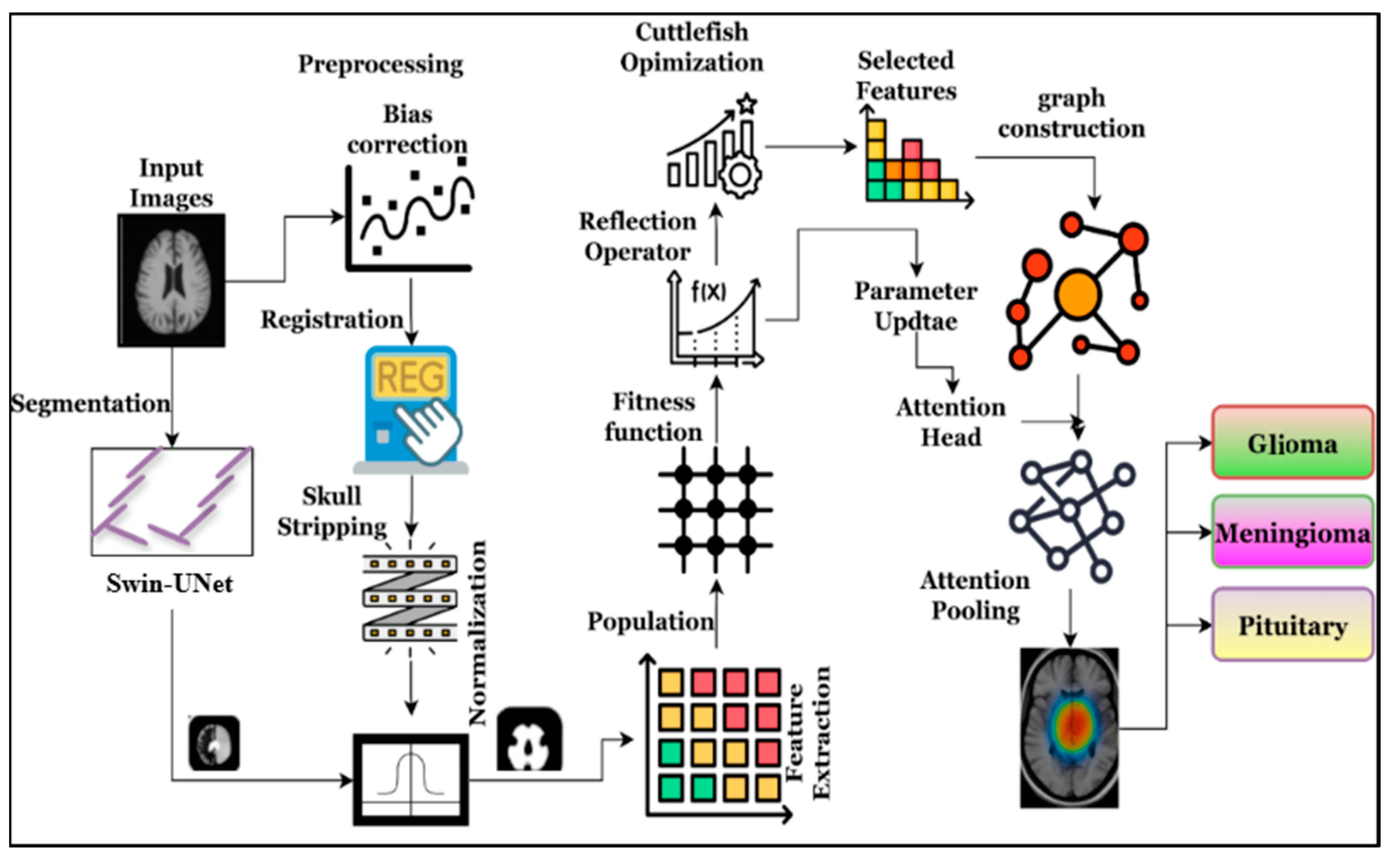
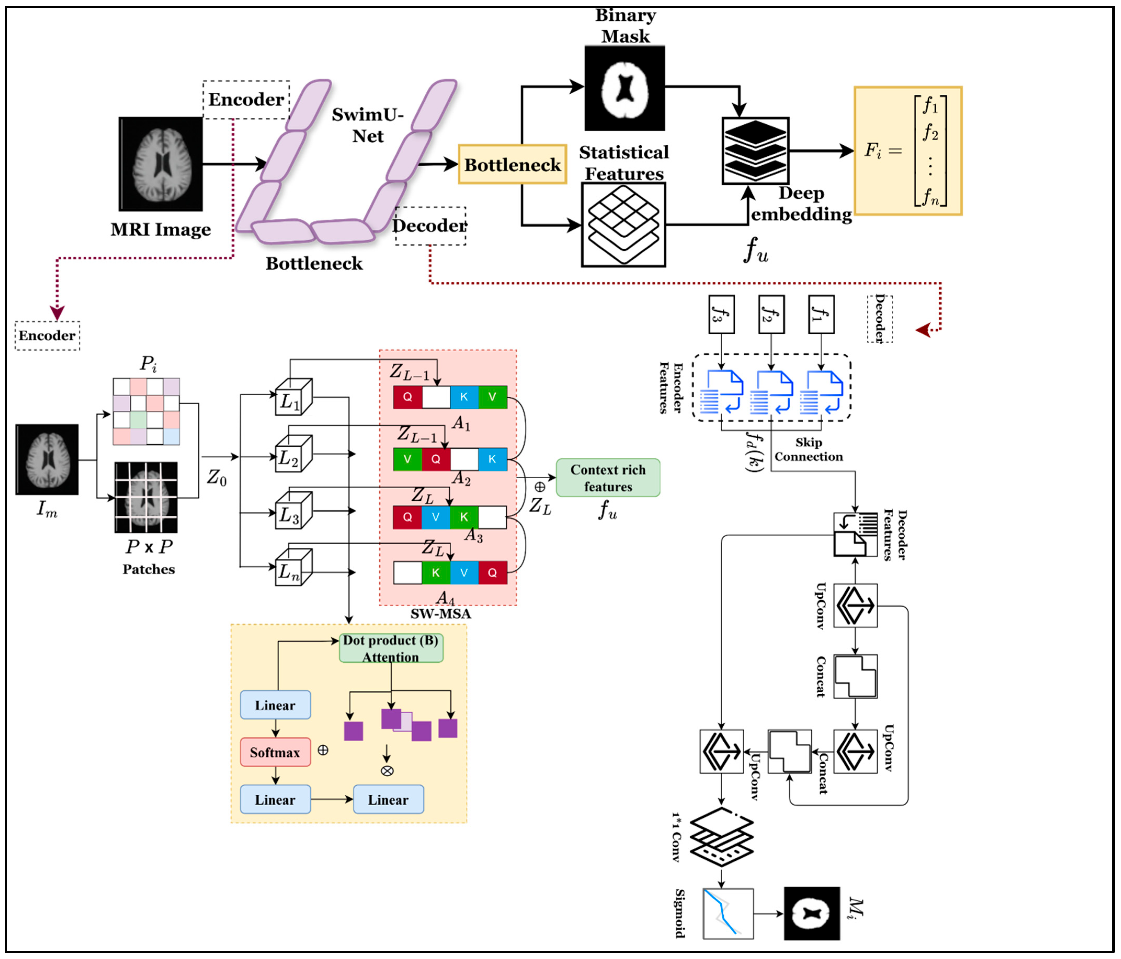
Background/Objectives: The earlier, more accurate, and more consistent prediction of the brain tumor recognition process requires automated systems to minimize diagnostic delays and human error. The automated system provides a platform for handling large medical images, speeding up clinical decision-making. However, the existing system is facing difficulties due to the high variability in tumor location, size, and shape, which leads to segmentation complexity. In addition, glioma-related tumors infiltrate the brain tissues, making it challenging to identify the exact tumor region. Method: The above-identified research difficulties are overcome by applying the Swin-UNet with cuttlefish-optimized attention-based Graph Neural Networks (SCAG-Net), thereby improving overall brain tumor recognition accuracy. This integrated approach is utilized to address infiltrative gliomas, tumor variability, and feature redundancy issues by improving diagnostic efficiency. Initially, the collected MRI images are processed using the Swin-UNet approach to identify the region, minimizing prediction error robustly. The region’s features are explored utilizing the cuttlefish algorithm, which minimizes redundant features and speeds up classification by improving accuracy. The selected features are further processed using the attention graph network, which handles structural and heterogeneous information across multiple layers, improving classification accuracy compared to existing methods. Results: The efficiency of the system, implemented with the help of public datasets such as BRATS 2018, BRATS 2019, BRATS 2020, and Figshare is ensured by the proposed SCAG-Net approach, which achieves maximum recognition accuracy. The proposed system achieved a Dice coefficient of 0.989, an Intersection over Union of 0.969, and a classification accuracy of 0.992. This performance surpassed the most recent benchmark models by margins of 1.0% to 1.8% and with statistically significant differences (p < 0.05). These findings present a statistically validated, computationally efficient, clinically deployable framework. Conclusions: The effective analysis of MRI complex structures is used in medical applications and clinical analysis. The proposed SCAG-Net framework significantly improves brain tumor recognition by addressing tumor heterogeneity and infiltrative gliomas using MRI images. The proposed approach provides a robust, efficient, and clinically deployable solution for brain tumor recognition from MRI images, supporting accurate and rapid diagnosis while maintaining expert-level performance.
Keywords: brain tumor; MRI; attention graph neural networks; Swin-UNet; cuttlefish; segmentation; redundant features brain tumor; MRI; attention graph neural networks; Swin-UNet; cuttlefish; segmentation; redundant features