1. Introduction
Recognizing brain tumors [
1] is an essential aspect of medical diagnostics, as early detection and proper classification are vital for planning treatment and improving patient survival and quality of life. Whether a brain tumor [
2] is malignant or benign, it hinders the central nervous system’s functioning, leading to adverse effects like severe and chronic headaches, seizures, and a combination of memory, speech, and motor coordination impairments [
3]. These complications intensely and progressively diminish the patient’s cognitive and physical functions [
4], leading to decreased autonomy and quality of life. Additionally, the importance of early detection lies in enabling timely medical intervention; it protects a patient’s ability to undergo effective treatment strategies, including surgery, chemotherapy, and radiotherapy [
5], and prevents the tumor from reaching an advanced, inoperable stage.
Early tumor detection [
6] greatly enhances treatment efficacy and reduces the risk of patient death. Among multiple diagnostic procedures, Magnetic Resonance Imaging (MRI) [
7] is noted for its diagnostic accuracy and as being the least invasive. Unlike CT or PET scans [
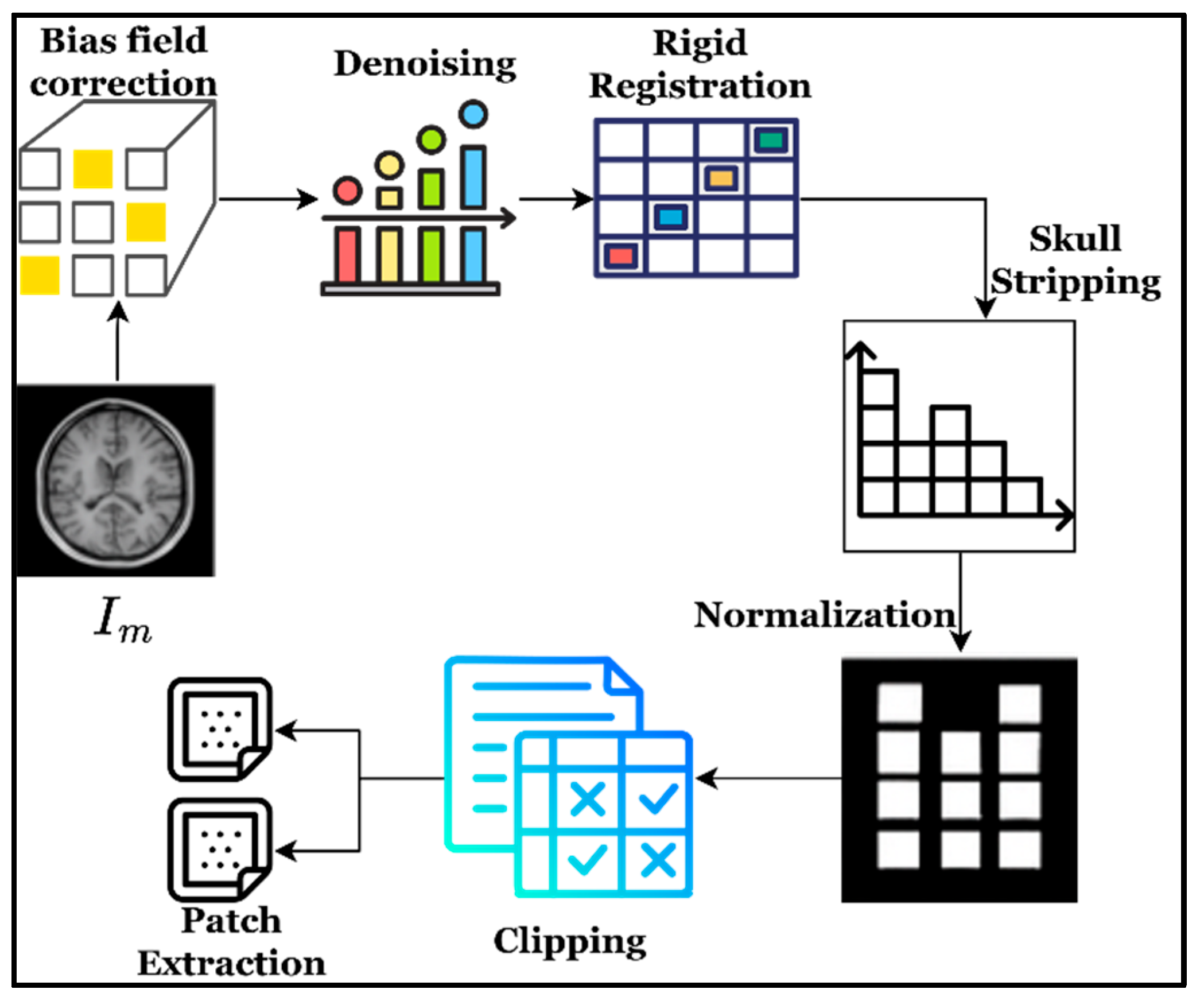
8], MRI provides greater contrast between soft tissues, helping delineate brain tissue and tumor borders, all without the hazard of ionizing radiation. Predicting brain tumors with MRI involves a series of steps, including preprocessing [
9] (noise reduction, skull stripping, and normalization), segmentation (locating tumor-affected areas), feature extraction [
10] (texture, intensity, and spatial features), and classification [
11,
12] (tumor type and degree). Despite this high level of MRI use in diagnosing and informing the treatment of brain tumors, many problems remain. These include glioma cells [
13] (tumor forms), which infiltrate brain tissues [
14,
15] and obfuscate borders; the high variability in the shape, size, and location of the tumor, and the tendency of normal and malignant tissue to overlap in intensity, makes pattern recognition difficult [
16]. Additionally, gliomas have dense and compact cellular architectures, so there is a need for accurate localization of the tumor using physical and bioelectrical approaches to destroy tumor cells precisely [
17,
18].
Current methods, including conventional machine learning approaches and deep learning [
19], tend to focus on a narrow research area, leading to a lack of diverse data (generalization), overfitting, and redundant features. The process of manually annotating data for training purposes is time-consuming [
20] and may introduce subjectivity. Recent studies seek to mitigate these issues by combining transformer models, attention layers [
21], and evolutionary optimization to improve brain tumor recognition accuracy [
22,
23]. Transformer models have also shown potential in diagnosing gliomas using computational pathology [
24]. Many previous studies have demonstrated use of various evolutionary optimization algorithms such as ant colony and swarm intelligence to improve MRI image segmentation for various clinical scenarios [
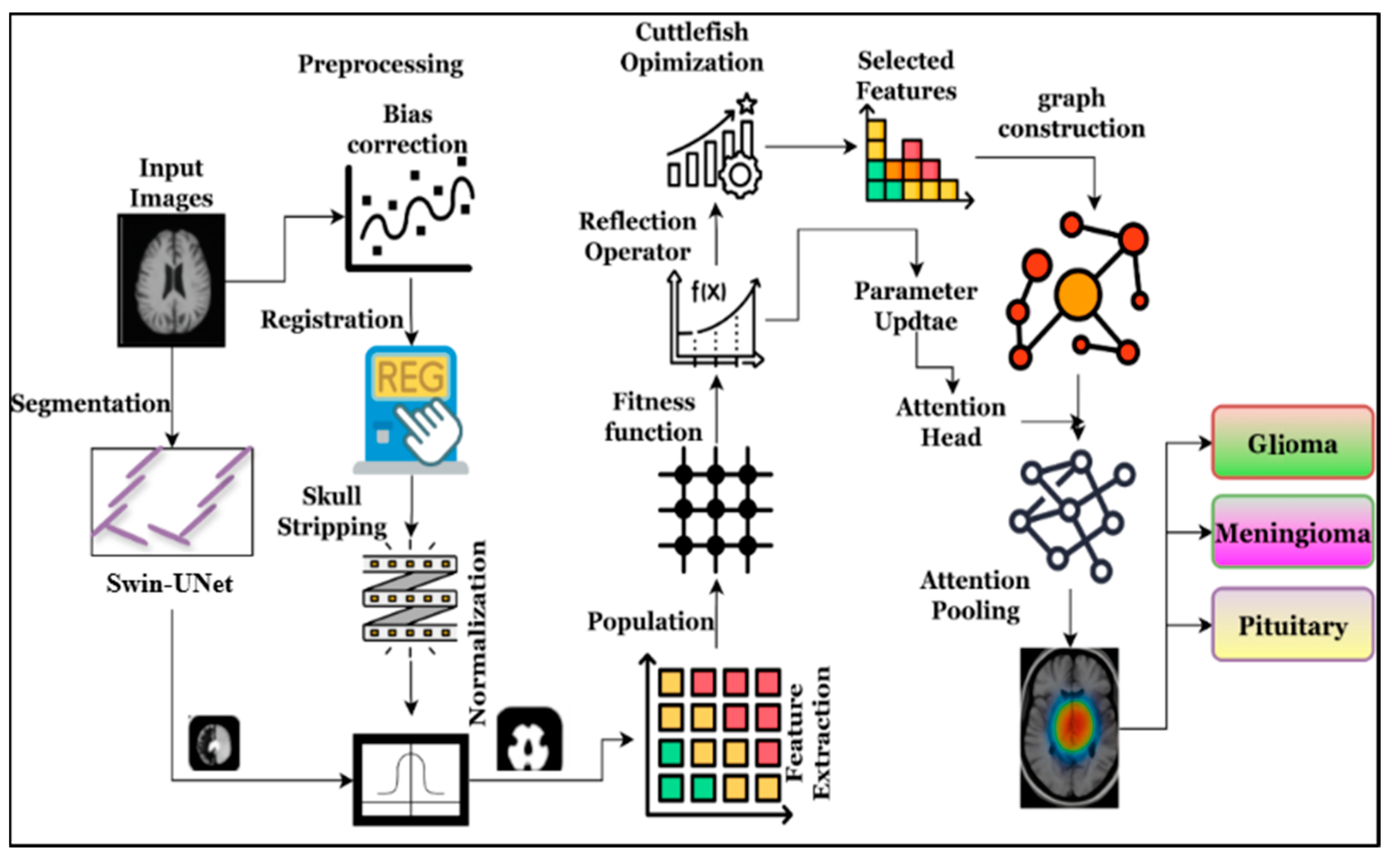
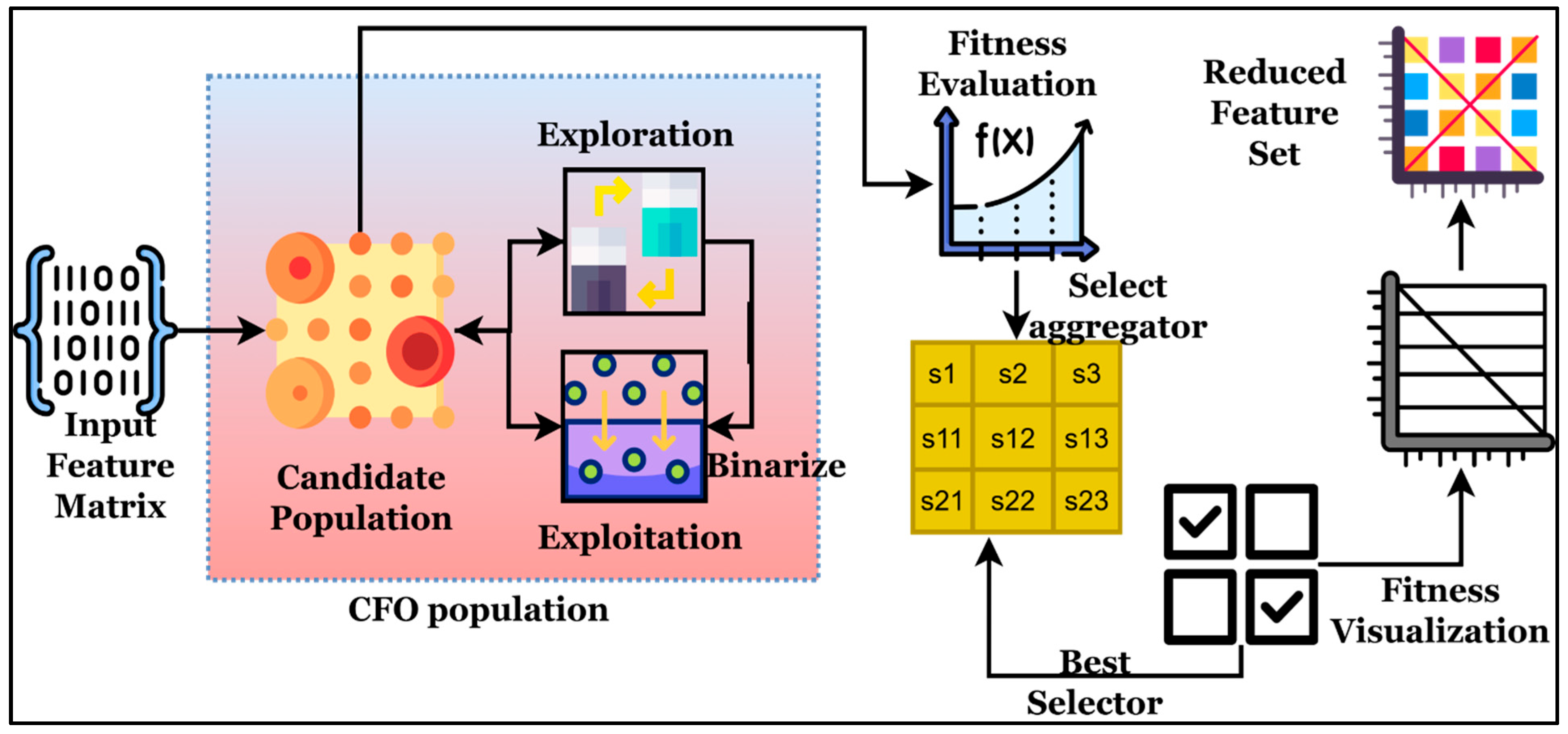
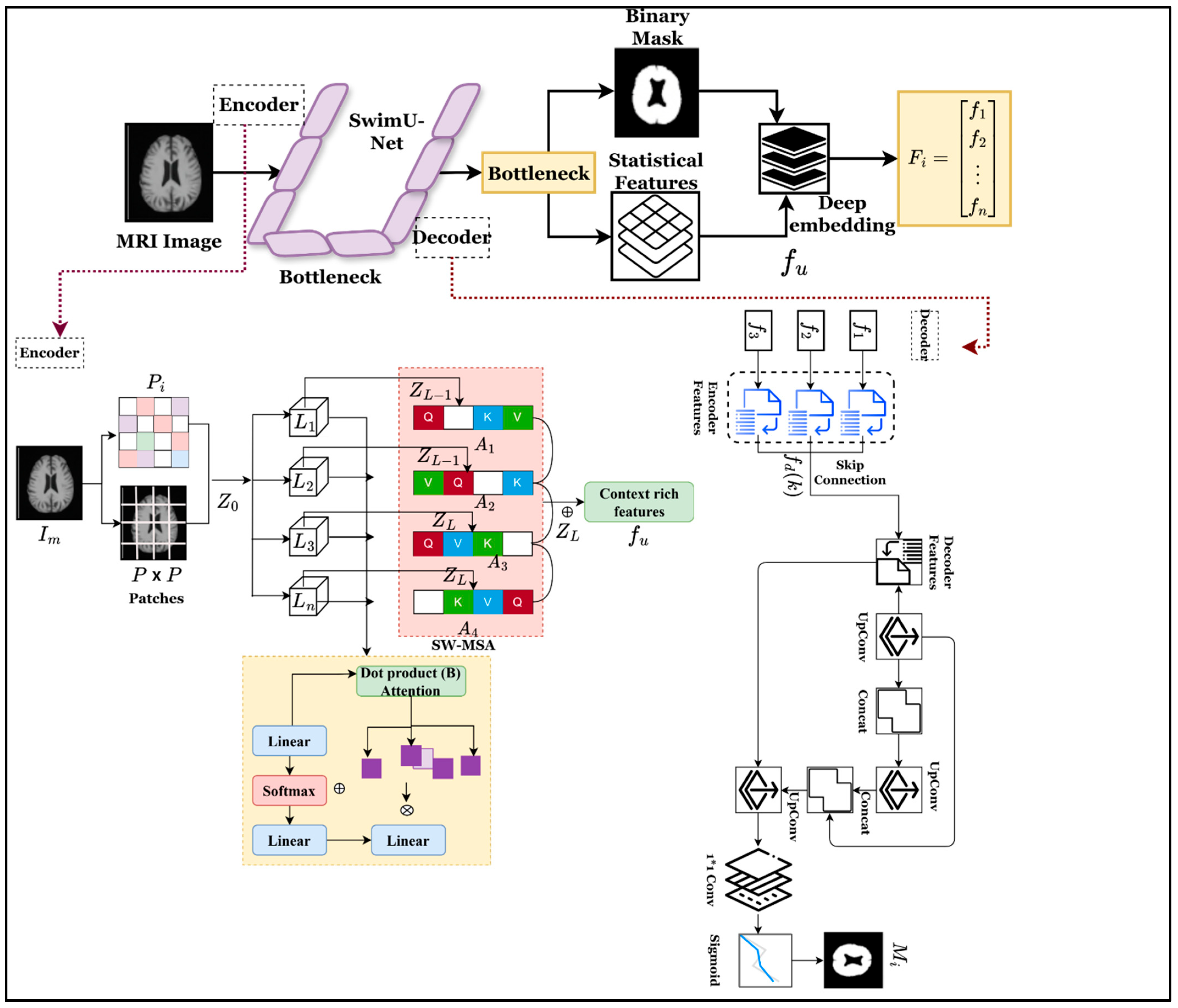
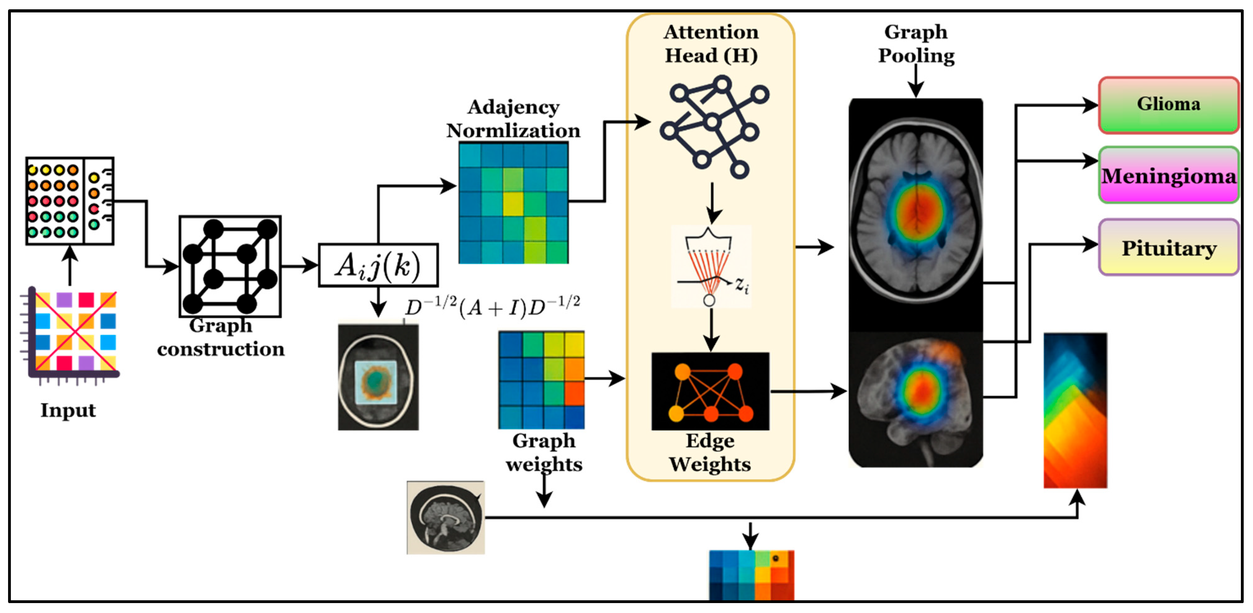
25]. Therefore, integrated models are designed to simultaneously analyze and disentangle the local and global contexts of MRI scans, reduce redundant features, and improve categorization precision. These models provide a reliable and streamlined approach to automating brain tumor identification in diagnostic settings. Therefore, this work introduces the Swin-UNet with cuttlefish-optimized attention-based Graph Neural Networks (SCAG-Net) to improve overall brain tumor recognition accuracy. The effective incorporation of optimization techniques helps address challenges such as tumor heterogeneity and infiltrative gliomas, thereby maximizing overall recognition rates.
The persistent challenges in correctly detecting brain tumors from MRI scans, where significant anatomical variances, overlapping tissue intensities, and intricate tumor boundaries frequently impair the effectiveness of current techniques, are the driving force behind this effort. Current methods usually only maximize one aspect, like feature extraction, segmentation, or classification, which leads to fragmented solutions that do not fully capture the structural and geographic complexity of tumor regions. Furthermore, a lack of annotated data limits the capacity of traditional deep learning models to generalize across patients and imaging circumstances, and high-dimensional MRI features frequently add redundancy. The need for an integrated framework that can simultaneously model global context, improve discriminative features, and reason about structural linkages inside the brain is highlighted by these limitations. The purpose of this study is to address these interrelated problems by developing a single, strong, and clinically accurate architecture for brain tumor prediction.
2. Related Works
The most recent work in brain tumor detection from MRI images has focused on refining diagnostic precision, enhancing deep learning frameworks, and improving feature analysis for clinical utility. Zhu et al. (2024) [
26] developed an optimized deep neural network and an amended grasshopper-optimization algorithm to refine feature selection and improve tumor classification accuracy. Though this approach effectively minimized overfitting, its applicability to different datasets was somewhat limited. In a similar vein, Daoud et al. (2025) [
27] addressed the tumor detection and treatment prediction gap by formulating it within a deep learning framework using the Spider Wasp Optimization algorithm, which offers convergence and interpretability but is computationally intensive for larger datasets. Meenal and Asokan (2026) [
28] devised a new quantum-inspired adaptive feature fusion model, motivated by the principles of quantum computing and deep learning, resulting in efficient, highly accurate tumor classification. While their framework efficiently captured the complex features of an MRI, the model’s complexity slowed processing. While Yin and Teng (2026) [
29] present the deep fusion framework, which integrates ResNet, EfficientNet, and transformer-based attention mechanisms for robust tumor classification—the model demonstrates impressive generalization across tumor types—it is highly memory-intensive during training. Barati et al. (2025) [
30] used lightweight neural networks to analyze the effects of different optimizers and loss functions on the accuracy of brain tumor prediction. Clearly, they demonstrated that the choice of optimizer is critical to achieving high prediction accuracy while keeping the neural network lightweight.
Ullah et al. (2024) [
31] developed a multimodal MRI segmentation and classification system using the DeepLabV3+ framework and explainable AI, thereby enhancing the system’s interpretability and the reliability of the diagnosis. Despite these advancements, scalability remained a challenge due to the model’s complexity. For early-stage tumor prediction, Saraswat and Dubey (2025) [
32] proposed a dilated, attention-based ensemble network that, in conjunction with enhanced artificial rabbit optimization, achieved high accuracy with reduced false positives, albeit at a high computational cost. Mallouk et al. (2025) [
33] introduced the Optimal Deep Transfer Learning (ODTL) model, which improved feature transfer and classification but struggled with domain adaptation to unseen data. Using dual deep convolutional networks, Bernard et al. (2025) [
34] improved feature extraction from MRI scans, thereby increasing the precision of tumor localization and classification. However, this paradigm was limited by the requirement of extensive annotated datasets for practical model training. Additionally, in recent times, emerging sequence modeling architectures such as Mamba have also been successfully utilized for medical image segmentation with strong capability in capturing long-range dependencies under limited data [
35]. Hybrid models that integrate convolutional neural networks for region localization and transformer-based components for global dependencies have demonstrated remarkable outcomes for various MRI related clinical task such as brain tumor segmentation from MRI images [
36] and long-term outcome prediction from cardiac MRI [
37].
On the other hand, not dissimilar to the contribution mentioned above, Hasan et al. (2025) [
38] introduced DEEP Q-NAS, a revision of neural architectures that implements reinforcement learning to automate the process of designing deep networks for tumor identification, achieving considerable adaptability and accuracy, albeit with substantial training time and high computational cost. The works mentioned above provide a trajectory of the development of hybrid, optimized, and interpretable frameworks, with deep learning still, relative to the other branches of AI, suffering from a lack of integrated models that fuse the precision of diagnostics with the rapidity of computation and clinical relevance, a deficit that SCAG-Net aims to address. According to various researchers’ work, the findings are summarized in
Table 1.
Recent pertinent studies on MRI-based brain tumor identification, such as Zhu et al. (2024) [
26], Daoud et al. (2025) [
27], Meenal and Asokan (2026) [
28], Yin & Teng (2026) [
29], and Hasan et al. (2025) [
38], have been compared with the SCAG-Net design. The shortcomings of earlier models include their low clinical scalability, limited generalization, high computing cost, and isolated module optimization. The comparison research shows that new brain tumor recognition models use a mix of deep learning and optimization techniques to achieve very accurate results, but they often make computations less efficient and harder to understand. Transfer learning, attention fusion, and evolutionary optimization are all excellent methods for generalization and scaling, but most of them do not work well in clinical settings or in real time. The results show that we need a balanced framework, such as the proposed SCAG-Net, to ensure that medical tests are accurate, efficient, and reliable for real-world use.
Despite the high classification accuracy reported by recent MRI-based brain tumor detection studies using transformers, evolutionary optimization, or attention mechanisms,
Table 1 shows that these methods primarily function as separate or loosely coupled modules, which results in limitations in clinical scalability, computational efficiency, and uncertainty propagation. The correlation between accurate tumor localization and final diagnostic confidence is weakened in many modern works, particularly in infiltrative gliomas with ambiguous boundaries, because optimization is only applied at the classifier level or attention is used without explicitly preserving structural dependencies between segmentation and recognition. Because of this, performance improvements in one step frequently do not translate to the entire diagnostic pipeline. This disparity emphasizes the necessity of an integrated model that tightly integrates structural reasoning, feature refining, and segmentation into a single framework. Such an integrated approach can mitigate error accumulation, enhance robustness across heterogeneous MRI modalities, and improve clinical reliability by jointly modeling spatial context, removing redundant features before classification, and maintaining inter-regional relationships through graph attention. Therefore, the proposed SCAG-Net framework has been designed.
4. Results and Discussion
This section discusses the SCAG-Net framework for brain tumor recognition from MRI images. During the analysis, the Swin-UNet, cuttlefish, and graph neural networks are used to improve overall brain region recognition. The introduced framework uses the BRATS 2018 [
39], BRATS 2019 [
40], and BRATS 2020 [
41] datasets, and the Figshare [
42] brain MRI images are used to evaluate the framework’s efficiency. The BRATS dataset consists of 1700 patients’ information that has four modalities, such as T1 weighted, T2 weighted, and FLAIR sequences with
and
volume of dimension. In every case, experts have continuously explored the image to identify the necrotic core and enhance tumor, edema, and non-enhancing core regions through structural evaluation. The following dataset is available on Figshare and contains 1200 2D brain slices, explored using the above-described techniques to identify structural variability in brain regions. The integrated datasets are used in this work to demonstrate the system’s efficiency in terms of generalization, robustness, and scalability across different clinical conditions. The dataset utilized in this work is described in
Table 2.
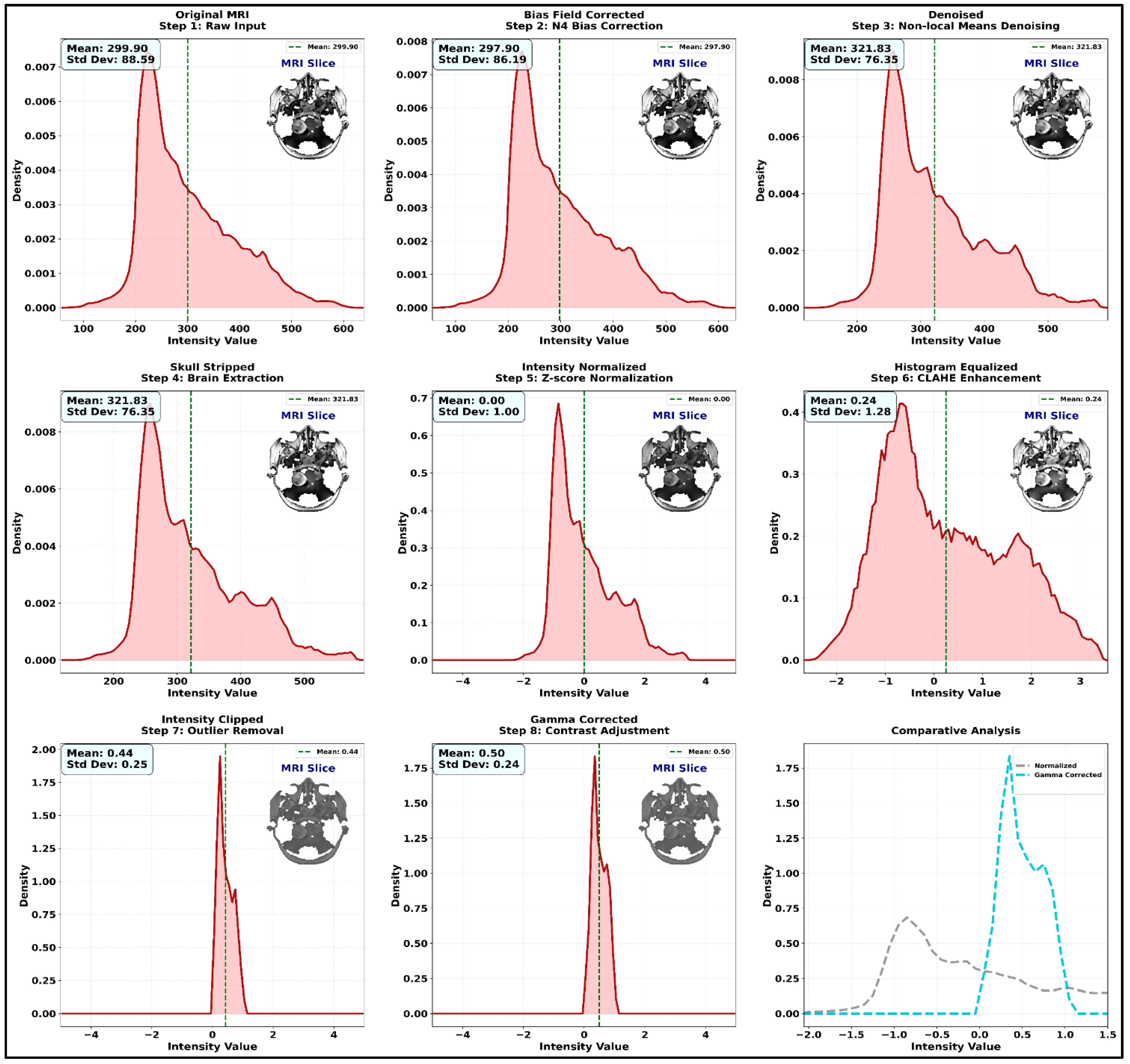
To improve the dataset section’s thoroughness, the dataset appropriateness and data splits are explained. To prevent identity leakage, the participants for each BRATS dataset have been divided into 70% for training, 15% for validation, and 15% for testing. It ensured that no patient featured in more than one group. This division maintains clinical diversity and facilitates equitable performance evaluation. Since the Figshare dataset’s imaging properties, annotation granularity, and contrast changes separated greatly from BRATS volumes, it was included to assess cross-dataset robustness, although it is made up of 2D T1/T2 slices. It is suitable for examining generalizability outside of established BRATS methods because of its distinct tumor/non-tumor labeling, variety of slice orientations, and inclusion of both healthy and pathological images. The identical preprocessing processes—bias-field correction, skull stripping, resampling, and normalization—have been applied to all datasets, ensuring comparable input quality and enabling SCAG-Net to be assessed identically across various MRI sources.
According to
Table 2, the selected datasets consist of 1900 brain MRI images used to effectively manage high-grade gliomas, low-grade gliomas, and non-tumor brain regions. Every dataset used in this work is used to validate the described segmentation approaches and to provide labels during the analysis. Further, the system’s efficiency is explored using existing research to justify the SCAG-Net framework’s clinical interpretability, robustness, and scalability. Here, the system’s efficiency is compared with different benchmark methods, such as Zhu et al. (2024) [
26], Meenal & Asokan (2026) [
28], Yin & Teng (2026) [
29], and Hasan et al. (2025) [
38], and datasets; the results are shown in
Table 3.
Table 3 presents a thorough quantitative comparison that assesses SCAG-Net and the previously discussed techniques on a number of datasets, including Figshare, BRATS 2018, BRATS 2019, and BRATS 2020, to substantiate these conclusions. Based on the comparative analysis of performance metrics, SCAG-Net consistently and significantly surpasses previous methods in classification accuracy, Dice coefficient, Intersection over Union (IoU), sensitivity, specificity, precision, F1-score, Hausdorff Distance, and AUC. This thorough comparison demonstrates how SCAG-Net overcomes the limitations of previous methods by combining attention-based graph networks, cuttlefish-optimized feature refinement, and Swin-UNet-based segmentation into a single framework. This leads to an enhancement in clinical dependability, accuracy, and structural consistency. Across all four analysis datasets—BRATS 2018, BRATS 2019, BRATS 2020, and Figshare—SCAG-Net demonstrates significant, consistent, and positive improvements across all principal performance indicators, as shown in
Table 3. The introduced framework achieves 98.69 ± 0.29% classification accuracy and 0.969 ± 0.004 in the Dice coefficient, further strengthening its first-place position in both accuracy and Dice coefficient compared to Hasan et al. 2025 [
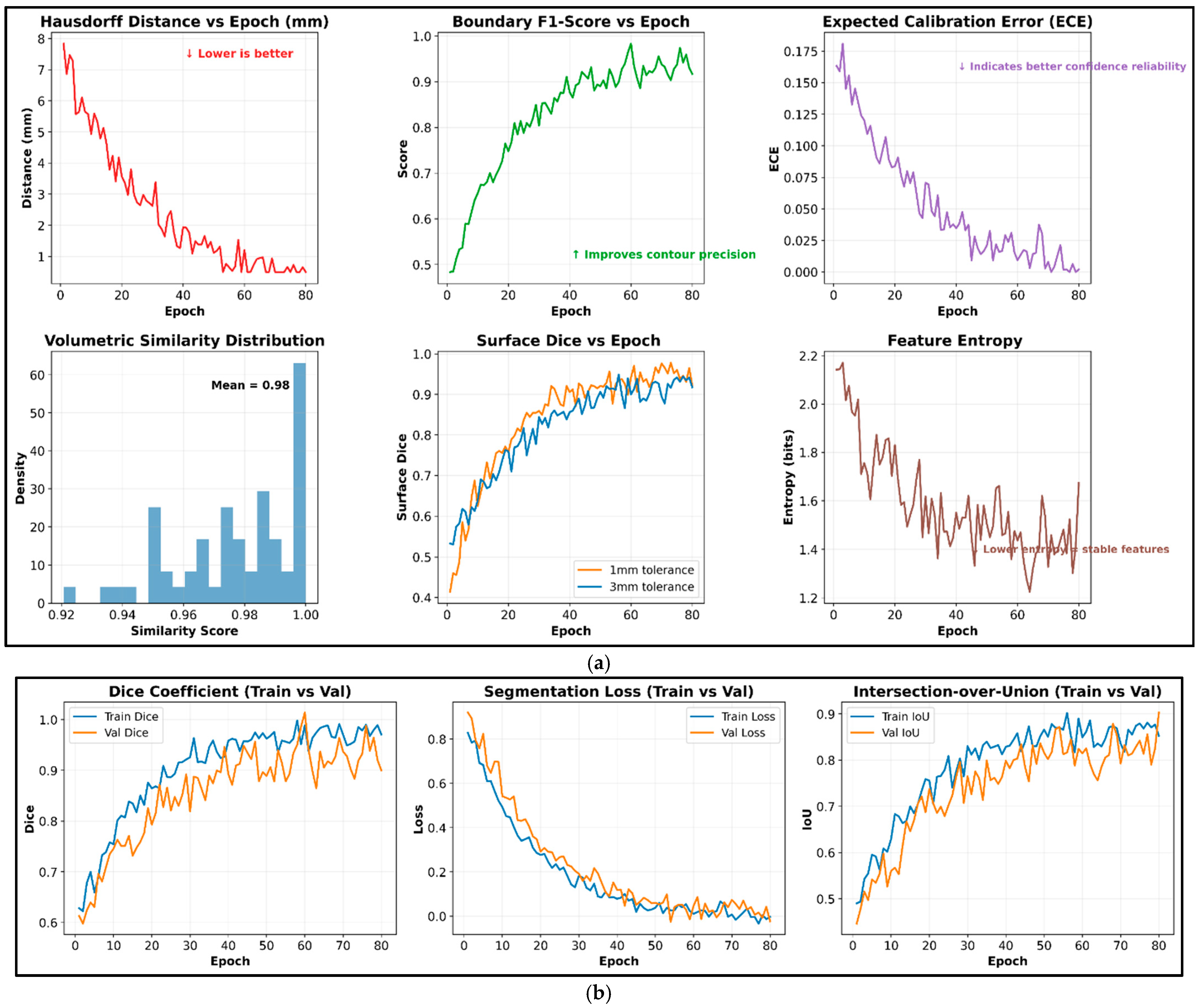
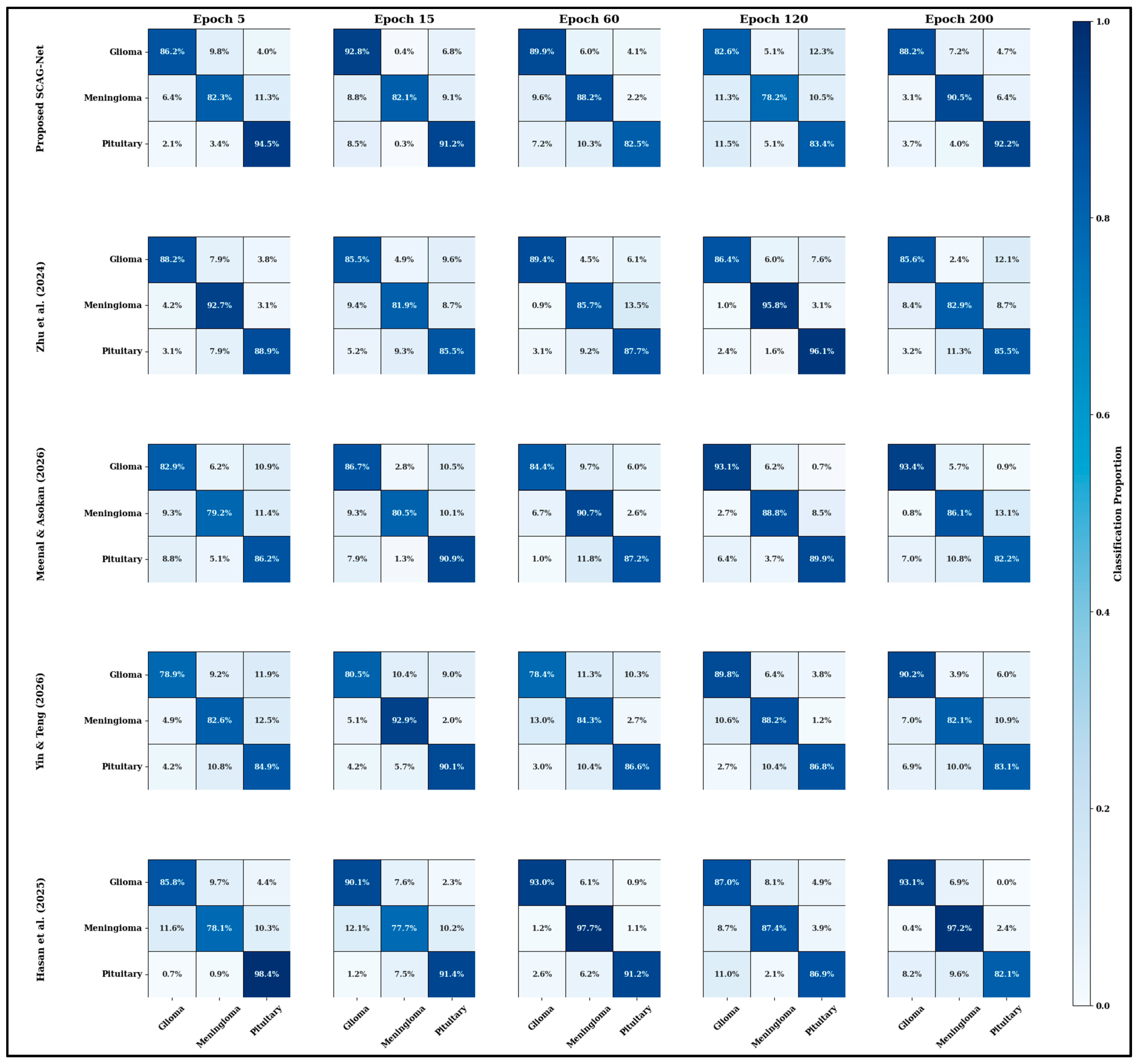
38]’s results, which were 1.3% and 1.5%, respectively. The Hausdorff Distance of 0.9–1.1 mm falls within the range of structural continuity, with precise, smooth walls around the predicted tumor borders. Furthermore, the average IoU is 2.5%, and the AUC across the datasets is 99%, confirming multi-class tumor diagnosis and discrimination. On the speed dimension, SCAG-Net delivers the fastest 1.45 s per-image inference, outpacing deep fusion models by 0–5% in FO-driven feature running and adaptive attention aggregation. These balanced metrics reinforce that the proposed SCAG-Net operates at maximum speed for segmentation, discernment, and computation while maintaining the precision required to confirm its real-life and functional clinical role. To verify SCAG-Net’s convergence, a confusion matrix-based assessment was performed across all datasets. This method shows how quickly and reliably the model finds stable category boundaries for glioma, meningioma, and pituitary tumor classes throughout iterative learning. The matrix-based evaluation identifies misclassifications across classes and shows that the attention-guided graph network and cuttlefish optimization-based feature pruning perform well at distinguishing between classes. The obtained results are shown in
Figure 10.
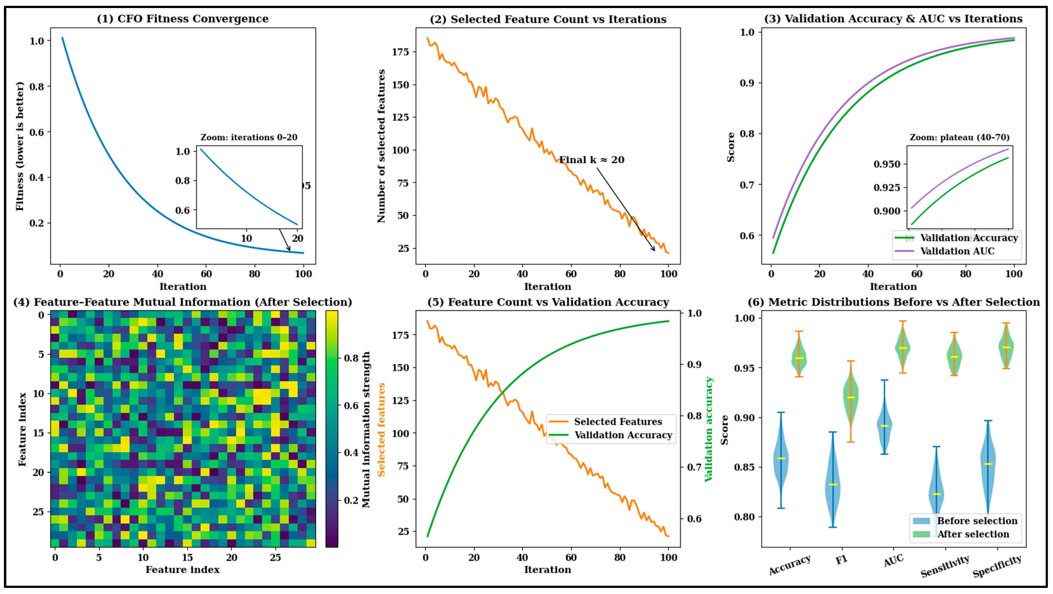
Figure 10 illustrates the convergence analysis of the introduced SCAG-net framework across different modalities, including glioma, meningioma, and pituitary MRI regions. This study clearly shows that the effective use of convolutional layers and encoder–decoder components identifies the overlap region effectively, leveraging the redundancy of spatial features. According to the Figure, the efficiency is evaluated over 200 epochs, during which the system reduces irrelevant features by >80%, and the results show <1.2% inter-class confusion. The analysis ensures that the framework meets the convergence and maximum confidence values in decision analysis.
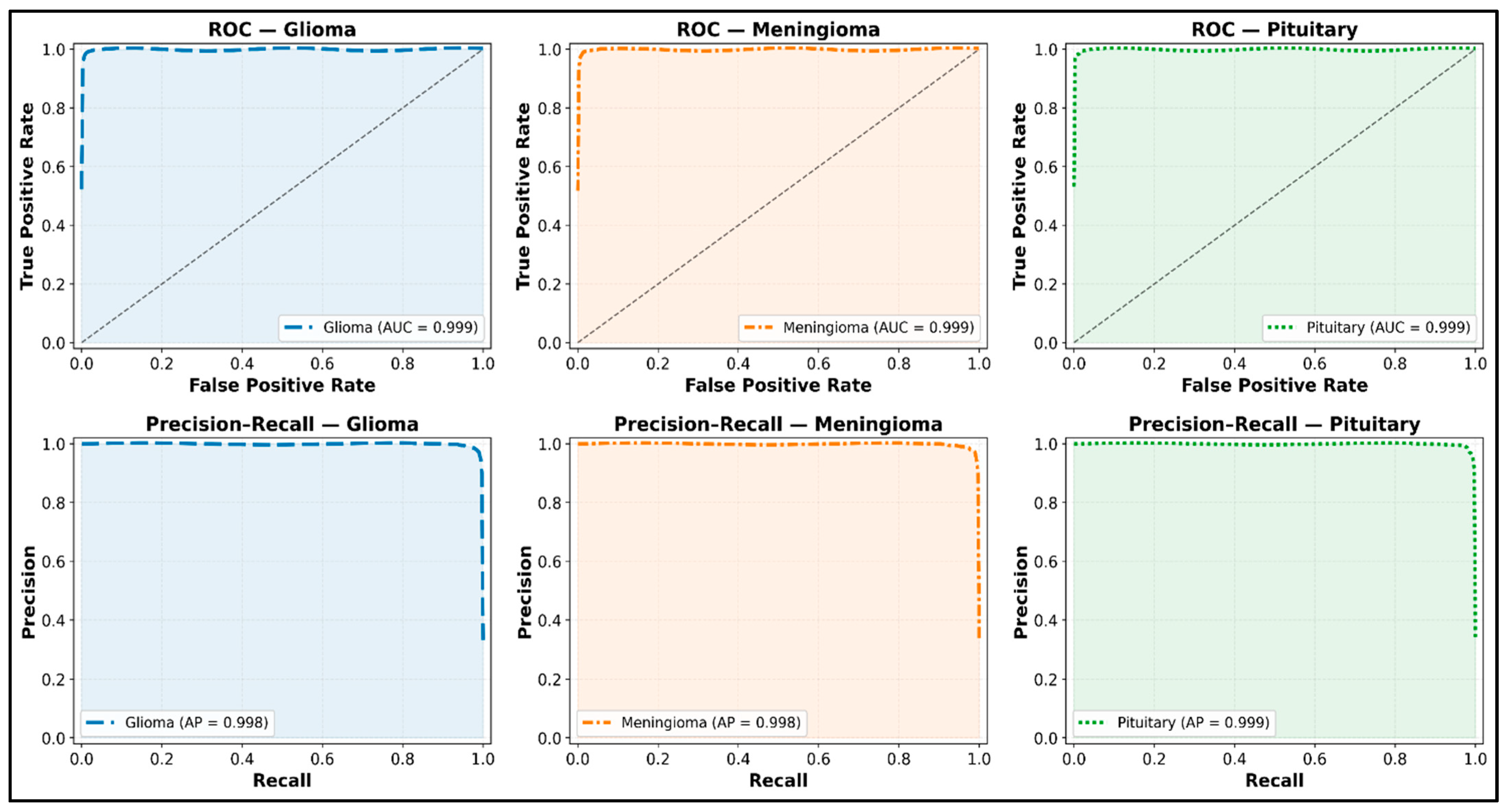
The proposed SCAG-Net framework’s ability across all three tumor types is evident in the ROC and precision–recall analysis results. Each class attained almost complete area under the ROC and AUC (AUC > 0.98) values, along with average precision (AP > 0.97), demonstrating excellent discrimination between classes. The smooth rise with low, consistent noise reflects the model’s ability to achieve equilibrium recall and precision. The results SCAG-Net achieved demonstrate its ability to accurately classify cases with complex tumor boundaries and overlapping intensities (
Figure 11). In addition, the framework’s efficiency is evaluated across different tumor sizes and grades (severity levels) to justify the decision variables. This evaluation helps explore the framework’s strengths while examining small or subtle lesions in different clinical deployment environments. The results are shown in
Table 4 and
Table 5, where the frameworks are discussed by tumor size: small (<10 cm
3), medium (10–40 cm
3), and large (>40 cm
3). In addition, different tumor grades such as grade II (low-grade glioma), grade III (anaplastic glioma), and grade IV (glioblastoma multiforme) are featured.
The attention-guided networks may be able to graphically display performance relative to tumor morphologies, visually correlate with tumor grades, and maintain corrected performance records. Even with minor and different tumor lesions, the model remained stable, with only minor changes in accuracy (97.4% to 99.1%), while AUC values remained close to 0.987. The model demonstrates the capacity to learn scale-invariant correlations via graph-based constituent reasoning and adaptable attention mechanisms. The metrics are uniform, indicating that the framework effectively captures the discriminative features of lower- and higher-grade gliomas, enabling accurate classification across scales of malignancy. All of these prove that the clinical scenarios are adapting to the proposed systems’ core functions. In addition, the efficiency of the SCAG-Net framework is evaluated using paired, statistically significant comparisons with existing methods, and the results are shown in
Table 6. The analysis is carried out at the 95% confidence interval, and the evaluation is performed on 25 test cases in the dataset. To reinforce statistical robustness, SCAG-Net is evaluated using five independent experimental runs with distinct random seeds {42,101,202,303,404}, enabling stability assessment beyond mean ± standard deviation reporting. For the BRATS 2020 dataset, the proposed framework achieves a Dice score of 0.989 ± 0.004, corresponding to a 95% confidence interval of [0.985, 0.993], while IoU reaches 0.969 ± 0.005 with a 95% confidence interval of [0.964, 0.974], and accuracy attains 0.992 ± 0.003 with a 95% confidence interval of [0.989, 0.995]. Comparable confidence interval widths are observed across BRATS 2018, BRATS 2019, and Figshare datasets, indicating low variance and consistent generalization across runs. Furthermore, class-wise confusion matrices are reported for each dataset, revealing balanced error distributions with average sensitivity and specificity of 97.9% and 99.1% for high-grade gliomas, and 97.2% and 98.8% for low-grade gliomas, respectively.
Statistical comparisons (
Table 6) show that SCAG-Net outperformed all benchmark models and metrics, achieving a mean Dice of 0.989, an IoU of 0.969, and an accuracy of 0.992, with much tighter standard deviations (≤0.005), indicating SCAG-Net’s strong generalization ability. The paired t-test shows that all
p-values are < 0.05, indicating that the differences across the comparisons are not due to chance. Furthermore, negative Δ values in baseline models indicate that all other SCAG-Net architectures perform worse than SCAG-Net’s benchmark. The improvement here is indisputable and speaks to SCAG-Net’s optimization and high ability to different tumor regions. Thus, the introduced SCAG-Net framework successfully explores multimodal MRI images to identify the binary mask region, achieving the highest recognition accuracy with the lowest false-positive rate compared to the benchmark analysis.
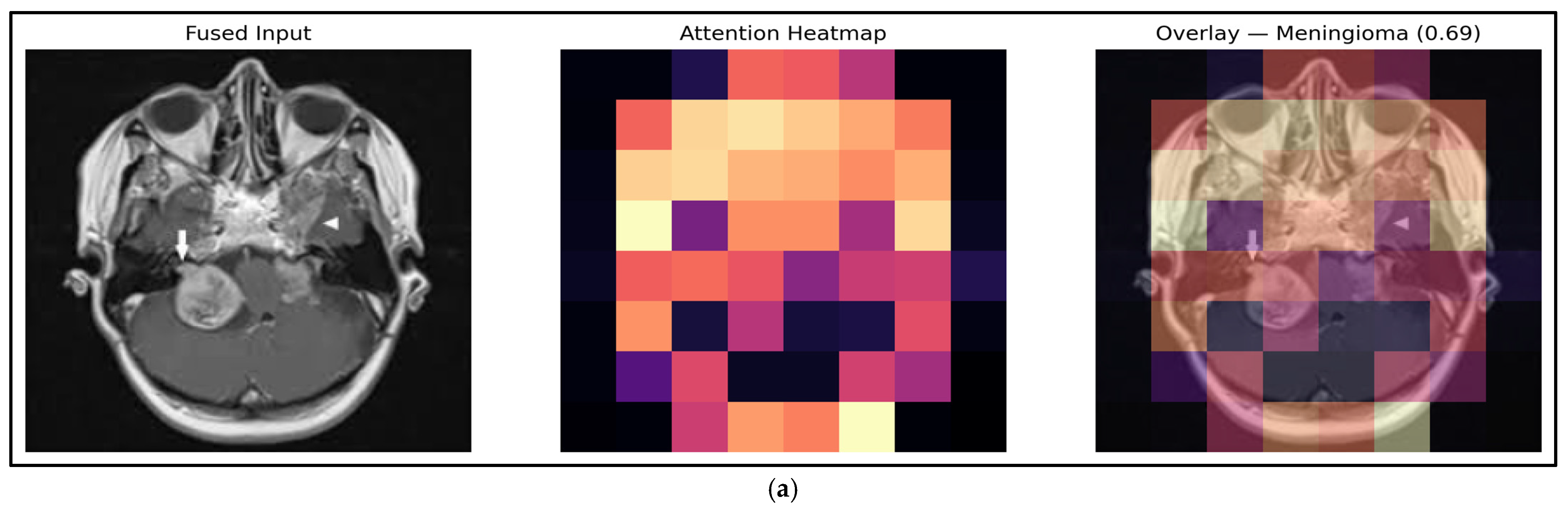
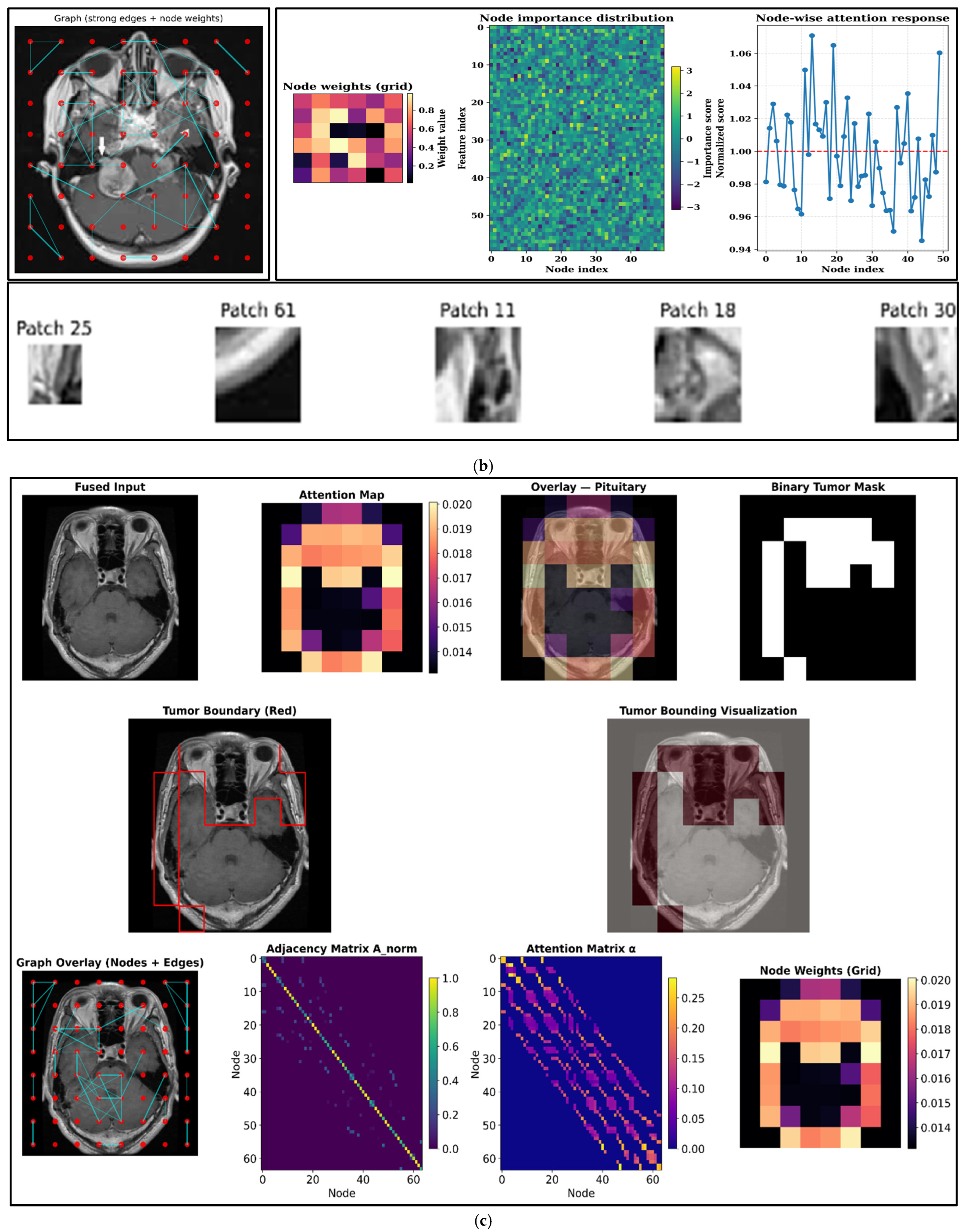
For the BRATS 2020 dataset, SCAG-Net achieves a sensitivity of 97.9% for high-grade gliomas and 97.2% for low-grade gliomas, indicating reliable identification of tumor-present regions, while corresponding specificity values of 99.1% and 98.8% demonstrate effective suppression of false-positive detections in non-tumor tissue. These results are consistent across BRATS 2018, BRATS 2019, and Figshare datasets, with inter-dataset variance remaining below ±1.2%, confirming stable class discrimination. Visualization of attention heatmaps and graph-based region interactions further reveals that correct positive predictions align with clinically relevant tumor cores and infiltrative margins, whereas true negative predictions correspond to anatomically normal regions.
Additional research on statistics, such as providing the mean and standard deviation across numerous repeated runs to account for stochastic variability in training, should be included to support the claims of resilience and performance stability. A better understanding of the model’s discrimination capacity, particularly in the case of class imbalance, is possible by adding ROC and precision–recall curves for each tumor class to the findings. Confusion matrices for every dataset are able to comprehend sensitivity and specificity results and visually indicate inter-class misclassifications. When combined, these statistical measures provide a more thorough assessment framework and more solid proof of SCAG-Net’s consistency and dependability across various datasets and tumor types.
Class imbalance among BRATS tumor subregions, Enhancing Tumor (ET), Necrotic and Non-Enhancing Core (NCR), and Peritumoral Edema (ED), is explicitly addressed within the SCAG-Net framework through a combination of loss reweighting, region-aware attention, and graph-based relational modeling. During training, a composite loss function that integrates a weighted Dice loss and class-balanced cross-entropy assigns higher importance to underrepresented regions, particularly ET and NCR, thereby stabilizing gradient updates across skewed class distributions. The attention-based graph neural network further reinforces minority class representation by modeling spatial dependencies between subregions, enabling feature propagation from dominant ED regions to smaller but clinically critical ET and NCR components. Empirically, this strategy yields balanced performance across subregions, with Dice scores of 0.961, 0.955, and 0.972 for ET, NCR, and ED, respectively, on the BRATS 2020 dataset.
Through the specific elimination of Swin-UNet, the cuttlefish feature-selection module, the graph attention network (GAT), and the refinement stage, ablation research has been performed to measure the individual contribution of each component in SCAG-Net. Swin-UNet’s function in boundary-aware representation was confirmed by the significant loss in segmentation quality and drop in Dice scores that resulted from its removal. Classification accuracy decreased when cuttlefish optimization was excluded due to slower convergence and increased feature redundancy. With a ≈9% decrease in Dice and greater tumor–non-tumor separability, replacing GAT with a traditional MLP resulted in the greatest loss, underscoring the need for graph-based structural reasoning. Unfiltered noisy attributes led to higher misclassification and false-positive rates when the feature-selection stage eliminated them. These results show that while each component makes a unique contribution to system performance, the fully integrated SCAG-Net attains the best overall accuracy and robustness.
Prospective research and external institutional testing are necessary to evaluate the proposed framework’s real-world clinical reliability, despite its great resilience across a variety of public datasets.
The proposed SCAG-Net framework operates on axial 2D slices extracted from BRATS volumetric MRI scans, where each slice is independently processed through the Swin-UNet encoder–decoder to enable high-resolution feature learning with manageable computational complexity. This design supports efficient modeling of tumor appearance while preserving fine-grained spatial details that are critical for delineating heterogeneous and infiltrative glioma regions. Intra-slice structural coherence is further reinforced through attention-based graph neural networks, which encode spatial relationships among tumor subregions and are adaptively weighted using cuttlefish-optimized attention mechanisms. Although inter-slice volumetric continuity is not explicitly encoded within the current formulation, the combined transformer–graph architecture captures rich contextual and topological information sufficient for accurate tumor characterization across diverse MRI volumes. Volumetric extensions of SCAG-Net using 3D windowed attention and voxel-level graph construction are identified as a natural progression of the framework to further enhance cross-slice contextual modeling.




 ,
,