The Diagnostic Value of Lung Ultrasound in Bronchopulmonary Dysplasia Among Preterm Infants with Gestational Age ≤ 28 Weeks

 , and
, and
Abstract
1. Introduction
2. Methods
2.1. Study Design
2.2. Subjects
2.3. Instrumental Examination
2.4. LUS Scanning
2.5. LUS Characteristics of BPD
- (1)
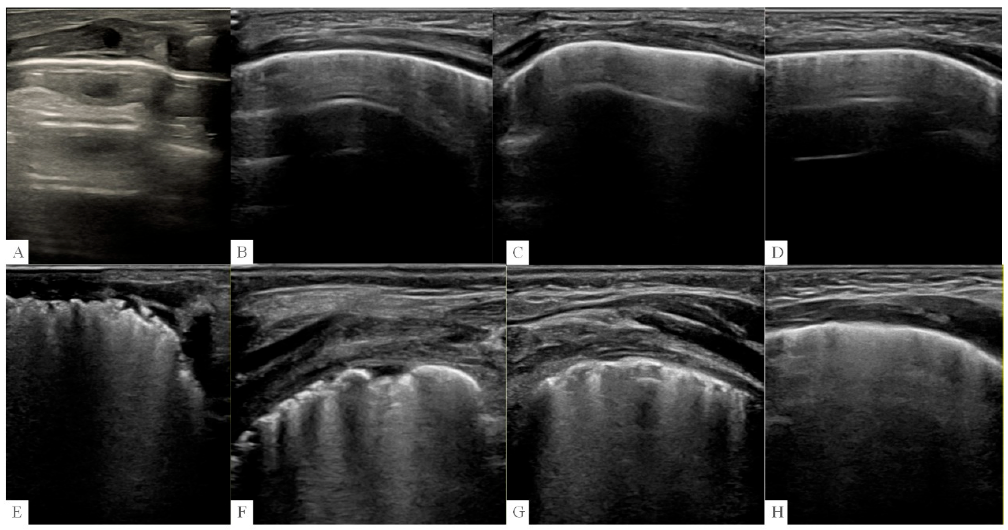
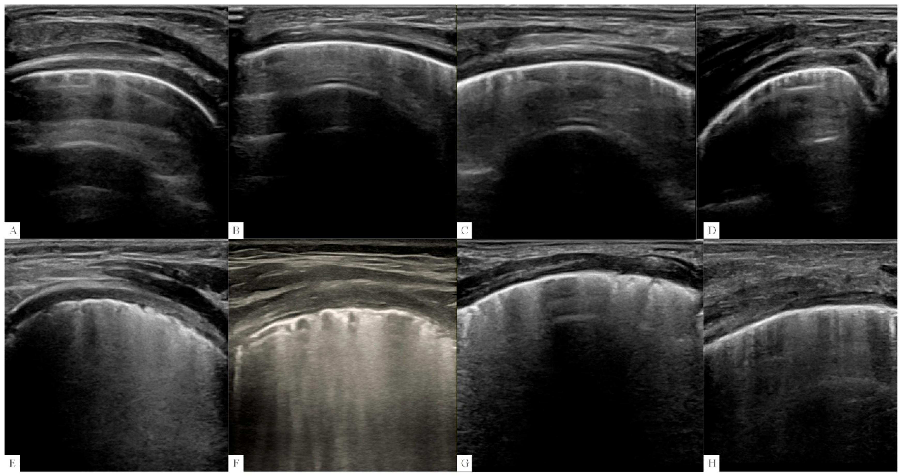
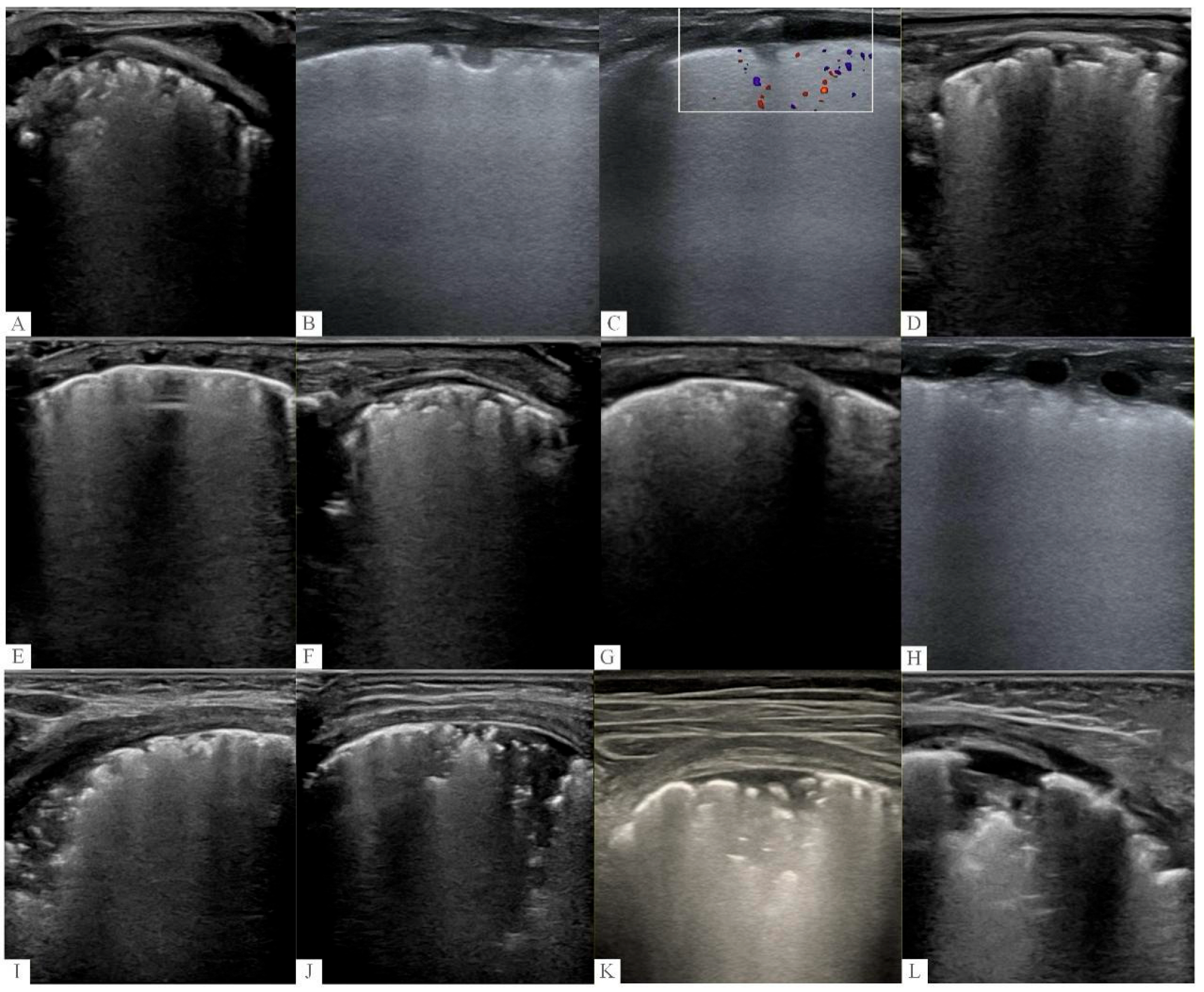
- Pleural line abnormalities: The pleural line was assessed for thickness, roughness, and clarity. A pleural line was considered “obviously thick, rough, and fuzzy” when it appeared widened, irregular in contour, and poorly defined compared with the normal thin, smooth, and echogenic pleural line. Pleural insect erosion (PIE)-like changes were defined as discontinuous and intermittent disruption of the pleural line, resembling a moth-eaten appearance.
- (2)
- lines: Horizontal echogenic lines parallel to the pleural line, representing reverberation artifacts from the pleural surface. A-lines were classified as “fuzzy, not clear, or invisible” when they were partially or completely obliterated by B-lines or other artifacts.
- (3)
- B-lines: Vertical hyperechoic artifacts arising from the pleural line, extending to the bottom of the screen without fading. B-lines were classified as “multiple, partial or completely fused” when they were too numerous to count and coalesced into confluent bands, indicating significant interstitial or alveolar fluid accumulation.
- (4)
- Lung consolidation: Defined as a tissue-like echogenicity region with air bronchograms (hyperechoic punctate or linear artifacts within the consolidation). Regarding the differentiation between atelectasis and pneumonia on LUS, atelectasis was differentiated from pneumonia/consolidation based on the following criteria: (1) the presence of dynamic air bronchograms (moving with respiration) was more suggestive of pneumonia, whereas static air bronchograms were more consistent with atelectasis; (2) the presence of associated pleural line abnormalities or B-lines in surrounding lung zones favored pneumonia; and (3) atelectasis typically resolved or improved with positive pressure ventilation or position changes, whereas pneumonic consolidation was more persistent.
- (5)
- Pleural effusion: Defined as an anechoic space between the parietal and visceral pleura.
- (6)
- Irregular patch-like anechoic or weakly echoic appearances on the pleural surface: Defined as irregular, well-demarcated areas of absent or reduced echogenicity on the pleural surface, with no detectable blood flow on color Doppler.
- (7)
- Subpleural speckled or patch-like hyperechogenicity: Defined as scattered, punctate or patchy bright echoes located immediately below the pleural line, which may correspond to microcysts, fibrosis, or inflammatory changes in the subpleural region.
2.6. Statistical Analysis
3. Results
4. Discussion
5. Conclusions
Author Contributions
Funding
Institutional Review Board Statement
Informed Consent Statement
Data Availability Statement
Acknowledgments
Conflicts of Interest
References
- Thébaud, B.; Goss, K.N.; Laughon, M.; Whitsett, J.A.; Abman, S.H.; Steinhorn, R.H.; Aschner, J.L.; Davis, P.G.; McGrath-Morrow, S.A.; Soll, R.F.; et al. Bronchopulmonary dysplasia. Nat. Rev. Dis. Prim. 2019, 5, 78. [Google Scholar] [CrossRef] [Scilit] [PubMed]
- Ambalavanan, N.; Deutsch, G.; Pryhuber, G.; Travers, C.P.; Willis, K.A. The evolving pathophysiology of bronchopulmonary dysplasia. Physiol. Rev. 2026, 106, 197–237. [Google Scholar] [CrossRef] [Scilit] [PubMed]
- Higgins, R.D.; Jobe, A.H.; Koso-Thomas, M.; Bancalari, E.; Viscardi, R.M.; Hartert, T.V.; Ryan, R.M.; Kallapur, S.G.; Steinhorn, R.H.; Konduri, G.G.; et al. Bronchopulmonary Dysplasia: Executive Summary of a Workshop. J. Pediatr. 2018, 197, 300–308. [Google Scholar] [CrossRef] [Scilit] [PubMed]
- Higano, N.S.; Bates, A.J.; Gunatilaka, C.C.; Hysinger, E.B.; Critser, P.J.; Hirsch, R.; Woods, J.C.; Fleck, R.J. Bronchopulmonary dysplasia from chest radio-graphs to magnetic resonance imaging and computed tomography: Adding value. Pediatr. Radiol. 2022, 52, 643–660, Erratum in Pediatr Radiol. 2022 52, 2442. https://doi.org/10.1007/s00247-022-05490-9. [Google Scholar] [CrossRef] [Scilit] [PubMed]
- Katz, S.L.; Parraga, G.; Luu, T.M.; Santyr, G.; Abdeen, N.; Deschenes, S.; Tse, S.M.; Couch, M.J.; Barrowman, N.; Hayawi, L.; et al. Pulmonary Magnetic Resonance Imaging of Ex-Preterm Children with and without Bronchopulmonary Dysplasia. Ann. Am. Thorac. Soc. 2022, 19, 1149–1157. [Google Scholar] [CrossRef] [Scilit] [PubMed]
- Sung, T.J.; Hwang, S.M.; Kim, M.Y.; Park, S.G.; Choi, K.Y. Relationship between clinical severity of “new” bronchopulmonary dysplasia and HRCT abnormalities in VLBW infants. Pediatr. Pulmonol. 2018, 53, 1391–1398. [Google Scholar] [CrossRef] [Scilit] [PubMed]
- Critser, P.J.; Higano, N.S.; Lang, S.M.; Kingma, P.S.; Fleck, R.J.; Hirsch, R.; Taylor, M.D.; Woods, J.C. Cardiovascular magnetic resonance imaging derived septal curvature in neonates with bronchopulmonary dysplasia associated pulmonary hypertension. J. Cardiovasc. Magn. Reson. 2020, 22, 50. [Google Scholar] [CrossRef] [Scilit] [PubMed]
- Sartorius, V.; Brunet, S.; De Luca, D. Characteristics of scores used for quantitative lung ultrasound in neonates: A systematic review. Eur. Respir. Rev. 2025, 34, 240232. [Google Scholar] [CrossRef] [Scilit] [PubMed]
- Francavilla, M.; Scarlato, L.; Camporesi, A.; Orlandi, A.; Cafagno, C.; Raguseo, C.L.; Santoiemma, V.; Russo, R.; Sacco, A.; Musolino, A.M.; et al. Lung ultrasound for the diagnosis and monitoring of community-acquired pneumonia in children: A prospective observational study. Front Pediatr. 2025, 13, 1702388. [Google Scholar] [CrossRef] [Scilit] [PubMed] [PubMed Central]
- Isac, R.; Cugerian-Ratiu, A.M.; Micsescu-Olah, A.M.; Bodescu, A.D.; Vlad, L.A.; Zaroniu, A.M.; Gafencu, M.; Doros, G. Can Lung Ultrasound Act as a Diagnosis and Monitoring Tool in Children with Community Acquired Pneumonia? Correlation with Risk Factors, Clinical Indicators and Biologic Results. J. Clin. Med. 2025, 14, 5304. [Google Scholar] [CrossRef] [Scilit] [PubMed] [PubMed Central]
- Gao, S.; Xiao, T.; Ju, R.; Ma, R.; Zhang, X.; Dong, W. The application value of lung ultrasound findings in preterm infants with bron-chopulmonary dysplasia. Transl. Pediatr. 2020, 9, 93–100. [Google Scholar] [CrossRef] [Scilit] [PubMed]
- Liu, J.; Chi, J.; Fu, W.; Zhang, L.; Qiu, R. Lung Ultrasonography to Diagnose Bronchopulmonary Dysplasia of Premature Infants. Inn. J. Pediatr. 2021, 31, e109598. [Google Scholar] [CrossRef] [Scilit]
- Aldecoa-Bilbao, V.; Velilla, M.; Teresa-Palacio, M.; Esponera, C.B.; Barbero, A.H.; Sin-Soler, M.; Sanz, M.I.; Roigés, M.D.S. Lung Ultrasound in Bronchopul-monary Dysplasia: Patterns and Predictors in Very Preterm Infants. Neonatology 2021, 118, 537–545. [Google Scholar] [CrossRef] [Scilit] [PubMed]
- Sun, Y.H.; Yuan, L.; Du, Y.; Zhou, J.G.; Lin, S.B.; Zhang, R.; Dong, Y.; Chen, C. Characterization of lung ultrasound imaging in preterm infants with bronchopulmonary dysplasia. Clin. Hemorheol. Microcirc. 2022, 80, 83–95. [Google Scholar] [CrossRef] [Scilit] [PubMed]
- Vanhaverbeke, K.; Van Eyck, A.; Van Hoorenbeeck, K.; De Winter, B.; Snoeckx, A.; Mulder, T.; Verhulst, S. Lung imaging in bronchopul-monary dysplasia: A systematic review. Respir. Med. 2020, 171, 106101. [Google Scholar] [CrossRef] [Scilit] [PubMed]
- Khetan, R.; Hurley, M.; Spencer, S.; Bhatt, J.M. Bronchopulmonary Dysplasia Within and Beyond the Neonatal Unit. Adv. Neonatal Care 2016, 16, 17–25, quiz E1–2. [Google Scholar] [CrossRef] [Scilit] [PubMed]
- Avni, E.F.; Cassart, M.; de Maertelaer, V.; Rypens, F.; Vermeylen, D.; Gevenois, P.A. Sonographic prediction of chronic lung disease in the premature undergoing mechanical ventilation. Pediatr. Radiol. 1996, 26, 463–469. [Google Scholar] [CrossRef] [Scilit] [PubMed]
- Pieper, C.H.; Smith, J.; Brand, E.J. The value of ultrasound examination of the lungs in predicting bronchopulmonary dysplasia. Pediatr. Radiol. 2004, 34, 227–231. [Google Scholar] [CrossRef] [Scilit] [PubMed]
- Volpicelli, G.; Melniker, L.A.; Cardinale, L.; Lamorte, A.; Frascisco, M.F. Lung ultrasound in diagnosing and monitoring pulmonary interstitial fluid. Radiol. Med. 2013, 118, 196–205. [Google Scholar] [CrossRef] [Scilit] [PubMed]
- Adams, E.W.; Harrison, M.C.; Counsell, S.J.; Allsop, J.M.; Kennea, N.L.; Hajnal, J.V.; Thornton, A.S.; Duggan, P.; Edwards, A.D. Increased lung water and tissue damage in bronchopulmonary dysplasia. J. Pediatr. 2004, 145, 503–507. [Google Scholar] [CrossRef] [Scilit] [PubMed]
- Zong, H.; Li, C.; Ye, N.; Song, J.; Yang, L.; Fang, S.; Shi, J.; Chen, X.; Yang, C. Lung ultrasound features during the evolution of BPD: A study from an animal model. J. Matern.-Fetal Neonatal Med. 2026, 39, 2611637. [Google Scholar] [CrossRef] [Scilit] [PubMed]
- Alonso-Ojembarrena, A.; Serna-Guerediaga, I.; Aldecoa-Bilbao, V.; Gregorio-Hernández, R.; Alonso-Quintela, P.; Concheiro-Guisán, A.; Ramos-Rodríguez, A.; Heras-Martín, M.d.L.; Rodeño-Fernández, L.; Oulego-Erroz, I.; et al. The Predictive Value of Lung Ultrasound Scores in Developing Bronchopulmonary Dysplasia: A Prospective Multicenter Diagnostic Accuracy Study. Chest 2021, 160, 1006–1016. [Google Scholar] [CrossRef] [Scilit] [PubMed]
- Zong, H.; Huang, Z.; Fu, Y.; Chen, X.; Yu, Y.; Huang, Y.; Sun, H.; Yang, C. Lung ultrasound score as a tool to predict severity of bronchopulmonary dysplasia in neonates born ≤25 weeks of gestational age. J. Perinatol. 2024, 44, 273–279. [Google Scholar] [CrossRef] [Scilit] [PubMed]
- Han, C.; Wang, X.; Pan, M.; Huang, X. Diagnostic accuracy of lung ultrasound score for bronchopulmonary dysplasia in preterm neonates: A systematic review and meta-analysis. Front. Pediatr. 2025, 13, 1694150. [Google Scholar] [CrossRef] [Scilit] [PubMed]



| Non–Mild BPD (n = 69) | Moderate–Severe BPD (n = 45) | p-Value | |
|---|---|---|---|
| GA (weeks) | 27.0 ± 1.4 | 26.3 ± 1.6 | 0.135 |
| Male (%) | 47 (68.1%) | 31 (68.9%) | 0.931 |
| Cesarean section (%) | 47 (68.1%) | 21 (46.7%) | 0.023 |
| Birth weight (g) | 969 ± 184 | 802 ± 228 | <0.001 |
| PROM > 18 h (%) | 58 (84.1%) | 32 (71.1%) | 0.097 |
| Antenatal steroids, full course (%) | 60 (87.0%) | 30 (66.7%) | 0.009 |
| Apgar score 1 min | 8 (7, 9) | 5 (5, 8) | <0.001 |
| Apgar score 5 min | 9 (9, 10) | 9 (7, 9) | <0.001 |
| hsPDA | 13 (18.8%) | 17 (37.8%) | 0.025 |
| IVH grade III–IV (%) | 2 (2.9%) | 4 (8.9%) | 0.211 |
| NEC or SIP | 3 (4.3%) | 3 (6.7%) | 0.679 |
| pulmonary surfactant therapy (%) | 43 (62.3%) | 37 (82.2%) | 0.023 |
| Lung Ultrasound Findings | Non–Mild BPD (n = 69) | Moderate–Severe BPD (n = 45) | p-Value |
|---|---|---|---|
| Pleural line obviously thick, rough, fuzzy (%) | 26 (37.7%) | 45 (100%) | <0.001 |
| Pleural insect erosion (PIE)-like changes (%) | 4 (5.8%) | 34 (75.6%) | <0.001 |
| Irregular patch-like anechoic or weakly echoic on the pleura surface (%) | 5 (7.2%) | 38 (84.4%) | <0.001 |
| Subpleural speckled or patch-like hyperechogenicity (%) | 6 (8.7%) | 33 (73.3%) | <0.001 |
| A-line fuzzy, not clear, even invisible (%) | 7 (10.1%) | 39 (86.7%) | <0.001 |
| Multiple, partial or complete fused B-lines (%) | 9 (13.0%) | 40 (88.9%) | <0.001 |
| Lung consolidation (≥0.5 cm) (%) | 8 (11.6%) | 36 (80.0%) | <0.001 |
| Pleural effusion (%) | 0 (0%) | 3 (6.7%) | 0.059 |
| LUS Features | Sensitivity (95%CI) | Specificity (95%CI) | PPV (95%CI) | NPV (95%CI) |
|---|---|---|---|---|
| Pleural line obviously thick, rough, fuzzy | 100.0 (92.1~100.0) | 62.3 (50.5~72.8) | 63.4 (51.8~73.6) | 100.0 (91.8~100.0) |
| Pleural insect erosion (PIE)-like changes | 75.6 (61.3~85.8) | 94.2 (86.0~97.7) | 89.5 (75.9~95.8) | 85.5 (75.9~91.7) |
| Irregular patch-like anechoic or weakly echoic on the pleura surface | 84.4 (71.2~92.3) | 92.8 (84.1~96.9) | 88.4 (75.5~94.9) | 90.1 (81.0~95.1) |
| Multiple, partial or complete fused B-lines | 88.9 (76.5~95.2) | 87.0 (77.0~93.0) | 81.6 (68.7~90.0) | 92.3 (83.2~96.7) |
| Lung consolidation (≥0.5 cm) | 80.0 (66.2~89.1) | 88.4 (78.8~94.0) | 81.8 (68.0~90.5) | 87.1 (77.3~93.1) |
| Subpleural speckled or patch-like hyperechogenicity | 73.3 (59.2~84.0) | 91.3 (82.4~96.0) | 84.6 (70.4~93.0) | 84.0 (74.2~90.6) |
Disclaimer/Publisher’s Note: The statements, opinions and data contained in all publications are solely those of the individual author(s) and contributor(s) and not of MDPI and/or the editor(s). MDPI and/or the editor(s) disclaim responsibility for any injury to people or property resulting from any ideas, methods, instructions or products referred to in the content. |
© 2026 by the authors. Licensee MDPI, Basel, Switzerland. This article is an open access article distributed under the terms and conditions of the Creative Commons Attribution (CC BY) license.
Share and Cite
Zong, H.; Lin, B.; Chen, X.; Huang, Y.; Song, J.; Sun, H.; Li, Q.; Zhang, S.; Yang, C. The Diagnostic Value of Lung Ultrasound in Bronchopulmonary Dysplasia Among Preterm Infants with Gestational Age ≤ 28 Weeks. Diagnostics 2026, 16, 2474. https://doi.org/10.3390/diagnostics16152474
Zong H, Lin B, Chen X, Huang Y, Song J, Sun H, Li Q, Zhang S, Yang C. The Diagnostic Value of Lung Ultrasound in Bronchopulmonary Dysplasia Among Preterm Infants with Gestational Age ≤ 28 Weeks. Diagnostics. 2026; 16(15):2474. https://doi.org/10.3390/diagnostics16152474
Chicago/Turabian StyleZong, Haifeng, Bingchun Lin, Xueyu Chen, Yichu Huang, Jingyu Song, Hongyan Sun, Qingling Li, Sue Zhang, and Chuanzhong Yang. 2026. "The Diagnostic Value of Lung Ultrasound in Bronchopulmonary Dysplasia Among Preterm Infants with Gestational Age ≤ 28 Weeks" Diagnostics 16, no. 15: 2474. https://doi.org/10.3390/diagnostics16152474
APA StyleZong, H., Lin, B., Chen, X., Huang, Y., Song, J., Sun, H., Li, Q., Zhang, S., & Yang, C. (2026). The Diagnostic Value of Lung Ultrasound in Bronchopulmonary Dysplasia Among Preterm Infants with Gestational Age ≤ 28 Weeks. Diagnostics, 16(15), 2474. https://doi.org/10.3390/diagnostics16152474