Tomographic 3D Ultrasound for Thyroid Volumetry—A Comparison Between Multiplanar 2D and 3D Imaging Methods

 , , , , , ,
Abstract
1. Introduction
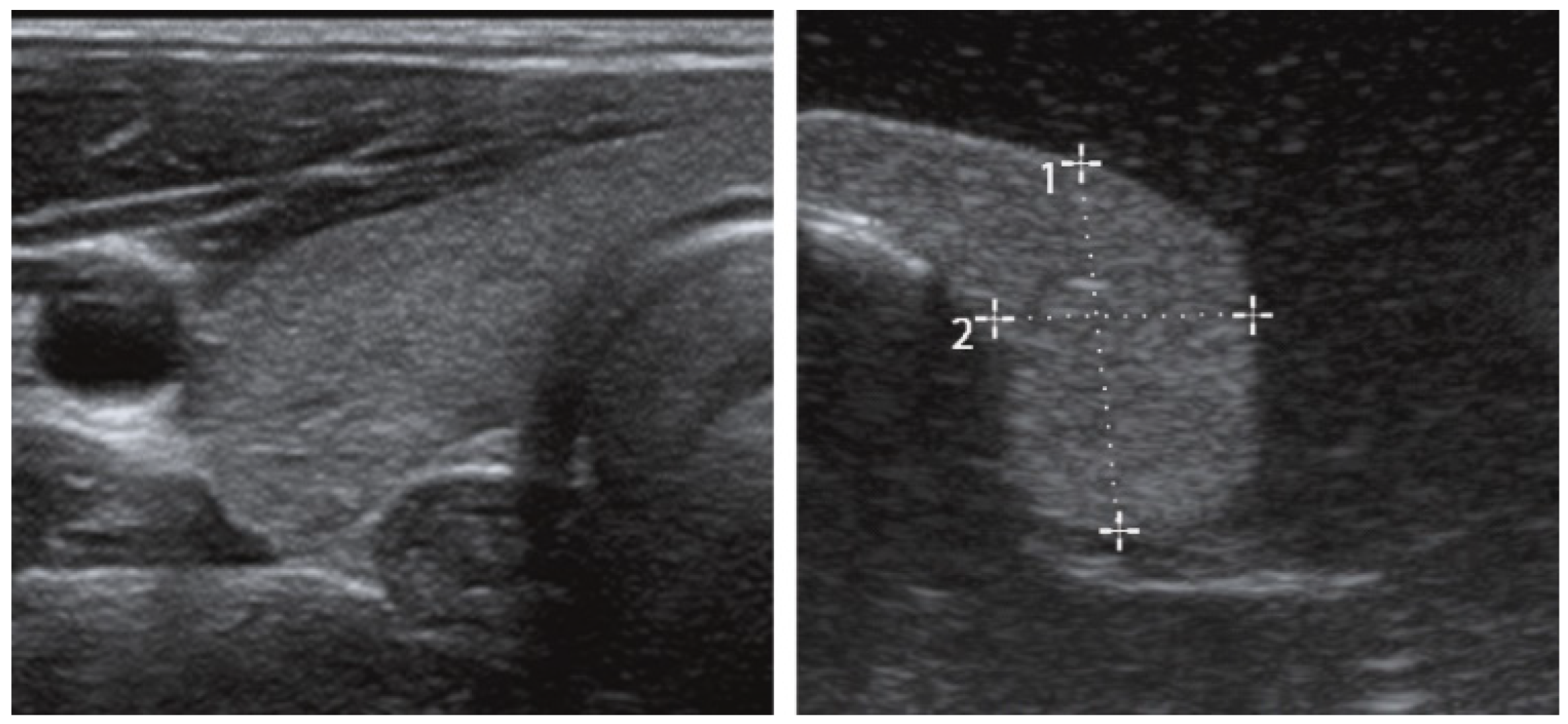
1.1. State of the Art: Thyroid Volume Measurement
1.2. Limitations of the State of the Art
1.3. Novel Approach
2. Materials and Methods
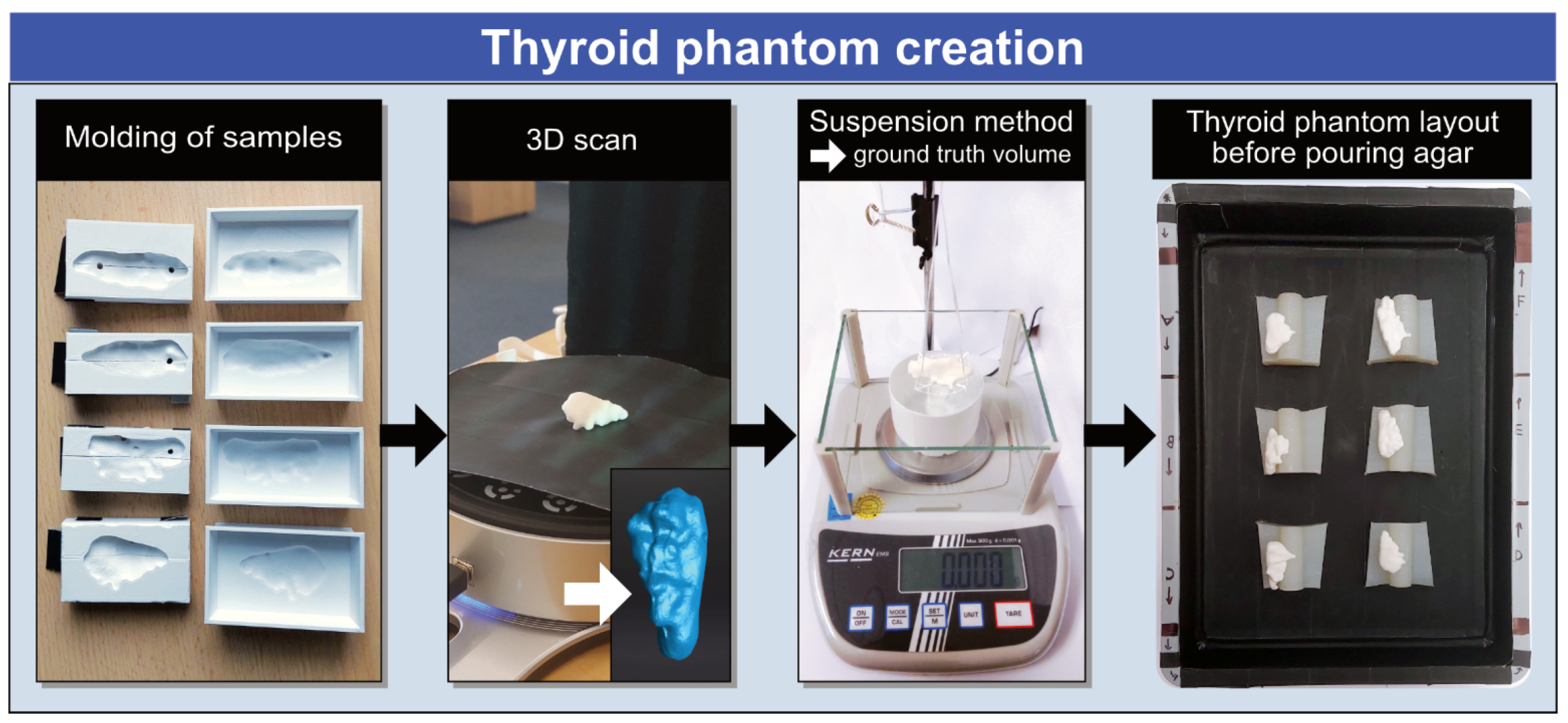
2.1. Thyroid Phantom Creation
2.1.1. Molding of the Samples
2.1.2. Suspension Method and 3D Scanning
2.1.3. Phantom Assembly
2.1.4. Tissue-Mimicking Material
2.2. Data Acquisition
2.2.1. Two-Dimensional B-Mode Ultrasound
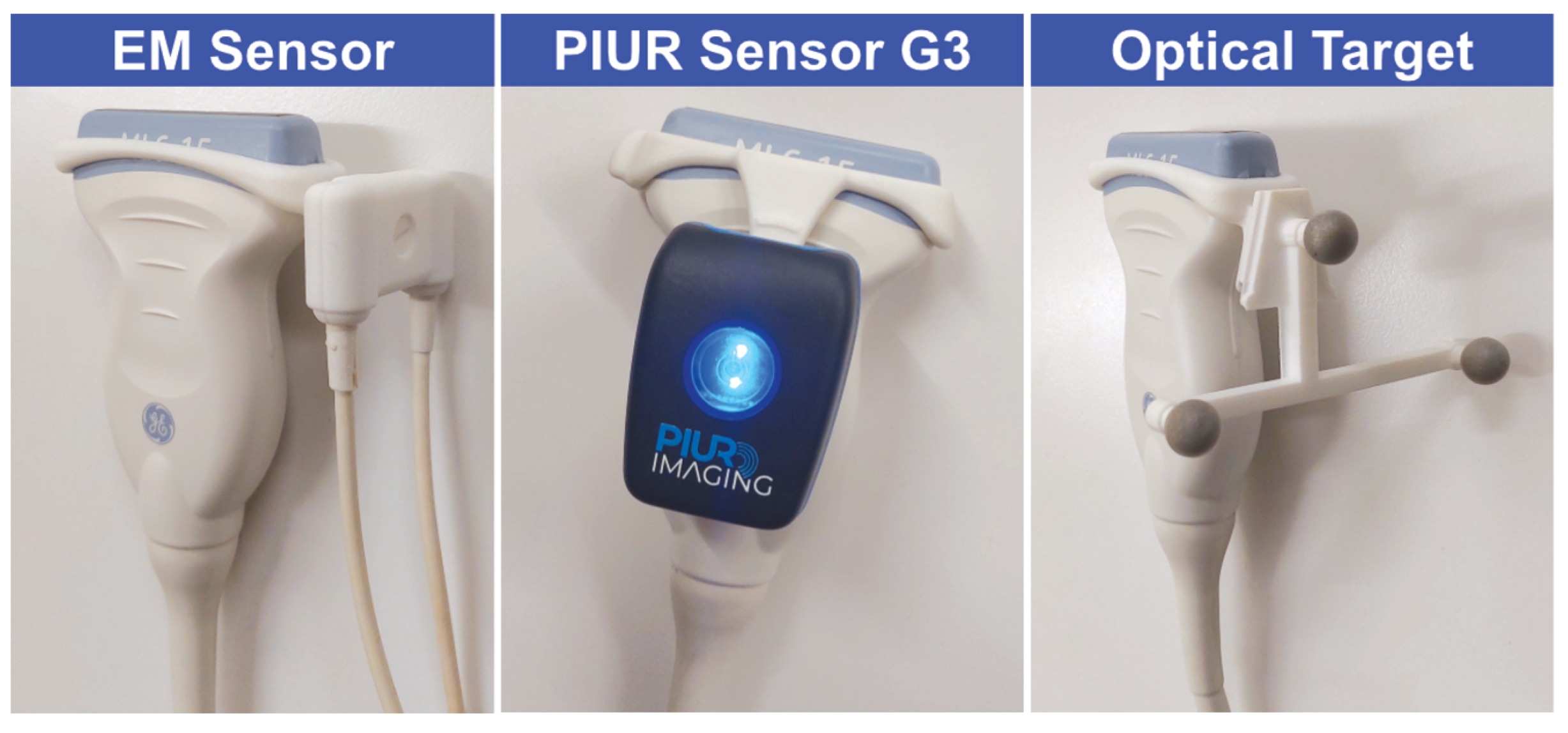
2.2.2. Electromagnetic Tracking
2.2.3. Inertial Tracking
2.2.4. Optical Tracking
2.2.5. Computed Tomography
2.2.6. Magnetic Resonance Imaging
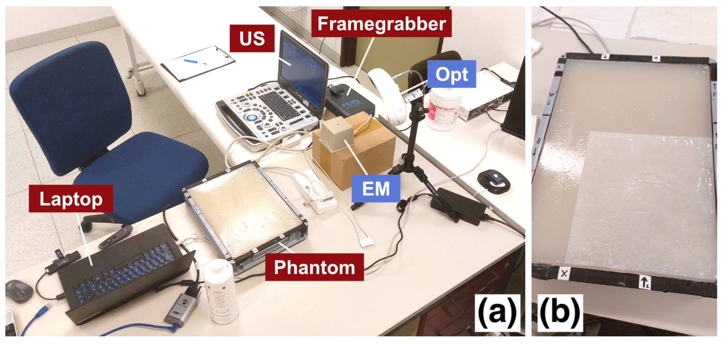
2.2.7. Acquisition Procedure
2.3. Segmentation
2.4. Statistical Methods
- Difference to ground truth:
- Intraobserver variability:
- Interobserver variability:
- 95% limits of agreement of :
3. Results
3.1. Difference Plot
3.2. Formal Statistical Comparisons
3.3. X-Y Plot
3.4. Intraobserver Plot
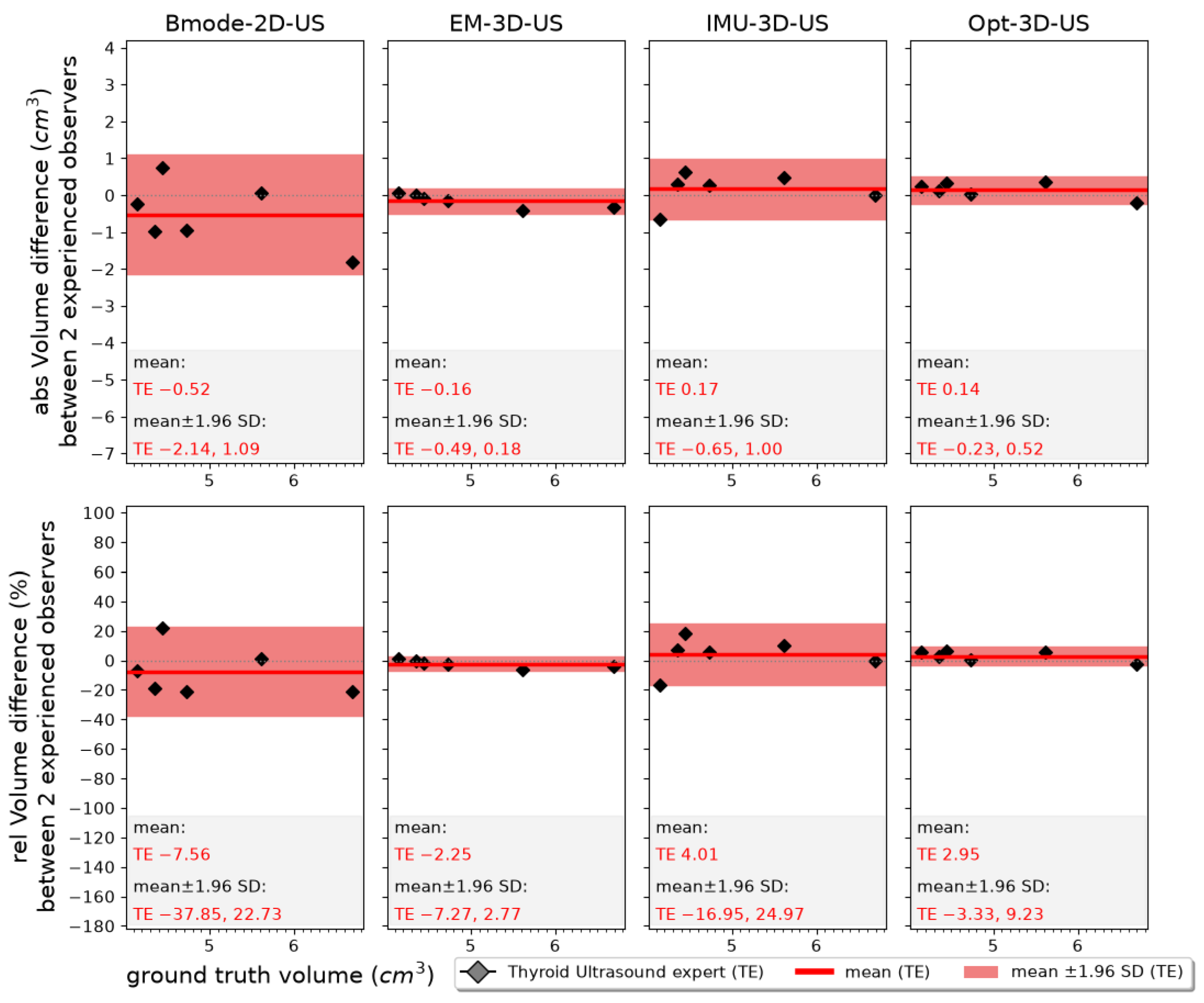
3.5. Interobserver Plot
4. Discussion
4.1. Quality of Ground Truth
4.2. Influence of Experience Level in Ultrasound
4.3. Comparison with the Literature
4.4. Discussion of Sample-Specific Bias
4.5. Limitations Regarding Segmentation Software and Potential Bias
4.6. General Limitations
4.7. Future Work
5. Conclusions
Author Contributions
Funding
Institutional Review Board Statement
Informed Consent Statement
Data Availability Statement
Conflicts of Interest
Abbreviations
| abs | Absolute |
| Bmode-2D-US | B-mode two-dimensional ultrasound |
| CT | Computed tomography |
| EM | Electromagnetic |
| EM-3D-US | Electromagnetically tracked three-dimensional ultrasound |
| ExFOV | Extended field of view |
| GT | Ground truth |
| IMU-3D-US | Inertial-measurement-unit-tracked three-dimensional ultrasound |
| MRI | Magnetic resonance imaging |
| Opt-3D-US | Optically tracked three-dimensional ultrasound |
| rel | Relative |
| SD | Standard deviation |
| TE | Thyroid ultrasound expert |
| TMM | Tissue-mimicking material |
| 3D-US | Three-dimensional ultrasound |
| US | Ultrasound |
| UU | Ultrasound user |
Appendix A. Additional Comparisons of Measurement Results


| EM-3D-US | IMU-3D-US | Opt-3D-US | CT | MRI | |
|---|---|---|---|---|---|
| Bmode-2D-US | 0.219 | 0.031 * | 0.563 | 0.031 * | 0.031 * |
| EM-3D-US | – | 0.031 * | 0.031 * | 0.031 * | 0.031 * |
| IMU-3D-US | – | 0.031 * | 1.000 | 0.219 | |
| Opt-3D-US | – | 0.031 * | 0.031 * | ||
| CT | – | 0.438 |


| Modality | UU2 (n = 6) | TE2 (n = 6) |
|---|---|---|
| Bmode-2D-US | 1.000 | 0.688 |
| EM-3D-US | 1.000 | 0.063 |
| IMU-3D-US | 0.031 * | 0.438 |
| Opt-3D-US | 0.031 * | 0.844 |
References
- Campennì, A.; Avram, A.M.; Verburg, F.A.; Iakovou, I.; Hänscheid, H.; de Keizer, B.; Petranović Ovčariček, P.; Giovanella, L. The EANM Guideline on Radioiodine Therapy of Benign Thyroid Disease. Eur. J. Nucl. Med. Mol. Imaging 2023, 50, 3324–3348. [Google Scholar] [CrossRef] [Scilit] [PubMed]
- Andermann, P.; Schlögl, S.; Mäder, U.; Luster, M.; Lassmann, M.; Reiners, C. Intra- and Interobserver Variability of Thyroid Volume Measurements in Healthy Adults by 2D versus 3D Ultrasound. Nuklearmedizin 2007, 46, 1–7. [Google Scholar] [CrossRef] [Scilit]
- Lyshchik, A.; Drozd, V.; Reiners, C. Accuracy of Three-Dimensional Ultrasound for Thyroid Volume Measurement in Children and Adolescents. Thyroid 2004, 14, 113–120. [Google Scholar] [CrossRef] [Scilit] [PubMed]
- Haugen, B.R.; Alexander, E.K.; Bible, K.C.; Doherty, G.M.; Mandel, S.J.; Nikiforov, Y.E.; Pacini, F.; Randolph, G.W.; Sawka, A.M.; Schlumberger, M.; et al. 2015 American Thyroid Association Management Guidelines for Adult Patients with Thyroid Nodules and Differentiated Thyroid Cancer: The American Thyroid Association Guidelines Task Force on Thyroid Nodules and Differentiated Thyroid Cancer. Thyroid 2016, 26, 1–133. [Google Scholar] [CrossRef] [Scilit] [PubMed]
- Brauer, V.F.H.; Eder, P.; Miehle, K.; Wiesner, T.D.; Hasenclever, H.; Paschke, R. Interobserver Variation for Ultrasound Determination of Thyroid Nodule Volumes. Thyroid 2005, 15, 1169–1175. [Google Scholar] [CrossRef] [Scilit] [PubMed]
- Freesmeyer, M.; Wiegand, S.; Schierz, J.-H.; Winkens, T.; Licht, K. Multimodal Evaluation of 2-D and 3-D Ultrasound, Computed Tomography and Magnetic Resonance Imaging in Measurements of the Thyroid Volume Using Universally Applicable Cross-Sectional Imaging Software: A Phantom Study. Ultrasound Med. Biol. 2014, 40, 1453–1462. [Google Scholar] [CrossRef] [Scilit] [PubMed]
- Krönke, M.; Eilers, C.; Dimova, D.; Köhler, M.; Buschner, G.; Schweiger, L.; Konstantinidou, L.; Makowski, M.; Nagarajah, J.; Navab, N.; et al. Tracked 3D Ultrasound and Deep Neural Network-Based Thyroid Segmentation Reduce Interobserver Variability in Thyroid Volumetry. PLoS ONE 2022, 17, e0268550. [Google Scholar] [CrossRef] [Scilit] [PubMed]
- Hughes, S.W. Archimedes Revisited: A Faster, Better, Cheaper Method of Accurately Measuring the Volume of Small Objects. Phys. Educ. 2005, 40, 468. [Google Scholar] [CrossRef] [Scilit]
- Rogers, S.; Carreira, J.; Thompson, R.; Morais, A.; Miller, C.; Wein, W.; Ghosh, J.; McCollum, C. An Ex Vivo Evaluation of Tomographic 3-D Ultrasound, B-Mode Ultrasound, CT and MR Imaging to Measure Artery Diameter, Length and Wall Volume. Ultrasound Med. Biol. 2019, 45, 2819–2829. [Google Scholar] [CrossRef] [Scilit] [PubMed]
- Deene, Y.D.; Wheatley, M.; Greig, T.; Hayes, D.; Ryder, W.; Loh, H. A Multi-Modality Medical Imaging Head and Neck Phantom: Part 1. Design and Fabrication. Phys. Med. 2022, 96, 166–178. [Google Scholar] [CrossRef] [Scilit] [PubMed]
- King, D.M.; Fagan, A.J.; Moran, C.M.; Browne, J.E. Comparative Imaging Study in Ultrasound, MRI, CT, and DSA Using a Multimodality Renal Artery Phantom. Med. Phys. 2011, 38, 565–573. [Google Scholar] [CrossRef] [Scilit] [PubMed]
- Souza, R.M.; Santos, T.Q.; Oliveira, D.P.; Souza, R.M.; Alvarenga, A.V.; Costa-Felix, R.P.B. Standard Operating Procedure to Prepare Agar Phantoms. J. Phys. Conf. Ser. 2016, 733, 012044. [Google Scholar] [CrossRef] [Scilit]
- Antoniou, A.; Georgiou, L.; Christodoulou, T.; Panayiotou, N.; Ioannides, C.; Zamboglou, N.; Damianou, C. MR Relaxation Times of Agar-Based Tissue-Mimicking Phantoms. J. Appl. Clin. Med. Phys. 2022, 23, e13533. [Google Scholar] [CrossRef] [Scilit] [PubMed]
- Drakos, T.; Antoniou, A.; Evripidou, N.; Alecou, T.; Giannakou, M.; Menikou, G.; Constantinides, G.; Damianou, C. Ultrasonic Attenuation of an Agar, Silicon Dioxide, and Evaporated Milk Gel Phantom. J. Med. Ultrasound 2021, 29, 239–249. [Google Scholar] [CrossRef] [Scilit] [PubMed]
- Antoniou, A.; Evripidou, N.; Georgiou, L.; Chrysanthou, A.; Ioannides, C.; Damianou, C. Tumor Phantom Model for MRI-Guided Focused Ultrasound Ablation Studies. Med. Phys. 2023, 50, 5956–5968. [Google Scholar] [CrossRef] [Scilit] [PubMed]
- Filippou, A.; Evripidou, N.; Damianou, C. Robotic System for Magnetic Resonance Imaging-Guided Focused Ultrasound Treatment of Thyroid Nodules. Int. J. Med. Robot 2023, 19, e2525. [Google Scholar] [CrossRef] [Scilit] [PubMed]
- Huber, J.S.; Peng, Q.; Moses, W.W. Multi-Modality Phantom Development. IEEE Trans. Nucl. Sci. 2009, 56, 2722–2727. [Google Scholar] [CrossRef]
- Kato, H.; Kuroda, M.; Yoshimura, K.; Yoshida, A.; Hanamoto, K.; Kawasaki, S.; Shibuya, K.; Kanazawa, S. Composition of MRI Phantom Equivalent to Human Tissues. Med. Phys. 2005, 32, 3199–3208. [Google Scholar] [CrossRef] [Scilit] [PubMed]
- Higgins, C.B.; McNamara, M.T.; Fisher, M.R.; Clark, O.H. MR Imaging of the Thyroid. AJR Am. J. Roentgenol. 1986, 147, 1255–1261. [Google Scholar] [CrossRef] [Scilit] [PubMed]
- Szomolanyi, P.; Rohrer, M.; Frenzel, T.; Noebauer-Huhmann, I.M.; Jost, G.; Endrikat, J.; Trattnig, S.; Pietsch, H. Comparison of the Relaxivities of Macrocyclic Gadolinium-Based Contrast Agents in Human Plasma at 1.5, 3, and 7 T, and Blood at 3 T. Investig. Radiol. 2019, 54, 559–564. [Google Scholar] [CrossRef] [Scilit] [PubMed]
- Yoshimura, K.; Kato, H.; Kuroda, M.; Yoshida, A.; Hanamoto, K.; Tanaka, A.; Tsunoda, M.; Kanazawa, S.; Shibuya, K.; Kawasaki, S.; et al. Development of a Tissue-Equivalent MRI Phantom Using Carrageenan Gel. Magn. Reson. Med. 2003, 50, 1011–1017. [Google Scholar] [CrossRef] [Scilit] [PubMed]
- Brunn, J.; Block, U.; Ruf, G.; Bos, I.; Kunze, W.P.; Scriba, P.C. Volumetrie der Schilddrüsenlappen mittels Real-time-Sonographie*1. Dtsch. Med. Wochenschr. 1981, 106, 1338–1340. [Google Scholar] [CrossRef] [Scilit] [PubMed]
- Shabana, W.; Peeters, E.; De Maeseneer, M. Measuring Thyroid Gland Volume: Should We Change the Correction Factor? AJR Am. J. Roentgenol. 2006, 186, 234–236. [Google Scholar] [CrossRef] [Scilit] [PubMed]
- Fedorov, A.; Beichel, R.; Kalpathy-Cramer, J.; Finet, J.; Fillion-Robin, J.-C.; Pujol, S.; Bauer, C.; Jennings, D.; Fennessy, F.; Sonka, M.; et al. 3D Slicer as an Image Computing Platform for the Quantitative Imaging Network. Magn. Reson. Imaging 2012, 30, 1323–1341. [Google Scholar] [CrossRef] [Scilit] [PubMed]
- Bland, J.M.; Altman, D.G. Statistical Methods for Assessing Agreement between Two Methods of Clinical Measurement. Lancet 1986, 1, 307–310. [Google Scholar] [CrossRef] [Scilit] [PubMed]
- Krouwer, J.S. Why Bland–Altman Plots Should Use X, Not (Y+X)/2 When X Is a Reference Method. Stat. Med. 2008, 27, 778–780. [Google Scholar] [CrossRef] [Scilit] [PubMed]
- Popović, Z.B.; Thomas, J.D. Assessing Observer Variability: A User’s Guide. Cardiovasc. Diagn. Ther. 2017, 7, 317–324. [Google Scholar] [CrossRef] [Scilit] [PubMed]
- Christensen, H.S.; Borgbjerg, J.; Børty, L.; Bøgsted, M. On Jones et al.’s Method for Extending Bland–Altman Plots to Limits of Agreement with the Mean for Multiple Observers. BMC Med. Res. Methodol. 2020, 20, 304. [Google Scholar] [CrossRef] [Scilit] [PubMed]
- Wilcoxon, F. Individual Comparisons by Ranking Methods. Biom. Bull. 1945, 1, 80–83. [Google Scholar] [CrossRef] [Scilit]


| Component | Sample | Background |
|---|---|---|
| Water | 1000 g | 1000 g |
| Agar | 60.0 g | 30.0 g |
| NaCl | 9.0 g | 9.0 g |
| TiO2 | 5.0 g | - |
| Gadoteridol 0.5 M | 1.0 g | - |
| A | B | C | D | E | F |
|---|---|---|---|---|---|
| 4.14 | 4.44 | 5.62 | 4.35 | 4.73 | 6.70 |
| Modality | Median DiffGT (cm3) | W | p-Value |
|---|---|---|---|
| Bmode-2D-US | +1.31 | 0.0 | 0.031 * |
| EM-3D-US | +0.94 | 0.0 | 0.031 * |
| Opt-3D-US | +1.02 | 0.0 | 0.031 * |
| IMU-3D-US | −0.31 | 0.0 | 0.031 * |
| CT | −0.24 | 0.0 | 0.031 * |
| MRI | +0.04 | 8.0 | 0.688 |
Disclaimer/Publisher’s Note: The statements, opinions and data contained in all publications are solely those of the individual author(s) and contributor(s) and not of MDPI and/or the editor(s). MDPI and/or the editor(s) disclaim responsibility for any injury to people or property resulting from any ideas, methods, instructions or products referred to in the content. |
© 2026 by the authors. Licensee MDPI, Basel, Switzerland. This article is an open access article distributed under the terms and conditions of the Creative Commons Attribution (CC BY) license.
Share and Cite
Roth, R.; Reinke, Z.; Siedlecki, M.; Luitjens, J.; Hügle, M.; Enke, J.S.; Liska, C.; Feitelson, L.-M.; Lapa, C.; Kroencke, T.; et al. Tomographic 3D Ultrasound for Thyroid Volumetry—A Comparison Between Multiplanar 2D and 3D Imaging Methods. Diagnostics 2026, 16, 2329. https://doi.org/10.3390/diagnostics16152329
Roth R, Reinke Z, Siedlecki M, Luitjens J, Hügle M, Enke JS, Liska C, Feitelson L-M, Lapa C, Kroencke T, et al. Tomographic 3D Ultrasound for Thyroid Volumetry—A Comparison Between Multiplanar 2D and 3D Imaging Methods. Diagnostics. 2026; 16(15):2329. https://doi.org/10.3390/diagnostics16152329
Chicago/Turabian StyleRoth, Robert, Zoe Reinke, Martin Siedlecki, Jan Luitjens, Martin Hügle, Johanna S. Enke, Christian Liska, Laura-Marie Feitelson, Constantin Lapa, Thomas Kroencke, and et al. 2026. "Tomographic 3D Ultrasound for Thyroid Volumetry—A Comparison Between Multiplanar 2D and 3D Imaging Methods" Diagnostics 16, no. 15: 2329. https://doi.org/10.3390/diagnostics16152329
APA StyleRoth, R., Reinke, Z., Siedlecki, M., Luitjens, J., Hügle, M., Enke, J. S., Liska, C., Feitelson, L.-M., Lapa, C., Kroencke, T., Bauer, R., & Wendler, T. (2026). Tomographic 3D Ultrasound for Thyroid Volumetry—A Comparison Between Multiplanar 2D and 3D Imaging Methods. Diagnostics, 16(15), 2329. https://doi.org/10.3390/diagnostics16152329