1. Introduction
Over the last decade, molecular imaging in oncology has undergone a profound transformation, particularly in prostate cancer, where the development of prostate-specific membrane antigen (PSMA)-targeted positron emission tomography/computed tomography (PET/CT) has dramatically reshaped diagnostic algorithms and clinical decision-making. Owing to its high tumor-to-background contrast and superior detection rates, especially in biochemical recurrence at low prostate-specific antigen (PSA) levels, PSMA PET/CT has progressively become the reference molecular imaging modality in several clinical settings and has largely replaced choline PET/CT in most routine prostate cancer imaging settings [
1,
2,
3].
Before the widespread adoption of PSMA-targeted imaging, radiolabeled choline tracers represented one of the most extensively used PET radiopharmaceuticals in prostate cancer imaging. By reflecting phospholipid membrane synthesis and cellular proliferation, choline PET/CT provided clinically meaningful information for disease localization, staging, and restaging, particularly in patients with biochemical recurrence [
4,
5,
6].
However, the transition from choline to PSMA imaging should not be interpreted simply as the replacement of a less sensitive tracer by a superior one. Rather, it reflects a broader evolution from metabolism-based imaging toward receptor-targeted and biology-driven imaging strategies [
7,
8]. Increasing evidence suggests that prostate cancer remains biologically heterogeneous, with variable PSMA expression across different disease states and tumor phenotypes, particularly in advanced, aggressive, or treatment-resistant disease [
9,
10]. At the same time, the role of choline PET/CT has progressively expanded beyond prostate cancer. Among extra-prostatic applications,
18F-fluorocholine PET/CT has emerged as a clinically established imaging modality in primary hyperparathyroidism, particularly after negative or inconclusive conventional imaging [
11,
12,
13].
Therefore, in the PSMA era, choline PET/CT should no longer be viewed as an obsolete imaging modality but rather as a selectively repositioned tracer with residual, complementary, and emerging clinical applications within modern biology-driven imaging pathways.
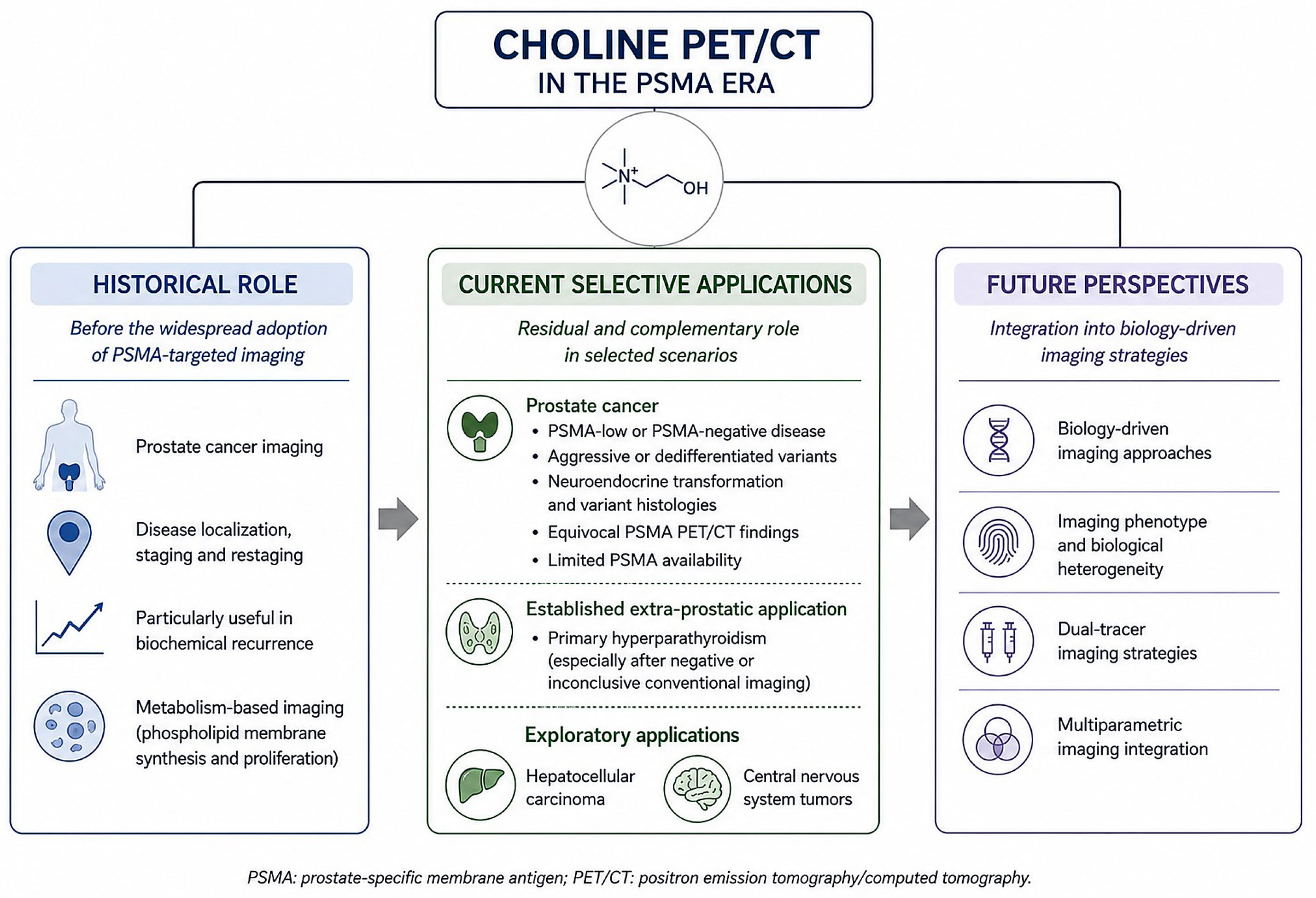
The progressive clinical and biological repositioning of choline PET/CT in the PSMA era is schematically illustrated in
Figure 1.
2. From Choline to PSMA: A Paradigm Shift
The clinical transition from choline PET/CT to PSMA-targeted imaging represents one of the most significant changes in prostate cancer molecular imaging. While choline PET/CT provided whole-body metabolic information and played an important role in the pre-PSMA era, particularly in biochemical recurrence, its diagnostic limitations became increasingly evident in patients with low PSA values and small-volume disease [
4,
5,
6].
PSMA PET/CT has substantially changed this scenario by providing higher lesion detectability, improved tumor-to-background contrast, and greater sensitivity for nodal and distant metastatic disease [
1,
2,
3]. These advantages explain why PSMA-targeted imaging has rapidly become the preferred molecular imaging approach in most prostate cancer settings [
8].
However, this paradigm shift should not be reduced to a simple hierarchy of tracer performance. Choline and PSMA tracers interrogate different biological processes: choline uptake reflects membrane metabolism and cellular proliferation [
4,
7], whereas PSMA-targeted tracers reflect receptor expression [
8]. These biological pathways may not evolve in parallel, particularly in advanced, heterogeneous, or treatment-resistant disease [
9,
10].
Accordingly, the relevant question in the contemporary PSMA era is no longer whether choline PET/CT can compete with PSMA PET/CT in routine prostate cancer imaging. Rather, it is whether choline PET/CT may still provide complementary metabolic information in selected clinical and biological contexts, especially when receptor-targeted imaging does not fully reflect disease phenotype.
3. Residual Role of Choline PET/CT in Prostate Cancer
Although PSMA-targeted PET imaging has become the dominant molecular imaging approach in prostate cancer, receptor-targeted imaging alone may not fully capture the biological complexity of selected advanced or treatment-resistant disease. PSMA PET/CT has demonstrated superior diagnostic performance in most standard clinical scenarios and is now incorporated into international recommendations and standardized reporting systems [
1,
2,
3,
8,
14,
15]. Nevertheless, increasing evidence suggests that PSMA expression may be heterogeneous, particularly in advanced, aggressive, or treatment-resistant prostate cancer [
9,
10]. In this context, the potential residual role of choline PET/CT should be interpreted as complementary and highly selective, rather than as an alternative to PSMA-targeted imaging in routine clinical practice.
The residual and emerging applications of choline PET/CT discussed in this review, together with their underlying biological rationale and relative strength of evidence, are summarized in
Table 1.
Although PSMA PET/CT represents the dominant molecular imaging modality in prostate cancer, it has important limitations that should be acknowledged. PSMA expression may be low, heterogeneous, or absent in selected disease phenotypes, particularly in advanced, dedifferentiated, neuroendocrine, or treatment-resistant prostate cancer. In addition, false-negative findings may occur in small-volume disease below PET spatial resolution, while equivocal uptake related to benign, inflammatory, traumatic, ganglionic, or treatment-related changes may reduce diagnostic confidence. Therefore, PSMA PET/CT should not be considered a universal representation of prostate cancer biology. In these settings, complementary imaging approaches may be useful, but their clinical role depends on the specific biological question: FDG PET/CT is the more established metabolic tracer for aggressive or dedifferentiated disease, whereas choline PET/CT has a more limited and selective role in prostate cancer.
3.1. PSMA-Negative or Low-PSMA Disease
Despite the excellent diagnostic performance of PSMA-targeted imaging, a subset of prostate cancers may demonstrate low, heterogeneous, or absent PSMA expression, particularly in advanced, aggressive, or treatment-resistant disease [
9,
10]. However, PSMA-negative or non-diagnostic findings should not be interpreted as a single biological entity. They may reflect true low or absent PSMA expression, tumor dedifferentiation, neuroendocrine transformation, inter- and intra-lesion heterogeneity, small-volume disease below the spatial resolution of PET imaging, or technical and interpretative factors.
The prevalence of clinically relevant PSMA-negative disease varies according to disease stage, patient selection, imaging criteria, and the tracer used. In advanced metastatic castration-resistant prostate cancer, discordant imaging phenotypes, particularly FDG-positive/PSMA-negative lesions, have been associated with aggressive biology and poorer outcomes. However, most of the available evidence concerns FDG/PSMA discordance rather than choline/PSMA discordance and cannot be directly extrapolated to support a routine role for choline PET/CT.
Unlike PSMA-targeted tracers, choline uptake reflects phospholipid membrane synthesis and cellular proliferation rather than receptor expression [
4,
7]. On this basis, choline PET/CT may theoretically provide complementary metabolic information in selected patients with persistent clinical suspicion and negative or non-diagnostic PSMA PET/CT findings. Nevertheless, this potential role is currently supported mainly by biological rationale and limited indirect evidence. Robust prospective studies specifically evaluating choline PET/CT in PSMA-negative or PSMA-low disease are lacking.
Therefore, choline PET/CT should not be proposed as a routine diagnostic step after negative PSMA PET/CT. Its use in this setting should be considered exploratory and limited to highly selected cases, ideally within multidisciplinary discussion or research-oriented imaging strategies.
3.2. Aggressive and Dedifferentiated Variants
Prostate cancer encompasses a broad biological spectrum, ranging from indolent androgen-sensitive disease to highly aggressive and poorly differentiated phenotypes. As tumor biology evolves, imaging characteristics may also change substantially.
Aggressive or dedifferentiated variants may exhibit heterogeneous or reduced PSMA uptake despite clinically significant disease burden [
9,
10]. In this setting, metabolism-based imaging may provide information that is not entirely captured by receptor-targeted tracers. Although most available evidence concerns FDG imaging rather than choline PET/CT, the same conceptual framework supports the potential value of complementary metabolic imaging in biologically aggressive disease [
9].
Accordingly, choline PET/CT may be considered a potential adjunctive imaging tool in selected cases characterized by discordance between clinical aggressiveness and PSMA-targeted imaging findings. Nevertheless, available evidence remains limited, and this role should currently be regarded as exploratory.
3.3. Neuroendocrine Transformation and Androgen-Independent Disease
Neuroendocrine transformation represents one of the most aggressive forms of prostate cancer progression and is frequently associated with reduced androgen receptor signaling and heterogeneous or low PSMA expression [
9,
10]. From an imaging perspective, this phenotype may result in reduced sensitivity of PSMA-targeted imaging and greater reliance on phenotype-oriented or metabolism-based imaging approaches.
Because choline uptake reflects membrane metabolism rather than receptor expression, choline PET/CT may theoretically provide complementary information in selected PSMA-low or PSMA-negative lesions [
4,
7]. However, evidence specifically supporting choline PET/CT in neuroendocrine prostate cancer remains scarce. Therefore, this potential application should currently be interpreted primarily as a biological rationale rather than as a validated clinical indication.
3.4. Equivocal PSMA Findings and Problem-Solving Scenarios
Equivocal PSMA findings remain a relevant issue in clinical practice. Mild or nonspecific uptake, inflammatory conditions, benign lesions, treatment-related changes, ganglia, fractures, and atypical lesion distribution may occasionally complicate image interpretation and reduce diagnostic confidence [
8,
14,
15,
16,
17]. Standardized reporting systems, such as E-PSMA and PSMA-RADS, have been developed to improve interpretation and communication of uncertain findings [
14,
15].
In highly selected cases, choline PET/CT may provide complementary metabolic information as a problem-solving tool. Discordant findings between PSMA-targeted and metabolism-based imaging may help refine interpretation, particularly in patients with biochemical progression, aggressive clinical features, or persistent clinical suspicion despite non-definitive PSMA PET/CT findings. However, the use of choline PET/CT in this setting should be individualized and cannot currently be considered a routine diagnostic strategy.
3.5. Limited PSMA Availability and Regulatory Issues
Despite the rapid global expansion of PSMA-targeted imaging and its incorporation into contemporary guidelines and procedure standards, access to PSMA PET/CT may remain heterogeneous across countries and healthcare systems [
3,
8]. Regulatory approval, reimbursement policies, tracer production, local infrastructure, and economic constraints may influence its implementation in routine clinical practice.
In several institutions, choline PET/CT may still represent a pragmatic imaging option, particularly in healthcare systems where access to PSMA-targeted imaging is limited or not consistently available. This residual role should not be interpreted as evidence of biological superiority, but rather as a real-world indication driven by logistical, regulatory, and economic factors.
3.6. FDG Versus Choline PET/CT as Complementary Metabolic Imaging
When discussing complementary metabolic imaging in the PSMA era, FDG PET/CT represents the most established comparator, particularly in advanced, aggressive, dedifferentiated, or neuroendocrine prostate cancer [
9,
18,
19]. FDG uptake reflects glucose metabolism and is more frequently associated with aggressive tumor biology, treatment resistance, and poorer prognosis. In metastatic castration-resistant prostate cancer, FDG-positive/PSMA-negative lesions have been increasingly recognized as a clinically relevant discordant phenotype and may identify patients with biologically aggressive disease [
9,
10]. Consistently, the TheraP trial required both PSMA PET/CT and FDG PET/CT for eligibility assessment and excluded patients with discordant FDG-positive/PSMA-negative lesions, supporting the clinical relevance of FDG as a complementary metabolic tracer in advanced prostate cancer [
18].
By contrast, choline PET/CT reflects phospholipid membrane synthesis and cellular proliferation rather than glycolytic metabolism [
4,
7]. Historically, this mechanism supported the use of choline PET/CT in biochemical recurrence and restaging of prostate cancer. However, in the contemporary PSMA era, choline PET/CT has substantially less evidence than FDG PET/CT for the characterization of aggressive PSMA-negative or dedifferentiated disease. Therefore, choline PET/CT should not be considered interchangeable with FDG PET/CT in this setting.
In practical terms, FDG PET/CT may be more appropriate when the clinical question concerns aggressive biology, suspected dedifferentiation, neuroendocrine transformation, or discordant progression despite low or heterogeneous PSMA expression [
9,
10]. Choline PET/CT may provide complementary information only in selected circumstances, particularly where prior choline-based imaging is available, where local tracer availability influences imaging pathways, or when membrane-metabolism imaging is considered relevant within a multidisciplinary assessment. Nevertheless, this remains a selective and insufficiently validated application in prostate cancer.
The distinction is different outside prostate cancer. In primary hyperparathyroidism,
18F-fluorocholine PET/CT has a specific and clinically established role for the localization of hyperfunctioning parathyroid tissue, whereas FDG PET/CT is not considered a relevant competing tracer for this indication [
11,
12,
13].
4. Beyond Prostate Cancer
Although prostate cancer historically represented the principal field of application for choline PET/CT, the inclusion of extra-prostatic applications in this review is intended to clarify how choline PET/CT has been clinically repositioned beyond its original onco>logic use. These indications are not equivalent in terms of evidence or clinical maturity. Primary hyperparathyroidism is discussed because it currently represents the most established and evidence-supported extra-prostatic application of
18F-fluorocholine PET/CT [
11,
12,
13,
20,
21,
22,
23,
24,
25,
26]. Hepatocellular carcinoma, by contrast, is included as an exploratory oncologic model in which FDG and choline uptake may reflect different aspects of tumor differentiation and metabolic heterogeneity [
27,
28,
29,
30,
31,
32,
33].
4.1. Hyperparathyroidism
Primary hyperparathyroidism has become one of the most relevant non-oncologic applications of choline PET/CT. Accurate preoperative localization of hyperfunctioning parathyroid tissue is essential to guide focused or minimally invasive parathyroidectomy, reduce operative time, and increase the likelihood of successful surgical treatment. Conventional imaging usually relies on neck ultrasonography and
99mTc-sestamibi scintigraphy, often combined with SPECT/CT; however, these techniques may show limited performance in patients with small adenomas, ectopic glands, multiglandular disease, coexisting thyroid abnormalities, previous neck surgery, or discordant imaging findings [
18,
21,
23].
In this context,
18F-fluorocholine PET/CT has demonstrated excellent diagnostic performance for the localization of hyperfunctioning parathyroid tissue. In a prospective study including 103 patients with primary hyperparathyroidism, Cuderman et al. reported a sensitivity of 92% for
18F-fluorocholine PET/CT, compared with 39–56% for individual conventional scintigraphic methods and 65% for combined conventional imaging. In patients with multiple hyperfunctioning glands,
18F-fluorocholine PET/CT showed a sensitivity of 88%, whereas conventional imaging showed substantially lower sensitivity, ranging from 22% to 34% for individual techniques and 44% when combined [
11].
Similarly, in a prospective dual-center study of 100 patients, Beheshti et al. reported a patient-based detection rate of 93% for
18F-fluorocholine PET/CT compared with 61% for
99mTc-MIBI/tetrofosmin SPECT/CT. In lesion-based analysis,
18F-fluorocholine PET/CT showed sensitivity, specificity, positive predictive value, negative predictive value, and accuracy of 93.7%, 96.0%, 90.2%, 97.4%, and 95.3%, respectively, compared with 60.8%, 98.5%, 94.1%, 86.3%, and 87.7% for conventional SPECT/CT imaging [
19].
The added value of
18F-fluorocholine PET/CT is particularly evident in patients with negative or inconclusive conventional imaging and in challenging pre-surgical localization scenarios. Grimaldi et al. specifically highlighted the clinical utility of
18F-fluorocholine PET/CT in difficult cases of primary hyperparathyroidism, supporting its role as a problem-solving tool when conventional imaging is insufficient [
20]. In the prospective APACH1 study, which enrolled patients with primary hyperparathyroidism and negative or inconclusive cervical ultrasound and
99mTc-sestamibi SPECT/CT,
18F-fluorocholine PET/CT achieved per-lesion and per-patient sensitivities of 91.3% and 90.5%, respectively, with corresponding positive predictive values of 87.5% and 86.4%. Moreover,
18F-fluorocholine PET/CT guided surgery in 88% of patients and allowed bilateral cervical exploration to be avoided in the majority of cases [
22].
These findings are supported by systematic reviews and meta-analyses reporting consistently high diagnostic performance of choline PET imaging for the detection of hyperfunctioning parathyroid glands [
12,
13]. The high spatial resolution of PET imaging, favorable lesion-to-background contrast, and relatively short acquisition protocols represent important technical and practical advantages of
18F-fluorocholine PET/CT [
11,
13,
20,
21,
23].
Current international recommendations generally position
18F-fluorocholine PET/CT as a second-line or problem-solving modality after negative or inconclusive conventional imaging [
21,
23]. However, increasing evidence and expert opinion support its potential use as a first-line imaging modality in selected centers with appropriate expertise, particularly when minimally invasive surgery is planned [
12,
13,
23]. Therefore, primary hyperparathyroidism represents the most clinically established and evidence-supported extra-prostatic application of
18F-fluorocholine PET/CT and provides a paradigmatic example of how choline-based imaging has evolved from a predominantly oncologic imaging modality into a multidisciplinary molecular imaging tool with established clinical utility beyond prostate cancer [
12,
21,
23].
4.2. Hepatocellular Carcinoma
Hepatocellular carcinoma (HCC) is characterized by marked biological and metabolic heterogeneity, which may partly explain the variable performance of different PET tracers. FDG PET/CT generally shows higher uptake in poorly differentiated and biologically aggressive tumors, whereas choline PET/CT tends to show higher uptake in well- and moderately differentiated HCC, reflecting differences in glucose metabolism and membrane lipid synthesis [
27,
28,
29,
30].
Early comparative studies supported the complementary value of these tracers. Yamamoto et al. reported that
11C-choline PET detected HCC lesions with higher sensitivity than FDG PET, particularly in well-differentiated tumors [
27]. In another prospective study including patients with chronic liver disease and suspected HCC, the sensitivity for HCC detection was 75% for
18F-fluorocholine PET/CT, 36% for FDG PET/CT, and 93% when both tracers were combined, suggesting that dual-tracer imaging may improve lesion detection compared with either tracer alone [
29].
However, the relationship between tracer uptake and tumor differentiation is not absolute. In a systematic review of dual
18F-FDG/
18F-choline PET behavior in histology-proven HCC, Ghidaglia et al. included 6 studies comprising 99 HCC lesions. Among well-differentiated HCCs, 51% were positive only for
18F-choline and 39% were positive for both
18F-FDG and
18F-choline. Among less-differentiated tumors, 37% were positive only for FDG, 36% were positive for both tracers, and 25% were positive only for
18F-choline. These data indicate substantial overlap between uptake patterns and histological differentiation, limiting the use of dual-tracer PET as a stand-alone surrogate marker of tumor differentiation [
28].
More recently, Sönmez et al. evaluated the clinical utility of dual
18F-FDG/
18F-choline PET/CT in a tertiary-center cohort of 168 HCC patients. Dual PET/CT findings increased the BCLC stage in 21 patients (12.5%) and modified the treatment strategy in 41 patients (24.4%). In addition, combined PET parameters and alpha-fetoprotein levels were associated with disease-free survival, suggesting a potential role for dual-tracer PET/CT in refining risk stratification and treatment planning in selected patients [
30].
Overall, these findings support the concept that FDG and choline PET/CT may provide complementary biological information in HCC. Nevertheless, the available evidence remains limited, heterogeneous, and insufficient to support routine clinical use. Therefore, choline PET/CT in HCC should be regarded as an exploratory and selective tool for biological characterization and multiparametric assessment rather than as a standard imaging modality.
4.3. Central Nervous System Tumors
In neuro-oncology, choline PET/CT has mainly been investigated in small and selected studies as a complementary imaging modality integrated with MRI, rather than as a standard imaging approach. Because choline uptake reflects membrane turnover and cellular proliferation, it may provide additional metabolic information in selected brain tumors and in challenging post-treatment settings [
7,
31,
32,
33].
One of the most relevant potential applications is the differentiation between tumor recurrence and radiation necrosis in previously treated gliomas or brain tumors, where conventional MRI may be inconclusive. Gao et al. reported the diagnostic accuracy of
11C-choline PET in differentiating glioma recurrence from radiation necrosis in a systematic review and meta-analysis [
32]. Data from Takenaka et al. also supported the potential utility of
11C-choline PET for differentiating recurrent glioma from radiation necrosis after radiotherapy, although
11C-methionine PET showed superior diagnostic performance [
33].
Choline PET/CT has also been explored for the characterization of selected intracranial lesions, including MRI-suspected low-grade gliomas, although available evidence remains limited and is generally based on small studies or heterogeneous patient populations [
31,
32,
33].
Nevertheless, amino acid PET tracers remain the preferred molecular imaging agents in several neuro-oncologic settings because of their more favorable physiological brain background and stronger evidence base, as reflected by joint EANM/EANO/RANO/SNMMI practice guidelines for PET imaging of gliomas [
34]. Consequently, choline PET/CT should not be regarded as a primary neuro-oncologic imaging modality, but rather as an adjunctive and highly selective tool when alternative tracers are unavailable or when additional metabolic information may be clinically useful.
4.4. Other Emerging Indications
Additional exploratory applications of choline PET/CT have been described in selected malignancies and incidental findings, further supporting the concept of choline imaging as a flexible metabolism-based imaging modality. However, most of these indications remain supported only by limited evidence, preliminary experiences, or isolated clinical reports.
Overall, the diversification of molecular imaging strategies suggests that future imaging pathways may progressively evolve toward multiparametric and biology-oriented approaches. Within this framework, choline PET/CT may retain value not as a universal tracer, but as a selective modality capable of providing complementary metabolic information in specific clinical and biological contexts.
5. Biological and Metabolic Perspectives
The transition from choline PET/CT to PSMA-targeted imaging reflects a broader evolution in oncologic molecular imaging, moving from metabolism-based approaches toward receptor-targeted and phenotype-oriented imaging strategies [
1,
7,
8,
35,
36,
37,
38,
39].
Radiolabeled choline tracers primarily reflect phospholipid membrane synthesis and cellular proliferation [
4,
7], whereas PSMA-targeted tracers evaluate receptor expression [
8]. Consequently, metabolism-based imaging and receptor-targeted imaging may provide partially different information regarding tumor phenotype and disease biology.
This concept is particularly relevant in advanced and biologically heterogeneous prostate cancer, where receptor expression, metabolic activity, dedifferentiation, and treatment-related changes may not evolve in parallel [
9,
10,
35,
36]. In this setting, discordant uptake patterns across different tracers may reflect underlying biological heterogeneity rather than simple technical differences.
The growing recognition of imaging heterogeneity has contributed to the concept of an “imaging phenotype”, whereby different radiopharmaceuticals may capture distinct molecular, metabolic, or functional characteristics of the same disease. Although this approach remains under investigation, it is increasingly relevant within precision oncology, where imaging is expected not only to localize disease but also to contribute to biological characterization and treatment selection [
35,
36,
37,
38,
39].
Within this evolving framework, choline PET/CT should not be interpreted as competing with PSMA PET/CT, but rather as a potential source of complementary metabolic information in selected clinical scenarios. This concept is consistent with other oncologic models in which multiple tracers are used to characterize different aspects of tumor biology, such as the complementary role of FDG and choline imaging in hepatocellular carcinoma [
27,
28,
29,
30].
Overall, the PSMA era has reinforced the importance of biology-driven imaging rather than eliminating the value of metabolism-based tracers. The future role of choline PET/CT will likely depend on its ability to contribute selectively to multiparametric imaging strategies in patients whose disease biology is not fully represented by receptor-targeted imaging alone.
6. Future Perspectives
Dual-tracer approaches combining receptor-targeted and metabolism-based radiopharmaceuticals may help characterize tumor heterogeneity and identify aggressive or biologically discordant disease phenotypes [
9,
10,
35,
36]. This concept is also supported by other oncologic models, such as hepatocellular carcinoma, in which FDG and choline PET/CT may provide complementary information on tumor biology [
27,
28,
29,
30]. In prostate cancer, this approach may be particularly relevant in advanced or treatment-resistant disease, where receptor expression and metabolic activity may not evolve in parallel [
9,
10,
35,
36].
Beyond visual interpretation, quantitative imaging approaches may further refine the clinical value of PET/CT. Radiomics and advanced computational tools may support the extraction of imaging features related to tumor heterogeneity, tracer distribution, and treatment response, potentially contributing to more personalized diagnostic and therapeutic pathways [
37,
38,
39].
However, these approaches remain investigational and require prospective validation, methodological standardization, and harmonization of imaging protocols before routine clinical implementation.
Future molecular imaging strategies will likely become increasingly phenotype-oriented and multiparametric, integrating metabolic, receptor-targeted, and functional information according to the specific clinical question, disease stage, and therapeutic context.
7. Conclusions
The widespread adoption of PSMA-targeted imaging has profoundly reshaped the role of choline PET/CT in contemporary molecular imaging. Although PSMA PET/CT has become the dominant imaging modality in prostate cancer because of its superior diagnostic performance, the transition from choline to PSMA imaging should not be interpreted simply as the replacement of one tracer by another.
Increasing evidence highlights the biological heterogeneity of advanced prostate cancer and the potential limitations of relying exclusively on receptor-targeted imaging. In selected scenarios, including PSMA-low or heterogeneous disease, aggressive variants, neuroendocrine transformation, equivocal PSMA findings, and limited tracer availability, choline PET/CT may still provide complementary metabolic information. However, these applications remain selective and are currently supported mainly by biological rationale, indirect evidence, and limited clinical data.
At the same time, 18F-fluorocholine PET/CT has emerged as a clinically relevant imaging modality in primary hyperparathyroidism, where evidence supporting its diagnostic performance is substantially stronger and increasingly consolidated.
Overall, the PSMA era has not eliminated the role of choline PET/CT, but rather contributed to its clinical and biological repositioning. Future molecular imaging strategies will likely become increasingly biology-driven and multiparametric, integrating receptor-targeted, metabolic, and functional information to better reflect disease complexity and heterogeneity.
Author Contributions
Conceptualization, V.R., P.C. and F.M.; methodology, V.R.; validation, V.R. and P.C.; formal analysis, V.R., M.S., L.F., I.M., M.C.; investigation, S.N., I.M., L.F., M.C., I.G.; data curation, F.M.; writing—original draft preparation, F.M.; writing—review and editing, V.R., F.M. and P.C.; visualization, V.R., L.F., I.M., M.C.,M.S., S.N., I.G.; supervision, P.C. All authors have read and agreed to the published version of the manuscript.
Funding
This research received no external funding.
Institutional Review Board Statement
Not applicable.
Informed Consent Statement
Not applicable.
Data Availability Statement
No new data were created or analyzed in this study. Data sharing is not applicable to this article.
Acknowledgments
During the preparation of this manuscript, the authors used ChatGPT 5.5, developed by OpenAI, for language editing, editorial refinement, and support in the preparation of the graphical summary. The tool was not used to generate original scientific data, perform analyses, select references autonomously, or draw conclusions. The authors reviewed, revised, and edited all AI-assisted output and take full responsibility for the content of this publication.
Conflicts of Interest
The authors declare no conflicts of interest.
References
- Panagiotidis, E.; Fragkiadaki, V.; Papathanasiou, N.; Kypraios, C.; Liatsikos, E.; Klampatsas, A.; Paschali, A.; Exarhos, D.; Zarokosta, F.; Chatzipavlidou, V.; et al. Comparison of 18F-PSMA-1007 and 18F-choline PET/CT in prostate cancer patients with biochemical recurrence: A phase 3, prospective, multicenter, randomized study. Nucl. Med. Commun. 2023, 44, 1126–1134. [Google Scholar] [CrossRef] [PubMed]
- García-Zoghby, L.; Lucas-Lucas, C.; Amo-Salas, M.; Soriano-Castrejón, Á.M.; García-Vicente, A.M. Head-to-head comparison of [18F]F-choline and imaging of prostate-specific membrane antigen, using [18F]DCFPyL PET/CT, in patients with biochemical recurrence of prostate cancer. Curr. Oncol. 2023, 30, 6271–6288. [Google Scholar] [CrossRef] [PubMed]
- Tilki, D.; van den Bergh, R.C.N.; Briers, E.; Van den Broeck, T.; Brunckhorst, O.; Darraugh, J.; Eberli, D.; De Meerleer, G.; De Santis, M.; Farolfi, A.; et al. EAU-EANM-ESTRO-ESUR-ISUP-SIOG Guidelines on Prostate Cancer. Part II—2024 update: Treatment of relapsing and metastatic prostate cancer. Eur. Urol. 2024, 86, 164–182. [Google Scholar] [CrossRef] [PubMed]
- Jadvar, H. Prostate cancer: PET with 18F-FDG, 18F- or 11C-acetate, and 18F- or 11C-choline. J. Nucl. Med. 2011, 52, 81–89. [Google Scholar] [CrossRef] [PubMed]
- Umbehr, M.H.; Müntener, M.; Hany, T.; Sulser, T.; Bachmann, L.M. The role of 11C-choline and 18F-fluorocholine positron emission tomography (PET) and PET/CT in prostate cancer: A systematic review and meta-analysis. Eur. Urol. 2013, 64, 106–117. [Google Scholar] [CrossRef] [PubMed]
- Evangelista, L.; Zattoni, F.; Guttilla, A.; Saladini, G.; Colletti, P.M.; Rubello, D. Choline PET or PET/CT and biochemical relapse of prostate cancer: A systematic review and meta-analysis. Clin. Nucl. Med. 2013, 38, 305–314. [Google Scholar] [CrossRef] [PubMed]
- Glunde, K.; Bhujwalla, Z.M.; Ronen, S.M. Choline metabolism in malignant transformation. Nat. Rev. Cancer 2011, 11, 835–848. [Google Scholar] [CrossRef] [PubMed]
- Fendler, W.P.; Eiber, M.; Beheshti, M.; Bomanji, J.; Calais, J.; Ceci, F.; Cho, S.; Fanti, S.; Giesel, F.L.; Goffin, K.; et al. PSMA PET/CT: Joint EANM procedure guideline/SNMMI procedure standard for prostate cancer imaging 2.0. Eur. J. Nucl. Med. Mol. Imaging 2023, 50, 1466–1486. [Google Scholar] [CrossRef] [PubMed]
- Jadvar, H. The VISION forward: Recognition and implication of PSMA−/18F-FDG+ metastatic castration-resistant prostate cancer. J. Nucl. Med. 2022, 63, 812–815. [Google Scholar] [CrossRef] [PubMed]
- Bakht, M.K.; Derecichei, I.; Li, Y.; Ferraiuolo, R.M.; Dunning, M.; Oh, S.W.; Hussein, A.; Youn, H.; Stringer, K.F.; Jeong, C.W.; et al. Neuroendocrine differentiation of prostate cancer leads to PSMA suppression. Endocr. Relat. Cancer. 2019, 26, 131–146. [Google Scholar] [CrossRef] [PubMed]
- Cuderman, A.; Senica, K.; Rep, S.; Hocevar, M.; Kocjan, T.; Sever, M.J.; Zaletel, K.; Lezaic, L. 18F-fluorocholine PET/CT in primary hyperparathyroidism: Superior diagnostic performance to conventional scintigraphic imaging for localization of hyperfunctioning parathyroid glands. J. Nucl. Med. 2020, 61, 577–583. [Google Scholar] [CrossRef] [PubMed]
- Giovanella, L.; Bacigalupo, L.; Treglia, G.; Piccardo, A. Will 18F-fluorocholine PET/CT replace other methods of preoperative parathyroid imaging? Endocrine 2021, 71, 285–297. [Google Scholar] [CrossRef] [PubMed]
- Treglia, G.; Piccardo, A.; Imperiale, A.; Strobel, K.; Kaufmann, P.A.; Prior, J.O.; Giovanella, L. Diagnostic performance of choline PET for detection of hyperfunctioning parathyroid glands in hyperparathyroidism: A systematic review and meta-analysis. Eur. J. Nucl. Med. Mol. Imaging 2019, 46, 751–765. [Google Scholar] [CrossRef] [PubMed]
- Ceci, F.; Oprea-Lager, D.E.; Emmett, L.; Adam, J.A.; Bomanji, J.; Czernin, J.; Eiber, M.; Haberkorn, U.; Hofman, M.S.; Hope, T.A.; et al. E-PSMA: The EANM standardized reporting guidelines v1.0 for PSMA-PET. Eur. J. Nucl. Med. Mol. Imaging 2021, 48, 1626–1638. [Google Scholar] [CrossRef] [PubMed]
- Rowe, S.P.; Pienta, K.J.; Pomper, M.G.; Gorin, M.A. PSMA-RADS version 1.0: A step towards standardizing the interpretation and reporting of PSMA-targeted PET imaging studies. Eur. Urol. 2018, 73, 485–487. [Google Scholar] [CrossRef] [PubMed]
- Shetty, D.; Patel, D.; Le, K.; Bui, C.; Mansberg, R. Pitfalls in gallium-68 PSMA PET/CT interpretation: A pictorial review. Tomography 2018, 4, 182–193. [Google Scholar] [CrossRef] [PubMed]
- Kuten, J.; Sarid, D.; Yossepowitch, O.; Mabjeesh, N.J.; Even-Sapir, E. The significance of equivocal bone findings in staging PSMA imaging in the preoperative setting: Validation of the PSMA-RADS version 1.0. EJNMMI Res. 2021, 11, 27. [Google Scholar] [CrossRef] [PubMed]
- Johnson, N.A.; Tublin, M.E.; Ogilvie, J.B. Parathyroid imaging: Technique and role in the preoperative evaluation of primary hyperparathyroidism. AJR Am. J. Roentgenol. 2007, 188, 1706–1715. [Google Scholar] [CrossRef] [PubMed]
- Beheshti, M.; Hehenwarter, L.; Paymani, Z.; Rendl, G.; Imamovic, L.; Rettenbacher, R.; Tsybrovskyy, O.; Langsteger, W.; Pirich, C. 18F-fluorocholine PET/CT in the assessment of primary hyperparathyroidism compared with 99mTc-MIBI or 99mTc-tetrofosmin SPECT/CT: A prospective dual-centre study in 100 patients. Eur. J. Nucl. Med. Mol. Imaging 2018, 45, 1762–1771. [Google Scholar] [CrossRef] [PubMed]
- Grimaldi, S.; Young, J.; Kamenicky, P.; Hartl, D.; Terroir, M.; Leboulleux, S.; Berdelou, A.; Hadoux, J.; Hescot, S.; Remy, H.; et al. Challenging pre-surgical localization of hyperfunctioning parathyroid glands in primary hyperparathyroidism: The added value of 18F-fluorocholine PET/CT. Eur. J. Nucl. Med. Mol. Imaging 2018, 45, 1772–1780. [Google Scholar] [CrossRef] [PubMed]
- Noskovicova, L.; Balogova, S.; Aveline, C.; Tassart, M.; Zhang-Yin, J.; Kerrou, K.; Jaksic, I.; Montravers, F.; Talbot, J.-N. 18F-fluorocholine-positron emission tomography/computerized tomography (FCH PET/CT) imaging for detecting abnormal parathyroid glands: Indication, practice, interpretation and diagnostic performance. Semin. Nucl. Med. 2024, 54, 875–895. [Google Scholar] [CrossRef] [PubMed]
- Quak, E.; Blanchard, D.; Houdu, B.; Le Roux, Y.; Ciappuccini, R.; Lireux, B.; de Raucourt, D.; Grellard, J.-M.; Licaj, I.; Bardet, S.; et al. F18-choline PET/CT guided surgery in primary hyperparathyroidism when ultrasound and MIBI SPECT/CT are negative or inconclusive: The APACH1 study. Eur. J. Nucl. Med. Mol. Imaging 2018, 45, 658–666. [Google Scholar] [CrossRef] [PubMed]
- Petranović Ovčariček, P.; Giovanella, L.; Carrió Gasset, I.; Hindié, E.; Huellner, M.W.; Luster, M.; Piccardo, A.; Weber, T.; Talbot, J.-N.; Verburg, F.A. The EANM practice guidelines for parathyroid imaging. Eur. J. Nucl. Med. Mol. Imaging 2021, 48, 2801–2822. [Google Scholar] [CrossRef] [PubMed]
- Piccardo, A.; Trimboli, P.; Rutigliani, M.; Puntoni, M.; Foppiani, L.; Bacigalupo, L.; Crescenzi, A.; Bottoni, G.; Treglia, G.; Paparo, F.; et al. Additional value of integrated 18F-choline PET/4D contrast-enhanced CT in the localization of hyperfunctioning parathyroid glands and correlation with molecular profile. Eur. J. Nucl. Med. Mol. Imaging 2019, 46, 766–775. [Google Scholar] [CrossRef] [PubMed]
- Hocevar, M.; Lezaic, L.; Rep, S.; Zaletel, K.; Kocjan, T.; Sever, M.J.; Zgajnar, J.; Peric, B. Focused parathyroidectomy without intraoperative parathormone testing is safe after pre-operative localization with 18F-fluorocholine PET/CT. Eur. J. Surg. Oncol. 2017, 43, 133–137. [Google Scholar] [CrossRef] [PubMed]
- Orevi, M.; Freedman, N.; Mishani, E.; Bocher, M.; Jacobson, O.; Krausz, Y. Localization of parathyroid adenoma by 11C-choline PET/CT: Preliminary results. Clin. Nucl. Med. 2014, 39, 1033–1038. [Google Scholar] [CrossRef] [PubMed]
- Yamamoto, Y.; Nishiyama, Y.; Kameyama, R.; Okano, K.; Kashiwagi, H.; Deguchi, A.; Kaji, M.; Ohkawa, M. Detection of hepatocellular carcinoma using 11C-choline PET: Comparison with 18F-FDG PET. J. Nucl. Med. 2008, 49, 1245–1248. [Google Scholar] [CrossRef] [PubMed]
- Ghidaglia, J.; Golse, N.; Pascale, A.; Sebagh, M.; Besson, F.L. 18F-FDG/18F-choline dual-tracer PET behavior and tumor differentiation in hepatocellular carcinoma: A systematic review. Front. Med. 2022, 9, 924824. [Google Scholar] [CrossRef] [PubMed]
- Talbot, J.N.; Fartoux, L.; Balogova, S.; Nataf, V.; Kerrou, K.; Gutman, F.; Huchet, V.; Ancel, D.; Grange, J.-D.; Rosmorduc, O. Detection of hepatocellular carcinoma with PET/CT: A prospective comparison of 18F-fluorocholine and 18F-FDG in patients with cirrhosis or chronic liver disease. J. Nucl. Med. 2010, 51, 1699–1706. [Google Scholar] [CrossRef] [PubMed]
- Sönmez, R.E.; Besson, F.L.; Ghidaglia, J.; Lewin, M.; Gomez, L.; Salloum, C.; Pittau, G.; Ciacio, O.; Allard, M.A.; Cherqui, D.; et al. Towards refining the utility of dual (18F-FDG/18F-choline) PET/CT for the management of hepatocellular carcinoma: A tertiary center study. Q. J. Nucl. Med. Mol. Imaging 2023, 67, 206–214. [Google Scholar] [CrossRef] [PubMed]
- Mišir Krpan, A.; Hodolič, M.; Golubić, A.T.; Baučić, M.; Nemir, J.; Mrak, G.; Žuvić, M.; Huić, D. 18F-FET and 18F-choline PET-CT in patients with MRI-suspected low-grade gliomas: A pilot study. Croat. Med. J. 2021, 62, 310–317. [Google Scholar] [CrossRef] [PubMed]
- Gao, L.; Xu, W.; Li, T.; Zheng, J.; Chen, G. Accuracy of 11C-choline positron emission tomography in differentiating glioma recurrence from radiation necrosis: A systematic review and meta-analysis. Medicine 2018, 97, e11556. [Google Scholar] [CrossRef] [PubMed]
- Takenaka, S.; Asano, Y.; Shinoda, J.; Nomura, Y.; Yonezawa, S.; Miwa, K.; Yano, H.; Iwama, T. Comparison of 11C-methionine, 11C-choline, and 18F-fluorodeoxyglucose-PET for distinguishing glioma recurrence from radiation necrosis. Neurol. Med. Chir. 2014, 54, 280–289. [Google Scholar] [CrossRef] [PubMed]
- Law, I.; Albert, N.L.; Arbizu, J.; Boellaard, R.; Drzezga, A.; Galldiks, N.; la Fougère, C.; Langen, K.-J.; Lopci, E.; Lowe, V.; et al. Joint EANM/EANO/RANO practice guidelines/SNMMI procedure standards for imaging of gliomas using PET with radiolabelled amino acids and [18F]FDG: Version 1.0. Eur. J. Nucl. Med. Mol. Imaging 2019, 46, 540–557. [Google Scholar] [CrossRef] [PubMed]
- Filippi, L.; Frantellizzi, V.; Chiaravalloti, A.; Pontico, M.; De Feo, M.S.; Corica, F.; Montebello, M.; Schillaci, O.; De Vincentis, G.; Bagni, O. Prognostic and theranostic applications of positron emission tomography for a personalized approach to metastatic castration-resistant prostate cancer. Int. J. Mol. Sci. 2021, 22, 3036. [Google Scholar] [CrossRef] [PubMed]
- Vetrone, L.; Fortunati, E.; Castellucci, P.; Fanti, S. Future imaging of prostate cancer: Do we need more than PSMA PET/CT? Semin. Nucl. Med. 2024, 54, 150–162. [Google Scholar] [CrossRef] [PubMed]
- Mayerhoefer, M.E.; Materka, A.; Langs, G.; Häggström, I.; Szczypiński, P.; Gibbs, P.; Cook, G. Introduction to radiomics. J. Nucl. Med. 2020, 61, 488–495. [Google Scholar] [CrossRef] [PubMed]
- Gillies, R.J.; Kinahan, P.E.; Hricak, H. Radiomics: Images are more than pictures, they are data. Radiology 2016, 278, 563–577. [Google Scholar] [CrossRef] [PubMed]
- Lambin, P.; Leijenaar, R.T.H.; Deist, T.M.; Peerlings, J.; de Jong, E.E.C.; van Timmeren, J.; Sanduleanu, S.; Larue, R.T.H.M.; Even, A.J.G.; Jochems, A.; et al. Radiomics: The bridge between medical imaging and personalized medicine. Nat. Rev. Clin. Oncol. 2017, 14, 749–762. [Google Scholar] [CrossRef] [PubMed]
| Disclaimer/Publisher’s Note: The statements, opinions and data contained in all publications are solely those of the individual author(s) and contributor(s) and not of MDPI and/or the editor(s). MDPI and/or the editor(s) disclaim responsibility for any injury to people or property resulting from any ideas, methods, instructions or products referred to in the content. |



 ,
,