Radiocapitellar Alignment in Suspected Pediatric Monteggia Lesions: Narrative Review and Imaging Interpretation Framework


Abstract
1. Introduction
2. Literature Search and Evidence Selection
2.1. Quality Safeguards for Narrative Synthesis
2.2. Use of Generative AI Tools
3. Existing Practice Guidance and Unmet Need
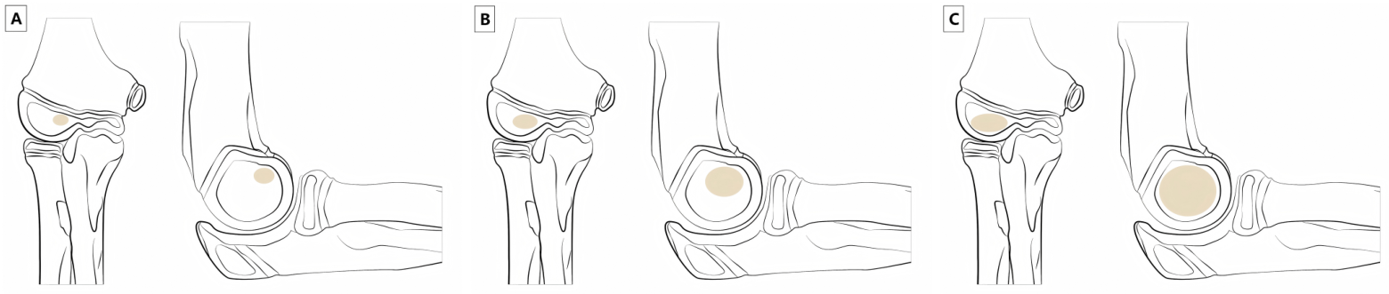
4. Developmental Anatomy and Ossification-Related Pitfalls
4.1. Ossification Sequence: Why Chronological Age Alone Is Insufficient
4.2. Eccentric Capitellar Ossification and Apparent RCL Abnormality
4.3. Diagnostic Implications
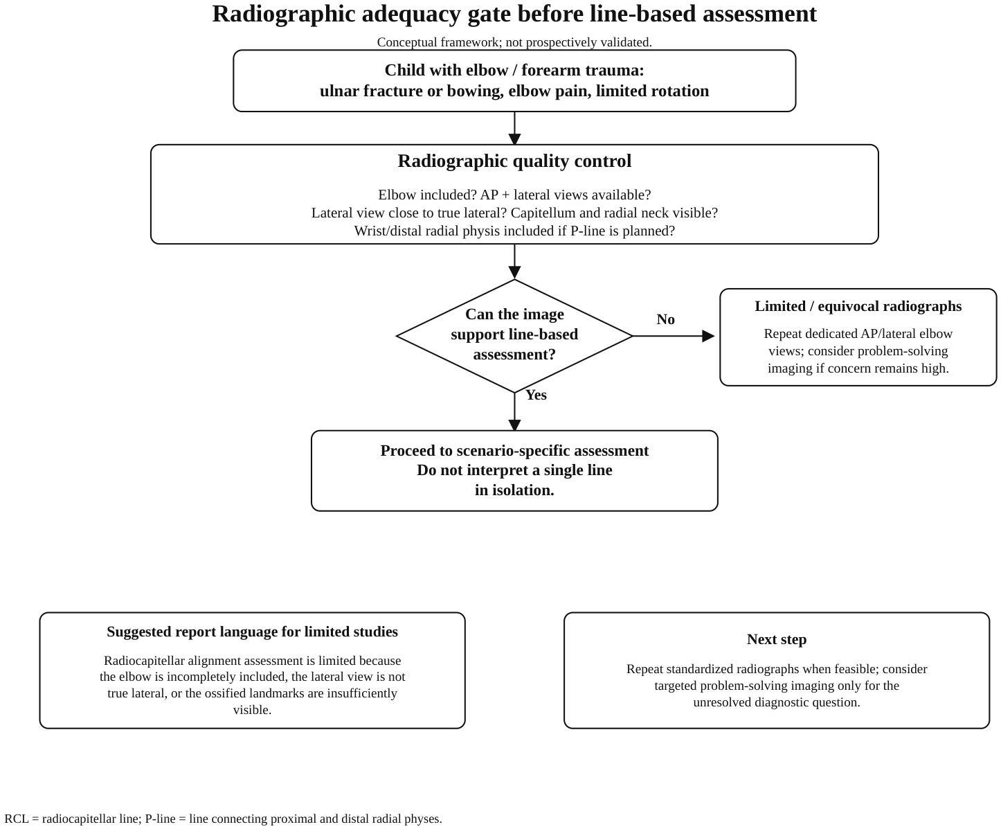
5. Radiographic Quality Control Before Drawing Lines
5.1. Minimum Radiographic Requirements
5.2. Projection, Beam Angle, and Lateral View Quality
5.3. AP Projection and Coronal-Plane Assessment
5.4. When Radiographs Should Be Considered Equivocal
- Elbow included when Monteggia lesion is suspected?
- AP and lateral elbow projections available, or limitation explicitly stated?
- Lateral view close to true lateral?
- Capitellum, radial neck, and radial head sufficiently visible for the intended line/sign assessment?
- Distal radial physis included if P-line assessment is planned?
- Imaging findings concordant with the clinical scenario?
6. Diagnostic Tools by Imaging Scenario
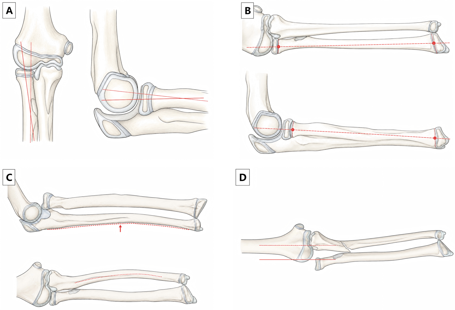
6.1. Standard Lateral Elbow Radiograph: Radial Neck-Axis RCL
6.2. Forearm Radiograph-Only Scenario: P-Line
6.3. AP or Coronal-Plane Concern: LHL and Other Adjunctive Lines
6.4. Ulnar Plastic Deformation: Ulnar Bow Sign
6.5. Equivocal Radiographs: Ultrasound, MRI, and Arthrography
6.6. Differential Considerations and the Role of CT
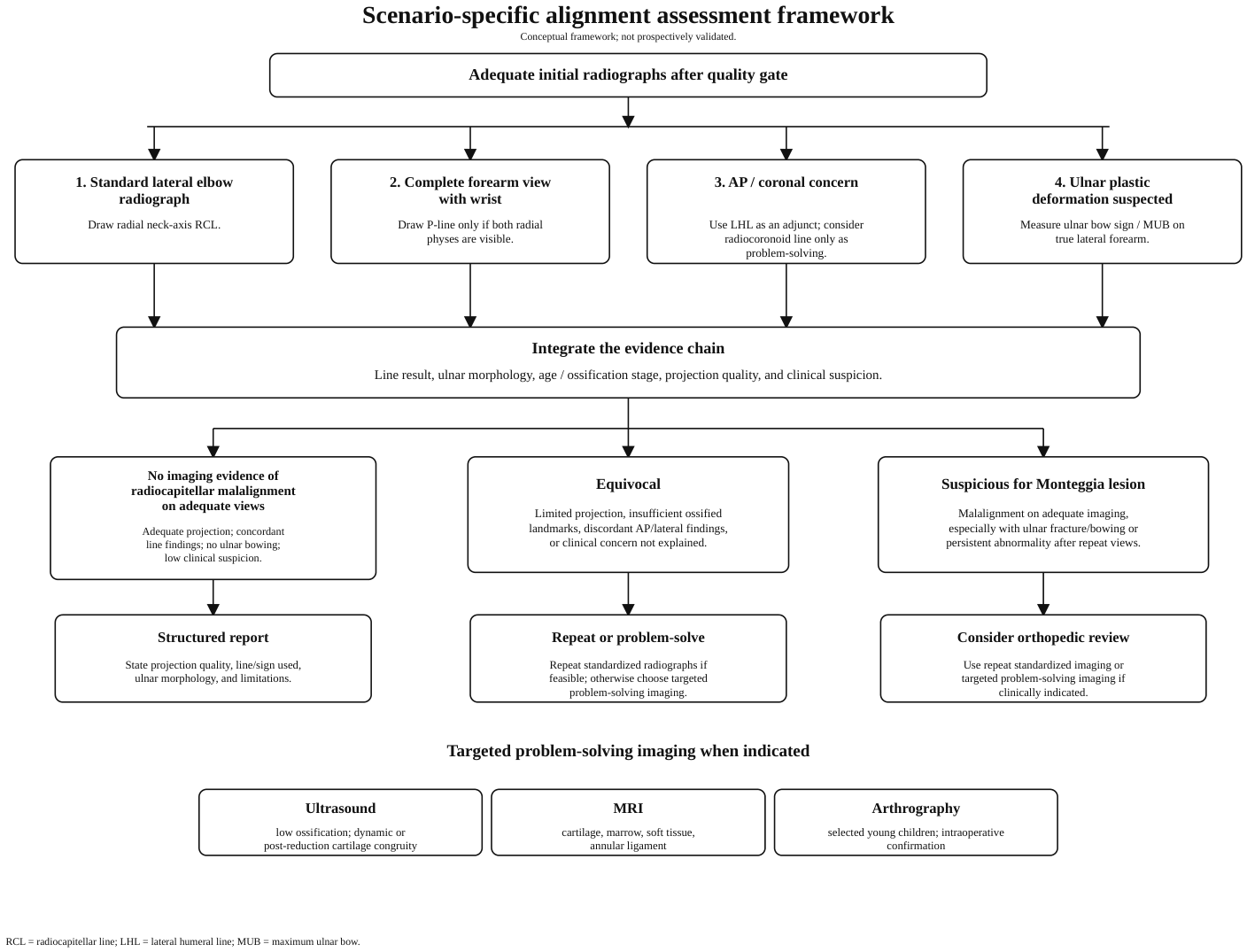
6.7. Practical Hierarchy for Discordant Alignment Findings
7. Diagnostic Errors and Reader Performance
8. Proposed Imaging Interpretation Framework
Operational Definitions for the Proposed Framework
Pitfall checklist. Do not exclude a Monteggia lesion when the elbow is incompletely included, the lateral view is not close to true lateral, ossified landmarks are insufficiently visible, the ulna shows plastic deformation, AP and lateral findings are discordant, or clinical suspicion persists despite apparently maintained alignment. In these settings, report the limitation and consider repeat standardized radiographs or targeted problem-solving imaging according to the unresolved question.
9. Structured Reporting Template
10. Future Directions: Explainable Automated Assessment
11. Discussion
12. Take-Home Messages for Clinical Practice
13. Applicability and Limitations
14. Conclusions
Supplementary Materials
Author Contributions
Funding
Institutional Review Board Statement
Informed Consent Statement
Data Availability Statement
Acknowledgments
Conflicts of Interest
Abbreviations
| AP | anteroposterior |
| LHL | lateral humeral line |
| MRI | magnetic resonance imaging |
| MUB | maximum ulnar bow |
| RCL | radiocapitellar line |
| US | ultrasound |
References
- Bado, J.L. The Monteggia lesion. Clin. Orthop. Relat. Res. 1967, 50, 71–86. [Google Scholar] [CrossRef] [PubMed]
- Giustra, P.E.; Killoran, P.J.; Furman, R.S. The missed Monteggia fracture. Radiology 1974, 110, 45–46. [Google Scholar] [CrossRef] [PubMed]
- Wintges, K.; Cramer, C.; Mader, K. Missed Monteggia Injuries in Children and Adolescents: A Treatment Algorithm. Children 2024, 11, 391. [Google Scholar] [CrossRef] [PubMed]
- Hubbard, J.; Chauhan, A.; Fitzgerald, R.; Abrams, R.; Mubarak, S.; Sangimino, M. Missed Pediatric Monteggia Fractures. JBJS Rev. 2018, 6, e2. [Google Scholar] [CrossRef] [PubMed]
- Li, G.; Zhang, Y. Pediatric Chronic Monteggia Fractures: Insights from a Comprehensive Review. Pediatr. Discov. 2025, 3, e70033. [Google Scholar] [CrossRef] [PubMed]
- Choi, M.H.; Moon, M.H.; Kang, S.W.; Kim, K.B.; Ahn, T.Y.; Bae, J.Y. Challenges in Diagnosing Pediatric Monteggia Fractures: Role of Clinical Experience and Imaging. Medicina 2025, 61, 1457. [Google Scholar] [CrossRef] [PubMed]
- Cepelik, M.; Pesl, T.; Hendrych, J.; Havranek, P. Monteggia lesion and its equivalents in children. J. Child. Orthop. 2019, 13, 560–569. [Google Scholar] [CrossRef] [PubMed]
- Xu, L.; Ye, W.; Li, H. Monteggia equivalent lesion in children: A narrative review. World J. Pediatr. Surg. 2021, 4, e000283. [Google Scholar] [CrossRef] [PubMed]
- Fader, L.M.; Laor, T.; Eismann, E.A.; Cornwall, R.; Little, K.J. Eccentric Capitellar Ossification Limits the Utility of the Radiocapitellar Line in Young Children. J. Pediatr. Orthop. 2016, 36, 161–166. [Google Scholar] [CrossRef] [PubMed]
- Fader, L.M.; Laor, T.; Eismann, E.A.; Cornwall, R.; Little, K.J. MR imaging of capitellar ossification: A study in children of different ages. Pediatr. Radiol. 2014, 44, 963–970. [Google Scholar] [CrossRef] [PubMed]
- Baskovic, M.; Gregov, A. Comprehensive Analysis of Pediatric Elbow Radiographic Lines and Angles. J. Orthop. Trauma 2024, 38, e55–e62. [Google Scholar] [CrossRef] [PubMed]
- Kunkel, S.; Cornwall, R.; Little, K.; Jain, V.; Mehlman, C.; Tamai, J. Limitations of the radiocapitellar line for assessment of pediatric elbow radiographs. J. Pediatr. Orthop. 2011, 31, 628–632. [Google Scholar] [CrossRef] [PubMed]
- Ramirez, R.N.; Ryan, D.D.; Williams, J.; Wren, T.A.L.; Ibrahim, D.; Weiss, J.M.; Kay, R.M.; Lightdale-Miric, N.; Skaggs, D.L. A line drawn along the radial shaft misses the capitellum in 16% of radiographs of normal elbows. J. Pediatr. Orthop. 2014, 34, 763–767. [Google Scholar] [CrossRef] [PubMed]
- Huang, Z.Q.; Zheng, X.H.; Yu, X.; Ou-Yang, C.Z. Eccentric rule of the radiocapitellar line from radiographs of normal elbows in young children. J. Pediatr. Orthop. B 2017, 26, 21–26. [Google Scholar] [CrossRef] [PubMed]
- Wang, C.; Su, Y. An Alternative to the Traditional Radiocapitellar Line for Pediatric Forearm Radiograph Assessment in Monteggia Fracture. J. Pediatr. Orthop. 2020, 40, e216–e221. [Google Scholar] [CrossRef] [PubMed]
- Souder, C.D.; Roocroft, J.H.; Edmonds, E.W. Significance of the Lateral Humeral Line for Evaluating Radiocapitellar Alignment in Children. J. Pediatr. Orthop. 2017, 37, e150–e155. [Google Scholar] [CrossRef] [PubMed]
- Lincoln, T.L.; Mubarak, S.J. “Isolated” traumatic radial-head dislocation. J. Pediatr. Orthop. 1994, 14, 454–457. [Google Scholar] [CrossRef] [PubMed]
- Liao, S.; Wang, T.; Huang, Q.; Liu, Y.; Lu, R.; Xu, Y.; Ding, X. Influence of ulnar bow sign on surgical treatment of missed Bado type I Monteggia fracture in children. Sci. Rep. 2022, 12, 10238. [Google Scholar] [CrossRef] [PubMed]
- Cepelik, M.; Hendrych, J.; Melinova, H.; Havranek, P.; Pesl, T. Ultrasound imaging in diagnostics of Monteggia lesion in children. J. Child. Orthop. 2022, 16, 262–268. [Google Scholar] [CrossRef] [PubMed]
- Lee, D.H.; Han, S.B.; Park, J.H.; Park, S.Y.; Jeong, W.K.; Lee, S.H. Elbow arthrography in children with an ulnar fracture and occult subluxation of the radial head. J. Pediatr. Orthop. B 2011, 20, 257–263. [Google Scholar] [CrossRef] [PubMed]
- Tan, J.W.; Mu, M.Z.; Liao, G.J.; Li, J.M. Pathology of the annular ligament in paediatric Monteggia fractures. Injury 2008, 39, 451–455. [Google Scholar] [CrossRef] [PubMed]
- Baethge, C.; Goldbeck-Wood, S.; Mertens, S. SANRA-a scale for the quality assessment of narrative review articles. Res. Integr. Peer Rev. 2019, 4, 5. [Google Scholar] [CrossRef] [PubMed]
- Pediatric Orthopaedic Society of North America (POSNA). Monteggia Fracture. Available online: https://posna.org/physician-education/study-guide/monteggia-fracture (accessed on 1 March 2026).
- Royal Children’s Hospital Melbourne. Clinical Practice Guidelines: Monteggia Fracture-Dislocations—Emergency Department. Available online: https://www.rch.org.au/clinicalguide/guideline_index/fractures/monteggia_fracturedislocations_emergency_department_setting/ (accessed on 1 March 2026).
- Kemnitz, S.; De Schrijver, F.; De Smet, L. Radial head dislocation with plastic deformation of the ulna in children. A rare and frequently missed condition. Acta Orthop. Belg. 2000, 66, 359–362. [Google Scholar] [PubMed]
- Phua, J.K.S.; Tan, T.S.E. Flexing ChatGPT-4o’s Diagnostic Muscle: Detection of Fractures in the Ossifying Pediatric Elbow on Radiographs. Diagnostics 2025, 15, 2882. [Google Scholar] [CrossRef] [PubMed]
- Tan, M.B.; Chua, Y.R.; Fan, Q.; Fortier, M.V.; Chang, P.P. Use of Deep Learning Model for Paediatric Elbow Radiograph Binomial Classification: Initial Experience, Performance and Lessons Learnt. Singap. Med. J. 2025, 66, 208–214. [Google Scholar] [CrossRef] [PubMed]
- Rayan, J.C.; Reddy, N.; Kan, J.H.; Zhang, W.; Annapragada, A.V. Binomial Classification of Pediatric Elbow Fractures Using a Deep Learning Multiview Approach Emulating Radiologist Decision Making. Radiol. Artif. Intell. 2019, 1, e180015. [Google Scholar] [CrossRef] [PubMed]
- Chakladar, S.; Pereira, D.E.; Tang, N.; Siddabattula, R.; Hosseinzadeh, P. Development and Validation of an Automated Pipeline for the Detection of Monteggia Fracture Dislocations in Pediatric Radiographs. J. Pediatr. Orthop. 2026, 46, 235–242. [Google Scholar] [CrossRef] [PubMed]
- Iyer, R.S.; Thapa, M.M.; Khanna, P.C.; Chew, F.S. Pediatric bone imaging: Imaging elbow trauma in children—A review of acute and chronic injuries. AJR Am. J. Roentgenol. 2012, 198, 1053–1068. [Google Scholar] [CrossRef] [PubMed]
- Vanmarsnille, T.; Laloo, F.; Herregods, N.; Jaremko, J.L.; Verstraete, K.L.; Jans, L. Pediatric Imaging of the Elbow: A Pictorial Review. Semin. Musculoskelet. Radiol. 2021, 25, 558–565. [Google Scholar] [CrossRef] [PubMed]
- Herman, M.J.; Boardman, M.J.; Hoover, J.R.; Chafetz, R.S. Relationship of the anterior humeral line to the capitellar ossific nucleus: Variability with age. J. Bone Jt. Surg. Am. 2009, 91, 2188–2193. [Google Scholar] [CrossRef] [PubMed]
- Goodwin, S.J.; Irwin, L.J.; Irwin, G.J. Gender differences in the order of appearance of elbow ossification centres. Scott. Med. J. 2019, 64, 2–9. [Google Scholar] [CrossRef] [PubMed]
- Kraus, R.; Dresing, K. Rational Usage of Fracture Imaging in Children and Adolescents. Diagnostics 2023, 13, 538. [Google Scholar] [CrossRef] [PubMed]
- McCann, F.; Canet, F.; Sandman, E.; Petit, Y.; Rouleau, D.M. Does radiographic beam angle affect the radiocapitellar ratio measurement of subluxation in the elbow? Clin. Orthop. Relat. Res. 2013, 471, 2556–2562. [Google Scholar] [CrossRef] [PubMed]
- Sandman, E.; Canet, F.; Petit, Y.; Laflamme, G.Y.; Athwal, G.S.; Rouleau, D.M. Effect of elbow position on radiographic measurements of radio-capitellar alignment. World J. Orthop. 2016, 7, 117–122. [Google Scholar] [CrossRef] [PubMed]
- Tan, D.J.; Tan, T.W.X.; Tay, G.M.L.H.; Lee, N.K.L.; Chew, E.M.; Mahadev, A.; Wong, K.P.L. Using the radiocoronoid line for diagnosis of elbow dislocation. J. Pediatr. Orthop. B 2022, 31, 442–448. [Google Scholar] [CrossRef] [PubMed]
- Huhtanen, J.T.; Nyman, M.; Sequeiros, R.B.; Koskinen, S.K.; Pudas, T.K.; Kajander, S.; Niemi, P.; Loyttyniemi, E.; Aronen, H.J.; Hirvonen, J. Discrepancies between Radiology Specialists and Residents in Fracture Detection from Musculoskeletal Radiographs. Diagnostics 2023, 13, 3207. [Google Scholar] [CrossRef] [PubMed]
- Busby, L.P.; Courtier, J.L.; Glastonbury, C.M. Bias in Radiology: The How and Why of Misses and Misinterpretations. Radiographics 2018, 38, 236–247. [Google Scholar] [CrossRef] [PubMed]
- Zhang, R.; Wang, X.; Xu, J.; Kang, Q.; Hamdy, R.C. Neglected Monteggia fracture: A review. EFORT Open Rev. 2022, 7, 287–294. [Google Scholar] [CrossRef] [PubMed]
- Yi, Y.; Liu, C.; Xu, Z.; Xie, Y.; Cao, S.; Wen, J.; Jian, X.; Li, Y. What do we need to address when we treat neglected Monteggia fracture in children. Front. Pediatr. 2024, 12, 1430549. [Google Scholar] [CrossRef] [PubMed]
- Letts, M.; Locht, R.; Wiens, J. Monteggia fracture-dislocations in children. J. Bone Jt. Surg. Br. 1985, 67, 724–727. [Google Scholar] [CrossRef]
- Wiley, J.J.; Galey, J.P. Monteggia injuries in children. J. Bone Jt. Surg. Br. 1985, 67, 728–731. [Google Scholar] [CrossRef]
- Dormans, J.P.; Rang, M. The problem of Monteggia fracture-dislocations in children. Orthop. Clin. N. Am. 1990, 21, 251–256. [Google Scholar] [CrossRef]
- Gleeson, A.P.; Beattie, T.F. Monteggia fracture-dislocation in children. Emerg. Med. J. 1994, 11, 192–194. [Google Scholar] [CrossRef] [PubMed]
- Olney, B.W.; Menelaus, M.B. Monteggia and equivalent lesions in childhood. J. Pediatr. Orthop. 1989, 9, 219–223. [Google Scholar] [CrossRef]
- Miles, K.A.; Finlay, D.B.L. Disruption of the radiocapitellar line in the normal elbow. Injury 1989, 20, 365–367. [Google Scholar] [CrossRef] [PubMed]
- Berbaum, K.S.; Schartz, K.M.; Caldwell, R.T.; El-Khoury, G.Y.; Ohashi, K.; Madsen, M.; Franken, E.A., Jr. Satisfaction of Search for Subtle Skeletal Fractures May Not Be Induced by More Serious Skeletal Injury. J. Am. Coll. Radiol. 2012, 9, 344–351. [Google Scholar] [CrossRef] [PubMed]
- Goyal, T.; Arora, S.S.; Banerjee, S.; Kandwal, P. Neglected Monteggia fracture dislocations in children: A systematic review. J. Pediatr. Orthop. B 2015, 24, 191–199. [Google Scholar] [CrossRef] [PubMed]
- Peiró, A.; Andrés, F.; Fernández-Esteve, F. Acute Monteggia lesions in children. J. Bone Jt. Surg. Am. 1977, 59, 92–97. [Google Scholar] [CrossRef]
- Fowles, J.V.; Sliman, N.; Kassab, M.T. The Monteggia lesion in children. Fracture of the ulna and dislocation of the radial head. J. Bone Jt. Surg. Am. 1983, 65, 1276–1282. [Google Scholar] [CrossRef]
- Bell Tawse, A.J. The treatment of malunited anterior Monteggia fractures in children. J. Bone Jt. Surg. Br. 1965, 47, 718–723. [Google Scholar] [CrossRef]
- Stoll, T.M.; Willis, R.B.; Paterson, D.C. Treatment of the missed Monteggia fracture in the child. J. Bone Jt. Surg. Br. 1992, 74, 436–440. [Google Scholar] [CrossRef] [PubMed]
- David-West, K.S.; Wilson, N.I.; Sherlock, D.A.; Bennet, G.C. Missed Monteggia injuries. Injury 2005, 36, 1206–1209. [Google Scholar] [CrossRef] [PubMed]
- Chin, K.; Kozin, S.H.; Herman, M.J.; Horn, B.D.; Eberson, C.P.; Bae, D.S. Pediatric Monteggia fracture-dislocations: Avoiding problems and managing complications. Instr. Course Lect. 2016, 65, 399–407. [Google Scholar] [PubMed]
- Bae, D.S. Successful Strategies for Managing Monteggia Injuries. J. Pediatr. Orthop. 2016, 36, S67–S70. [Google Scholar] [CrossRef] [PubMed]
- Tan, S.H.S.; Low, J.Y.; Chen, H.; Tan, J.Y.; Lim, A.K.S.; Hui, J.H.P. Surgical Management of Missed Pediatric Monteggia Fractures: A Systematic Review and Meta-Analysis. J. Orthop. Trauma 2022, 36, 65–73. [Google Scholar] [CrossRef] [PubMed]
- DeFroda, S.F.; Hansen, H.; Gil, J.A.; Hawari, A.H.; Cruz, A.I., Jr. Radiographic Evaluation of Common Pediatric Elbow Injuries. Orthop. Rev. 2017, 9, 7030. [Google Scholar] [CrossRef] [PubMed]
- Elhusseiny, K.; El-Sobky, T.A. Imaging Pitfalls of the Acutely Traumatized Pediatric Elbow. Oman Med. J. 2018, 33, 444–446. [Google Scholar] [CrossRef] [PubMed]
- Lloyd-Roberts, G.C.; Bucknill, T.M. Anterior dislocation of the radial head in children: Aetiology, natural history and management. J. Bone Jt. Surg. Br. 1977, 59, 402–407. [Google Scholar] [CrossRef]




| Framework Component | Representative Evidence and Basic Data | Directness Category | Main Contribution | Major Limitation for This Framework |
|---|---|---|---|---|
| RCL and ossification-related pitfalls | Fader et al. [9,10]; Kunkel et al. [12]; Ramirez et al. [13]; Huang et al. [14]; Baskovic and Gregov [11] | Indirect normal-elbow/ developmental evidence | Demonstrates age-, ossification-, projection-, and line-placement dependence of RCL; supports caution with a middle-third rule in young children. | Does not provide Monteggia-specific sensitivity, specificity, or outcome benefit of a framework. |
| P-line for forearm radiographs | Wang and Su [15]; 170 normal pediatric forearm radiographic examinations reported in the method study | Projection or measurement- method evidence | Provides a forearm-radiograph method when both proximal and distal radial physes are visible. | Validated mainly in normal forearm radiographs; not a substitute for dedicated elbow views. |
| LHL and AP/coronal-plane adjuncts | Souder et al. [16]; 37 normal AP elbow radiographs with Bado III comparison cases as reported | Projection or measurement- method evidence with limited Monteggia comparison | Provides an AP/coronal-plane adjunct when AP RCL is unreliable or lateral displacement is suspected. | Small method study; AP rotation and incomplete lateral ossification margins may limit interpretation. |
| Ulnar bow sign/MUB | Lincoln and Mubarak [17]; Kemnitz et al. [25]; Liao et al. [18] | Monteggia-related clinical/missed-injury evidence | Emphasizes ulnar plastic deformation as part of the injury unit and a clue to occult Monteggia-equivalent injury. | Requires true lateral whole-forearm imaging; MUB threshold is a warning sign rather than a universal diagnostic cutoff. |
| Ultrasound | Cepelik et al. [19]; prospective problem-solving diagnostic study of pediatric elbow ultrasonography | Problem-solving imaging evidence | Visualizes cartilaginous congruity and may help in low-ossification or post-reduction assessment. | Operator- and position-dependent; does not replace radiographs for fracture characterization. |
| MRI | Fader et al. [9,10]; Tan et al. [21] | Problem-solving imaging evidence | Clarifies cartilage, ossification–center relationship, marrow, annular ligament, and soft-tissue interposition. | Availability, cost, scan time, and sedation considerations limit routine acute use. |
| Arthrography | Lee et al. [20]; small series of children with ulnar fracture and occult radial head subluxation | Problem-solving imaging evidence | Clarifies occult subluxation in selected young children with insufficient ossification, often in procedural settings. | Invasive and institutional-practice dependent; not a routine screening test. |
| Diagnostic error and automation | Choi et al. [6]; observer-performance study; generic pediatric elbow AI studies [26,27,28]; Chakladar et al. [29] Monteggia-specific automation | Observer-performance/ contextual evidence and AI/ automation development evidence | Supports structured search, cautious reporting, and future explainable measurement tools. | Does not prospectively validate the proposed framework or replace human clinical judgment. |
| Age/Ossification Context | Main Interpretive Risk | More Informative Assessment | Acceptable Uncertainty | When Not to Conclude Normal Alignment |
|---|---|---|---|---|
| Young child or low ossification | Ossified capitellum may not represent the cartilaginous center; AP RCL may appear eccentric. | Radiographic adequacy check; lateral radial neck-axis RCL when possible; ulnar morphology; consider US/MRI if unresolved. | RCL eccentricity relative to the ossified nucleus may be developmental, especially on AP/coronal assessment. | Non-true lateral view, poor landmark visibility, ulnar bowing/fracture, AP/lateral discordance, or persistent clinical concern. |
| Partial ossification/ school-age child | Landmarks are more visible but projection, rotation, and line-axis selection remain important. | True lateral radial neck-axis RCL with AP/coronal adjuncts (LHL or radiocoronoid line) when lateral displacement is suspected. | Mild line discordance should be interpreted with projection quality and ulnar morphology rather than in isolation. | Forearm-only imaging without elbow, wrist absent when P-line is needed, non-true lateral view, or discordant line/sign findings. |
| Older child/adolescent or more mature ossification | Line methods become more adult-like, but ulnar plastic deformation or subtle malalignment may still be missed. | Standard elbow AP/lateral radiographs; radial neck-axis RCL; LHL/P-line/MUB according to imaging scenario. | Less uncertainty is expected when projections and landmarks are adequate, but clinical discordance still matters. | Ulnar bowing, persistent malalignment after repeat standardized views, or symptoms inconsistent with an isolated forearm fracture. |
| Unresolved Question | Possible Modality | Evidence Basis | Key Limitation | Not Intended for |
|---|---|---|---|---|
| Incomplete or non-standard radiographs | Repeat standardized radiographs | Practice guidance and radiographic-quality principles | Pain, immobilization, and cooperation may still limit positioning | Replacing problem-solving imaging when cartilage or soft tissue remains the unresolved question |
| Uncertain ossification or symmetry | Contralateral comparison radiographs | Practice-resource suggestion and pediatric elbow developmental context | Additional radiation and imperfect symmetry; use should be targeted | Routine use when ipsilateral views are already adequate and concordant |
| Reduction or stability concern during orthopedic management | Fluoroscopy/dynamic assessment | Practice-resource and intraoperative problem-solving context | Operator-dependent; often in orthopedic or procedural setting | Routine screening of uncomplicated radiographs |
| Low ossification or dynamic cartilage congruity question | Ultrasound | Problem-solving imaging evidence in children | Operator- and position-dependent; requires appropriate expertise | Replacing standard radiographs for fracture characterization |
| Cartilage, marrow, annular ligament, or soft-tissue interposition | MRI | Developmental MRI and soft-tissue pathology evidence | Availability, cost, scan time, and possible sedation | Routine first-line acute screening |
| Occult subluxation in selected young children, especially during anesthesia or surgery | Arthrography | Small problem-solving series in ulnar fracture with uncertain alignment | Invasive; institutional practice dependent | Routine noninvasive screening |
| Complex osseous injury or preoperative bony anatomy | CT | General pediatric trauma imaging context | Ionizing radiation; does not directly solve cartilaginous non-ossification | Low-ossification cartilage congruity questions when US/MRI is more appropriate |
| Imaging Scenario | Primary or Adjunctive Assessment | Evidence Type | Key Limitations | Suggested Next Step If Equivocal |
|---|---|---|---|---|
| Standard lateral elbow radiograph | Radial neck-axis RCL | Normal pediatric elbow alignment studies; indirect Monteggia relevance | Age-, projection-, and technique-dependent; shaft-axis lines may be misleading | Recheck projection quality; assess ulnar bow; consider US/MRI if discordant |
| Forearm radiograph with wrist included | P-line | Normal pediatric forearm radiograph method study | Requires visible proximal and distal radial physes; not a substitute for dedicated elbow views | Obtain dedicated elbow AP/lateral views if abnormal or incomplete |
| AP/coronal-plane concern | LHL; radiocoronoid line as optional adjunct | Small method studies; limited Monteggia-specific validation | AP rotation and unclear ossification margins may limit interpretation | Correlate with lateral view and ulnar morphology |
| Ulnar plastic deformation suspected | Ulnar bow sign/MUB | Classic diagnostic sign and missed-injury literature | Requires true lateral whole-forearm view; MUB threshold is a warning sign, not a universal cutoff | Combine with alignment findings; escalate if clinical concern persists |
| Low ossification or discordant radiographs | Ultrasound | Prospective diagnostic/problem-solving study | Operator- and position-dependent | MRI if still indeterminate or soft-tissue question persists |
| Persistent uncertainty or soft-tissue concern | MRI | Developmental MRI evidence and soft-tissue pathology studies | Availability, cost, and possible sedation in young children | Use for cartilage/soft-tissue clarification and orthopedic decision-making |
| Young child requiring intraoperative confirmation | Arthrography | Small arthrography problem-solving series | Invasive; not routine screening | Confirm reduction, congruity, and stability intraoperatively |
| Step | Question | Preferred Action | Reporting Implication |
|---|---|---|---|
| 1 | Are the images adequate? | Check elbow inclusion, AP/lateral views, true lateral quality, visible capitellum/radial neck, and wrist/distal radial physis if P-line is planned. | If inadequate, report the assessment as limited or equivocal rather than normal. |
| 2 | Is a standard lateral elbow view available? | Use radial neck-axis RCL as the primary line-based assessment when landmarks are visible. | Avoid rigid application of the ossified-capitellum middle-third rule in young children. |
| 3 | Is the study a complete forearm radiograph? | Use P-line only if proximal and distal radial physes are both visible. | Abnormal or unavailable P-line should prompt dedicated elbow views when clinical concern persists. |
| 4 | Is AP/coronal lateral displacement suspected? | Use LHL as an adjunct; consider radiocoronoid line only as problem-solving. | LHL is limited if the lateral distal humeral margin or radial neck cortex is poorly visualized. |
| 5 | Is ulnar plastic deformation present? | Describe ulnar bowing or MUB on a true lateral forearm radiograph. | Ulnar bowing increases suspicion even when one alignment line appears reassuring. |
| 6 | Are findings discordant or clinically inconsistent? | Repeat standardized views or use US/MRI/arthrography according to the unresolved question. | Do not force a normal/abnormal binary conclusion. |
| Category | Operational Meaning | Suggested Reporting or Next Step |
|---|---|---|
| Radiographically adequate | Elbow included; AP and lateral views available; lateral view close to true lateral; capitellum/radial neck sufficiently visible; distal radial physis visible if P-line assessment is planned. | Proceed to scenario-specific line or sign assessment. |
| No imaging evidence of radiocapitellar malalignment on adequate views | Projection adequate; line findings concordant across available projections; no ulnar bowing or plastic deformation; age-related eccentricity considered; clinical suspicion low or not discordant with imaging. | Structured report describing projection quality, line/sign used, ulnar morphology, limitations, and the need for clinical correlation if concern persists. |
| Equivocal | Projection limited; AP/lateral findings discordant; ossified landmarks insufficient; clinical findings discordant with apparent alignment; ulnar bowing present but line-based assessment uncertain. | Repeat standardized radiographs if feasible; otherwise use targeted problem-solving imaging based on the unresolved question. |
| Suspicious for Monteggia lesion | Clear malalignment on adequate imaging; malalignment with ulnar fracture or bowing; persistent abnormality after repeat standardized imaging; or high clinical concern with unresolved radiographic abnormality. | Consider orthopedic correlation/review and confirmation of alignment with repeat standardized imaging or targeted problem-solving imaging as clinically indicated. |
| Scenario | First Question | Preferred Assessment | If Equivocal | Suggested Wording |
|---|---|---|---|---|
| Forearm trauma with ulnar fracture | Is the elbow included? | Dedicated elbow AP/lateral views; assess ulnar morphology. | Repeat elbow views if incomplete. | Radiocapitellar alignment cannot be confidently assessed on the current views. |
| Young child or low ossification | Are ossified landmarks reliable? | Lateral radial neck-axis RCL with ulnar morphology. | Consider US or MRI if findings remain discordant. | Alignment assessment is limited by incomplete ossification and should be correlated clinically. |
| Complete forearm radiograph with wrist | Are both radial physes visible? | P-line as a forearm-based adjunct. | Obtain dedicated elbow views if abnormal or unavailable. | P-line assessment does/does not suggest radiocapitellar malalignment on the available forearm views. |
| AP/coronal concern | Is lateral displacement suspected? | LHL as an AP/coronal adjunct. | Repeat standardized AP/lateral views or use problem-solving imaging. | AP adjunctive line findings are suspicious/limited; lateral-view correlation is required. |
| Ulnar bowing suspected | Is a true lateral forearm view available? | Ulnar bow sign/MUB. | Orthopedic review or targeted imaging if suspicion persists. | Ulnar plastic deformation raises concern for a Monteggia-equivalent injury. |
| Post-reduction or persistent concern | Is congruity or stability unresolved? | US, MRI, arthrography, or dynamic assessment according to the question. | Orthopedic correlation. | Targeted problem-solving imaging is suggested for the unresolved alignment question. |
| Reporting Element | What to Document | Example Wording |
|---|---|---|
| Radiographic adequacy | Whether elbow AP/lateral views are available, the lateral view is close to true lateral, and key landmarks are visible. | “Radiocapitellar assessment is limited because the lateral projection is not true lateral or ossified landmarks are insufficiently visible.” |
| Age/ossification context | Visibility of the capitellum and radial head, and whether age-related eccentricity may affect interpretation. | “RCL interpretation is limited by incomplete ossification; correlate with ulnar morphology and consider repeat or targeted problem-solving imaging.” |
| Line or sign used | RCL axis, P-line, LHL, radiocoronoid line, or ulnar bow sign/MUB. | “RCL was assessed using the radial neck axis on the lateral elbow view.” |
| Ulnar morphology | Ulnar fracture, bowing, or plastic deformation on a true lateral forearm view. | “Subtle dorsal ulnar bowing is present and should be interpreted with radiocapitellar alignment.” |
| Assessment category | No imaging evidence of radiocapitellar malalignment on adequate views, equivocal, or suspicious. | “Findings are equivocal because line findings and clinical concern are discordant.” |
| Suggested next step | Repeat radiographs, ultrasound, MRI, arthrography, or orthopedic review as clinically indicated. | “Repeat standardized views or targeted ultrasound/MRI may be considered for the unresolved alignment question. Persistent clinical concern may prompt orthopedic correlation.” |
Disclaimer/Publisher’s Note: The statements, opinions and data contained in all publications are solely those of the individual author(s) and contributor(s) and not of MDPI and/or the editor(s). MDPI and/or the editor(s) disclaim responsibility for any injury to people or property resulting from any ideas, methods, instructions or products referred to in the content. |
© 2026 by the authors. Licensee MDPI, Basel, Switzerland. This article is an open access article distributed under the terms and conditions of the Creative Commons Attribution (CC BY) license.
Share and Cite
Li, X.; Gao, F.; Wang, J.; Chen, B.; Li, T. Radiocapitellar Alignment in Suspected Pediatric Monteggia Lesions: Narrative Review and Imaging Interpretation Framework. Diagnostics 2026, 16, 2105. https://doi.org/10.3390/diagnostics16132105
Li X, Gao F, Wang J, Chen B, Li T. Radiocapitellar Alignment in Suspected Pediatric Monteggia Lesions: Narrative Review and Imaging Interpretation Framework. Diagnostics. 2026; 16(13):2105. https://doi.org/10.3390/diagnostics16132105
Chicago/Turabian StyleLi, Xiaoyue, Fei Gao, Jingmiao Wang, Baisong Chen, and Taichun Li. 2026. "Radiocapitellar Alignment in Suspected Pediatric Monteggia Lesions: Narrative Review and Imaging Interpretation Framework" Diagnostics 16, no. 13: 2105. https://doi.org/10.3390/diagnostics16132105
APA StyleLi, X., Gao, F., Wang, J., Chen, B., & Li, T. (2026). Radiocapitellar Alignment in Suspected Pediatric Monteggia Lesions: Narrative Review and Imaging Interpretation Framework. Diagnostics, 16(13), 2105. https://doi.org/10.3390/diagnostics16132105