Multiplatform Morphometric Profiling of Whole-Brain, Cerebellar Subregional, and Thalamic Nuclei Alterations in Pediatric Migraine Without Aura


Abstract
1. Introduction
2. Methods
2.1. Study Design and Ethical Approval
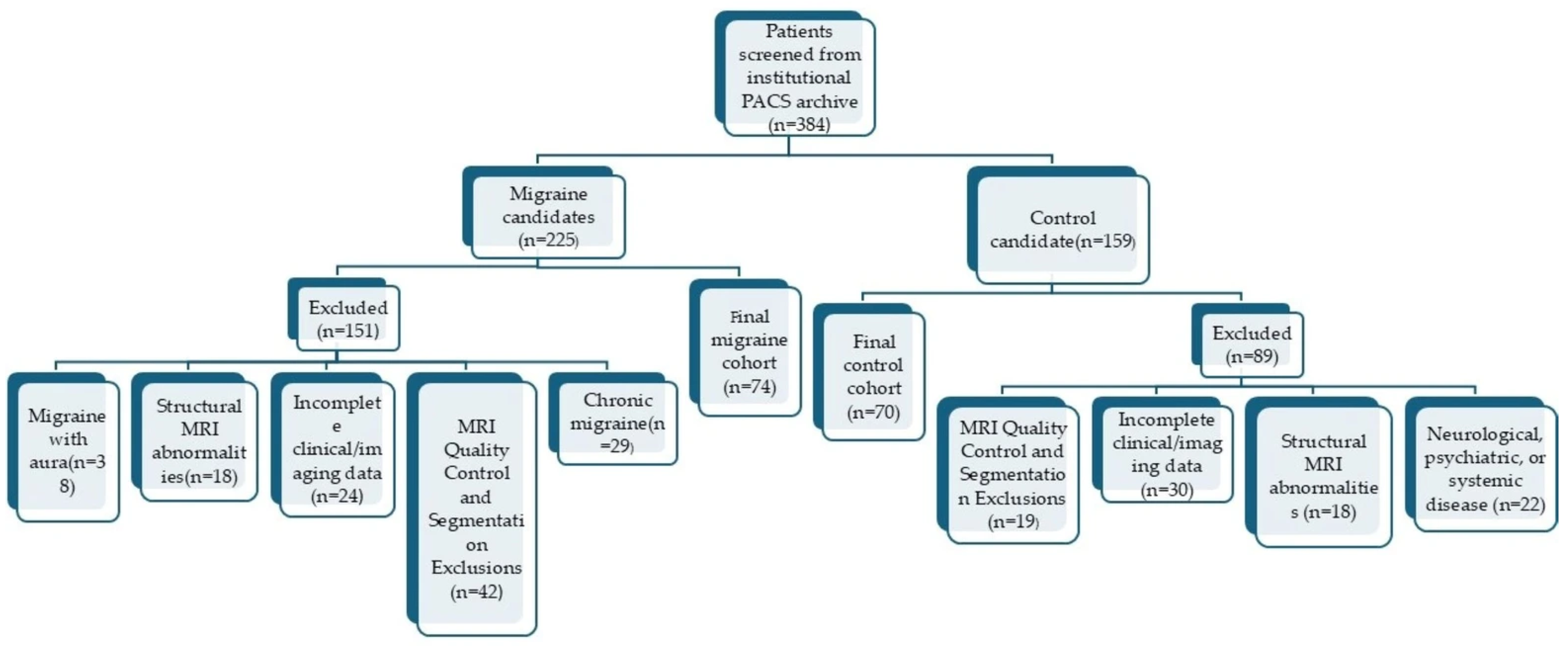
2.2. Study Population
- •
- Age between 12 and 17 years;
- •
- A diagnosis of migraine without aura according to the criteria defined in the third edition of the International Classification of Headache Disorders (ICHD-3) [27];
- •
- Confirmation of the migraine diagnosis by a pediatric neurologist with 10 years of experience in pediatric headache disorders based on clinical history, headache characteristics, attack pattern, and associated symptoms;
- •
- Normal neurological examination findings;
- •
- Availability of a diagnostically adequate cranial MRI examination acquired at the same institution;
- •
- Availability of high-resolution isotropic three-dimensional (3D) T1-weighted images suitable for volumetric analysis;
- •
- Absence of structural, developmental, or signal abnormalities on conventional MRI that could affect volumetric assessment;
- •
- Complete clinical and imaging data.
- •
- Age between 12 and 17 years;
- •
- Undergoing cranial MRI because of syncope, transient alteration of consciousness, or benign nonfocal neurological complaints unrelated to migraine or other primary headache disorders;
- •
- No history of migraine or any other primary headache disorder;
- •
- Normal neurological examination findings;
- •
- Structurally normal cranial MRI findings;
- •
- No history of epilepsy, neurodevelopmental disorders, psychiatric disease, or chronic systemic illness;
- •
- No history of any neurological disorder requiring regular neurological follow-up;
- •
- Availability of a diagnostically adequate cranial MRI examination acquired at the same institution.
- •
- Diagnosis of migraine with aura or documentation of aura symptoms in the clinical records;
- •
- Diagnosis of chronic migraine according to ICHD-3 criteria;
- •
- Reporting ≥ 15 headache days per month during the preceding three months;
- •
- Diagnosis of, or clinical suspicion for, a secondary headache syndrome;
- •
- Diagnosis of, or clinical suspicion for, medication-overuse headache;
- •
- History of long-term prophylactic migraine treatment;
- •
- Regular use of antiepileptic drugs, antidepressants, antipsychotics, psychostimulants, long-term sedatives, or other medications that could affect central nervous system function;
- •
- Use of hormonal therapy;
- •
- Presence of endocrine disorders, hormonal diseases, or systemic conditions that could influence pubertal development;
- •
- Cases in which the migraine subtype could not be reliably verified from clinical records.
- •
- Presence of a chronic systemic disease requiring long-term medical treatment;
- •
- Known vascular or demyelinating disease;
- •
- History of previous neurosurgical intervention;
- •
- History of traumatic brain injury;
- •
- Presence of congenital brain malformations or incidental intracranial structural lesions;
- •
- Incomplete clinical or imaging data;
- •
- MRI examinations performed outside the institution;
- •
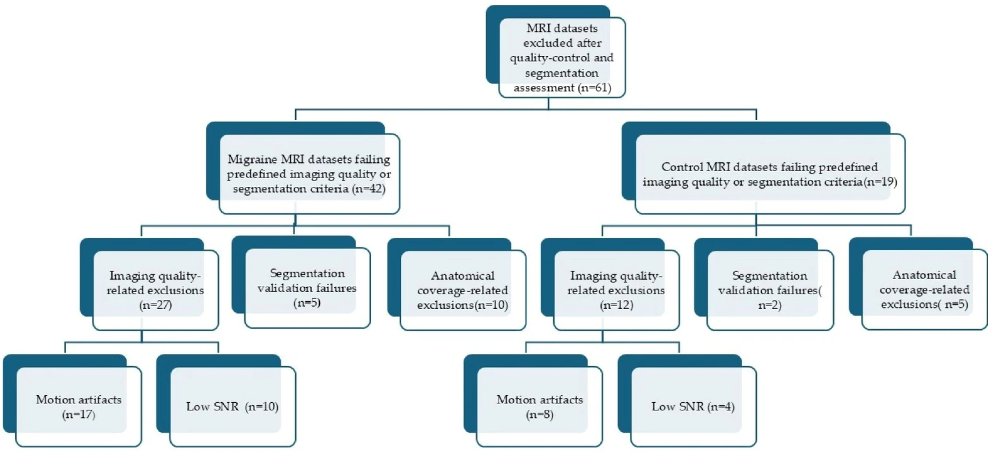
- Imaging datasets unsuitable for reliable automated segmentation because of substantial motion artifacts, inadequate image quality, or failure to achieve anatomical verification during the quality control process.
2.3. Magnetic Resonance Imaging Protocol
- •
- Axial T1-weighted spin-echo (SE): repetition time/echo time (TR/TE), 450/15 ms; field of view (FOV), 230 mm; slice thickness, 5 mm; matrix, 308 × 183.
- •
- Axial fat-saturated T1-weighted SE: TR/TE, 633/15 ms; FOV, 230 mm; slice thickness, 5 mm; matrix, 308 × 183.
- •
- Axial T2-weighted turbo spin-echo (TSE): TR/TE, 5240/100 ms; FOV, 230 mm; slice thickness, 5 mm; matrix, 384 × 237.
- •
- Coronal fluid-attenuated inversion recovery (FLAIR): TR/TE, 11,000/130 ms; FOV, 230 mm; slice thickness, 5 mm; matrix, 256 × 157.
- •
- Coronal T2-weighted TSE: TR/TE, 3027/100 ms; FOV, 200 mm; slice thickness, 3 mm; matrix, 336 × 217.
- •
- Coronal T1-weighted inversion recovery: TR/TE, 3079/15 ms; FOV, 200 mm; slice thickness, 3.5 mm; matrix, 336 × 211.
- •
- Coronal FLAIR: TR/TE, 11,000/130 ms; FOV, 230 mm; slice thickness, 3 mm; matrix, 256 × 157.
- •
- 3D FLAIR: TR/TE, 4800/315 ms; FOV, 250 mm; slice thickness, 1.04 mm; matrix, 216 × 218.
- •
- Axial diffusion-weighted imaging (DWI): b-values, 0 and 1000 s/mm2; FOV, 230 mm; slice thickness, 5 mm; matrix, 152 × 106.
- •
- Isotropic 3D T1-weighted imaging: TR/TE, 2500/46 ms; FOV, 230 mm; slice thickness, 1.0 mm; matrix, 256 × 256.
2.4. Magnetic Resonance Imaging Quality Control Pipeline
2.5. Neuroimaging Processing Workflow
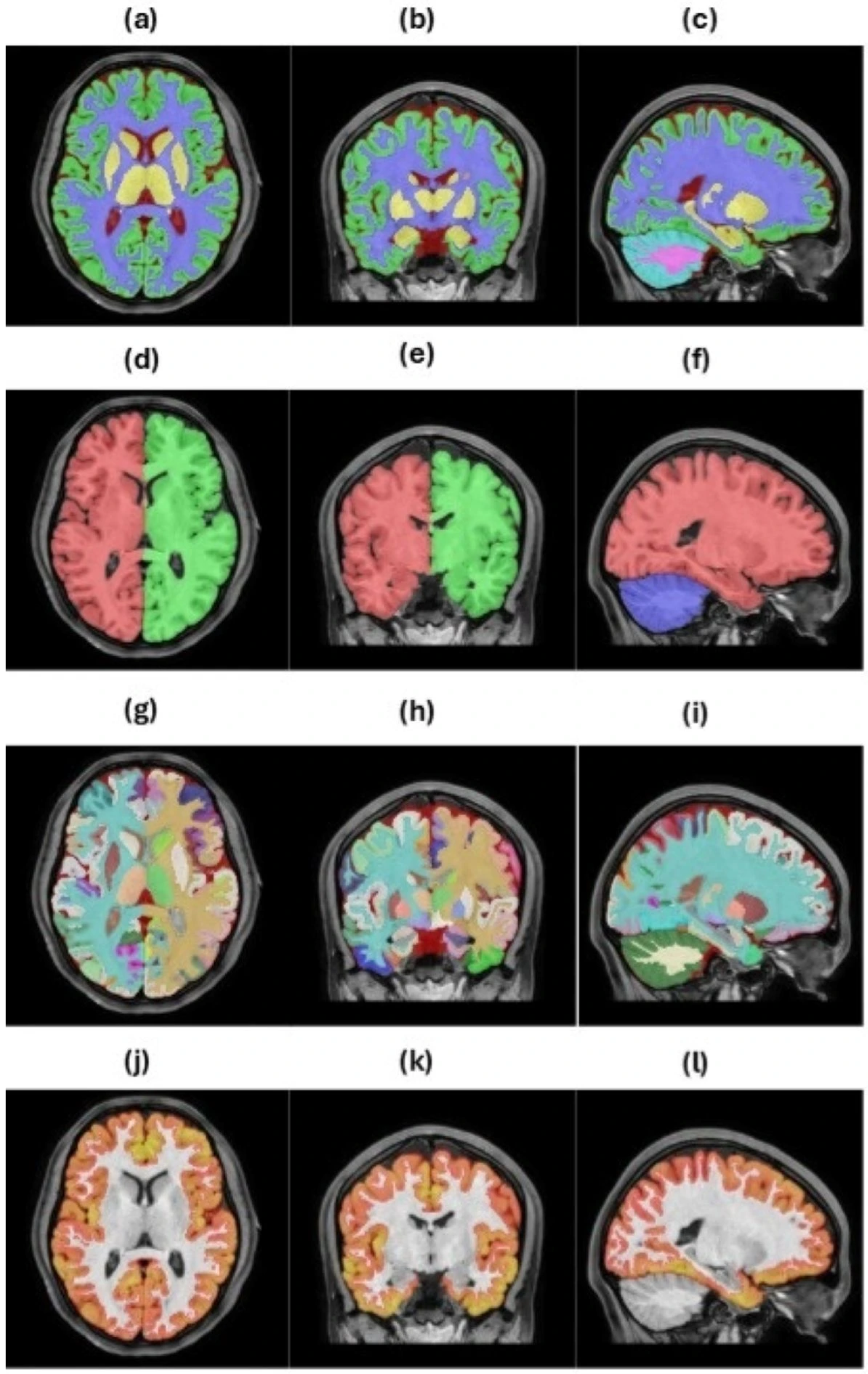
2.5.1. vol2Brain Processing Pipeline
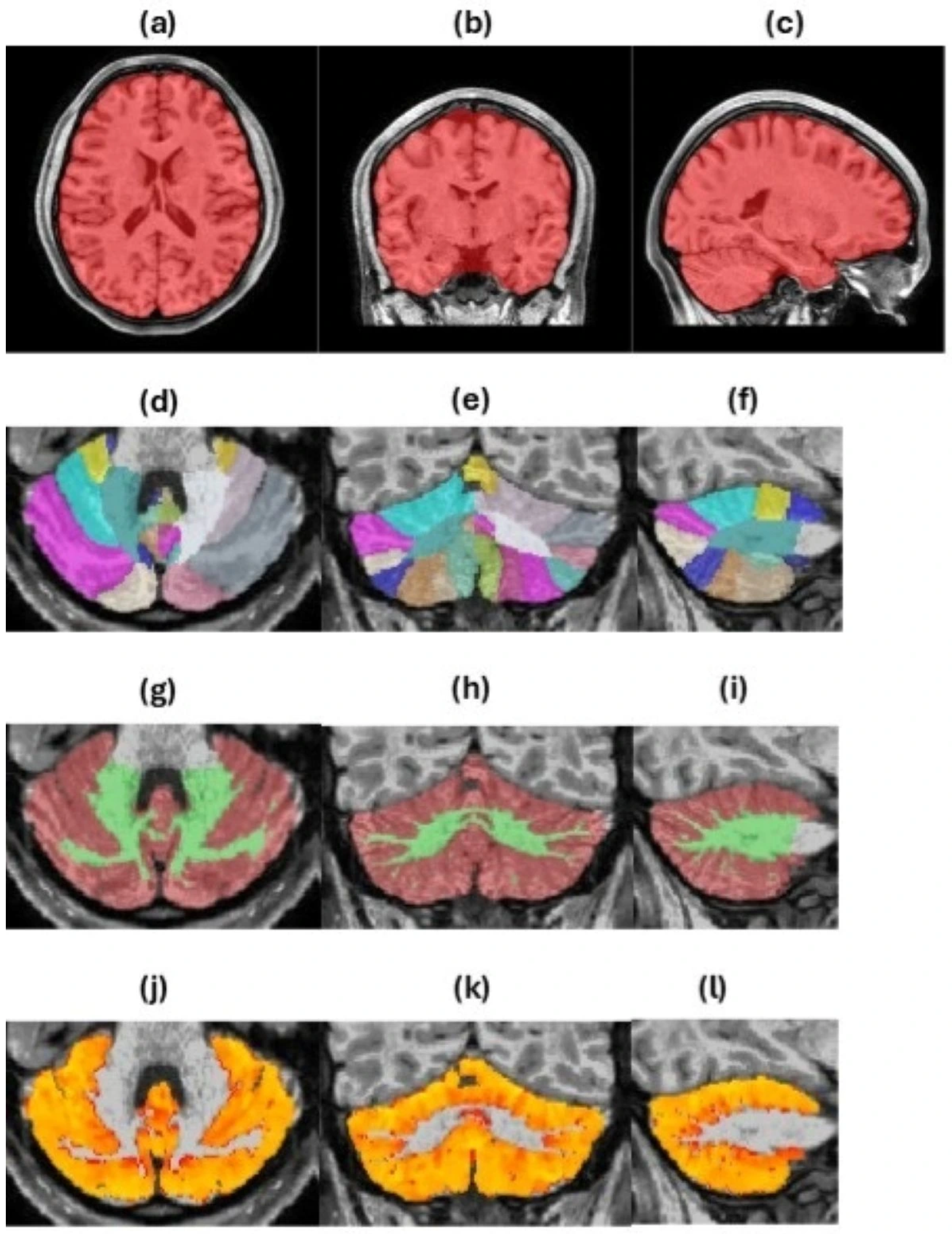
2.5.2. FreeSurfer Processing Pipeline
2.5.3. Visual Validation and Volumetric Analysis Using 3D Slicer
2.6. Statistical Analysis
3. Results
3.1. Study Population and Demographic Characteristics
3.2. Magnetic Resonance Imaging Quality Control and Segmentation Reliability
3.3. Whole-Brain Volumetric Findings
3.4. Cerebellar Subregional Analysis
3.5. Thalamic Nuclei Findings
3.6. Total Intracranial Volume-Normalized Volumetric Findings
4. Discussion
5. Conclusions
Supplementary Materials
Author Contributions
Funding
Institutional Review Board Statement
Informed Consent Statement
Data Availability Statement
Acknowledgments
Conflicts of Interest
References
- Khan, A.; Liu, S.; Tao, F. Current trends in pediatric migraine: Clinical insights and therapeutic strategies. Brain Sci. 2025, 15, 280. [Google Scholar] [CrossRef] [PubMed]
- Onofri, A.; Pensato, U.; Rosignoli, C.; Caponnetto, V.; Charlotte, C.; Gabriel, M.; Ornello, R.; Sacco, S. Primary headache epidemiology in children and adolescents: A systematic review and meta-analysis. J. Headache Pain 2023, 24, 8. [Google Scholar] [CrossRef] [PubMed]
- Konrad, K.; Firk, C.; Uhlhaas, P.J. Brain development during adolescence: Neuroscientific insights into this developmental period. Dtsch. Arztebl. Int. 2013, 110, 425–431. [Google Scholar] [CrossRef] [PubMed]
- Rocca, M.A.; Messina, R.; Colombo, B.; Falini, A.; Comi, G.; Filippi, M. Structural brain MRI abnormalities in pediatric patients with migraine. J. Neurol. 2014, 261, 350–357. [Google Scholar] [CrossRef] [PubMed]
- Xu, W.J.; Barisano, G.; Phung, D.; Chou, B.; Pinto, S.N.; Lerner, A.; Sheikh-Bahaei, N. Structural MRI in migraine: A review of migraine vascular and structural changes in brain parenchyma. J. Cent. Nerv. Syst. Dis. 2023, 15, 11795735231167868. [Google Scholar] [CrossRef] [PubMed]
- Bell, T.; Khaira, A.; Stokoe, M.; Webb, M.; Noel, M.; Amoozegar, F.; Harris, A.D. Age-related differences in resting state functional connectivity in pediatric migraine. J. Headache Pain 2021, 22, 65. [Google Scholar] [CrossRef] [PubMed]
- Goadsby, P.J.; Holland, P.R.; Martins-Oliveira, M.; Hoffmann, J.; Schankin, C.; Akerman, S. Pathophysiology of migraine: A disorder of sensory processing. Physiol. Rev. 2017, 97, 553–622. [Google Scholar] [CrossRef] [PubMed]
- Zhou, Y.; Gong, L.; Yang, Y.; Zhang, X.; Liu, J.; Wang, C.; Zhang, J.; Wei, X.; Yu, S. Spatio-temporal dynamics of resting-state brain networks are associated with migraine disability. J. Headache Pain 2023, 24, 13. [Google Scholar] [CrossRef] [PubMed]
- Schramm, S.; Börner, C.; Reichert, M.; Baum, T.; Zimmer, C.; Heinen, F.; Bonfert, M.V.; Sollmann, N. Functional magnetic resonance imaging in migraine: A systematic review. Cephalalgia 2023, 43, 3331024221128278. [Google Scholar] [CrossRef] [PubMed]
- Deodato, M.; Granato, A.; Martini, M.; Sabot, R.; Buoite Stella, A.; Manganotti, P. Instrumental assessment of pressure pain threshold over trigeminal and extra-trigeminal area in people with episodic and chronic migraine: A cross-sectional observational study. Neurol. Sci. 2024, 45, 3923–3929. [Google Scholar] [CrossRef] [PubMed]
- Coppola, G.; Di Lorenzo, C.; Schoenen, J.; Pierelli, F. Habituation and sensitization in primary headaches. J. Headache Pain 2013, 14, 65. [Google Scholar] [CrossRef] [PubMed]
- Noseda, R. Cerebro-cerebellar networks in migraine symptoms and headache. Front. Pain Res. 2022, 3, 940923. [Google Scholar] [CrossRef] [PubMed]
- Guarnera, A.; Bottino, F.; Napolitano, A.; Sforza, G.; Cappa, M.; Chioma, L.; Pasquini, L.; Rossi-Espagnet, M.C.; Lucignani, G.; Figà-Talamanca, L.; et al. Early alterations of cortical thickness and gyrification in migraine without aura: A retrospective MRI study in pediatric patients. J. Headache Pain 2021, 22, 79. [Google Scholar] [CrossRef] [PubMed]
- Webb, M.E.; Amoozegar, F.; Harris, A.D. Magnetic resonance imaging in pediatric migraine. Can. J. Neurol. Sci. 2019, 46, 653–665. [Google Scholar] [CrossRef] [PubMed]
- Rudolph, S.; Badura, A.; Lutzu, S.; Pathak, S.S.; Thieme, A.; Verpeut, J.L.; Wagner, M.J.; Yang, Y.M.; Fioravante, D. Cognitive-affective functions of the cerebellum. J. Neurosci. 2023, 43, 7554–7564. [Google Scholar] [CrossRef] [PubMed]
- Schwedt, T.J.; Chong, C.D.; Chiang, C.C.; Baxter, L.; Schlaggar, B.L.; Dodick, D.W. Enhanced pain-induced activity of pain-processing regions in a case-control study of episodic migraine. Cephalalgia 2014, 34, 947–958. [Google Scholar] [CrossRef] [PubMed]
- Guell, X.; Schmahmann, J. Cerebellar functional anatomy: A didactic summary based on human fMRI evidence. Cerebellum 2020, 19, 1–5. [Google Scholar] [CrossRef] [PubMed]
- Akerman, S.; Holland, P.R.; Goadsby, P.J. Diencephalic and brainstem mechanisms in migraine. Nat. Rev. Neurosci. 2011, 12, 570–584. [Google Scholar] [CrossRef] [PubMed]
- Akçay, H.İ. Comparison of brain volumes in episodic and chronic migraine using automated whole-brain volumetry. Front. Neurol. 2026, 17, 1772869. [Google Scholar] [CrossRef] [PubMed]
- Harkey, T.; Baker, D.; Hagen, J.; Scott, H.; Palys, V. Practical methods for segmentation and calculation of brain volume and intracranial volume: A guide and comparison. Quant. Imaging Med. Surg. 2022, 12, 3748–3761. [Google Scholar] [CrossRef] [PubMed]
- Lee, J.; Lee, J.Y.; Oh, S.W.; Chung, M.S.; Park, J.E.; Moon, Y.; Jeon, H.J.; Moon, W.J. Evaluation of reproducibility of brain volumetry between commercial software, Inbrain and established research purpose method, FreeSurfer. J. Clin. Neurol. 2021, 17, 307–316. [Google Scholar] [CrossRef] [PubMed]
- Monereo-Sánchez, J.; de Jong, J.J.A.; Drenthen, G.S.; Beran, M.; Backes, W.H.; Stehouwer, C.D.A.; Schram, M.T.; Linden, D.E.J.; Jansen, J.F.A. Quality control strategies for brain MRI segmentation and parcellation: Practical approaches and recommendations—Insights from the Maastricht study. NeuroImage 2021, 237, 118174. [Google Scholar] [CrossRef] [PubMed]
- Herting, M.M.; Sowell, E.R. Puberty and structural brain development in humans. Front. Neuroendocrinol. 2017, 44, 122–137. [Google Scholar] [CrossRef] [PubMed]
- Arain, M.; Haque, M.; Johal, L.; Mathur, P.; Nel, W.; Rais, A.; Sandhu, R.; Sharma, S. Maturation of the adolescent brain. Neuropsychiatr. Dis. Treat. 2013, 9, 449–461. [Google Scholar] [CrossRef] [PubMed]
- Blakemore, S.J. Imaging brain development: The adolescent brain. NeuroImage 2012, 61, 397–406. [Google Scholar] [CrossRef] [PubMed]
- Asato, M.R.; Terwilliger, R.; Woo, J.; Luna, B. White matter development in adolescence: A DTI study. Cereb. Cortex 2010, 20, 2122–2131. [Google Scholar] [CrossRef] [PubMed]
- Headache Classification Committee of the International Headache Society. The International Classification of Headache Disorders, 3rd edition. Cephalalgia 2018, 38, 1–211. [Google Scholar] [CrossRef] [PubMed]
- Mills, K.L.; Goddings, A.L.; Herting, M.M.; Meuwese, R.; Blakemore, S.J.; Crone, E.A.; Dahl, R.E.; Güroğlu, B.; Raznahan, A.; Sowell, E.R.; et al. Structural brain development between childhood and adulthood: Convergence across four longitudinal samples. NeuroImage 2016, 141, 273–281. [Google Scholar] [CrossRef] [PubMed]
- Evans, A.C.; Brain Development Cooperative Group. The NIH MRI study of normal brain development. NeuroImage 2006, 30, 184–202. [Google Scholar] [CrossRef] [PubMed]
- Giedd, J.N.; Blumenthal, J.; Jeffries, N.O.; Castellanos, F.X.; Liu, H.; Zijdenbos, A.; Paus, T.; Evans, A.C.; Rapoport, J.L. Brain development during childhood and adolescence: A longitudinal MRI study. Nat. Neurosci. 1999, 2, 861–863. [Google Scholar] [CrossRef] [PubMed]
- Šišić, N.; Rogelj, P. Deep learning for brain MRI tissue and structure segmentation: A comprehensive review. Algorithms 2025, 18, 636. [Google Scholar] [CrossRef]
- Manjón, J.V.; Coupé, P. volBrain: An online MRI brain volumetry system. Front. Neuroinform. 2016, 10, 30. [Google Scholar] [CrossRef] [PubMed]
- Iglesias, J.E.; Insausti, R.; Lerma-Usabiaga, G.; Bocchetta, M.; Van Leemput, K.; Greve, D.N.; van der Kouwe, A.; Alzheimer’s Disease Neuroimaging Initiative; Fischl, B.; Caballero-Gaudes, C.; et al. A probabilistic atlas of the human thalamic nuclei combining ex vivo MRI and histology. NeuroImage 2018, 183, 314–326. [Google Scholar] [CrossRef] [PubMed]
- Gaser, C.; Dahnke, R.; Thompson, P.M.; Kurth, F.; Luders, E. CAT: A computational anatomy toolbox for the analysis of structural MRI data. GigaScience 2024, 13, giae049. [Google Scholar] [CrossRef] [PubMed]
- Bürkle, E.; Nazzal, A.; Debolski, A.; Ernemann, U.; Lindig, T.; Bender, B. Scan–rescan reliability assessment of brain volumetric analysis across scanners and software solutions. Sci. Rep. 2025, 15, 29843. [Google Scholar] [CrossRef] [PubMed]
- Li, X.; Morgan, P.S.; Ashburner, J.; Smith, J.; Rorden, C. The first step for neuroimaging data analysis: DICOM to NIfTI conversion. J. Neurosci. Methods 2016, 264, 47–56. [Google Scholar] [CrossRef] [PubMed]
- Rushmore, R.J.; Bouix, S.; Kubicki, M.; Rathi, Y.; Yeterian, E.; Makris, N. HOA2.0-ComPaRe: A next generation Harvard–Oxford Atlas comparative parcellation reasoning method for human and macaque individual brain parcellation and atlases of the cerebral cortex. Front. Neuroanat. 2022, 16, 1035420. [Google Scholar] [CrossRef] [PubMed]
- Çetin, S. Evaluation of brain structures’ volume using vol2Brain software in patients with idiopathic sudden sensorineural hearing loss. Indian J. Otol. 2024, 30, 56–59. [Google Scholar] [CrossRef]
- Fischl, B. FreeSurfer. NeuroImage 2012, 62, 774–781. [Google Scholar] [CrossRef] [PubMed]
- Dale, A.M.; Fischl, B.; Sereno, M.I. Cortical surface-based analysis: I. Segmentation and surface reconstruction. NeuroImage 1999, 9, 179–194. [Google Scholar] [CrossRef] [PubMed]
- Fedorov, A.; Beichel, R.; Kalpathy-Cramer, J.; Finet, J.; Fillion-Robin, J.C.; Pujol, S.; Bauer, C.; Jennings, D.; Fennessy, F.; Sonka, M.; et al. 3D Slicer as an image computing platform for the Quantitative Imaging Network. Magn. Reson. Imaging 2012, 30, 1323–1341. [Google Scholar] [CrossRef] [PubMed]
- de Tommaso, M.; Vecchio, E.; Quitadamo, S.G.; Coppola, G.; Di Renzo, A.; Parisi, V.; Silvestro, M.; Russo, A.; Tedeschi, G. Pain-related brain connectivity changes in migraine: A narrative review and proof of concept about possible novel treatments interference. Brain Sci. 2021, 11, 234. [Google Scholar] [CrossRef] [PubMed]
- Karsan, N.; Goadsby, P.J. Neuroimaging in the pre-ictal or premonitory phase of migraine: A narrative review. J. Headache Pain 2023, 24, 106. [Google Scholar] [CrossRef] [PubMed]
- Matoso, A.; Fouto, A.R.; Esteves, I.; Caetano, G.; Vilela, P.; Gil-Gouveia, R.; Figueiredo, P. Involvement of the cerebellum in structural connectivity enhancement in episodic migraine. J. Headache Pain 2024, 25, 154. [Google Scholar] [CrossRef] [PubMed]
- Casillo, F.; Sebastianelli, G.; Abagnale, C.; Di Renzo, A.; Ziccardi, L.; Parisi, V.; Coppola, G. Brain imaging in migraine with and without aura: Similarities and differences. Cephalalgia 2025, 45, 3331024251365807. [Google Scholar] [CrossRef] [PubMed]
- Bethlehem, R.A.I.; Seidlitz, J.; White, S.R.; Vogel, J.W.; Anderson, K.M.; Adamson, C.; Adler, S.; Alexopoulos, G.S.; Anagnostou, E.; Areces-Gonzalez, A.; et al. Brain charts for the human lifespan. Nature 2022, 604, 525–533. [Google Scholar] [CrossRef] [PubMed]
- Buyanova, I.S.; Arsalidou, M. Cerebral white matter myelination and relations to age, gender, and cognition: A selective review. Front. Hum. Neurosci. 2021, 15, 662031. [Google Scholar] [CrossRef] [PubMed]
- Liang, X.; Sun, L.; Liao, X.; Lei, T.; Xia, M.; Duan, D.; Zeng, Z.; Li, Q.; Xu, Z.; Men, W.; et al. Structural connectome architecture shapes the maturation of cortical morphology from childhood to adolescence. Nat. Commun. 2024, 15, 784. [Google Scholar] [CrossRef] [PubMed]
- van Drunen, L.; Dobbelaar, S.; Crone, E.A.; Wierenga, L.M. Genetic and environmental influences on structural brain development from childhood to adolescence: A longitudinal twin study on cortical thickness, surface area, and subcortical volume. Dev. Cogn. Neurosci. 2024, 68, 101407. [Google Scholar] [CrossRef] [PubMed]
- Kosuge, S.; Masaoka, Y.; Kasai, H.; Honma, M.; Murakami, K.; Yoshii, N.; Watanabe, K.; Naito, T.; Kosuge, M.; Matsui, M.; et al. The right amygdala and migraine: Analyzing volume reduction and its relationship with symptom severity. PLoS ONE 2024, 19, e0301543. [Google Scholar] [CrossRef] [PubMed]
- Ou, Y.; Ni, X.; Gao, X.; Yu, Y.; Zhang, Y.; Wang, Y.; Liu, J.; Yin, Z.; Rong, J.; Sun, M.; et al. Structural and functional changes of anterior cingulate cortex subregions in migraine without aura: Relationships with pain sensation and pain emotion. Cereb. Cortex 2024, 34, bhae040. [Google Scholar] [CrossRef] [PubMed]
- Younis, S.; Hougaard, A.; Noseda, R.; Ashina, M. Current understanding of thalamic structure and function in migraine. Cephalalgia 2019, 39, 1675–1682. [Google Scholar] [CrossRef] [PubMed]
- Schmitz, N.; Admiraal-Behloul, F.; Arkink, E.B.; Kruit, M.C.; Schoonman, G.G.; Ferrari, M.D.; van Buchem, M.A. Attack frequency and disease duration as indicators for brain damage in migraine. Headache 2008, 48, 1044–1055. [Google Scholar] [CrossRef] [PubMed]
- Rocca, M.A.; Ceccarelli, A.; Falini, A.; Colombo, B.; Tortorella, P.; Bernasconi, L.; Comi, G.; Scotti, G.; Filippi, M. Brain gray matter changes in migraine patients with T2-visible lesions: A 3-T MRI study. Stroke 2006, 37, 1765–1770. [Google Scholar] [CrossRef] [PubMed]
- Chong, C.D.; Schwedt, T.J. Migraine affects white-matter tract integrity: A diffusion-tensor imaging study. Cephalalgia 2015, 35, 1162–1171. [Google Scholar] [CrossRef] [PubMed]
- Zhang, J.; Su, J.; Wang, M.; Zhao, Y.; Yao, Q.; Zhang, Q.; Lu, H.; Zhang, H.; Wang, S.; Li, G.-F.; et al. Increased default mode network connectivity and increased regional homogeneity in migraineurs without aura. J. Headache Pain 2016, 17, 98. [Google Scholar] [CrossRef] [PubMed]
- Tessitore, A.; Russo, A.; Giordano, A.; Conte, F.; Corbo, D.; De Stefano, M.; Cirillo, S.; Cirillo, M.; Esposito, F.; Tedeschi, G. Disrupted default mode network connectivity in migraine without aura. J. Headache Pain 2013, 14, 89. [Google Scholar] [CrossRef] [PubMed]
- Coppola, G.; Di Renzo, A.; Tinelli, E.; Di Lorenzo, C.; Scapeccia, M.; Parisi, V.; Serrao, M.; Evangelista, M.; Ambrosini, A.; Colonnese, C.; et al. Resting state connectivity between default mode network and insula encodes acute migraine headache. Cephalalgia 2018, 38, 846–854. [Google Scholar] [CrossRef] [PubMed]
- Martinelli, D.; Castellazzi, G.; De Icco, R.; Bacila, A.; Allena, M.; Faggioli, A.; Sances, G.; Pichiecchio, A.; Borsook, D.; Wheeler-Kingshott, C.A.M.G.; et al. Thalamocortical connectivity in experimentally induced migraine attacks: A pilot study. Brain Sci. 2021, 11, 165. [Google Scholar] [CrossRef] [PubMed]
- Shin, K.J.; Lee, H.J.; Park, K.M. Alterations of individual thalamic nuclei volumes in patients with migraine. J. Headache Pain 2019, 20, 112. [Google Scholar] [CrossRef] [PubMed]
- Giardina, I.; Di Renzo, A.; Chiffi, D.; Giuliani, G.; Sebastianelli, G.; Casillo, F.; Abagnale, C.; Ziccardi, L.; Pucci, A.; Parisi, V.; et al. 3T MRI thalamic segmentation reveals no macrostructural changes in interictal episodic migraine without aura compared to healthy controls. J. Headache Pain 2025, 26, 243. [Google Scholar] [CrossRef] [PubMed]



| Group | Nuclei Included |
|---|---|
| Anterior | Anteroventral nucleus (AVN) |
| Intralaminar/Midline | Centromedian nucleus (CN), Midline thalamic nuclei (MTN), Intralaminar nuclei (ISN) |
| Medial | Mediodorsal nucleus (MN) |
| Posterior | Pulvinar nucleus (PN), Medial geniculate nucleus (MGN), Lateral geniculate nucleus (LGN) |
| Ventral | Ventral anterior nucleus (VAN), Ventrolateral anterior nucleus (VLAN), Ventrolateral posterior nucleus (VLPN), Ventral posterolateral nucleus (VPLN) |
| Habenular | Habenular nucleus (HN) |
| Whole Thalamus | Right and left thalamus |
| Variable | Migraine Without Aura (n = 74) | Control (n = 70) | p-Value |
|---|---|---|---|
| Age, years | 14.8 ± 1.6 | 14.3 ± 1.5 | 0.071 |
| Sex | 0.337 | ||
| Female, n (%) | 46 (62.2) | 38 (54.3) | |
| Male, n (%) | 28 (37.8) | 32 (45.7) | |
| Body mass index, kg/m2 | 21.4 ± 3.2 | 20.8 ± 3.0 | 0.249 |
| Total intracranial volume, cm3 | 1398 ± 118 | 1432 ± 122 | 0.12 |
| History of syncope/nonfocal complaint, n (%) | — | 43 (61.4) | — |
| Disease duration, years | 2.6 ± 1.4 | — | — |
| Age at migraine onset, years | 12.2 ± 1.8 | — | — |
| Monthly attack frequency, attacks/month | 4.1 ± 2.3 | — | — |
| Attack duration, hours | 13.6 ± 8.4 | — | — |
| Nausea/vomiting, n (%) | 42 (56.8) | — | — |
| Photophobia, n (%) | 34 (45.9) | — | — |
| Phonophobia, n (%) | 45 (60.8) | — | — |
| Unilateral headache, n (%) | 49 (66.2) | — | — |
| Pulsating headache quality, n (%) | 51 (68.9) | — | — |
| Analgesic use during attacks, n (%) | 58 (78.4) | — | — |
| Anatomical Region | Measurement Type | Migraine Without Aura Mean ± SD | Control Mean ± SD | p-Value | q (FDR) | Effect Size (Hedges’ g) [95% CI] |
|---|---|---|---|---|---|---|
| Amygdala (R) | Volume (mm3) | 1712 ± 221 | 1846 ± 238 | 0.004 | 0.029 | 0.58 [0.25, 0.92] |
| Amygdala (L) | Volume (mm3) | 1695 ± 214 | 1818 ± 226 | 0.006 | 0.032 | 0.56 [0.22, 0.89] |
| Anterior Insula (R) | Cortical Thickness (mm) | 3.37 ± 0.24 | 3.52 ± 0.25 | 0.009 | 0.038 | 0.61 [0.27, 0.95] |
| Anterior Cingulate Gyrus (L) | Cortical Thickness (mm) | 4.01 ± 0.31 | 4.19 ± 0.30 | 0.012 | 0.041 | 0.59 [0.25, 0.93] |
| Precuneus | Cortical Thickness (mm) | 2.81 ± 0.27 | 2.96 ± 0.28 | 0.016 | 0.045 | 0.54 [0.21, 0.88] |
| Thalamus (R) | Volume (mm3) | 5635 ± 612 | 5878 ± 641 | 0.026 | 0.049 | 0.39 [0.06, 0.72] |
| Remaining 129 regions | — | — | — | N.S. | — | — |
| Anatomical Region | Side | Migraine Without Aura Mean ± SD (cm3) | Control Mean ± SD (cm3) | p-Value | q (FDR) | Effect Size Hedges’ g [95% CI] |
|---|---|---|---|---|---|---|
| Lobule I–II | Right | 0.060 ± 0.009 | 0.061 ± 0.008 | 0.72 | 0.86 | 0.05 [−0.34, 0.44] |
| Lobule I–II | Left | 0.051 ± 0.008 | 0.053 ± 0.008 | 0.76 | 0.88 | 0.04 [−0.35, 0.43] |
| Lobule III | Right | 0.61 ± 0.08 | 0.63 ± 0.08 | 0.49 | 0.72 | 0.11 [−0.28, 0.50] |
| Lobule III | Left | 0.56 ± 0.07 | 0.58 ± 0.08 | 0.53 | 0.75 | 0.10 [−0.29, 0.49] |
| Lobule IV | Right | 1.84 ± 0.19 | 1.89 ± 0.20 | 0.41 | 0.68 | 0.14 [−0.25, 0.53] |
| Lobule IV | Left | 2.18 ± 0.22 | 2.24 ± 0.23 | 0.38 | 0.65 | 0.15 [−0.24, 0.54] |
| Lobule V | Right | 3.94 ± 0.37 | 4.03 ± 0.39 | 0.36 | 0.63 | 0.17 [−0.22, 0.56] |
| Lobule V | Left | 4.01 ± 0.38 | 4.08 ± 0.40 | 0.40 | 0.66 | 0.16 [−0.23, 0.55] |
| Lobule VI | Right | 8.88 ± 0.71 | 9.29 ± 0.76 | 0.011 | 0.041 | 0.46 [0.12, 0.80] |
| Lobule VI | Left | 8.93 ± 0.69 | 9.35 ± 0.74 | 0.014 | 0.046 | 0.43 [0.09, 0.77] |
| Lobule VIIA (Crus I) | Right | 11.24 ± 1.08 | 11.46 ± 1.12 | 0.18 | 0.43 | 0.20 [−0.19, 0.59] |
| Lobule VIIA (Crus I) | Left | 11.31 ± 1.10 | 11.55 ± 1.14 | 0.21 | 0.48 | 0.19 [−0.20, 0.58] |
| Lobule VIIA (Crus II) | Right | 7.08 ± 0.74 | 7.23 ± 0.76 | 0.23 | 0.50 | 0.18 [−0.21, 0.57] |
| Lobule VIIA (Crus II) | Left | 7.14 ± 0.75 | 7.28 ± 0.77 | 0.28 | 0.57 | 0.16 [−0.23, 0.55] |
| Lobule VIIB | Right | 4.71 ± 0.46 | 4.82 ± 0.47 | 0.31 | 0.59 | 0.16 [−0.23, 0.55] |
| Lobule VIIB | Left | 5.02 ± 0.49 | 5.12 ± 0.50 | 0.34 | 0.61 | 0.15 [−0.24, 0.54] |
| Lobule VIIIA | Right | 6.32 ± 0.61 | 6.45 ± 0.63 | 0.35 | 0.63 | 0.14 [−0.25, 0.53] |
| Lobule VIIIA | Left | 5.71 ± 0.56 | 5.83 ± 0.58 | 0.37 | 0.65 | 0.13 [−0.26, 0.52] |
| Lobule VIIIB | Right | 3.98 ± 0.39 | 4.07 ± 0.40 | 0.054 | 0.081 | 0.30 [−0.09, 0.69] |
| Lobule VIIIB | Left | 3.58 ± 0.35 | 3.66 ± 0.36 | 0.40 | 0.66 | 0.14 [−0.25, 0.53] |
| Lobule IX | Right | 4.08 ± 0.42 | 4.16 ± 0.43 | 0.51 | 0.73 | 0.10 [−0.29, 0.49] |
| Lobule IX | Left | 3.80 ± 0.39 | 3.88 ± 0.40 | 0.55 | 0.76 | 0.09 [−0.30, 0.48] |
| Lobule X | Right | 0.62 ± 0.08 | 0.64 ± 0.08 | 0.45 | 0.69 | 0.12 [−0.27, 0.51] |
| Lobule X | Left | 0.59 ± 0.07 | 0.61 ± 0.08 | 0.48 | 0.71 | 0.11 [−0.28, 0.50] |
| Anatomical Region | Side | Migraine Without Aura Mean ± SD (cm3) | Control Mean ± SD (cm3) | p-Value | q (FDR) | Effect Size Hedges’ g [95% CI] |
|---|---|---|---|---|---|---|
| Lobule I–II | Right | 0.035 ± 0.006 | 0.036 ± 0.006 | 0.68 | 0.84 | 0.07 [−0.26, 0.40] |
| Lobule I–II | Left | 0.022 ± 0.005 | 0.023 ± 0.005 | 0.72 | 0.86 | 0.05 [−0.28, 0.38] |
| Lobule III | Right | 0.53 ± 0.07 | 0.55 ± 0.07 | 0.44 | 0.68 | 0.13 [−0.20, 0.46] |
| Lobule III | Left | 0.49 ± 0.07 | 0.51 ± 0.07 | 0.47 | 0.70 | 0.12 [−0.21, 0.45] |
| Lobule IV | Right | 1.69 ± 0.18 | 1.75 ± 0.19 | 0.28 | 0.55 | 0.19 [−0.14, 0.52] |
| Lobule IV | Left | 2.03 ± 0.21 | 2.10 ± 0.22 | 0.25 | 0.52 | 0.20 [−0.13, 0.53] |
| Lobule V | Right | 3.41 ± 0.33 | 3.52 ± 0.35 | 0.22 | 0.48 | 0.22 [−0.11, 0.55] |
| Lobule V | Left | 3.55 ± 0.34 | 3.66 ± 0.36 | 0.24 | 0.50 | 0.21 [−0.12, 0.54] |
| Lobule VI | Right | 8.19 ± 0.64 | 8.73 ± 0.68 | 0.003 | 0.018 | 0.64 [0.30, 0.98] |
| Lobule VI | Left | 8.24 ± 0.63 | 8.78 ± 0.67 | 0.005 | 0.023 | 0.60 [0.26, 0.94] |
| Lobule VIIA (Crus I) | Right | 9.86 ± 0.91 | 10.01 ± 0.94 | 0.19 | 0.45 | 0.23 [−0.10, 0.56] |
| Lobule VIIA (Crus I) | Left | 9.95 ± 0.92 | 10.10 ± 0.95 | 0.21 | 0.47 | 0.22 [−0.11, 0.55] |
| Lobule VIIA (Crus II) | Right | 6.16 ± 0.66 | 6.28 ± 0.68 | 0.16 | 0.42 | 0.24 [−0.09, 0.57] |
| Lobule VIIA (Crus II) | Left | 6.55 ± 0.69 | 6.67 ± 0.70 | 0.18 | 0.44 | 0.23 [−0.10, 0.56] |
| Lobule VIIB | Right | 4.48 ± 0.44 | 4.58 ± 0.45 | 0.26 | 0.53 | 0.19 [−0.14, 0.52] |
| Lobule VIIB | Left | 4.55 ± 0.44 | 4.64 ± 0.46 | 0.31 | 0.58 | 0.17 [−0.16, 0.50] |
| Lobule VIIIA | Right | 5.84 ± 0.56 | 5.96 ± 0.58 | 0.24 | 0.51 | 0.20 [−0.13, 0.53] |
| Lobule VIIIA | Left | 5.47 ± 0.53 | 5.58 ± 0.55 | 0.29 | 0.56 | 0.18 [−0.15, 0.51] |
| Lobule VIIIB | Right | 3.54 ± 0.35 | 3.63 ± 0.36 | 0.061 | 0.088 | 0.29 [−0.04, 0.62] |
| Lobule VIIIB | Left | 3.24 ± 0.33 | 3.31 ± 0.34 | 0.34 | 0.60 | 0.16 [−0.17, 0.49] |
| Lobule IX | Right | 3.56 ± 0.37 | 3.65 ± 0.38 | 0.39 | 0.64 | 0.14 [−0.19, 0.47] |
| Lobule IX | Left | 3.25 ± 0.34 | 3.33 ± 0.35 | 0.42 | 0.66 | 0.13 [−0.20, 0.46] |
| Lobule X | Right | 0.60 ± 0.08 | 0.62 ± 0.08 | 0.33 | 0.59 | 0.16 [−0.17, 0.49] |
| Lobule X | Left | 0.56 ± 0.07 | 0.58 ± 0.07 | 0.37 | 0.62 | 0.15 [−0.18, 0.48] |
| Anatomical Region | Side | Migraine Without Aura Mean ± SD (mm) | Control Mean ± SD (mm) | p-Value | q (FDR) | Effect Size Hedges’ g [95% CI] |
|---|---|---|---|---|---|---|
| Lobule I–II | Right | 3.22 ± 0.36 | 3.26 ± 0.37 | 0.54 | 0.72 | 0.10 [−0.23, 0.43] |
| Lobule I–II | Left | 3.14 ± 0.35 | 3.18 ± 0.36 | 0.51 | 0.70 | 0.11 [−0.22, 0.44] |
| Lobule III | Right | 3.91 ± 0.40 | 3.97 ± 0.41 | 0.39 | 0.61 | 0.15 [−0.18, 0.48] |
| Lobule III | Left | 4.12 ± 0.41 | 4.18 ± 0.42 | 0.42 | 0.64 | 0.14 [−0.19, 0.47] |
| Lobule IV | Right | 5.21 ± 0.43 | 5.28 ± 0.45 | 0.33 | 0.56 | 0.16 [−0.17, 0.49] |
| Lobule IV | Left | 5.19 ± 0.44 | 5.26 ± 0.45 | 0.35 | 0.58 | 0.15 [−0.18, 0.48] |
| Lobule V | Right | 4.72 ± 0.38 | 4.80 ± 0.39 | 0.26 | 0.49 | 0.20 [−0.13, 0.53] |
| Lobule V | Left | 4.87 ± 0.40 | 4.94 ± 0.41 | 0.32 | 0.55 | 0.17 [−0.16, 0.50] |
| Lobule VI | Right | 4.96 ± 0.36 | 5.05 ± 0.37 | 0.17 | 0.38 | 0.24 [−0.09, 0.57] |
| Lobule VI | Left | 5.01 ± 0.37 | 5.09 ± 0.38 | 0.21 | 0.44 | 0.21 [−0.12, 0.54] |
| Lobule VIIA (Crus I) | Right | 4.84 ± 0.31 | 5.06 ± 0.34 | 0.006 | 0.031 | 0.66 [0.32, 1.00] |
| Lobule VIIA (Crus I) | Left | 4.91 ± 0.33 | 4.99 ± 0.34 | 0.18 | 0.39 | 0.23 [−0.10, 0.56] |
| Lobule VIIA (Crus II) | Right | 4.72 ± 0.30 | 4.91 ± 0.32 | 0.011 | 0.044 | 0.61 [0.27, 0.95] |
| Lobule VIIA (Crus II) | Left | 4.78 ± 0.31 | 4.85 ± 0.33 | 0.25 | 0.48 | 0.21 [−0.12, 0.54] |
| Lobule VIIB | Right | 5.05 ± 0.37 | 5.13 ± 0.38 | 0.20 | 0.43 | 0.21 [−0.12, 0.54] |
| Lobule VIIB | Left | 5.03 ± 0.36 | 5.10 ± 0.37 | 0.28 | 0.52 | 0.19 [−0.14, 0.52] |
| Lobule VIIIA | Right | 4.91 ± 0.35 | 4.98 ± 0.36 | 0.29 | 0.53 | 0.18 [−0.15, 0.51] |
| Lobule VIIIA | Left | 5.03 ± 0.36 | 5.10 ± 0.37 | 0.30 | 0.54 | 0.18 [−0.15, 0.51] |
| Lobule VIIIB | Right | 4.93 ± 0.34 | 5.01 ± 0.36 | 0.19 | 0.41 | 0.22 [−0.11, 0.55] |
| Lobule VIIIB | Left | 5.09 ± 0.36 | 5.15 ± 0.37 | 0.36 | 0.59 | 0.16 [−0.17, 0.49] |
| Lobule IX | Right | 4.96 ± 0.39 | 5.02 ± 0.40 | 0.43 | 0.65 | 0.14 [−0.19, 0.47] |
| Lobule IX | Left | 4.99 ± 0.40 | 5.04 ± 0.41 | 0.48 | 0.68 | 0.12 [−0.21, 0.45] |
| Lobule X | Right | 3.91 ± 0.37 | 3.98 ± 0.38 | 0.34 | 0.57 | 0.17 [−0.16, 0.50] |
| Lobule X | Left | 3.96 ± 0.38 | 4.02 ± 0.39 | 0.41 | 0.63 | 0.15 [−0.18, 0.48] |
| Anatomical Region | Side | Migraine Without Aura Mean ± SD (cm3) | Control Mean ± SD (cm3) | p-Value | q (FDR) | Effect Size Hedges’ g [95% CI] |
|---|---|---|---|---|---|---|
| AVN | Right | 0.101 ± 0.018 | 0.105 ± 0.019 | 0.28 | 0.56 | 0.18 [−0.15, 0.51] |
| AVN | Left | 0.099 ± 0.017 | 0.103 ± 0.018 | 0.34 | 0.62 | 0.15 [−0.18, 0.48] |
| VAN | Right | 0.232 ± 0.036 | 0.252 ± 0.039 | 0.014 | 0.041 | 0.53 [0.19, 0.87] |
| VAN | Left | 0.229 ± 0.035 | 0.242 ± 0.038 | 0.071 | 0.108 | 0.31 [−0.02, 0.64] |
| VLAN | Right | 0.091 ± 0.014 | 0.095 ± 0.015 | 0.22 | 0.49 | 0.22 [−0.11, 0.55] |
| VLAN | Left | 0.096 ± 0.014 | 0.101 ± 0.015 | 0.18 | 0.44 | 0.25 [−0.08, 0.58] |
| VLPN | Right | 0.795 ± 0.078 | 0.820 ± 0.083 | 0.083 | 0.127 | 0.29 [−0.04, 0.62] |
| VLPN | Left | 0.808 ± 0.079 | 0.834 ± 0.084 | 0.092 | 0.136 | 0.28 [−0.05, 0.61] |
| VPLN | Right | 0.281 ± 0.041 | 0.305 ± 0.045 | 0.011 | 0.038 | 0.56 [0.22, 0.90] |
| VPLN | Left | 0.314 ± 0.044 | 0.329 ± 0.047 | 0.081 | 0.122 | 0.29 [−0.04, 0.62] |
| PN | Right | 1.108 ± 0.126 | 1.196 ± 0.132 | 0.006 | 0.027 | 0.68 [0.34, 1.02] |
| PN | Left | 1.236 ± 0.135 | 1.321 ± 0.142 | 0.009 | 0.034 | 0.61 [0.27, 0.95] |
| LGN | Right | 0.097 ± 0.018 | 0.101 ± 0.019 | 0.31 | 0.60 | 0.16 [−0.17, 0.49] |
| LGN | Left | 0.088 ± 0.016 | 0.091 ± 0.017 | 0.39 | 0.66 | 0.13 [−0.20, 0.46] |
| MGN | Right | 0.076 ± 0.011 | 0.079 ± 0.012 | 0.27 | 0.55 | 0.18 [−0.15, 0.51] |
| MGN | Left | 0.067 ± 0.010 | 0.070 ± 0.011 | 0.25 | 0.53 | 0.19 [−0.14, 0.52] |
| CN | Right | 0.118 ± 0.017 | 0.123 ± 0.018 | 0.19 | 0.45 | 0.24 [−0.09, 0.57] |
| CN | Left | 0.116 ± 0.017 | 0.121 ± 0.018 | 0.23 | 0.50 | 0.22 [−0.11, 0.55] |
| MN | Right | 0.615 ± 0.071 | 0.664 ± 0.076 | 0.004 | 0.021 | 0.66 [0.32, 1.00] |
| MN | Left | 0.618 ± 0.070 | 0.660 ± 0.074 | 0.012 | 0.039 | 0.58 [0.24, 0.92] |
| HN | Right | 0.019 ± 0.004 | 0.020 ± 0.004 | 0.42 | 0.68 | 0.12 [−0.21, 0.45] |
| HN | Left | 0.020 ± 0.004 | 0.021 ± 0.004 | 0.45 | 0.70 | 0.11 [−0.22, 0.44] |
| MTN | Right | 0.010 ± 0.003 | 0.011 ± 0.003 | 0.48 | 0.72 | 0.10 [−0.23, 0.43] |
| MTN | Left | 0.011 ± 0.003 | 0.012 ± 0.003 | 0.51 | 0.74 | 0.09 [−0.24, 0.42] |
| ISN | Right | 2.170 ± 0.210 | 2.218 ± 0.224 | 0.19 | 0.45 | 0.23 [−0.10, 0.56] |
| ISN | Left | 2.158 ± 0.208 | 2.205 ± 0.219 | 0.21 | 0.48 | 0.22 [−0.11, 0.55] |
| Anatomical Region | Measurement Type | Migraine Without Aura Mean ± SD | Control Mean ± SD | p-Value | q (FDR) | Effect Size Hedges’ g [95% CI] |
|---|---|---|---|---|---|---|
| Right Amygdala | TIV-adjusted volume (cm3) | 1.70 ± 0.21 | 1.83 ± 0.24 | 0.011 | 0.032 | 0.55 [0.22, 0.88] |
| Left Amygdala | TIV-adjusted volume (cm3) | 1.68 ± 0.20 | 1.80 ± 0.22 | 0.014 | 0.037 | 0.52 [0.19, 0.85] |
| Right Thalamus | TIV-adjusted volume (cm3) | 5.60 ± 0.60 | 5.84 ± 0.64 | 0.021 | 0.046 | 0.42 [0.09, 0.75] |
| Right Lobule VI | TIV-adjusted volume (cm3) | 8.84 ± 0.70 | 9.25 ± 0.75 | 0.017 | 0.041 | 0.45 [0.12, 0.78] |
| Left Lobule VI | TIV-adjusted volume (cm3) | 8.89 ± 0.69 | 9.30 ± 0.74 | 0.019 | 0.044 | 0.43 [0.10, 0.76] |
| Right Lobule VI | TIV-adjusted gray matter volume (cm3) | 8.15 ± 0.63 | 8.68 ± 0.67 | 0.005 | 0.019 | 0.62 [0.29, 0.95] |
| Left Lobule VI | TIV-adjusted gray matter volume (cm3) | 8.20 ± 0.62 | 8.73 ± 0.66 | 0.007 | 0.024 | 0.59 [0.26, 0.92] |
| Right VAN | TIV-adjusted volume (cm3) | 0.229 ± 0.035 | 0.249 ± 0.038 | 0.018 | 0.043 | 0.50 [0.17, 0.83] |
| Right VPLN | TIV-adjusted volume (cm3) | 0.278 ± 0.040 | 0.302 ± 0.044 | 0.014 | 0.036 | 0.54 [0.21, 0.87] |
| Right PN | TIV-adjusted volume (cm3) | 1.097 ± 0.123 | 1.184 ± 0.129 | 0.008 | 0.026 | 0.66 [0.33, 0.99] |
| Left PN | TIV-adjusted volume (cm3) | 1.225 ± 0.132 | 1.309 ± 0.139 | 0.011 | 0.031 | 0.60 [0.27, 0.93] |
| Right MN | TIV-adjusted volume (cm3) | 0.608 ± 0.070 | 0.656 ± 0.074 | 0.006 | 0.021 | 0.64 [0.31, 0.97] |
| Left MN | TIV-adjusted volume (cm3) | 0.611 ± 0.069 | 0.652 ± 0.073 | 0.013 | 0.034 | 0.57 [0.24, 0.90] |
| Anatomical Domain | Anatomical Region | Measurement Type | Direction of Change in Migraine Group | q (FDR) | Effect Size (Hedges’ g) [95% CI] |
|---|---|---|---|---|---|
| Whole-brain morphometry | Right amygdala | Volume | ↓ | 0.029 | 0.58 [0.25, 0.92] |
| Whole-brain morphometry | Left amygdala | Volume | ↓ | 0.032 | 0.56 [0.22, 0.89] |
| Whole-brain morphometry | Right thalamus | Volume | ↓ | 0.049 | 0.39 [0.06, 0.72] |
| Whole-brain morphometry | Right anterior insula | Cortical thickness | ↓ | 0.038 | 0.61 [0.27, 0.95] |
| Whole-brain morphometry | Left anterior cingulate gyrus | Cortical thickness | ↓ | 0.041 | 0.59 [0.25, 0.93] |
| Whole-brain morphometry | Precuneus | Cortical thickness | ↓ | 0.045 | 0.54 [0.21, 0.88] |
| Cerebellar subregional analysis | Right Lobule VI | Volume | ↓ | 0.041 | 0.46 [0.12, 0.80] |
| Cerebellar subregional analysis | Left Lobule VI | Volume | ↓ | 0.046 | 0.43 [0.09, 0.77] |
| Cerebellar subregional analysis | Right Lobule VI | Gray matter volume | ↓ | 0.018 | 0.64 [0.30, 0.98] |
| Cerebellar subregional analysis | Left Lobule VI | Gray matter volume | ↓ | 0.023 | 0.60 [0.26, 0.94] |
| Cerebellar subregional analysis | Right Crus I | Cortical thickness | ↓ | 0.031 | 0.66 [0.32, 1.00] |
| Cerebellar subregional analysis | Right Crus II | Cortical thickness | ↓ | 0.044 | 0.61 [0.27, 0.95] |
| Thalamic nuclei analysis | Right ventral anterior nucleus (VAN) | Volume | ↓ | 0.041 | 0.53 [0.19, 0.87] |
| Thalamic nuclei analysis | Right ventral posterolateral nucleus (VPLN) | Volume | ↓ | 0.038 | 0.56 [0.22, 0.90] |
| Thalamic nuclei analysis | Right pulvinar nucleus (PN) | Volume | ↓ | 0.027 | 0.68 [0.34, 1.02] |
| Thalamic nuclei analysis | Left pulvinar nucleus (PN) | Volume | ↓ | 0.034 | 0.61 [0.27, 0.95] |
| Thalamic nuclei analysis | Right mediodorsal nucleus (MN) | Volume | ↓ | 0.021 | 0.66 [0.32, 1.00] |
| Thalamic nuclei analysis | Left mediodorsal nucleus (MN) | Volume | ↓ | 0.039 | 0.58 [0.24, 0.92] |
Disclaimer/Publisher’s Note: The statements, opinions and data contained in all publications are solely those of the individual author(s) and contributor(s) and not of MDPI and/or the editor(s). MDPI and/or the editor(s) disclaim responsibility for any injury to people or property resulting from any ideas, methods, instructions or products referred to in the content. |
© 2026 by the authors. Licensee MDPI, Basel, Switzerland. This article is an open access article distributed under the terms and conditions of the Creative Commons Attribution (CC BY) license.
Share and Cite
Aytaç, A.; Aydın, H.; Akay, E. Multiplatform Morphometric Profiling of Whole-Brain, Cerebellar Subregional, and Thalamic Nuclei Alterations in Pediatric Migraine Without Aura. Diagnostics 2026, 16, 2085. https://doi.org/10.3390/diagnostics16132085
Aytaç A, Aydın H, Akay E. Multiplatform Morphometric Profiling of Whole-Brain, Cerebellar Subregional, and Thalamic Nuclei Alterations in Pediatric Migraine Without Aura. Diagnostics. 2026; 16(13):2085. https://doi.org/10.3390/diagnostics16132085
Chicago/Turabian StyleAytaç, Adil, Hilal Aydın, and Emrah Akay. 2026. "Multiplatform Morphometric Profiling of Whole-Brain, Cerebellar Subregional, and Thalamic Nuclei Alterations in Pediatric Migraine Without Aura" Diagnostics 16, no. 13: 2085. https://doi.org/10.3390/diagnostics16132085
APA StyleAytaç, A., Aydın, H., & Akay, E. (2026). Multiplatform Morphometric Profiling of Whole-Brain, Cerebellar Subregional, and Thalamic Nuclei Alterations in Pediatric Migraine Without Aura. Diagnostics, 16(13), 2085. https://doi.org/10.3390/diagnostics16132085