2. Materials and Methods
This study was approved by the Ethics Committee of Antalya Training and Research Hospital (approval date: 11 July 2024; approval no: 10/14). Because of the retrospective design and the use of existing imaging and clinical records, the requirement for informed consent was waived by the Ethics Committee.
2.1. Study Design
The present study was designed as a retrospective case–control study. Adult patients who presented to the otolaryngology outpatient clinic and underwent paranasal sinus computed tomography (PNS CT) between 3 April 2023, and 30 May 2024, were retrospectively screened from the hospital records. CT images were reviewed jointly by an experienced otolaryngologist and radiologist. Patients meeting the inclusion and exclusion criteria and showing frontal or maxillary sinus hypoplasia/aplasia were included in the sinus variation groups. The control group consisted of adults who underwent paranasal sinus CT during the same study period, had no radiologic evidence of frontal or maxillary sinus hypoplasia/aplasia, and met the same exclusion criteria. Controls were not individually matched to cases by age or sex. The study population consisted of 117 patients with sinus variation (63 women and 54 men) and 53 healthy controls (21 women and 32 men).
Inclusion criteria were as follows: (1) adults aged 18 years and older, (2) patients with paranasal CT scans, and (3) patients with frontal or maxillary sinus variation. The exclusion criteria were as follows: (1) history of craniofacial syndrome, (2) endocrine diseases affecting the facial skeleton, (3) history of previous aesthetic or traumatic surgery in the facial region, (4) tumor in the paranasal sinuses, and (5) technical inadequacy affecting image quality. Information on prior facial trauma or previous facial/aesthetic surgery was screened from the documented medical history available in the electronic records associated with the CT examination. Only patients without a recorded history of facial trauma or prior facial/aesthetic surgery before CT acquisition were eligible for inclusion.
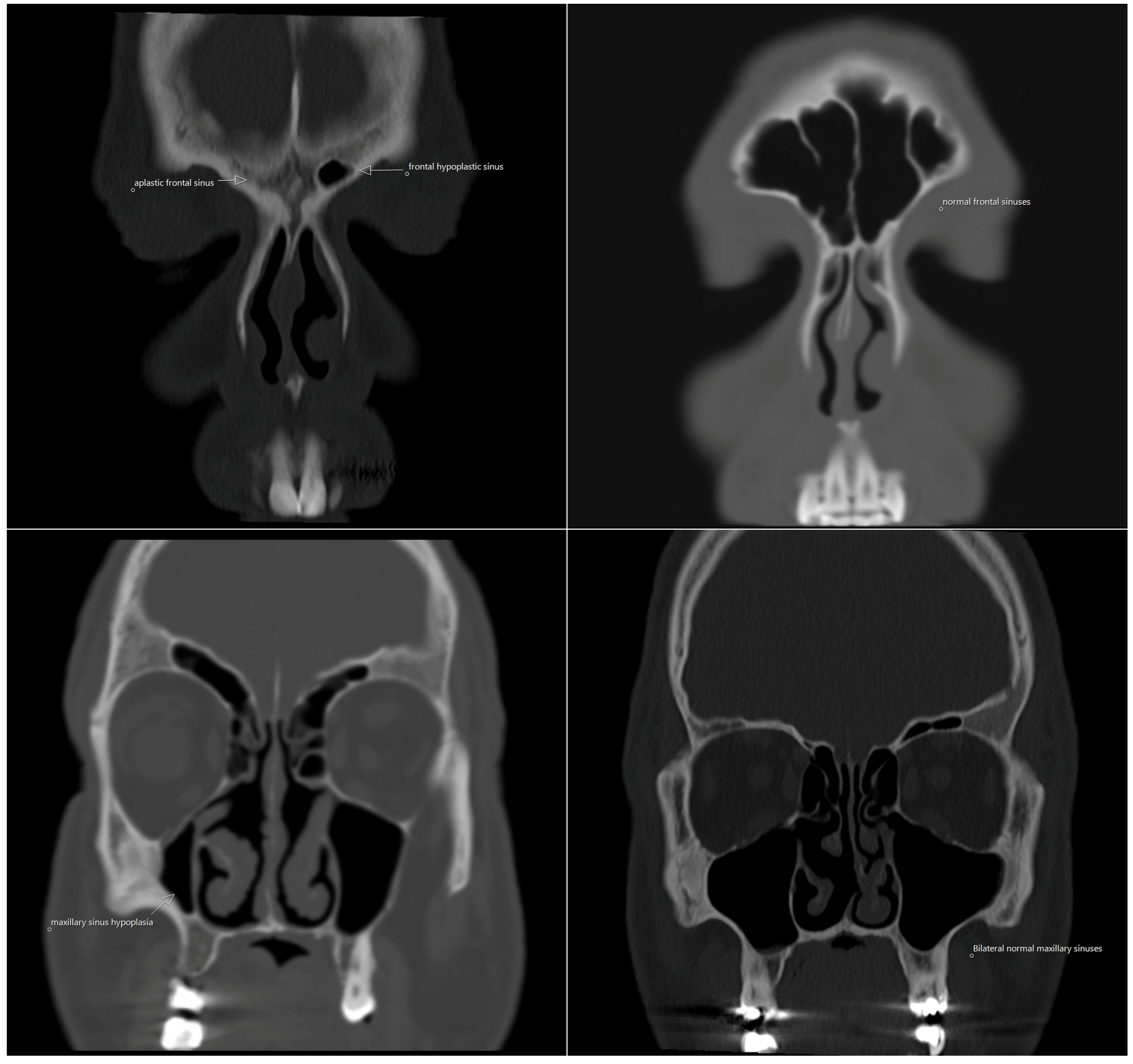
We evaluated 3000 paranasal CT examinations obtained during the study period. Among these, all eligible adults with frontal and/or maxillary sinus hypoplasia/aplasia were included in the study. The sinus variation groups consisted of unilateral frontal sinus variation (UFSV), unilateral maxillary sinus variation (UMSV), bilateral frontal sinus variation (BFSV), and bilateral maxillary sinus variation (BMSV). Sinus variation groups included both hypoplastic and aplastic sinuses. These entities were analyzed together because both represent reduced or absent sinus development within the same anatomical region, and further subdivision into separate hypoplasia and aplasia subgroups would have resulted in very small subgroup sizes, particularly for maxillary sinus variation, limiting the robustness of statistical comparisons. The bilateral maxillary sinus variation (BMSV) subgroup consisted of only 3 cases. Because this number was too small for reliable inferential comparison, and because pooling bilateral and unilateral maxillary sinus variation would have introduced anatomical heterogeneity, the BMSV subgroup was described descriptively but was not included in the main statistical analyses. The healthy control group was formed from eligible adults from the same imaging pool who had no radiologic evidence of frontal or maxillary sinus hypoplasia/aplasia. The CT images obtained in this study are shown in
Figure 1.
2.2. Imaging Protocol
A total of 3000 contrast-free paranasal sinus CT scans, acquired between 3 April 2023, and 30 May 2024, from the Picture Archiving and Communication Systems (PACS) Sectra PACS system of our institution, were examined. A CT 5300 (Philips Healthcare, Suzhou Co. Ltd., Suzhou, China) equipped with 64-row detectors and 128 slices was used for the CT imaging. The scan generated non-contrast continuous slices with characteristics including 1.0 mm slice thickness, 120 kV tube voltage, 200 mA tube current, and a field of view (FOV) of 140–160 mm. The scanning range encompassed the hard palate to the superior region of the frontal sinus. Subsequent coronal reconstruction was performed. The sections were reconstructed with a standard 512 × 512 matrix; for the 140–160 mm field of view, this corresponded to an in-plane pixel size of approximately 0.27–0.31 mm at a 1.0 mm reconstructed section thickness, so that the resulting voxels were not strictly isotropic. From this volumetric dataset, three-dimensional surface-rendered (volume-rendered) reconstructions were generated and used for all craniofacial measurements, while multiplanar reformatted images (axial, coronal, and sagittal planes) were available for anatomical orientation.
2.3. Image Analysis
Paranasal sinus CT images were reconstructed in three-dimensional format using 3D reconstruction on PACS. The reconstructions obtained were analyzed using the integrated measurement modules of the system, and linear distance measurements were performed between specific anthropometric reference points. All anatomical landmarks were identified and all linear craniofacial distances were measured directly on these three-dimensional surface-rendered (volume-rendered) reconstructions of the bony craniofacial framework, rather than on two-dimensional multiplanar reformatted images.
All craniofacial measurements were performed by two experienced anatomists using a consensus-based approach. Each CT image was examined jointly, and all anthropometric points were identified and measured by mutual agreement between the observers. The identification of each reference point and any discrepancies in the measurements were resolved by consensus until agreement was reached.
2.4. Measurements
The craniofacial distances measured were as follows: right and left orbital breadth, orbital height, interorbital breadth, biorbital breadth, minimum frontal breadth, upper facial breadth, bizygomatic breadth, nasal breadth, nasal aperture height, nasal height, and nasion–prosthion height. The definitions of these distances are given below.
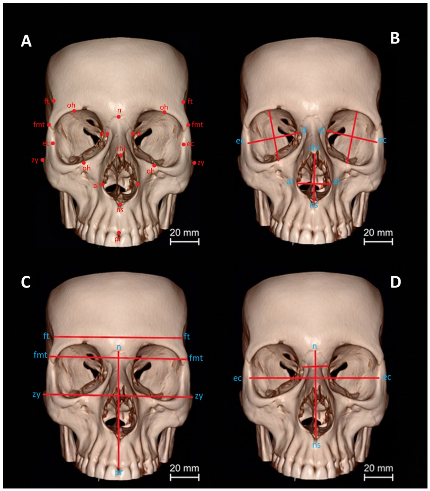
Orbital Breadth: The distance from dacryon to ectoconchion.
Orbital Height: The distance between the superior and inferior orbital margins perpendicular to the orbital breadth, bisecting the orbit into equal medial and lateral halves.
lnterorbital Breadth: The distance between right and left dacryon.
Biorbital Breadth: The distance from left to right ectoconchion.
Minimum Frontal Breadth: The distance between the right and left frontotemporale.
Upper Facial Breadth: The distance between the right and left frontomalare temporale.
Bizygomatic Breadth: The distance between right and left zygion.
Nasal Breadth: The distance between right and left alare.
Nasal Aperture Height: The distance from rhinion to nasospinale.
Nasal Height: The distance from nasion to nasospinale.
Nasion–Prosthion Height: The distance from nasion to prosthion.
Craniofacial distances were determined using Data Collection Procedures for Forensic Skeletal Material 2.0 book [
15]. The measurements are shown in
Figure 2 and definitions of the craniofacial landmarks used for morphometric measurements are provided in
Supplementary Table S1.
2.5. Statistical Analysis
Statistical analyses were performed using SAS version 9.4 statistical software package (Statistical Analysis Software SAS 9.4; SAS Institute, Cary, NC, USA). Means and standard deviations were used as descriptive statistics for quantitative variables, and numbers and percentages were used as descriptive statistics for qualitative variables. The Kolmogorov–Smirnov test and skewness coefficients were used to test whether the distribution of each quantitative variable was normally distributed. Skewness values between −2 and +2 are considered acceptable to prove a normal univariate distribution. The skewness values of each variable ranged from −2 to +2, and the results of Kolmogorov–Smirnov analyses showed a normal distribution; therefore, parametric statistical analyses were performed. Because controls were not individually matched to cases and the sex distribution differed across groups, age and sex were included as covariates in the ANCOVA models to reduce potential confounding. Analysis of covariance (ANCOVA) was used to compare group means in the craniofacial distances while controlling or adjusting for the influence of age and sex. Bonferroni corrected post hoc tests were used if a significant mean difference was found in the least-square group means. Bonferroni correction was applied across all multiple comparisons. The paired sample t-test was used for right-left comparisons of craniofacial distances.
Pearson correlation analysis was performed to reveal the association between craniofacial distance variables, and chi-square analysis was performed to reveal the relationship between qualitative variables. No a priori power or sample-size calculation was performed because this was a retrospective case–control study based on all eligible cases identified during the study period. Effect sizes (Cohen’s d) were reported to aid interpretation of the magnitude of group differences. Cohen’s d value was calculated to evaluate the effect size. An effect size (Cohen’s d) value was defined as small if it was less than 0.2, medium if it was 0.5, and large if it was greater than 0.8. Throughout the study, the threshold for statistical significance was set at p < 0.05.
3. Results
Our study included 117 patients with sinus variations and 53 healthy controls. The patients were divided into four groups. There were 46, 13, 55 in the UFSV, UMSV, BFSV group and three patients in the BMSV group, respectively. Among the patients in the UFSV group, 30 had right-sided sinus variations and 16 had left-sided sinus variations. Among the patients in the UMSV group, 11 had right-sided sinus variations and 2 had left-sided sinus variations. The BMSV subgroup included only 3 patients and was therefore not entered into the main comparative statistical analyses; these cases were considered descriptively only. The demographic characteristics of the study groups are presented in
Table 1.
Analysis of covariance with the subject’s sex and age as covariates was performed to compare groups for each of the craniofacial distances; the results are presented in
Table 2. Unadjusted means and standard errors for all craniofacial distances in different groups are presented in
Supplementary Table S2.
When the least square mean values of the measured parameters were examined, the left orbital breadth, right orbital breadth, and biorbital breadth were found to be the highest in the control group, whereas the left orbital height, upper facial breadth, and bizygomatic breadth were highest in the UMSV group. On the other hand, the least square mean values of the left orbital height, biorbital breadth, upper facial breadth, and bizygomatic breadth were lowest in the BFSV group, and the left orbital breadth and right orbital breadth had the lowest least square mean values in the UFSV group. The least square mean values of orbital height, interorbital breadth, minimum frontal breadth, nasal breadth, nasal aperture height, nasal height, and nasion–prosthion height were similar between the groups (p > 0.05).
In this analysis, statistically significant differences were found between the least squares means of the groups in the left orbital breadth (p = 0.0001), left orbital height (p = 0.0250), right orbital breadth (p = 0.0000), biorbital breadth (p = 0.0000), upper facial breadth (p = 0.0204), and bizygomatic breadth (p = 0.0026). In all other measured variables (right orbital height, orbital breadth, minimum frontal breadth, nasal breadth, nasal aperture height, nasal height, and nasion–prosthion height), no statistically significant differences were found between the groups (p > 0.05).
For all the significant craniofacial distances, Bonferroni-corrected pairwise comparisons showed that there were no significant differences between the least square means of HC and UMSV and of BFSV and UFSV, and the least square means of HC and UMSV subjects were significantly higher then those of BFSV and UFSV patients.
The right and left orbital measurements of the groups with unilateral sinus variation according to the variation side were compared, and the results are presented in
Table 3.
As can be seen from this table, no statistically significant differences were found between the right and left sides in orbital breadth and height measurements for both the UFSV and UMSV groups. Similarly, no statistically significant differences were observed in the right and left orbital breadth and orbital height measurements in the BFSV group. However, in the HC group, however, the right orbital height measurements were significantly lower than those on the left side (p = 0.0051).
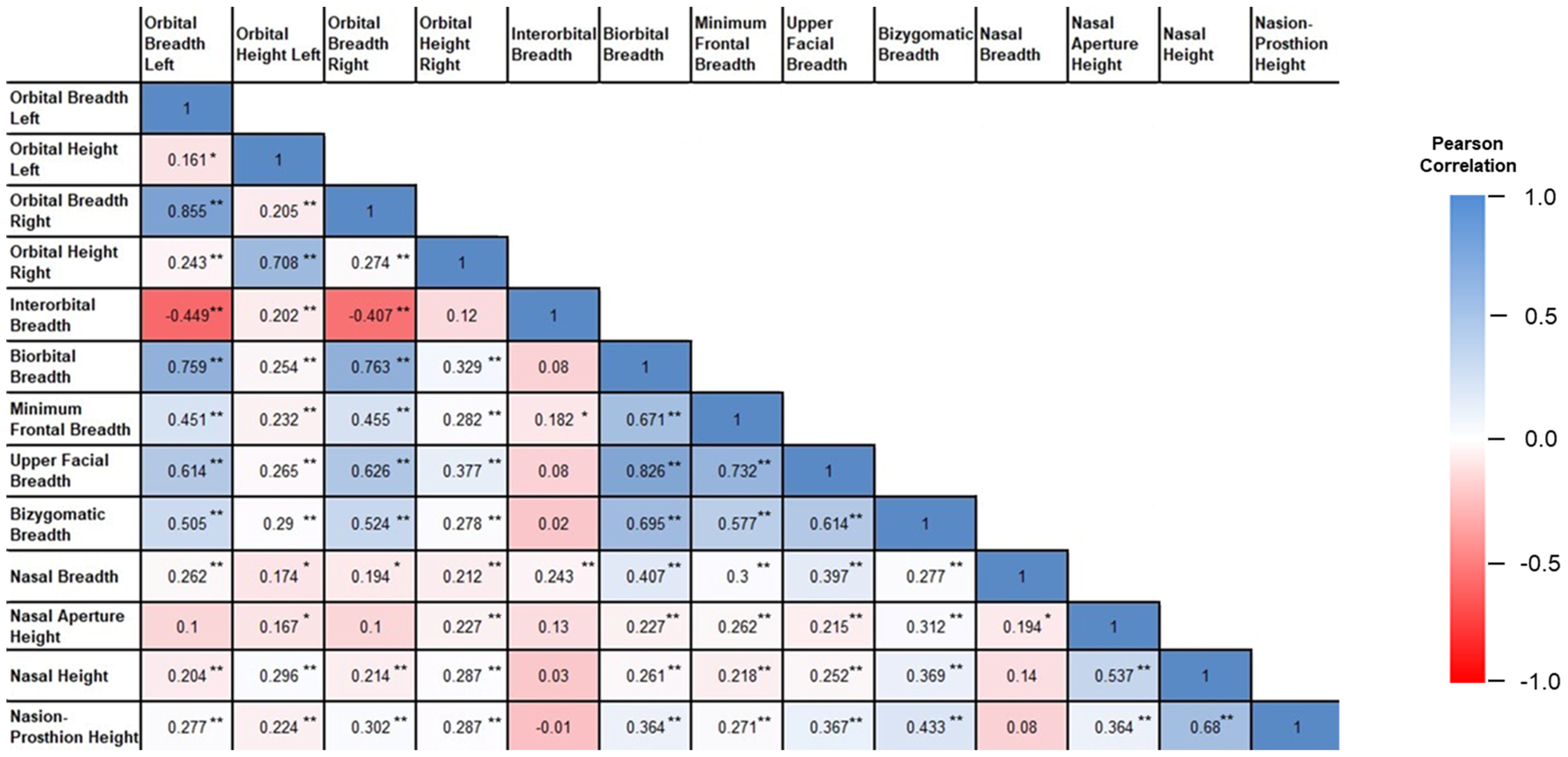
Analysis of the relationships among the craniofacial parameters revealed that the majority of craniofacial dimensions were significantly correlated. When the parameters were compared, the following pairs demonstrated a very strong positive correlation: right orbital breadth with left orbital breadth, right orbital height with left orbital height, both orbital breadths with biorbital breadth, upper facial breadth with biorbital breadth, and upper facial breadth with minimum frontal breadth. In contrast, both orbital breadths showed a moderate negative correlation with the interorbital breadth. The results of the Pearson correlation analysis for the 13 measured craniofacial distances are shown in
Figure 3. The complete correlation matrix, including all correlation coefficients and corresponding
p values are presented in
Supplementary Table S3.
4. Discussion
Preoperative endoscopic evaluation of sinus anatomy can be performed using CT, which is the gold standard, providing information about bone structures and air, thus providing a highly sensitive imaging method [
2].
The frontal sinus is the most complex of the paranasal sinuses owing to its location, anatomical variations, and multiple clinical presentations. Frontal sinuses are not present at birth and are usually well developed during childhood. They continue to grow gradually until reaching their maximum size after puberty [
16].
The maxillary sinuses are the largest among the paranasal sinuses. The maxillary sinus has a significant impact on the diagnosis and semiology of craniofacial structures [
17]. Maxillary sinus hypoplasia is less common than sphenoid or frontal sinus hypoplasia, and may be acquired or congenital. Uncinate process abnormalities associated with maxillary sinus hypoplasia may inadvertently cause orbital complications during surgery [
18].
The development of the paranasal sinuses is closely related to the growth of the facial skeleton and is an important factor to be considered in surgical planning [
10]. Additionally, many studies have shown that growth of the maxilla and nasal cavities is closely related to the development of the sinuses, and these structures ultimately determine the morphology of the face [
19].
Although the possible relationships between frontal and maxillary sinus variations and facial morphology have been reported in the literature, to our knowledge, no studies have specifically evaluated how these variations are associated with facial morphometry.
In the current study, we analyzed the differences in facial morphometry at some anthropometric points between individuals with sinus variations and individuals with healthy sinuses.
Our findings suggest that frontal sinus hypoplasia/aplasia may be associated with selected differences in craniofacial measurements, particularly in the orbital region.
Because of the retrospective and cross-sectional nature of this study, the observed relationships should be interpreted as associations rather than evidence of a developmental causal effect. It is possible that frontal sinus hypoplasia/aplasia and the measured craniofacial features reflect shared developmental determinants rather than a direct influence of one structure on the other. Alternative explanations include common genetic or embryologic factors affecting craniofacial growth, as well as measurement variability or selection-related bias inherent to retrospective imaging-based studies.
In our study, when we compared the HC group with other groups, we found that left orbital breadth, left orbital height, right orbital breadth, biorbital breadth, upper facial breadth, and bizygomatic breadth were significantly lower in the UFSV and BFSV groups than in the HC group. There was no statistically significant difference between the HC and UMSV groups. In the comparison of each parameter, the measurements related to the orbit and upper face region generally showed a very strong positive correlation with each other.
Aslier et al. investigated the effects of craniofacial structure and nasal septum deviation on frontal sinus morphology in 3 D using 74 dry skulls. As a result of their craniofacial measurements, they found the minimum frontal breadth as 94.42 ± 4.66 mm, bizygomatic breadth as 125.88 ± 6.92 mm, nasal breadth as 23.90 ± 2.05 mm, and nasal height as 50.12 ± 3.82 mm. In our study, we found the minimum frontal breadth as 96.58 ± 0.68 mm, bizygomatic breadth as 125.71 ± 0.89 mm, nasal breadth as 23.89 ± 0.28 mm, and nasal height as 53.29 ± 0.55 mm in healthy individuals. In the UFSV group, we found the minimum frontal breadth as 97.48 ± 0.72 mm, bizygomatic breadth as 124.36 ± 0.95 mm, nasal breadth as 23.65 ± 0.30 mm, and nasal height as 53.61 ± 0.59 mm. In the BFSV group, we found the minimum frontal breadth as 96.90 ± 0.67 mm, bizygomatic breadth as 123.87 ± 0.88 mm, nasal breadth as 23.23 ± 0.28 mm, and nasal height as 53.24 ± 0.55 mm. Accordingly, we observed that nasal breadth was not affected by sinus variations. In addition, bizygomatic breadth showed a significant decrease especially in the UFSV and BFSV groups, suggesting that sinus variations may reflect shared developmental patterns.
Aslier et al. investigated the relationship between different pneumatized frontal sinuses and cranial variables in groups with craniofacial and bony nasal septum deviation, and found a relationship between maxillary sinus breadth and nasal height in the right frontal sinus group, maximum head length, upper face height, and nasal height in the left frontal sinus group, zygomatic breadth, and nasal height. These studies have reported associations between sinus dimensions and facial height; however, such findings do not establish causality, and similar relationships may reflect shared developmental or genetic influences [
14]. In our study, we found no difference in nasal height between the control and frontal sinus groups with varied sinuses.
Abeta et al. investigated the relationship between morphometric measurements of the frontal sinuses and facial growth patterns by selecting individuals who had undergone 80-cone beam computed tomography (CBCT) scans. To evaluate the relationship between the craniofacial features and frontal sinus characteristics, cephalometric measurements were performed for each individual. They demonstrated a correlation between frontal sinus dimensions and craniofacial features [
17].
Gursoy et al. evaluated three-dimensional frontal sinus morphology, taking into account different vertical facial developments, and reported that the anteroposterior dimension of the frontal sinuses decreased in individuals with a vertical growth pattern and that significant correlations emerged with vertical craniofacial parameters [
20]. Furthermore, Albitar et al. evaluated facial measurements in relation to facial aesthetics in patients with a vertical growth pattern. In their study, they found that among individuals with a vertical growth pattern, lower mouth width/face height ratios in women and increased facial convexity angles in men were associated with higher attractiveness, and these findings highlighted the importance of including these variables in orthodontic diagnosis and treatment planning to improve facial aesthetics. Although our study focused on skeletal craniofacial distances rather than aesthetic evaluation, these findings suggest that evaluating facial morphometry in individuals with different craniofacial patterns can provide valuable clinical and anatomical information [
21].
Oksayan et al. divided 60 adults equally into three groups (high-angle, low-angle, and normal-angle groups) according to skeletal vertical facial growth patterns and compared maxillary sinus volume and dimensions using CBCT with cephalometric measurements. The researchers’ findings indicated that subjects with a high angle exhibited statistically lower values with regard to maxillary sinus length and breadth than subjects with a low angle. This study demonstrated that vertical facial growth had no impact on the right and left maxillary sinus volumes [
13]. In addition, Azizia et al. evaluated the relationship between sagittal skeletal discrepancies and maxillary sinus volume in adults using CBCT. Their study found no significant difference in maxillary sinus volume, surface area, and height among skeletal classes; however, they reported a significant increase in maxillary sinus width and depth in skeletal class I compared to skeletal class III [
22].
In the present study, we investigated the relationship between craniofacial distances in individuals with sinus variation and healthy individuals. The investigation revealed a positive correlation between the transverse craniofacial distances and a negative correlation between the vertical distances.
In summary, we observed no clear morphological impact of sinus variations on facial symmetry. These variations may affect more bone structures but may not change the general facial structure. However, the presence of similar variations may lead to more pronounced morphological changes in individuals during the growth period, suggesting that the observed associations may vary across developmental stages; however, the present adult cross-sectional dataset does not allow conclusions regarding growth-related effects over time.
From an otolaryngological perspective, these anatomical variants may be relevant during radiologic evaluation and preoperative assessment; however, their clinical implications require confirmation in larger studies. Although several group differences reached statistical significance, the absolute magnitude of these differences was small and measured in millimetres. Therefore, these findings should be interpreted primarily as morphometric associations at the group level rather than as differences with clear standalone clinical significance for individual patients. In particular, the observed differences are unlikely to directly change radiologic interpretation or surgical decision-making on their own, but they may contribute to a broader anatomical understanding of craniofacial patterns associated with sinus variation. In this context, awareness of such patterns may still be useful during preoperative imaging review, particularly in patients undergoing endoscopic sinus surgery or procedures involving the orbit or anterior skull base. However, these morphometric differences should be regarded as adjunctive anatomical information rather than independent determinants of surgical strategy.
The cranial base is at the intersection of many sub disciplines because its proximity to the orbit and maxilla. Therefore, it is important to understand the morphology of the frontal and maxillary sinuses, which are anatomical structures.
Frontal sinuses sometimes become excessively enlarged, but in pneumatosel types, they have been reported to cause facial asymmetry and changes in facial contours [
23].
Overly wide frontal sinuses can be observed together with facial asymmetry and can cause serious aesthetic problems. Forehead and frontal sinus surgeries may be necessary for some rare craniofacial malformations, such as pneumosinus dilatans. In such patients, the aim is to correct the protruding forehead/frontal boss by minimizing the overly wide frontal sinuses [
24].
Surgeons sometimes enter the skull through the frontal sinus, and awareness of the asymmetrical frontal sinuses is very important in minimizing surgical complications. In frontal sinus surgery, especially in patients with narrow or medium-breadth head/upper face structures and asymmetric skull base openings, detailed anatomical knowledge and preoperative evaluation with CT are of great importance because of the frequent occurrence of anatomical variations and proximity to the orbit and anterior skull base.
We believe this study may contribute to the anatomical characterization of craniofacial morphometric patterns associated with sinus variation. However, the observed differences were small in absolute magnitude and should be interpreted cautiously with respect to direct clinical application.
This study has several limitations. First, its retrospective design does not allow causal inference and may be subject to incomplete or unevenly documented clinical information. Many factors known to influence craniofacial morphology and dimensions—such as ethnic origin, body measurements (height and weight), dental condition, history of previous orthodontic or orthognathic treatment, and skeletal classification—were not available in the database and, due to the retrospective nature of this study, could not be systematically recorded. Consequently, in the statistical analyses, only age and sex were controlled for using ANCOVA as covariates. Consequently, these variables may have a potential effect on craniofacial measurements. Second, the study was conducted at a single center, which may limit the representativeness of the sample. Third, because participants were selected from patients who underwent paranasal CT in a clinical setting, selection bias cannot be excluded, and the findings may therefore have limited generalizability beyond similar adult hospital-based populations. In addition, the sex distribution of the groups was not homogeneous. To reduce potential confounding, age and sex were included as covariates in the ANCOVA models; however, residual confounding cannot be excluded. Controls were selected from eligible individuals imaged during the same study period rather than being individually matched to cases. Furthermore, no a priori power or sample-size calculation was performed because the study was based on retrospectively available eligible cases and controls. Another limitation is that hypoplasia and aplasia were analyzed together within the same variation groups. Although these entities are biologically distinct, separate subgroup analyses were not feasible because of the limited number of cases in several subgroups, particularly those involving maxillary sinus variation. Lastly, all measurements were obtained through a consensus-based assessment carried out jointly by two experienced anatomists. Although this approach aimed to minimise variability in the determination of reference points and measurement procedures, as the measurements were not recorded independently, formal inter-observer and intra-observer reliability analyses, such as intraclass correlation coefficient (ICC) calculations or Bland–Altman plots, could not be performed.
Future studies evaluating morphometric variables in facial asymmetry with a larger number of subjects are necessary to confirm our findings. Furthermore, we believe that the findings of this study can serve as a starting point for future studies that could advance our understanding of facial morphology development and its contribution to the presence of sinus variations and healthy controls.
In conclusion, individuals with unilateral and bilateral frontal sinus variation showed significantly lower left orbital breadth, right orbital breadth, and biorbital breadth compared with healthy controls, whereas no significant differences were observed for most other measurements or in the maxillary sinus variation groups. These findings indicate an association between frontal sinus hypoplasia/aplasia and selected craniofacial morphometric differences, particularly in the orbital region. However, the present study does not support causal inference, and the observed relationships may also reflect shared developmental factors or methodological limitations. Further studies with larger and more balanced samples are needed to clarify the biological and clinical relevance of these associations.



 ,
,