Intra-Individual Variability of Myocardial Blood Flow and Flow Reserve Assessed by [15O]H2O-PET in Patients with Angina and No Obstructive Coronary Disease

 , , ,
, , ,  , and
, and
Abstract
1. Introduction
2. Methods
2.1. Study Population
2.2. Eligibility Criteria
2.3. PET-CT Scans and Imaging Protocol
2.4. Diurnal Variation and Atrial Fibrillation
2.5. Analysis and Readers
2.6. Statistics
3. Results and Analysis
3.1. Study Population
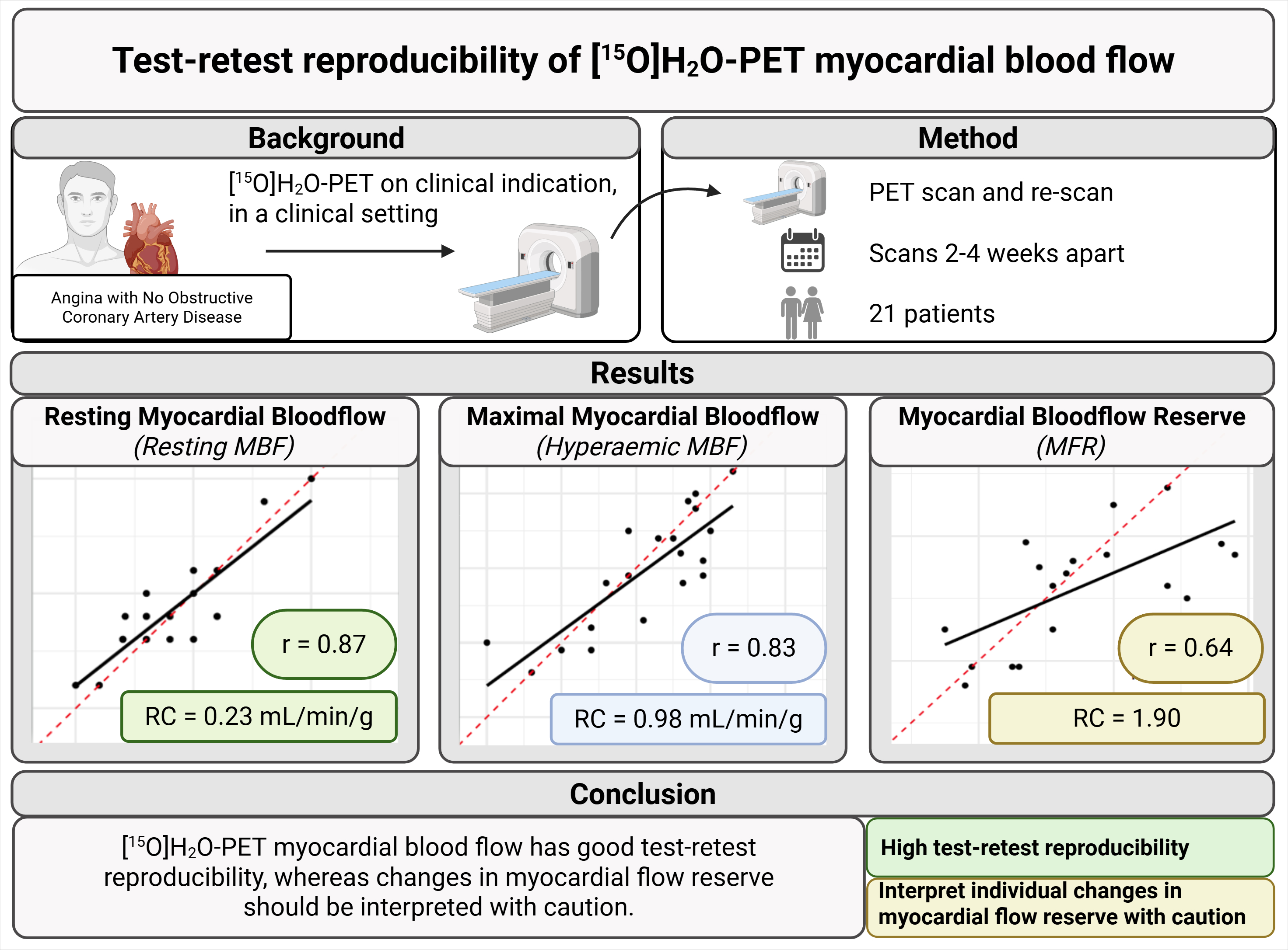
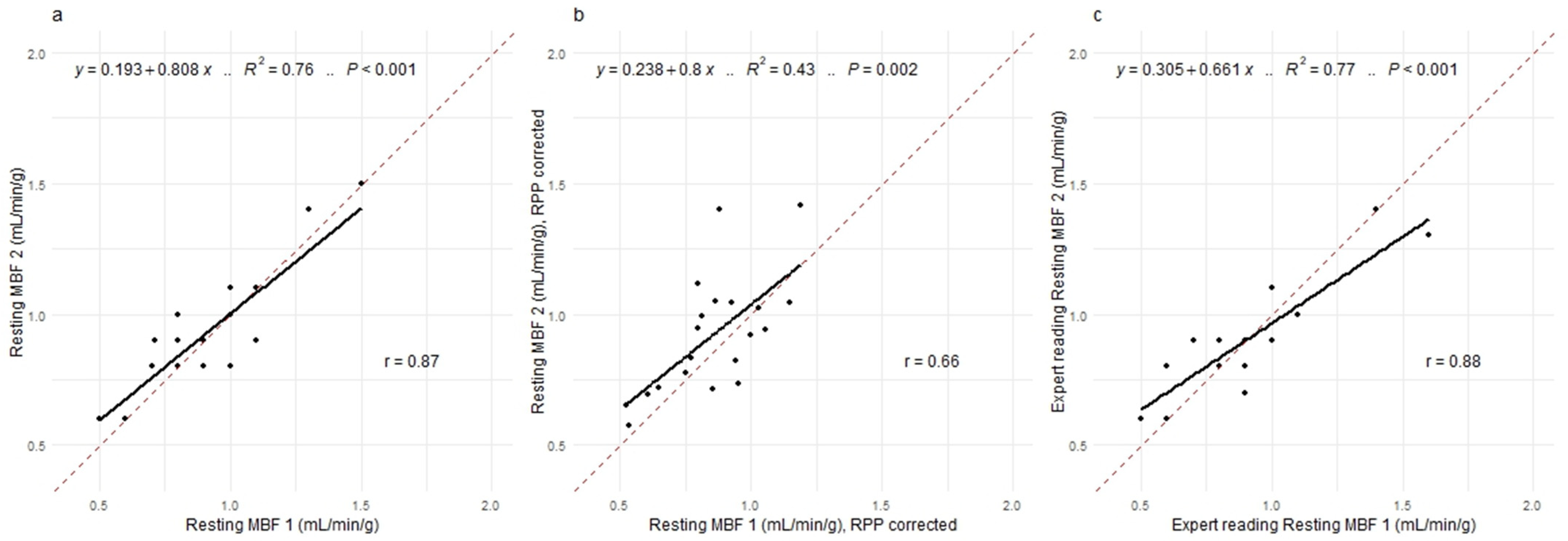
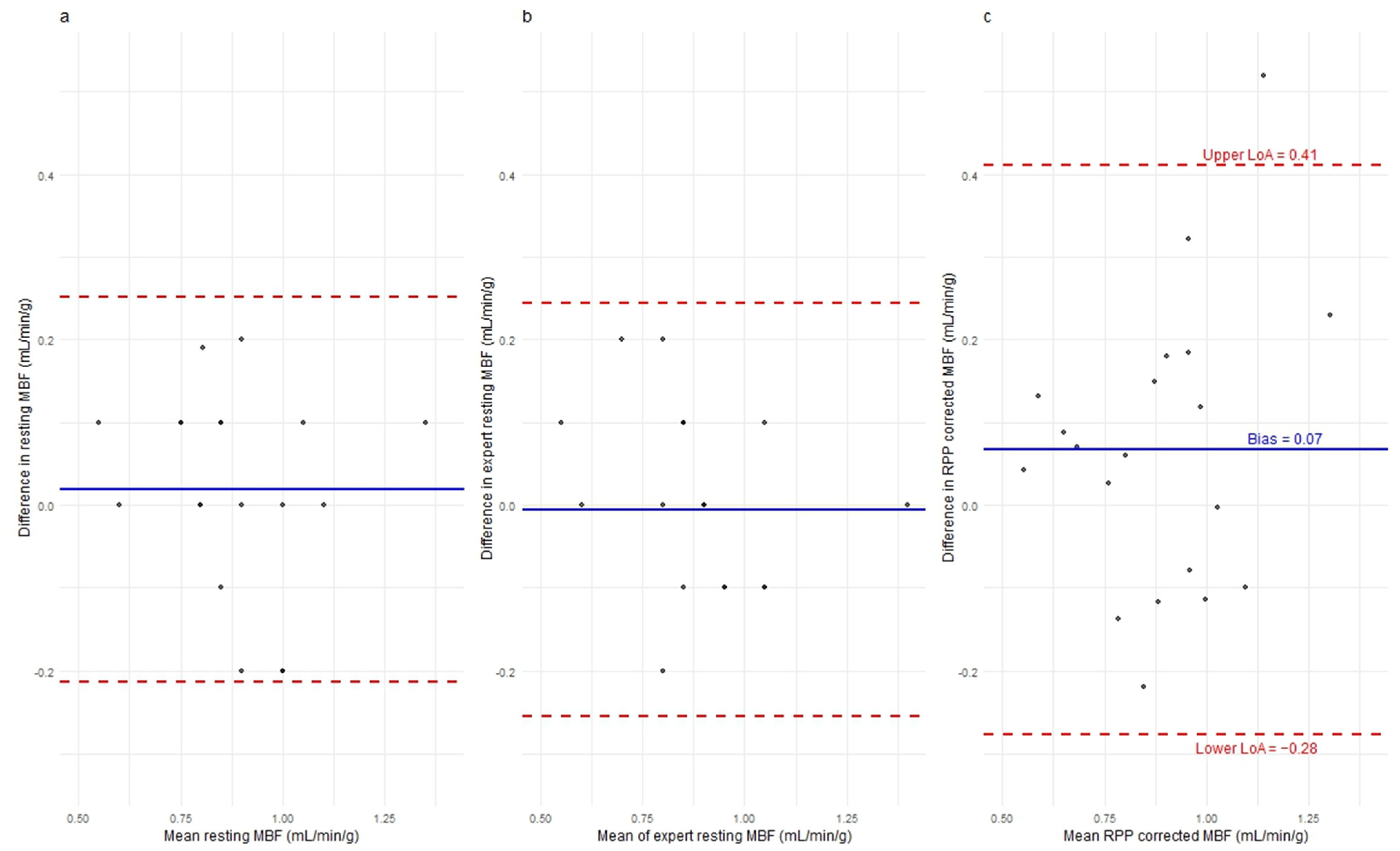
3.2. Resting MBF
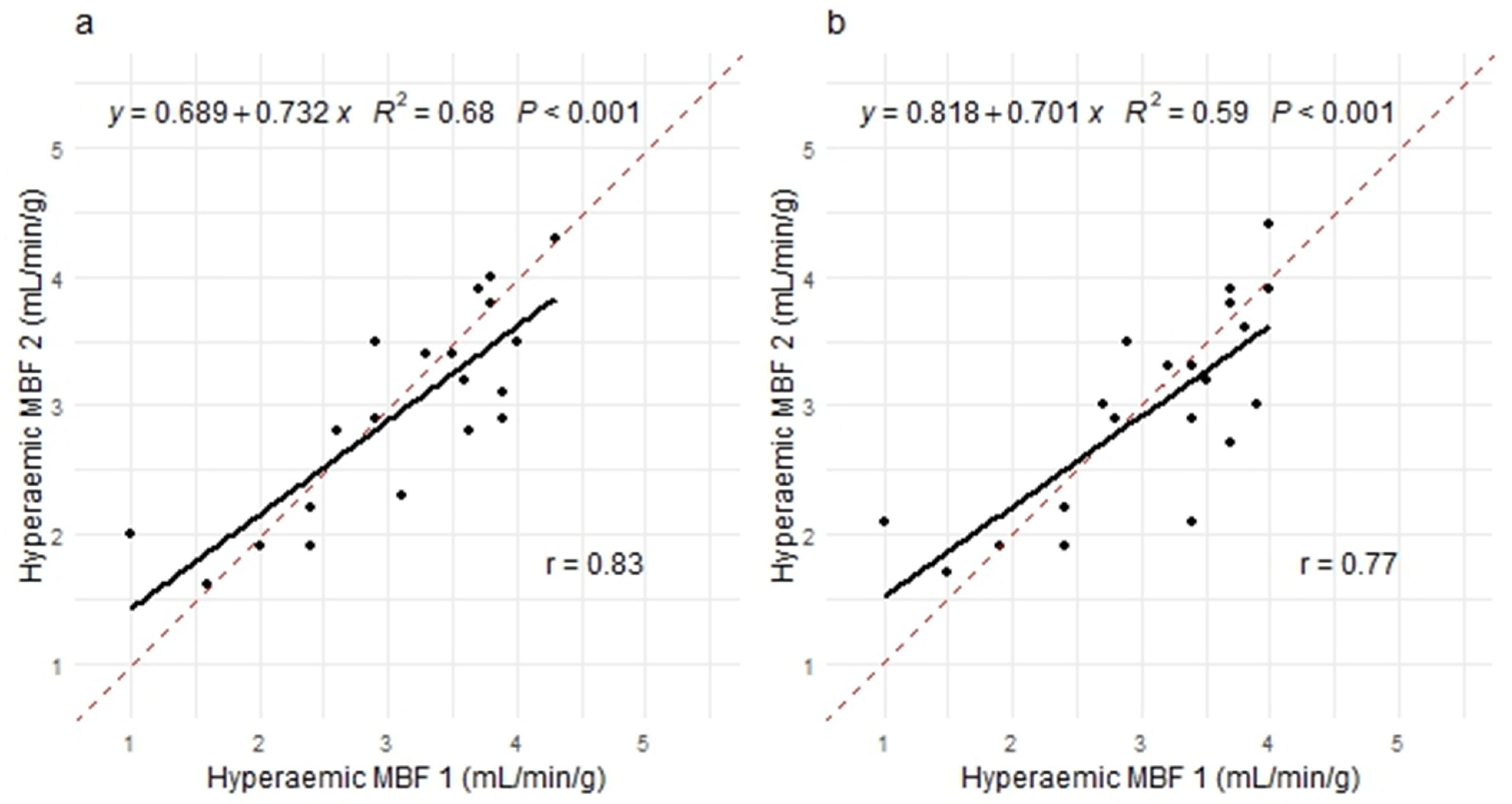
3.3. Hyperaemic MBF
3.4. Myocardial Flow Reserve
3.5. Atrial Fibrillation and Diurnal Variation
4. Discussion
4.1. Resting and Hyperaemic MBF Reproducibility
4.2. MFR Reproducibility
4.3. Rate–Pressure Product Variability
4.4. Technical and Biological Variation
4.5. Clinical Implications
4.6. Limitations
5. Conclusions
Supplementary Materials
Author Contributions
Funding
Institutional Review Board Statement
Informed Consent Statement
Data Availability Statement
Acknowledgments
Conflicts of Interest
Abbreviations
| CFVR | Coronary Flow Velocity Reserve |
| CV | Coefficient of Variation |
| CMR | Cardiac Magnetic Resonance |
| LoA | Limit of Agreement |
| MBF | Myocardial Blood Flow |
| MFR | Myocardial Flow Reserve |
| MPI | Myocardial Perfusion Imaging |
| PET | Positron Emission Tomography |
| RC | Repeatability Coefficient |
| RPP | Rate–Pressure Product |
References
- Schindler, T.H.; Fearon, W.F.; Pelletier-Galarneau, M.; Ambrosio, G.; Sechtem, U.; Ruddy, T.D.; Patel, K.K.; Bhatt, D.L.; Bateman, T.M.; Gewirtz, H.; et al. Myocardial Perfusion PET for the Detection and Reporting of Coronary Microvascular Dysfunction: A JACC: Cardiovascular Imaging Expert Panel Statement. JACC Cardiovasc. Imaging 2023, 16, 536–548. [Google Scholar] [CrossRef] [Scilit] [PubMed]
- Taqueti, V.R.; Hachamovitch, R.; Murthy, V.L.; Naya, M.; Foster, C.R.; Hainer, J.; Dorbala, S.; Blankstein, R.; Di Carli, M.F. Global coronary flow reserve is associated with adverse cardiovascular events independently of luminal angiographic severity and modifies the effect of early revascularization. Circulation 2015, 131, 19–27. [Google Scholar] [CrossRef] [Scilit] [PubMed]
- Rehan, R.; Yong, A.; Ng, M.; Weaver, J.; Puranik, R. Coronary microvascular dysfunction: A review of recent progress and clinical implications. Front. Cardiovasc. Med. 2023, 10, 1111721. [Google Scholar] [CrossRef] [Scilit] [PubMed]
- Højstrup, S.; Hansen, K.W.; Talleruphuus, U.; Marner, L.; Bjerking, L.; Jakobsen, L.; Christiansen, E.H.; Bouchelouche, K.; Wiinberg, N.; Guldbrandsen, K.; et al. Myocardial Flow Reserve, an Independent Prognostic Marker of All-Cause Mortality Assessed by 82Rb PET Myocardial Perfusion Imaging: A Danish Multicenter Study. Circ. Cardiovasc. Imaging 2023, 16, E015184. [Google Scholar] [CrossRef] [Scilit] [PubMed]
- D’Antonio, A.; Assante, R.; Zampella, E.; Cantoni, V.; Green, R.; Gaudieri, V.; Mannarino, T.; Falzarano, M.; Volpicelli, F.; Cutillo, P.; et al. Prognostic value of myocardial flow reserve by PET imaging in patients with suspected coronary artery disease: A systematic review and meta-analysis. IJC Heart Vasc. 2025, 58, 101677. [Google Scholar] [CrossRef] [Scilit] [PubMed]
- Vrints, C.; Andreotti, F.; Koskinas, K.C.; Rossello, X.; Adamo, M.; Ainslie, J.; Banning, A.P.; Budaj, A.; Buechel, R.R.; Chiariello, G.A.; et al. 2024 ESC Guidelines for the management of chronic coronary syndromes. Eur. Heart J. 2024, 45, 3415–3537. [Google Scholar] [CrossRef] [Scilit] [PubMed]
- Hsu, B. PET tracers and techniques for measuring myocardial blood flow in patients with coronary artery disease. J. BioMed. Res. 2013, 27, 452–459. [Google Scholar] [CrossRef] [Scilit] [PubMed]
- Maaniitty, T.; Knuuti, J.; Saraste, A. 15O-Water PET MPI: Current Status and Future Perspectives. Semin Nucl. Med. 2020, 50, 238–247. [Google Scholar] [CrossRef] [Scilit] [PubMed]
- Sciagrà, R.; Lubberink, M.; Hyafil, F.; Saraste, A.; Slart, R.H.J.A.; Agostini, D.; Nappi, C.; Georgoulias, P.; Bucerius, J.; Rischpler, C.; et al. EANM procedural guidelines for PET/CT quantitative myocardial perfusion imaging. Eur. J. Nucl. Med. Mol. Imaging 2021, 48, 1040–1069. [Google Scholar] [CrossRef] [Scilit] [PubMed]
- Manabe, O.; Naya, M.; Aikawa, T.; Yoshinaga, K. 15O-labeled Water is the Best Myocardial Blood Flow Tracer for Precise MBF Quantification. Ann. Nucl. Cardiol. 2019, 5, 69–72. [Google Scholar] [CrossRef] [Scilit]
- Siegrist, P.T.; Gaemperli, O.; Koepfli, P.; Schepis, T.; Namdar, M.; Valenta, I.; Aiello, F.; Fleischmann, S.; Alkadhi, H.; Kaufmann, P.A. Repeatability of Cold Pressor Test-Induced Flow Increase Assessed with H2 15O and PET. J. Nucl. Med. 2006, 47, 1420–1426. [Google Scholar] [CrossRef] [Scilit] [PubMed]
- Kaufmann, P.A.; Gnecchi-Ruscone, T.; Yap, J.T.; Rimoldi, O.; Camici, P.G. Assessment of the reproducibility of baseline and hyperemic myocardial blood flow measurements with 15O-labeled water and PET. J. Nucl. Med. 1999, 40, 1848–1856. [Google Scholar] [PubMed]
- Taqueti, V.R.; Di Carli, M.F. Coronary Microvascular Disease Pathogenic Mechanisms and Therapeutic Options: JACC State-of-the-Art Review. J. Am. Coll. Cardiol. 2018, 72, 2625–2641. [Google Scholar] [CrossRef] [Scilit] [PubMed]
- Camici, P.G.; Crea, F. Coronary microvascular dysfunction. N. Engl. J. Med. 2007, 356, 830–840. [Google Scholar] [CrossRef] [Scilit] [PubMed]
- Kaufmann, P.A.; Camici, P.G. Myocardial Blood Flow Measurement by PET: Technical Aspects and Clinical Applications. J. Nucl. Med. 2005, 46, 75–88. [Google Scholar] [PubMed]
- Danad, I.; Uusitalo, V.; Kero, T.; Saraste, A.; Raijmakers, P.G.; Lammertsma, A.A.; Heymans, M.W.; Kajander, S.A.; Pietilä, M.; James, S.; et al. Quantitative assessment of myocardial perfusion in the detection of significant coronary artery disease: Cutoff values and diagnostic accuracy of quantitative [15O]H2O PET imaging. J. Am. Coll. Cardiol. 2014, 64, 1464–1475. [Google Scholar] [CrossRef] [Scilit] [PubMed]
- Nesterov, S.V.; Han, C.; Mäki, M.; Kajander, S.; Naum, A.G.; Helenius, H.; Lisinen, I.; Ukkonen, H.; Pietilä, M.; Joutsiniemi, E.; et al. Myocardial perfusion quantitation with 15O-labelled water PET: High reproducibility of the new cardiac analysis software (Carimas). Eur. J. Nucl. Med. Mol. Imaging 2009, 36, 1594–1602. [Google Scholar] [CrossRef] [Scilit] [PubMed]
- Huck, D.M.; Weber, B.N.; Brown, J.M.; Lopez, D.; Hainer, J.; Blankstein, R.; Dorbala, S.; Divakaran, S.; Di Carli, M.F. Prognostic value of myocardial flow reserve vs corrected myocardial flow reserve in patients without obstructive coronary artery disease. J. Nucl. Cardiol. 2024, 37, 101854. [Google Scholar] [CrossRef] [Scilit] [PubMed]
- Bland, J.M.; Altman, D.G. Statistical methods for assessing agreement between two methods of clinical measurement. Lancet 1986, 327, 307–310. [Google Scholar] [CrossRef] [Scilit]
- Cicchetti, D.V.; Sparrow, S.A. Developing criteria for establishing interrater reliability of specific items: Applications to assessment of adaptive behavior. Am. J. Ment. Defic. 1981, 86, 127–137. [Google Scholar] [PubMed]
- Królikowska, A.; Reichert, P.; Karlsson, J.; Mouton, C.; Becker, R.; Prill, R. Improving the reliability of measurements in orthopaedics and sports medicine. Knee Surg. Sports Traumatol. Arthrosc. 2023, 31, 5277–5285. [Google Scholar] [CrossRef] [Scilit] [PubMed]
- Manabe, O.; Yoshinaga, K.; Katoh, C.; Naya, M.; deKemp, R.A.; Tamaki, N. Repeatability of rest and hyperemic myocardial blood flow measurements with 82Rb dynamic PET. J. Nucl. Med. 2009, 50, 68–71. [Google Scholar] [CrossRef] [Scilit] [PubMed]
- El Fakhri, G.; Kardan, A.; Sitek, A.; Dorbala, S.; Abi-Hatem, N.; Lahoud, Y.; Fischman, A.; Coughlan, M.; Yasuda, T.; Di Carli, M.F. Reproducibility and accuracy of quantitative myocardial blood flow assessment with 82Rb PET: Comparison with 13N-ammonia PET. J. Nucl. Med. 2009, 50, 1062–1071. [Google Scholar] [CrossRef] [Scilit] [PubMed]
- Wyss, C.A.; Koepfli, P.; Mikolajczyk, K.; Burger, C.; Von Schulthess, G.K.; Kaufmann, P.A. Bicycle Exercise Stress in PET for Assessment of Coronary Flow Reserve: Repeatability and Comparison with Adenosine Stress. J. Nucl. Med. 2003, 44, 146–154. [Google Scholar] [PubMed]
- Efseaff, M.; Klein, R.; Ziadi, M.C.; Beanlands, R.S.; Dekemp, R.A. Short-term repeatability of resting myocardial blood flow measurements using rubidium-82 PET imaging. J. Nucl. Cardiol. 2012, 19, 997–1006. [Google Scholar] [CrossRef] [Scilit] [PubMed]
- Chareonthaitawee, P.; Christenson, S.D.; Anderson, J.L.; Kemp, B.J.; Hodge, D.O.; Ritman, E.L.; Gibbons, R.J. Reproducibility of Measurements of Regional Myocardial Blood Flow in a Model of Coronary Artery Disease: Comparison of H215O and 13NH3 PET Techniques. J. Nucl. Med. 2006, 47, 1193–1201. [Google Scholar] [PubMed]
- Kitkungvan, D.; Johnson, N.P.; Roby, A.E.; Patel, M.B.; Kirkeeide, R.; Gould, K.L. Routine Clinical Quantitative Rest Stress Myocardial Perfusion for Managing Coronary Artery Disease: Clinical Relevance of Test-Retest Variability. JACC Cardiovasc. Imaging 2017, 10, 565–577. [Google Scholar] [CrossRef] [Scilit] [PubMed]
- Lubberink, M.; Nordstrom, J.; Sigfridsson, J.; Svanstrom, P.; Eggers, K.M.; Sorensen, J.; Kero, T. Routine clinical reproducibility of hyperemic myocardial blood flow measurements with 15O-water-PET. Eur. Heart J. Cardiovasc. Imaging 2024, 25, jeae142. [Google Scholar] [CrossRef] [Scilit]
- Morton, G.; Jogiya, R.; Plein, S.; Schuster, A.; Chiribiri, A.; Nagel, E. Quantitative cardiovascular magnetic resonance perfusion imaging: Inter-study reproducibility. Eur. Heart J. Cardiovasc. Imaging 2012, 13, 954–960. [Google Scholar] [CrossRef] [Scilit] [PubMed]
- Olsen, R.H.; Pedersen, L.R.; Snoer, M.; Christensen, T.E.; Ghotbi, A.A.; Hasbak, P.; Prescott, E. Coronary flow velocity reserve by echocardiography: Feasibility, reproducibility and agreement with PET in overweight and obese patients with stable and revascularized coronary artery disease. Cardiovasc. Ultrasound 2016, 14, 22. [Google Scholar] [CrossRef] [Scilit] [PubMed]
- Murthy, V.L.; Bateman, T.M.; Beanlands, R.S.; Berman, D.S.; Borges-Neto, S.; Chareonthaitawee, P.; Cerqueira, M.D.; Dekemp, R.A.; DePuey, E.G.; Dilsizian, V.; et al. Clinical quantification of myocardial blood flow using PET: Joint position paper of the SNMMI cardiovascular council and the ASNC. J. Nucl. Med. 2018, 59, 273–293. [Google Scholar] [PubMed]
- Al Rifai, M.; Ahmed, A.I.; Nayfeh, M.; Nabi, F.; Al-Mallah, M.H. The prognostic significance of rate pressure product corrected myocardial flow reserve. J. Nucl. Cardiol. 2025, 49, 102222. [Google Scholar] [CrossRef] [Scilit] [PubMed]
- Gewirtz, H. Coronary circulation: Pressure/flow parameters for assessment of ischemic heart disease. J. Nucl. Cardiol. 2019, 26, 459–470. [Google Scholar] [CrossRef] [Scilit] [PubMed]
- Mcginn, A.L.; White, C.W.; Wilson, R.F. Interstudy Variability of Coronary Flow Reserve Influence of Heart Rate, Arterial Pressure, and Ventricular Preload. Circulation 1990, 81, 1319–1330. [Google Scholar] [CrossRef] [Scilit] [PubMed]
- Vohra, S.; Sakul, S.; Agarwal, V.; Patel, K.K. Quantification of myocardial blood flow using PET: Current status, clinical applications, and future directions. Prog. Cardiovasc. Dis. 2025, 93, 10–20. [Google Scholar] [CrossRef] [Scilit] [PubMed]
- Krakauer, M.; Ismail, A.; Talleruphuus, U.; Henriksen, A.C.; Lonsdale, M.N.; Rasmussen, I.L.; Fuglsang, S.; Prescott, E.; Hovind, P.; Marner, L. 82Rb and [15O]H2O myocardial perfusion PET imaging: A prospective head to head comparison. J. Nucl. Cardiol. 2023, 30, 2790–2802. [Google Scholar] [CrossRef] [Scilit] [PubMed]
- Prior, J.O.; Allenbach, G.; Valenta, I.; Kosinski, M.; Burger, C.; Verdun, F.R.; Delaloye, A.B.; Kaufmann, P.A. Quantification of myocardial blood flow with 82Rb positron emission tomography: Clinical validation with 15O-water. Eur. J. Nucl. Med. Mol. Imaging 2012, 39, 1037–1047. [Google Scholar] [CrossRef] [Scilit] [PubMed]
- Ziadi, M.C.; Dekemp, R.A.; Williams, K.A.; Guo, A.; Chow, B.J.W.; Renaud, J.M.; Sarveswaran, N.; Tee, R.E.; Beanlands, R.S. Impaired myocardial flow reserve on rubidium-82 positron emission tomography imaging predicts adverse outcomes in patients assessed for myocardial ischemia. J. Am. Coll. Cardiol. 2011, 58, 740–748. [Google Scholar] [CrossRef] [Scilit] [PubMed]
- Rauf, M.; Hansen, K.W.; Galatius, S.; Wiinberg, N.; Brinth, L.S.; Højstrup, S.; Talleruphuus, U.; Prescott, E. Prognostic implications of myocardial perfusion imaging by 82-rubidium positron emission tomography in male and female patients with angina and no perfusion defects. Eur. Heart J. Cardiovasc. Imaging 2023, 24, 212–222. [Google Scholar] [CrossRef] [Scilit] [PubMed]
- Knuuti, J.; Wijns, W.; Saraste, A.; Capodanno, D.; Barbato, E.; Funck-Brentano, C.; Prescott, E.; Storey, R.F.; Deaton, C.; Cuisset, T.; et al. 2019 ESC Guidelines for the diagnosis and management of chronic coronary syndromes. Eur. Heart J. 2020, 41, 407–477. [Google Scholar] [CrossRef] [Scilit] [PubMed]




| Patient characteristics | N = 21 (10 female/11 male) |
| Age (years) | 69.8 (8.21) |
| Sex (Female) | 10 (47.6%) |
| BMI | 28.7 (6.31) |
| Smoking status: Current Former Never | 1 (4.8%) 12 (57.1%) 8 (38.1%) |
| Hypertension | 11 (52.4%) |
| Hypercholesterolemia | 10 (47.6%) |
| Diabetes type II | 4 (19.0%) |
| Atrial fibrillation | 5 (23.8%) |
| Medicine changes b | 2 (9.5%) |
| Interval between scans (days) | 21 (5.37) [10;29] |
| Scan time of day similarity a | 12 (57.1%) |
| Scan 1 Mean (SD) | Scan 2 Mean (SD) | p-Value d | Mean Difference (SD) | RC *, mL/min/g (95% CI) | CV **, % (95% CI) | ICC *** (95% CI) | Difference Median (IQR) [Range] | |
|---|---|---|---|---|---|---|---|---|
| Resting MBF, Global a | ||||||||
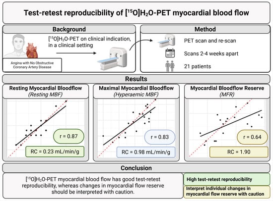
| Original | 0.91 (0.24) | 0.92 (0.22) | 0.47 | 0.02 (0.12) | 0.23 (0.18;0.34) | 9.2 (7.0;13.4) | 0.87 (0.71;0.95) | 0 (0.1) [0.4] |
| Expert | 0.92 (0.26) | 0.91 (0.19) | 0.86 | −0.01 (0.13) | 0.25 (0.19;0.37) | 9.9 (7.5;14.4) | 0.85 (0.67;0.94) | 0 (0.2) [0.5] |
| RPP-corrected c | 0.85 (0.19) | 0.92 (0.22) | 0.10 | 0.07 (0.18) | 0.34 (0.26;0.50) | 14.0 (10.6;20.4) | 0.62 (0.27;0.83) | 0.07 (0.24) [0.7] |
| Hyperaemic MBF, Global a | ||||||||
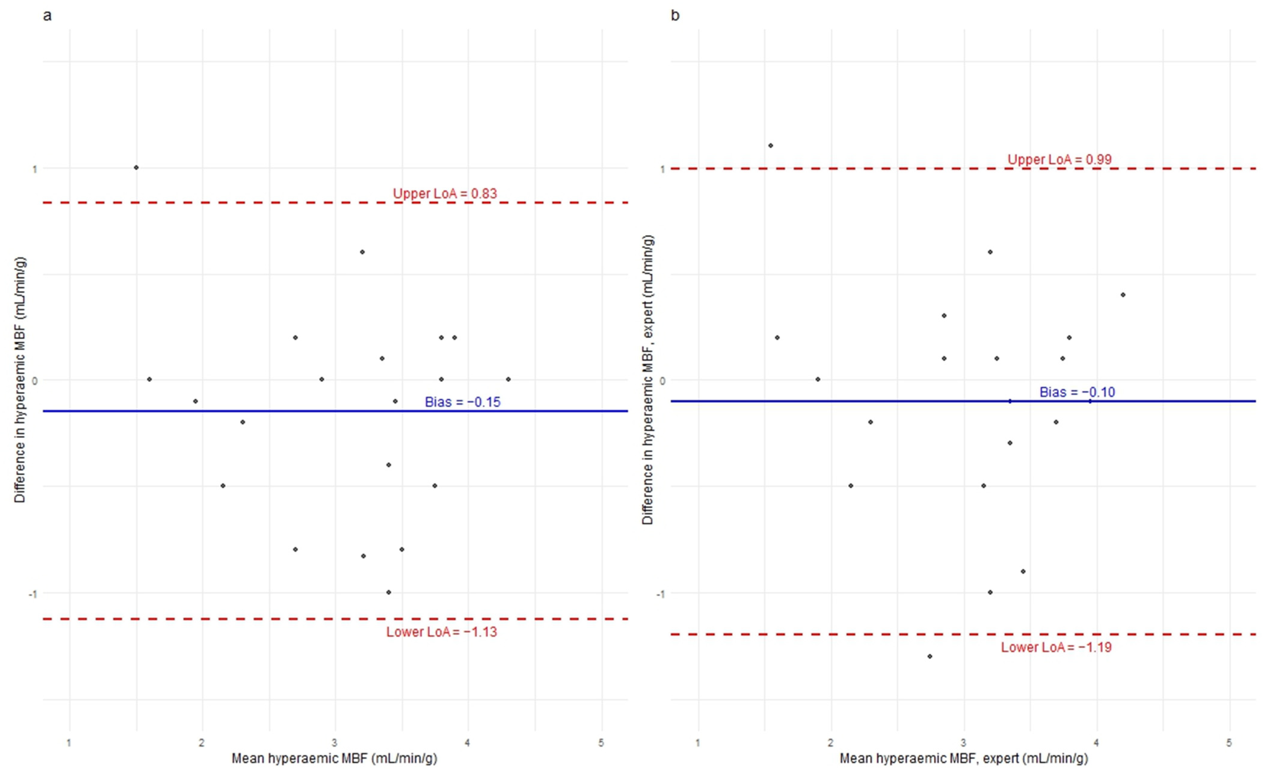
| Original | 3.06 (0.90) | 2.97 (0.78) | 0.21 | −0.15 (0.50) | 0.98 (0.75;1.43) | 11.6 (8.8;17.0) | 0.81 (0.60;0.92) | −0.05 (0.625) [2] |
| Expert | 3.0 (0.88) | 2.97 (0.78) | 0.21 | −0.10 (0.56) | 1.09 (0.83;1.60) | 13.1 (10.0;19.1) | 0.77 (0.51;0.90) | −0.05 (0.55) [2.4] |
| Myocardial Flow Reserve b | ||||||||
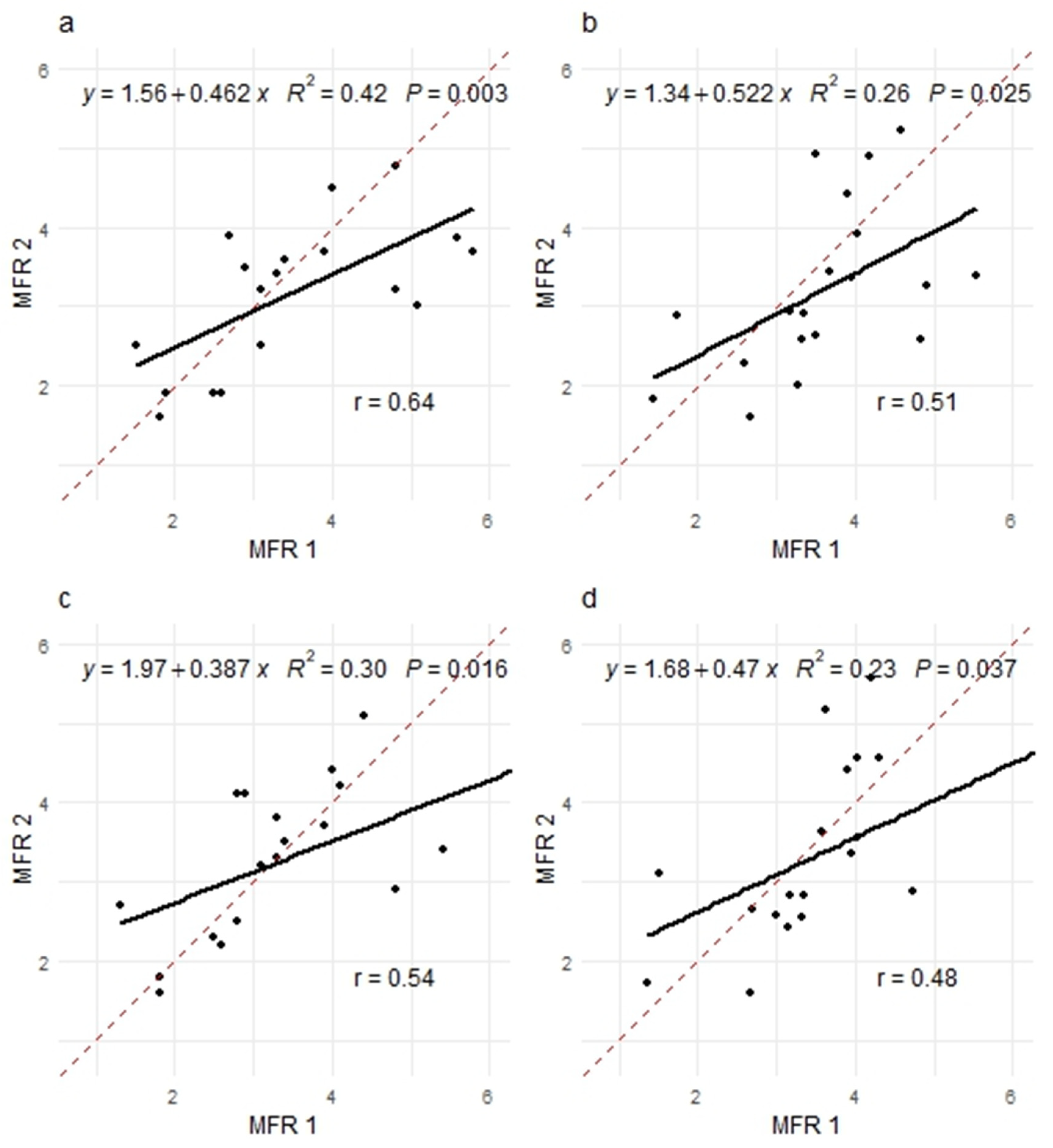
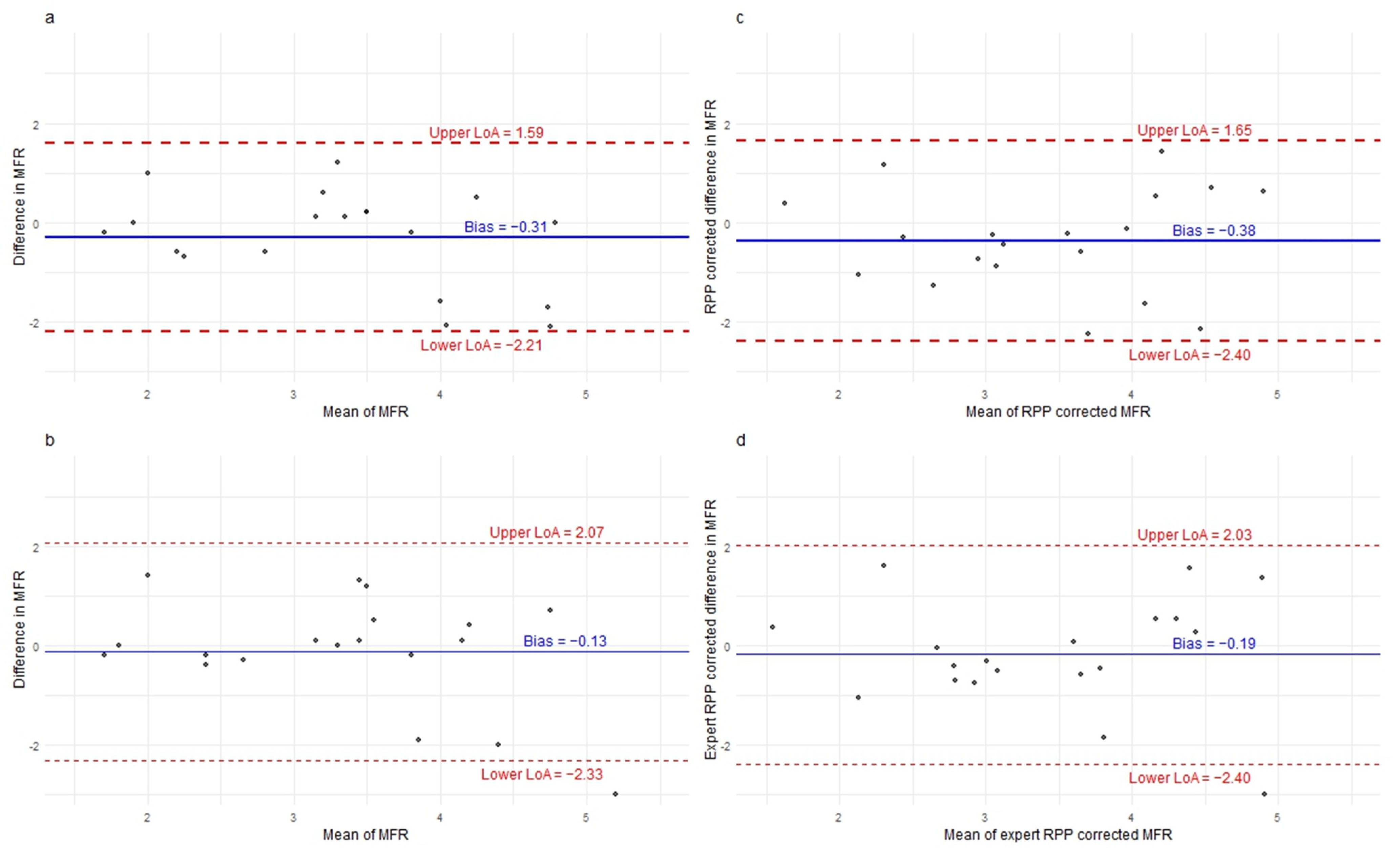
| Original | 3.47 (1.23) | 3.23 (0.92) | 0.18 | −0.31 (0.97) | 1.90 (1.43;2.81) | 20.6 (15.5;30.4) | 0.60 (0.23;0.82) | −0.02 (0.85) [3.3] |
| RPP-corrected c | 3.59 (1.01) | 3.26 (1.05) | 0.13 | −0.38 (1.03) | 2.03 (1.53;3.00) | 21.5 (16.2;31.8) | 0.49 (0.08;0.79) | −0.31 (1.42) [3.68] |
| Expert | 3.40 (1.28) | 3.23 (0.92) | 0.63 | −0.13 (1.12) | 2.20 (1.66;3.25) | 23.7 (17.9;35.0) | 0.53 (0.12;0.79) | 0 (0.7) [4.4] |
| Expert RPP-corrected c | 3.52 (1.09) | 3.36 (1.07) | 0.48 | −0.19 (1.13) | 2.21 (1.67;3.27) | 23.3 (17.6;34.4) | 0.49 (0.07;0.77) | −0.33 (1.09) [4.63] |
Disclaimer/Publisher’s Note: The statements, opinions and data contained in all publications are solely those of the individual author(s) and contributor(s) and not of MDPI and/or the editor(s). MDPI and/or the editor(s) disclaim responsibility for any injury to people or property resulting from any ideas, methods, instructions or products referred to in the content. |
© 2026 by the authors. Licensee MDPI, Basel, Switzerland. This article is an open access article distributed under the terms and conditions of the Creative Commons Attribution (CC BY) license.
Share and Cite
Seidelin, L.; Prescott, E.; Fischer, M.; Haahr, R.; Hovind, P.; Rauf, M.; Krakauer, M. Intra-Individual Variability of Myocardial Blood Flow and Flow Reserve Assessed by [15O]H2O-PET in Patients with Angina and No Obstructive Coronary Disease. Diagnostics 2026, 16, 1975. https://doi.org/10.3390/diagnostics16131975
Seidelin L, Prescott E, Fischer M, Haahr R, Hovind P, Rauf M, Krakauer M. Intra-Individual Variability of Myocardial Blood Flow and Flow Reserve Assessed by [15O]H2O-PET in Patients with Angina and No Obstructive Coronary Disease. Diagnostics. 2026; 16(13):1975. https://doi.org/10.3390/diagnostics16131975
Chicago/Turabian StyleSeidelin, Laila, Eva Prescott, Mads Fischer, Rasmus Haahr, Peter Hovind, Maira Rauf, and Martin Krakauer. 2026. "Intra-Individual Variability of Myocardial Blood Flow and Flow Reserve Assessed by [15O]H2O-PET in Patients with Angina and No Obstructive Coronary Disease" Diagnostics 16, no. 13: 1975. https://doi.org/10.3390/diagnostics16131975
APA StyleSeidelin, L., Prescott, E., Fischer, M., Haahr, R., Hovind, P., Rauf, M., & Krakauer, M. (2026). Intra-Individual Variability of Myocardial Blood Flow and Flow Reserve Assessed by [15O]H2O-PET in Patients with Angina and No Obstructive Coronary Disease. Diagnostics, 16(13), 1975. https://doi.org/10.3390/diagnostics16131975