Hydrogel-Coated Clips Are Associated with a Higher Risk of Dislocation After Ultrasound-Guided Breast Biopsy
 , , , , ,
Abstract
1. Introduction
2. Materials and Methods
2.1. Study Design
2.2. Inclusion and Exclusion Criteria
2.3. Ultrasound Examination
2.4. Biopsy Procedure and Follow-Up Examinations
- UltraClip Dual Trigger Biodur 108 Coil Marker (UC), Becton Dickinson, Franklin Lakes, NJ, USA;
- TUMARK Professional (TP), Somatex Medical Technologies, Berlin, Germany;
- TUMARK Vision (TV), Somatex Medical Technologies, Berlin, Germany;
- HydroMARK Breast Biopsy Site Marker (HM), Mammatome, Quickborn, Germany.
2.5. Histopathologic Evaluation
2.6. Statistical Analysis
3. Results
3.1. Patient and Lesion Demographics
3.2. Dislocation/Migration Rates and Time Courses
3.3. Analysis of Cofactors of Clip Dislocation
4. Discussion
Limitations
5. Conclusions
Author Contributions
Funding
Institutional Review Board Statement
Informed Consent Statement
Data Availability Statement
Acknowledgments
Conflicts of Interest
Abbreviations
| UC | UltraClip Dual Trigger Biodur 108 Coil Marker |
| TP | TUMARK Professional |
| TV | TUMARK Vision |
| HM | HydroMARK Breast Biopsy Site Marker |
| SWE | Shear Wave elastography |
| ACR | American College of Radiology |
| SE | Strain elastography |
| CNB | Core needle biopsy |
References
- Xiong, X.; Zheng, L.W.; Ding, Y.; Chen, Y.F.; Cai, Y.W.; Wang, L.P.; Huang, L.; Liu, C.C.; Shao, Z.M.; Yu, K.D. Breast cancer: Pathogenesis and treatments. Signal Transduct. Target. Ther. 2025, 10, 49. [Google Scholar] [CrossRef] [PubMed]
- Kopicky, L.; Fan, B.; Valente, S.A. Intraoperative evaluation of surgical margins in breast cancer. Semin. Diagn. Pathol. 2024, 41, 293–300. [Google Scholar] [CrossRef] [PubMed]
- Liberman, L.; Dershaw, D.D.; Morris, E.A.; Abramson, A.F.; Thornton, C.M.; Rosen, P.P. Clip placement after stereotactic vacuum-assisted breast biopsy. Radiology 1997, 205, 417–422. [Google Scholar] [CrossRef] [PubMed]
- Shaukat, S.; Haroon, J.; Afzal, F.; Mazhar, S.; Shaukat, Z.; Ghaffar, Z.; Masood, M.J. Role of Ultrasound-Guided Marker Placement in Breast Lesion for Precise Localization, Surgical Resection, Neoadjuvant Chemotherapy Response, and Post-Chemotherapy Follow-Up. Cureus 2025, 17, e89402. [Google Scholar] [CrossRef] [PubMed]
- Minella, C.; Villasco, A.; D’Alonzo, M.; Cellini, L.; Accomasso, F.; Actis, S.; Biglia, N. Surgery after Neoadjuvant Chemotherapy: A Clip-Based Technique to Improve Surgical Outcomes, a Single-Center Experience. Cancers 2022, 14, 2229. [Google Scholar] [CrossRef] [PubMed]
- Couto, H.L.; Hassan, A.T.; Steinmacher, D.I.; Pessoa, E.C.; Millen, E.C.; Zerwes, F.; Cavalcante, F.P.; Tosello, G.; Novita, G.; Machado Badan, G.; et al. Breast and axillary marking in the neoadjuvant setting: Survey results from experts of the Brazilian society of mastology. Front. Oncol. 2024, 14, 1393417. [Google Scholar] [CrossRef] [PubMed]
- Shalaby, L.A.S.E.D.; Khallaf, E.S.e.d.; Moussa, M.M. Clip and wire localization of locally advanced malignant breast masses in patients undergoing neoadjuvant chemotherapy and breast conservation therapy. Egypt. J. Radiol. Nucl. Med. 2019, 50, 96. [Google Scholar] [CrossRef]
- Alamdaran, S.A.; Gharib, M.; Homaee Shandiz, F.; Bizhani, O.; Forghani Torghaban, M.N.; Farrokh Tehrani, D.; Keshtgar, M. Accuracy Assessment of Surgical Clip Marker/Wire Localization in Advanced Breast Cancer. Rep. Radiother. Oncol. 2018, 4, e80855. [Google Scholar] [CrossRef]
- Lee, I.T.L.; Ma, K.S.K.; Luan, Y.Z.; Chen, J.L. Immediate clip migration after breast biopsy: A meta-analysis for potential risk factors. Br. J. Radiol. 2022, 95, 20220195. [Google Scholar] [CrossRef] [PubMed]
- Jaszewski, P.; Tejero, H. Core Versus Vacuum-Assisted Postbiopsy Breast Clip Migration Rates. Cureus 2025, 17, e84113. [Google Scholar] [CrossRef] [PubMed]
- Schulz-Wendtland, R.; Dankerl, P.; Bani, M.R.; Fasching, P.A.; Heusinger, K.; Lux, M.P.; Jud, S.M.; Rauh, C.; Bayer, C.M.; Schrauder, M.G.; et al. Evaluation of a Marker Clip System in Sonographically Guided Core Needle Biopsy for Breast Cancer Localization Before and After Neoadjuvant Chemotherapy. Geburtshilfe Frauenheilkd. 2017, 77, 169–175. [Google Scholar] [CrossRef] [PubMed]
- Pinkney, D.M.; Mychajlowycz, M.; Shah, B.A. A prospective comparative study to evaluate the displacement of four commercially available breast biopsy markers. Br. J. Radiol. 2016, 89, 20160149. [Google Scholar] [CrossRef] [PubMed]
- Smith, S.; Taylor, C.R.; Kanevsky, E.; Povoski, S.P.; Hawley, J.R. Long-term safety and efficacy of breast biopsy markers in clinical practice. Expert Rev. Med. Devices 2021, 18, 121–128. [Google Scholar] [CrossRef] [PubMed]
- Schünemann, H.J.; Lerda, D.; Quinn, C.; Follmann, M.; Alonso-Coello, P.; Rossi, P.G.; Lebeau, A.; Nyström, L.; Broeders, M.; Ioannidou-Mouzaka, L.; et al. Breast Cancer Screening and Diagnosis: A Synopsis of the European Breast Guidelines. Ann. Intern. Med. 2020, 172, 46. [Google Scholar] [CrossRef] [PubMed]
- Winkler, N.S. Ultrasound Guided Core Breast Biopsies. Tech. Vasc. Interv. Radiol. 2021, 24, 100776. [Google Scholar] [CrossRef] [PubMed]
- Youn, I.; Choi, S.H.; Kook, S.H.; Choi, Y.J.; Park, C.H.; Park, Y.L.; Kim, D.H. Ultrasonography-Guided Surgical Clip Placement for Tumor Localization in Patients Undergoing Neoadjuvant Chemotherapy for Breast Cancer. J. Breast Cancer 2015, 18, 44. [Google Scholar] [CrossRef] [PubMed]
- Alshafeiy, T.I.; Matich, A.; Rochman, C.M.; Harvey, J.A. Advantages and Challenges of Using Breast Biopsy Markers. J. Breast Imaging 2022, 4, 78–95. [Google Scholar] [CrossRef] [PubMed]
- Klein, R.L.; Mook, J.A.; Euhus, D.M.; Rao, R.; Wynn, R.T.; Eastman, A.B.; Leitch, A.M. Evaluation of a hydrogel based breast biopsy marker (HydroMARK®) as an alternative to wire and radioactive seed localization for non-palpable breast lesions. J. Surg. Oncol. 2012, 105, 591–594. [Google Scholar] [CrossRef] [PubMed]
- Gentile, L.F.; Himmler, A.; Shaw, C.M.; Bouton, A.; Vorhis, E.; Marshall, J.; Spiguel, L.R. Ultrasound-Guided Segmental Mastectomy and Excisional Biopsy Using Hydrogel-Encapsulated Clip Localization as an Alternative to Wire Localization. Ann. Surg. Oncol. 2016, 23, 3284–3289. [Google Scholar] [CrossRef] [PubMed]
- Sakamoto, N.; Fukuma, E.; Tsunoda, Y.; Teraoka, K.; Koshida, Y. Evaluation of the dislocation and long-term sonographic detectability of a hydrogel-based breast biopsy site marker. Breast Cancer 2018, 25, 575–582. [Google Scholar] [CrossRef] [PubMed]
- Shah, A.D.; Mehta, A.K.; Talati, N.; Brem, R.; Margolies, L.R. Breast tissue markers: Why? What’s out there? How do I choose? Clin. Imaging 2018, 52, 123–136. [Google Scholar] [CrossRef] [PubMed]
- Portnow, L.H.; Thornton, C.M.; Milch, H.S.; Mango, V.L.; Morris, E.A.; Saphier, N.B. Biopsy Marker Standardization: What’s in a Name? Am. J. Roentgenol. 2019, 212, 1400–1405. [Google Scholar] [CrossRef] [PubMed]
- Aloufi, A.S.; Abujamea, A.H.; Fakhouri, F.S.; Albeshan, S.M.; Alohaly, N.A.; Sabir, R.A.; Aldawsari, A.M.; Altokhais, R.S.; Malik, M.A.; Alruwaili, A.R. Differentiating Breast Tissue Stiffness With Magnetic Resonance Elastography (MRE): A Focus on Fibroglandular and Fatty Tissues. NMR Biomed. 2025, 38, e70134. [Google Scholar] [CrossRef] [PubMed]
- Guo, R.; Lu, G.; Qin, B.; Fei, B. Ultrasound Imaging Technologies for Breast Cancer Detection and Management: A Review. Ultrasound Med. Biol. 2018, 44, 37–70. [Google Scholar] [CrossRef] [PubMed]
- Martinez, J.; Smith, P.C. The Dynamic Interaction between Extracellular Matrix Remodeling and Breast Tumor Progression. Cells 2021, 10, 1046. [Google Scholar] [CrossRef] [PubMed]
- Selim, Y.A.Y.; Sabit, H.; Arneth, B.; Shaaban, M.A. Diagnostic Accuracy of Sonoelastography for Breast Lesions: A Meta-Analysis Comparing Strain and Shear Wave Elastography. J. Imaging 2025, 11, 221. [Google Scholar] [CrossRef] [PubMed]
- Sigrist, R.M.S.; Liau, J.; Kaffas, A.E.; Chammas, M.C.; Willmann, J.K. Ultrasound Elastography: Review of Techniques and Clinical Applications. Theranostics 2017, 7, 1303–1329. [Google Scholar] [CrossRef] [PubMed]
- Barr, R.G. Future of breast elastography. Ultrasonography 2019, 38, 93–105. [Google Scholar] [CrossRef] [PubMed]
- Andrade, G.; Pereira, A.; Gonçalves, L.; Videira, C. Intraductal Migration of a Breast Tissue Marker Placed under Ultrasound Guidance during COVID-Induced Delay of Surgery. J. Breast Cancer 2021, 24, 402–408. [Google Scholar] [CrossRef] [PubMed]
- Jain, A.; Khalid, M.; Qureshi, M.M.; Georgian-Smith, D.; Kaplan, J.A.; Buch, K.; Grinstaff, M.W.; Hirsch, A.E.; Hines, N.L.; Anderson, S.W.; et al. Stereotactic core needle breast biopsy marker migration: An analysis of factors contributing to immediate marker migration. Eur. Radiol. 2017, 27, 4797–4803. [Google Scholar] [CrossRef] [PubMed]
- McCarty, C.; Yi, M.; Sous, S.; Leslie, M.; Tariq, E.; Dondapati, P.; Kameyama, H.; Nuguri, S.; Chakraborty, N.; Wilkerson, M.; et al. Sustained Inflammation of Breast Tumors after Needle Biopsy. Pathobiology 2023, 90, 114–122. [Google Scholar] [CrossRef] [PubMed]
- Shirazi, S.; Hajiesmaeili, H.; Khosla, M.; Taj, S.; Sircar, T.; Vidya, R. Comparison of Wire and Non-Wire Localisation Techniques in Breast Cancer Surgery: A Review of the Literature with Pooled Analysis. Medicina 2023, 59, 1297. [Google Scholar] [CrossRef] [PubMed]
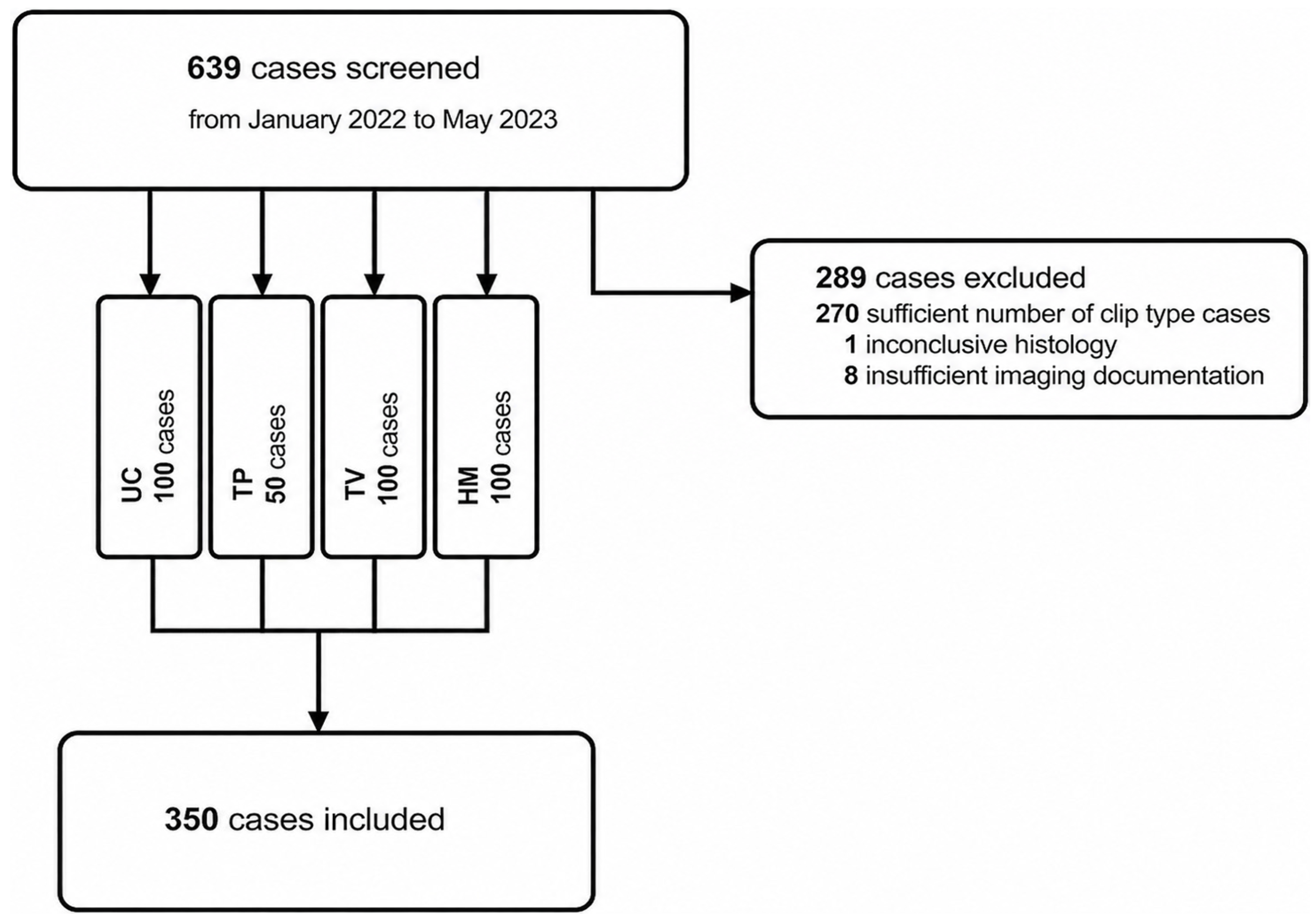


| Inclusion Criteria | Exclusion Criteria |
|---|---|
| Ultrasound-guided biopsy for a newly diagnosed breast tumour with same-day clip confirmation mammogram | Patient age under 18 years |
| Sufficient ultrasound documentation, including B-mode and shear-wave elastography imaging | Prior history of ipsilateral breast cancer |
| Follow-up imaging, including post-biopsy mammogram and (a) at least one follow-up examination, including post-wire mammogram or (b) one follow-up examination at least 3 months apart from the initial placement. | Insufficient ultrasound documentation |
| Available histological diagnosis | Patients undergoing neoadjuvant Therapy |
| Inconclusive histology |
| Variables | n (%) |
|---|---|
| Sex | |
| Female | 346 (98.9) |
| Male | 4 (1.1) |
| Age group | |
| 18–39 | 39 (11.1) |
| 40–59 | 141 (40.3) |
| ≥60 | 170 (48.6) |
| Breast lesions localisation | |
| Left breast | 187 (53.4) |
| Right breast | 163 (46.6) |
| Upper outer quadrant | 196 (56.0) |
| Lower outer quadrant | 72 (20.6) |
| Upper inner quadrant | 38 (10.9) |
| Lower inner quadrant | 44 (12.5) |
| Overall breast density | |
| ACR A | 61 (18.9) |
| ACR B | 83 (25.7) |
| ACR C | 114 (35.3) |
| ACR D | 65 (20.1) |
| Perilesional breast density | |
| Mostly glandular tissue | 38 (10.9) |
| Mixed glandular and fatty tissue | 123 (35.1) |
| Predominantly fatty tissue | 189 (54.0) |
| Maximal lesion diameter | [mm] |
| Mean ± SD | 16.5 ± 10.8 |
| Range | 2.0 to 70.0 |
| Lesion volume | [cm3] |
| Mean ± SD | 2.2 ± 4.9 |
| range | 0.1 to 47.0 |
| Histopathological Diagnosis | n (%) |
|---|---|
| Benign lesions | 107 (30.6) |
| Fibrous-cystic mastopathy | 60 (56.1) |
| Fibroadenoma | 33 (30.8) |
| Focal mastitis/fatty necrosis | 7 (6.5) |
| other | 7 (6.5) |
| High-risk lesions | 13 (3.7) |
| Papillomatous neoplasia | 7 (53.8) |
| ADH/epithelial proliferation | 4 (30.8) |
| other | 2 (15.4) |
| Malignant lesions | 230 (65.7) |
| Invasive carcinoma, no special type (NST) | 183 (79.6) |
| Ductal carcinoma in-situ | 20 (8.7) |
| Invasive lobular carcinoma | 15 (6.5) |
| other | 12 (5.2) |
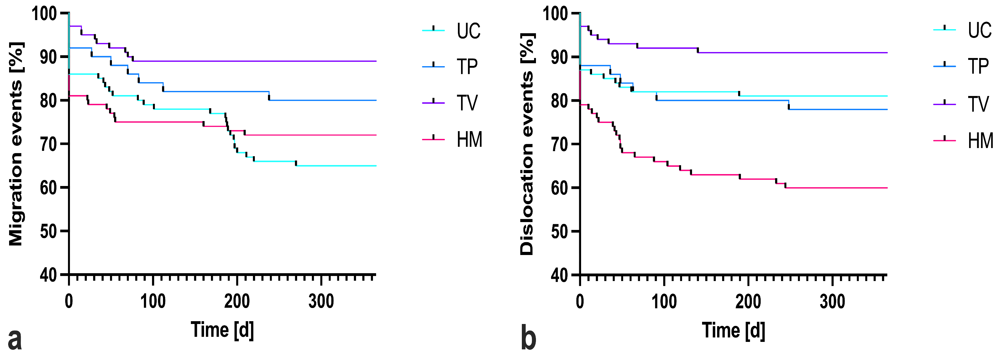
| Clip Type | Overall Clips (n) | Migration Rate (%) | Dislocation Rate (%) | Clip Displacement Distance [mm] |
|---|---|---|---|---|
| UC | 100 | 26 | 14 | 4.7 ± 4.7 mm |
| TP | 50 | 18 | 20 | 3.6 ± 3.6 mm |
| TV | 100 | 10 | 9 | 4.9 ± 2.6 mm |
| HM | 100 | 25 | 38 | 4.0 ± 4.3 mm |
| OR (95% CI) | ||||||
|---|---|---|---|---|---|---|
| Variables | b | SE | p-Value | OR | Lower Value | Upper Value |
| Predominantly fatty tissue perilesional | 1.105 | 0.553 | 0.046 | 3.020 | 1.021 | 8.932 |
| Distance Lesion—Skin [mm] | 0.007 | 0.054 | 0.901 | 1.007 | 0.905 | 1.119 |
| Distance Lesion—Pectoralis [mm] | 0.053 | 0.069 | 0.435 | 1.055 | 0.922 | 1.207 |
| Lesion volume [cm3] | 0.215 | 0.142 | 0.131 | 1.240 | 0.938 | 1.638 |
| Vascularity | ||||||
| None * | 0.177 | |||||
| Sparse | 0.922 | 1.065 | 0.386 | 2.515 | 0.312 | 20.267 |
| Moderate | −1.553 | 1.157 | 0.179 | 0.212 | 0.022 | 2.042 |
| Strong | −1.538 | 1.056 | 0.145 | 0.215 | 0.027 | 1.703 |
| Contour | ||||||
| Smooth * | 0.041 | |||||
| Lobulated | −4.335 | 1.721 | 0.012 | 0.013 | 0.000 | 0.382 |
| Irregular | −4.648 | 2.006 | 0.021 | 0.010 | 0.000 | 0.489 |
| Margin | ||||||
| Circumscribed * | 0.264 | |||||
| Partially circumscribed | −0.867 | 1.346 | 0.519 | 0.420 | 0.030 | 5.875 |
| Diffuse | 0.977 | 1.448 | 0.500 | 2.656 | 0.155 | 45.384 |
| Strain Elastography | ||||||
| Soft * | 0.001 | |||||
| Hard | 4.677 | 1.591 | 0.003 | 107.408 | 4.748 | 2429.799 |
| Same as surrounding | −1.239 | 1.631 | 0.448 | 0.290 | 0.012 | 7.082 |
| Shear wave (SWE) assessment | ||||||
| Intralesional SWE [m/s] | −0.005 | 0.030 | 0.860 | 0.995 | 0.938 | 1.055 |
| Perilesional SWE [m/s] | −0.501 | 0.260 | 0.054 | 0.606 | 0.364 | 1.008 |
| Clip type | ||||||
| UC * | 0.128 | |||||
| TP | 2.385 | 1.687 | 0.158 | 10.854 | 0.398 | 296.232 |
| TV | 2.100 | 1.575 | 0.182 | 8.170 | 0.373 | 179.095 |
| HM | 3.285 | 1.531 | 0.032 | 26.719 | 1.329 | 537.001 |
| Procedure related | ||||||
| CNB samples [n] | 0.145 | 0.487 | 0.766 | 1.156 | 0.445 | 3.006 |
| Needle gauge | −0.263 | 0.624 | 0.674 | 0.769 | 0.226 | 2.613 |
| Clip-to-lesion-surface | −0.581 | 0.185 | 0.002 | 0.559 | 0.389 | 0.803 |
| Distance [mm] | ||||||
| Perilesional hematoma (yes) | −0.816 | 0.922 | 0.376 | 0.442 | 0.073 | 2.694 |
Disclaimer/Publisher’s Note: The statements, opinions and data contained in all publications are solely those of the individual author(s) and contributor(s) and not of MDPI and/or the editor(s). MDPI and/or the editor(s) disclaim responsibility for any injury to people or property resulting from any ideas, methods, instructions or products referred to in the content. |
© 2026 by the authors. Licensee MDPI, Basel, Switzerland. This article is an open access article distributed under the terms and conditions of the Creative Commons Attribution (CC BY) license.
Share and Cite
Swoboda, M.; Deeg, J.; Panczel, M.; Amort, B.; Haushammer, S.; Ladenhauf, V.; Galijasevic, M.; Lacaita, P.G.; Egle, D.; Soleiman, A.; et al. Hydrogel-Coated Clips Are Associated with a Higher Risk of Dislocation After Ultrasound-Guided Breast Biopsy. Diagnostics 2026, 16, 1915. https://doi.org/10.3390/diagnostics16121915
Swoboda M, Deeg J, Panczel M, Amort B, Haushammer S, Ladenhauf V, Galijasevic M, Lacaita PG, Egle D, Soleiman A, et al. Hydrogel-Coated Clips Are Associated with a Higher Risk of Dislocation After Ultrasound-Guided Breast Biopsy. Diagnostics. 2026; 16(12):1915. https://doi.org/10.3390/diagnostics16121915
Chicago/Turabian StyleSwoboda, Michael, Johannes Deeg, Mark Panczel, Birgit Amort, Silke Haushammer, Valentin Ladenhauf, Malik Galijasevic, Pietro G. Lacaita, Daniel Egle, Afschin Soleiman, and et al. 2026. "Hydrogel-Coated Clips Are Associated with a Higher Risk of Dislocation After Ultrasound-Guided Breast Biopsy" Diagnostics 16, no. 12: 1915. https://doi.org/10.3390/diagnostics16121915
APA StyleSwoboda, M., Deeg, J., Panczel, M., Amort, B., Haushammer, S., Ladenhauf, V., Galijasevic, M., Lacaita, P. G., Egle, D., Soleiman, A., Kluckner, M., & Gruber, L. (2026). Hydrogel-Coated Clips Are Associated with a Higher Risk of Dislocation After Ultrasound-Guided Breast Biopsy. Diagnostics, 16(12), 1915. https://doi.org/10.3390/diagnostics16121915