Deep Learning Based on B-Mode and Color Doppler Ultrasound for Differentiation of Primary Thyroid Lymphoma and Hashimoto’s Thyroiditis: A Retrospective Single-Center Study

Abstract
1. Introduction
2. Materials and Methods
2.1. Study Design and Population
2.2. Ultrasound Images Acquisition and Reader Comparison
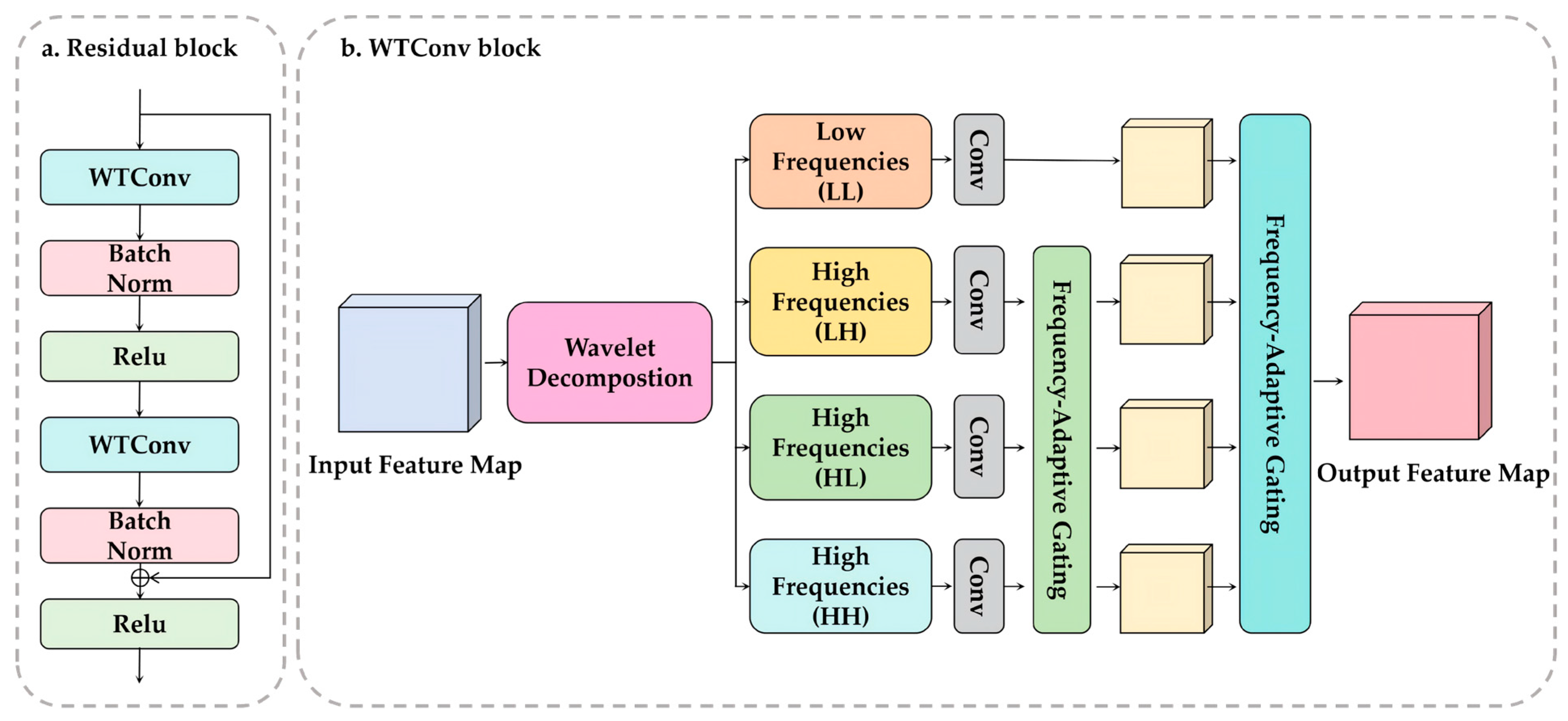
2.3. Deep Learning Model Development
2.4. Model Training
2.5. Statistical Analysis
3. Results
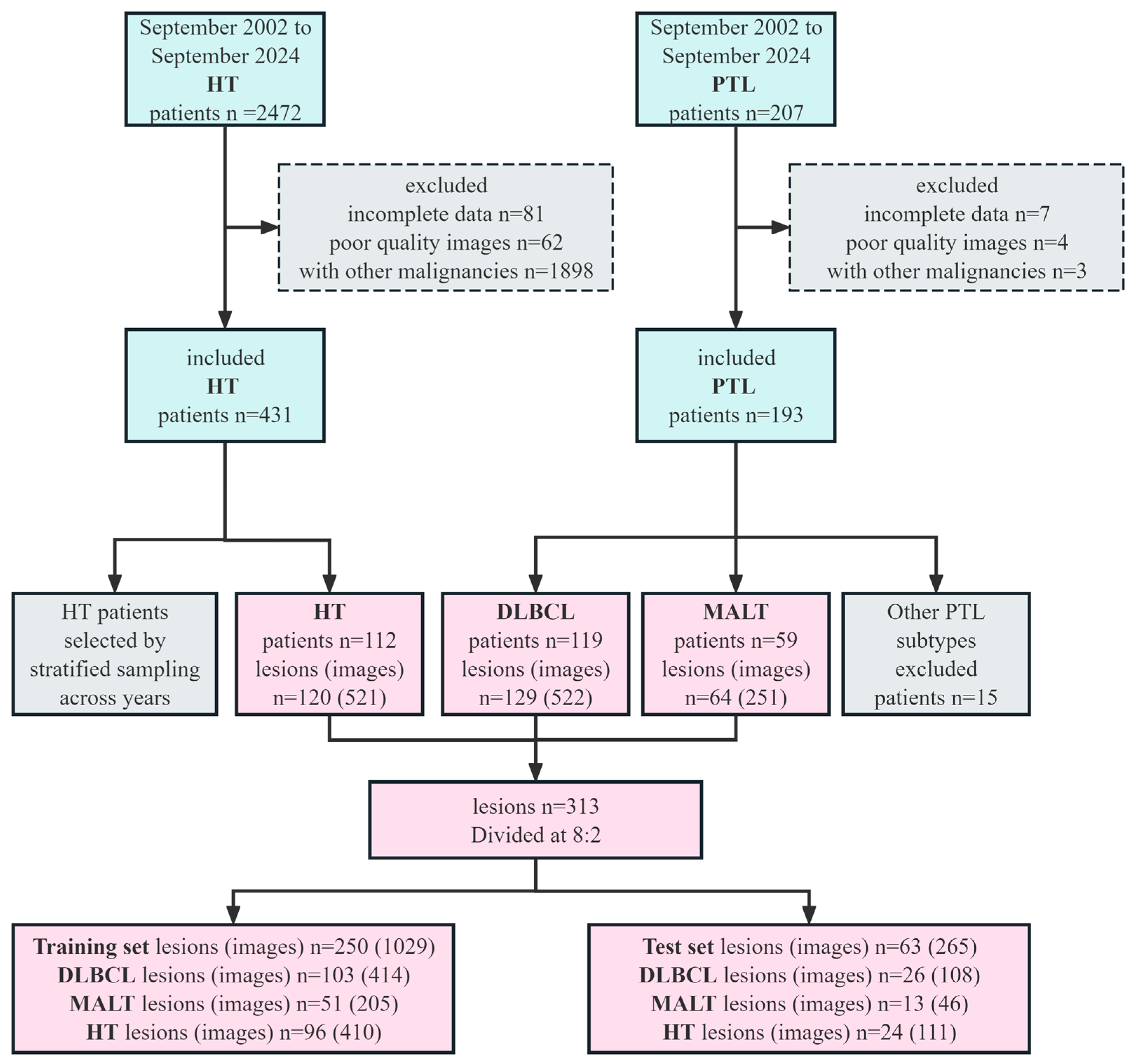
3.1. Cohort and Dataset Characteristics
3.2. Clinical and Ultrasound Feature Analysis
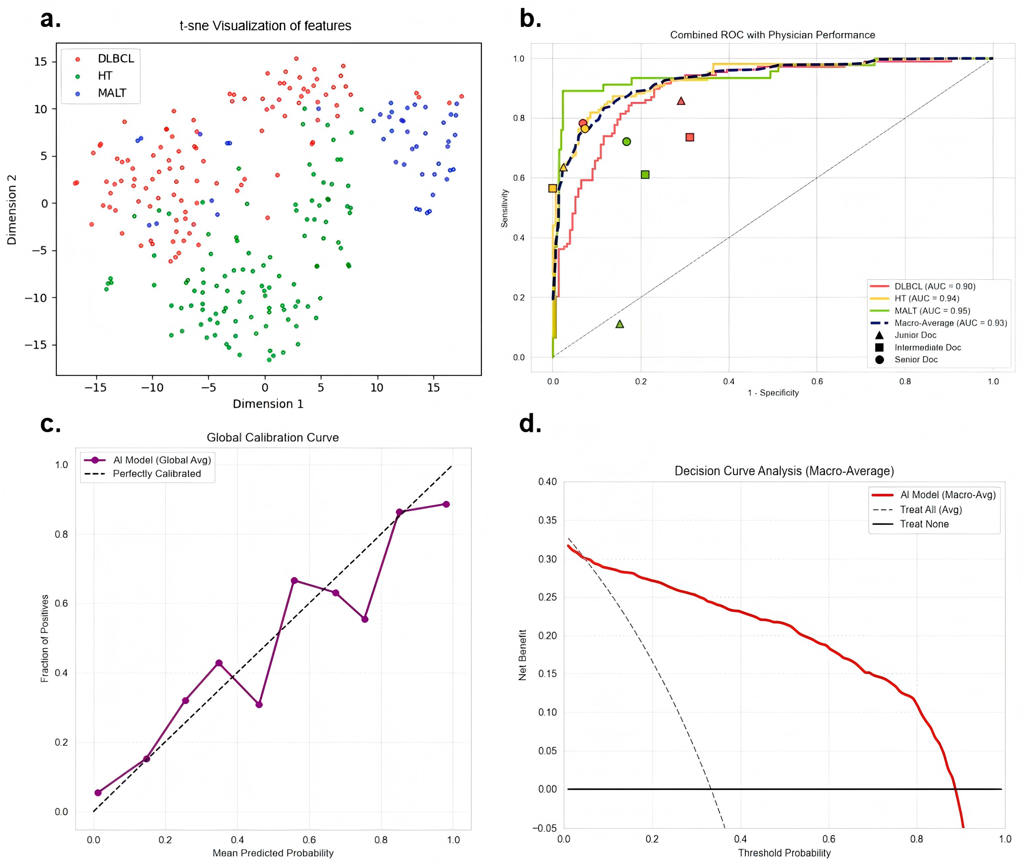
3.3. Image-Level Diagnostic Performance of the Model
3.4. Physician Performance
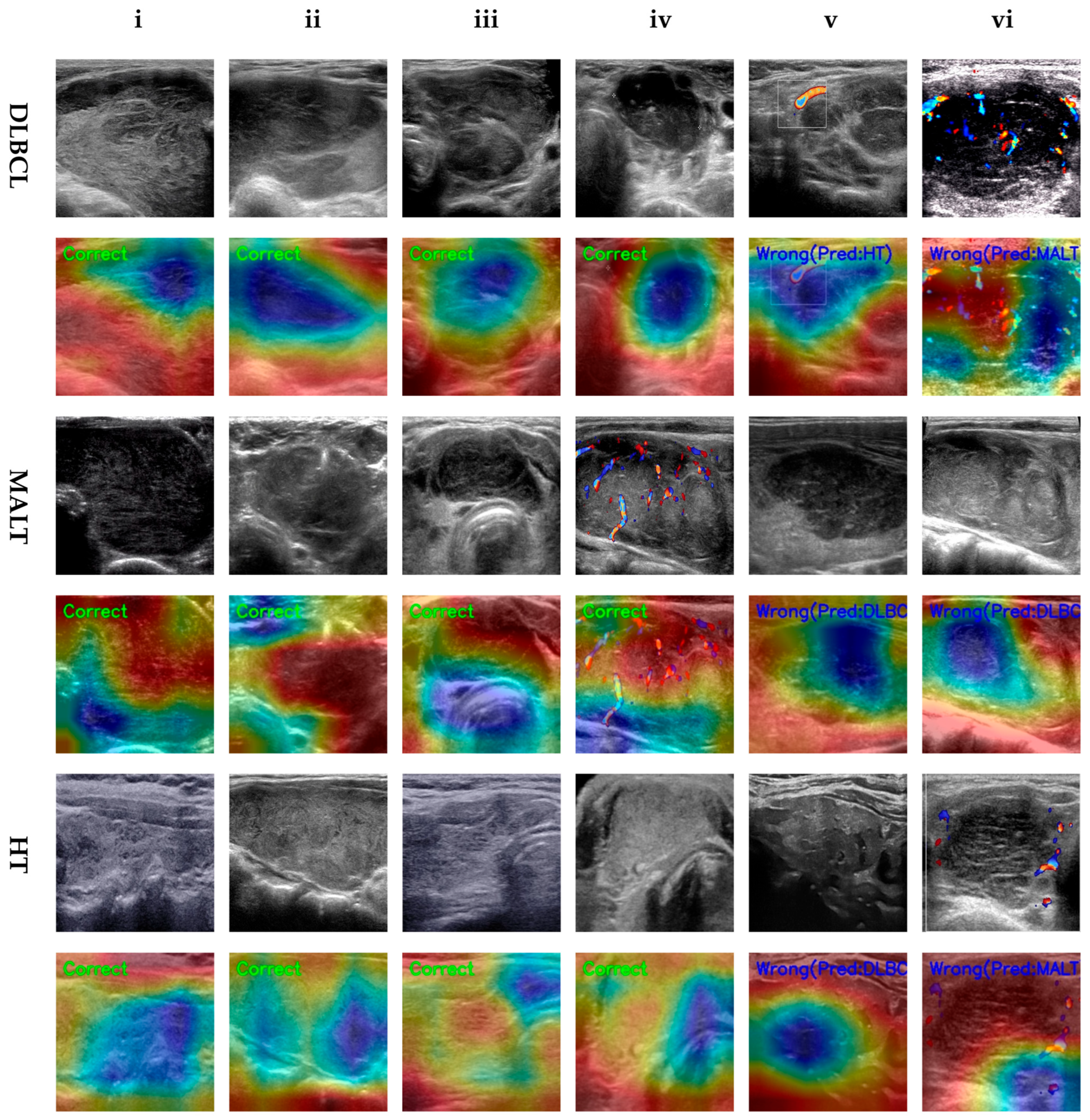
3.5. Grad-CAM Visualization
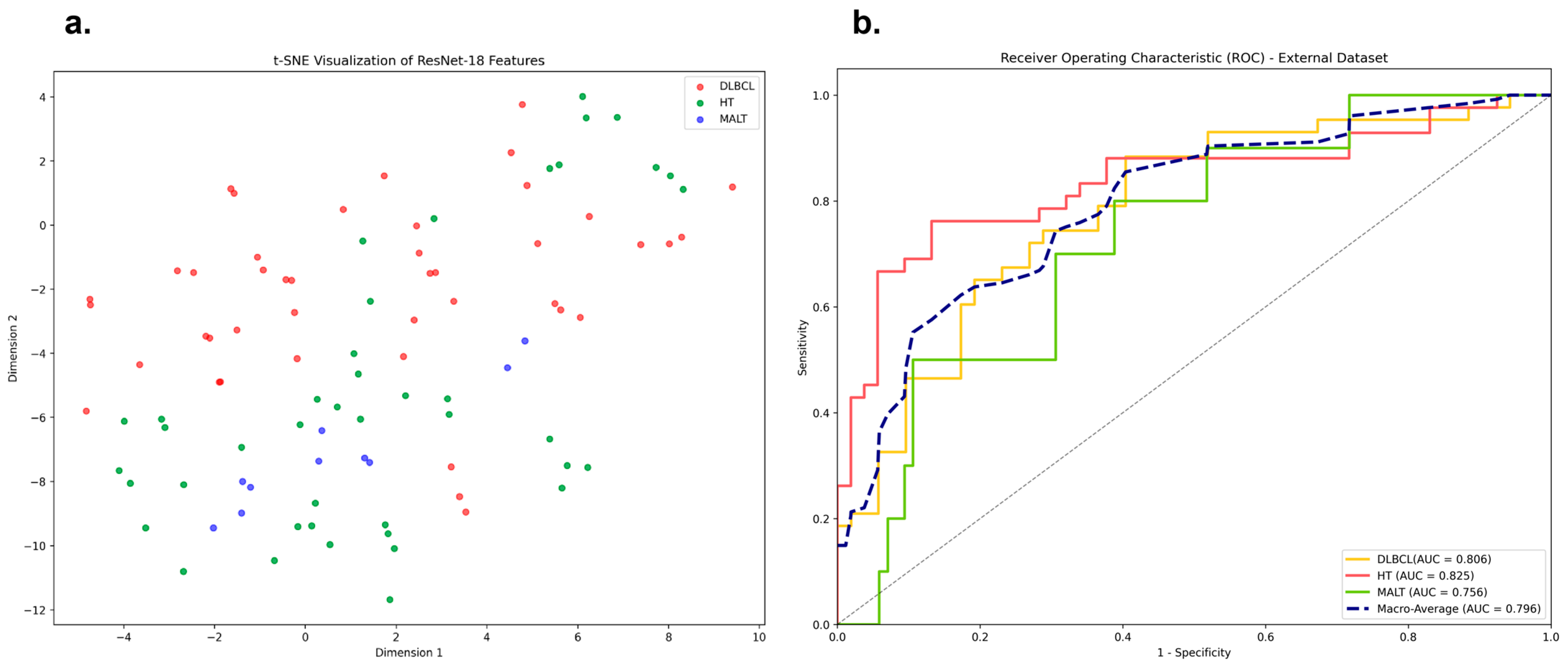
3.6. Exploratory External Validation
4. Discussion
5. Conclusions
Supplementary Materials
Author Contributions
Funding
Institutional Review Board Statement
Informed Consent Statement
Data Availability Statement
Conflicts of Interest
Abbreviations
| PTL | Primary Thyroid Lymphoma |
| DLBCL | Diffuse Large B-cell Lymphoma |
| MALT | Mucosa-Associated Lymphoid Tissue |
| HT | Hashimoto’s Thyroiditis |
| BMUS | B-mode Ultrasound |
| CDUS | Color Doppler Ultrasound |
| WTConv | Wavelet Transform Convolution |
| ResNet | Residual Network |
| AUC | Area under the receiver operating characteristic curve |
| ROC | Receiver Operating Characteristic |
| Grad-CAM | Gradient-weighted Class Activation Mapping |
| FNA | Fine-Needle Aspiration |
| CNB | Core-Needle Biopsy |
| H&E | Hematoxylin and Eosin |
| 18F-FDG PET | 18F-Fluorodeoxyglucose Positron Emission Tomography |
| PPV | Positive Predictive Value |
| NPV | Negative Predictive Value |
| Acc | Accuracy |
| CI | Confidence Interval |
| t-SNE | t-distributed Stochastic Neighbor Embedding |
| DCA | Decision Curve Analysis |
| CT | Computed Tomography |
| MRI | Magnetic Resonance Imaging |
References
- Stein, S.A.; Wartofsky, L. Primary Thyroid Lymphoma: A Clinical Review. J. Clin. Endocrinol. Metab. 2013, 98, 3131–3138. [Google Scholar] [CrossRef] [PubMed]
- Xiang, N.; Dong, F.; Zhan, X.; Wang, S.; Wang, J.; Sun, E.; Chen, B. Incidence and prognostic factors of primary thyroid lymphoma and construction of prognostic models for post-chemotherapy and postoperative patients: A population-based study. BMC Endocr. Disord. 2021, 21, 68. [Google Scholar] [CrossRef] [PubMed]
- Travaglino, A.; Pace, M.; Varricchio, S.; Insabato, L.; Giordano, C.; Picardi, M.; Pane, F.; Staibano, S.; Mascolo, M. Hashimoto Thyroiditis in Primary Thyroid Non-Hodgkin Lymphoma. Am. J. Clin. Pathol. 2020, 153, 156–164. [Google Scholar] [CrossRef] [PubMed]
- Xue, X.; Wu, L.; Zhang, J.; Sun, W.; Jiang, S.; Chu, X.; Sun, Y. Ultrasound scanning in diagnosing primary thyroid lymphoma. Clin. Hemorheol. Microcirc. 2024, 88, 385–393. [Google Scholar] [CrossRef] [PubMed]
- Xiao, L.; Zhao, Z.; Zhou, L.; Yan, J.; Luo, D.; Liu, F.; Zhou, Q.; Huang, D. Clinical and imaging features of primary thyroid MALT lymphoma. Front. Oncol. 2025, 15, 1498609. [Google Scholar] [CrossRef] [PubMed]
- Tan, H.; Ge, H.; Qin, Z.; Jiang, Y.; Chang, S.; Tang, N. Evaluating the diagnostic efficiency of ultrasound and serum autoantibodies in Hashimoto’s thyroiditis: A cross-sectional study. Sci. Rep. 2025, 15, 12674. [Google Scholar] [CrossRef] [PubMed]
- Durante, C.; Hegedüs, L.; Czarniecka, A.; Paschke, R.; Russ, G.; Schmitt, F.; Soares, P.; Solymosi, T.; Papini, E. 2023 European Thyroid Association Clinical Practice Guidelines for thyroid nodule management. Eur. Thyroid J. 2023, 12, e230067. [Google Scholar] [CrossRef] [PubMed]
- Sharma, A.; Jasim, S.; Reading, C.C.; Ristow, K.M.; Villasboas Bisneto, J.C.; Habermann, T.M.; Fatourechi, V.; Stan, M. Clinical Presentation and Diagnostic Challenges of Thyroid Lymphoma: A Cohort Study. Thyroid 2016, 26, 1061–1067. [Google Scholar] [CrossRef] [PubMed]
- Su, M.W.; Beck, T.N.; Knepprath, J.; Romero-Velez, G.; Heiden, K.B.; McHenry, C.R. Primary thyroid lymphoma: A multi-center retrospective review. Am. J. Surg. 2024, 237, 115927. [Google Scholar] [CrossRef] [PubMed]
- Rovira, A.; Carroll, P.; Simo, R. Thyroid lymphoma. Curr. Opin. Otolaryngol. Head Neck Surg. 2023, 31, 83–88. [Google Scholar] [CrossRef] [PubMed]
- Ji, J.; Gao, L.; Liu, R.; Shi, X.; Ma, L.; Pan, A.; Li, N.; Liu, C.; Li, X.; Yang, M.; et al. The potential value of ultrasound in predicting local refractory/relapse events in primary thyroid lymphoma patients. Cancer Imaging 2024, 24, 39. [Google Scholar] [CrossRef] [PubMed]
- Cai, X.; Zhou, Y.; Ren, J.; Wei, J.; Lu, S.; Gu, H.; Xu, W.; Zhu, X. Intelligent diagnosis of thyroid nodules with AI ultrasound assistance and cytology classification. Front. Endocrinol. 2025, 16, 1546983. [Google Scholar] [CrossRef] [PubMed]
- Cao, C.; Li, Q.; Tong, J.; Shi, L.; Li, W.; Xu, Y.; Cheng, J.; Du, T.; Li, J.; Cui, X. Artificial intelligence in thyroid ultrasound. Front. Oncol. 2023, 13, 1060702. [Google Scholar] [CrossRef] [PubMed]
- Zhang, Q.; Zhang, S.; Pan, Y.; Sun, L.; Li, J.; Qiao, Y.; Zhao, J.; Wang, X.; Feng, Y.; Zhao, Y.; et al. Deep learning to diagnose Hashimoto’s thyroiditis from sonographic images. Nat. Commun. 2022, 13, 3759. [Google Scholar] [CrossRef]
- Yamaguchi, S.; Isokawa, T.; Matsui, N.; Kamiura, N.; Tsuruyama, T. AI system for diagnosing mucosa-associated lymphoid tissue lymphoma and diffuse large B cell lymphoma using ImageNet and hematoxylin and eosin–stained specimens. PNAS Nexus 2025, 4, pgaf137. [Google Scholar] [CrossRef] [PubMed]
- Kim, S.; Park, J.H.; Kim, C.; You, S.; Choi, J.; Chang, J.W.; Jo, I.Y.; Lee, B.; Park, I.; Kim, H.S.; et al. Robust Multimodal Deep Learning for Lymphoma Subtype Classification Using 18F-FDG PET Maximum Intensity Projection Images and Clinical Data: A Multi-Center Study. Cancers 2026, 18, 210. [Google Scholar] [CrossRef] [PubMed]
- Tao, Y.; Yu, Y.; Wu, T.; Xu, X.; Dai, Q.; Kong, H.; Zhang, L.; Yu, W.; Leng, X.; Qiu, W.; et al. Deep learning for the diagnosis of suspicious thyroid nodules based on multimodal ultrasound images. Front. Oncol. 2022, 12, 1012724. [Google Scholar] [CrossRef] [PubMed]
- Collins, G.S.; Moons, K.G.M.; Dhiman, P.; Riley, R.D.; Beam, A.L.; Van Calster, B.; Ghassemi, M.; Liu, X.; Reitsma, J.B.; van Smeden, M.; et al. TRIPOD+AI statement: Updated guidance for reporting clinical prediction models that use regression or machine learning methods. BMJ 2024, 385, e78378. [Google Scholar] [CrossRef] [PubMed]
- Ota, H.; Ito, Y.; Matsuzuka, F.; Kuma, S.; Fukata, S.; Morita, S.; Kobayashi, K.; Nakamura, Y.; Kakudo, K.; Amino, N.; et al. Usefulness of ultrasonography for diagnosis of malignant lymphoma of the thyroid. Thyroid 2006, 16, 983–987. [Google Scholar] [CrossRef] [PubMed]
- Adler, D.D.; Carson, P.L.; Rubin, J.M.; Quinn-Reid, D. Doppler ultrasound color flow imaging in the study of breast cancer: Preliminary findings. Ultrasound Med. Amp. Biol. 1990, 16, 553–559. [Google Scholar] [CrossRef] [PubMed]
- Yang, J.; Shi, X.; Wang, B.; Qiu, W.; Tian, G.; Wang, X.; Wang, P.; Yang, J. Ultrasound Image Classification of Thyroid Nodules Based on Deep Learning. Front. Oncol. 2022, 12, 905955. [Google Scholar] [CrossRef] [PubMed]
- He, K.; Zhang, X.; Ren, S.; Sun, J. Deep Residual Learning for Image Recognition. In Proceedings of the 2016 IEEE Conference On Computer Vision and Pattern Recognition (Cvpr), Las Vegas, NV, USA, 26 June–1 July 2016. [Google Scholar] [CrossRef]
- Finder, S.E.; Amoyal, R.; Treister, E.; Freifeld, O. Wavelet Convolutions for Large Receptive Fields. In Proceedings of the Computer Vision—ECCV 2024, Milan, Italy, 29 September–4 October 2024; Springer: Cham, Switzerland, 2024; pp. 363–380. [Google Scholar]
- Liu, P.; Zhang, H.; Lian, W.; Zuo, W. Multi-Level Wavelet Convolutional Neural Networks. IEEE Access 2019, 7, 74973–74985. [Google Scholar] [CrossRef]
- Hyjek, E.; Isaacson, P.G. Primary B cell lymphoma of the thyroid and its relationship to Hashimoto’s thyroiditis. Hum. Pathol. 1988, 19, 1315–1326. [Google Scholar] [CrossRef] [PubMed]
- He, X.; Bai, Y.; Wang, S.; Chen, Q.; Cao, H.; Xu, P.; Yao, M.; Zhang, W.; Wang, Z.; Wang, X.; et al. Ultrasonographic differentiation of diffuse large B-cell lymphoma and mucosa-associated lymphoid tissue lymphoma in primary thyroid lymphoma. Front. Endocrinol. 2026, 17, 1743975. [Google Scholar] [CrossRef] [PubMed]
- Kariya, A.; Tachibana, T.; Hiramatsu, Y.; Wani, Y.; Matsumoto, J.; Furukawa, C.; Sato, A.; Naoi, Y.; Orita, Y.; Sato, Y.; et al. Primary Thyroid Lymphoma: Clinical Factors Predicting the Possibility of Diffuse Large B-Cell Lymphoma. Ear Nose Throat J. 2023. [Google Scholar] [CrossRef] [PubMed]
- Zhang, X.; Wei, B.; Nong, L.; Zhang, H.; Gao, Y.; Ou, J. The usefulness of serial ultrasound in thyroid mucosa-associated lymphoid tissue lymphoma. Front. Endocrinol. 2022, 13, 1054584. [Google Scholar] [CrossRef] [PubMed]
- Yang, L.; Zhao, H.; He, Y.; Zhu, X.; Yue, C.; Luo, Y.; Ma, B. Contrast-Enhanced Ultrasound in the Differential Diagnosis of Primary Thyroid Lymphoma and Nodular Hashimoto’s Thyroiditis in a Background of Heterogeneous Parenchyma. Front. Oncol. 2021, 10, 597975. [Google Scholar] [CrossRef] [PubMed]
- Yamashita, R.; Kapoor, T.; Alam, M.N.; Galimzianova, A.; Syed, S.A.; Akdogan, M.U.; Alkim, E.; Wentland, A.L.; Madhuripan, N.; Goff, D.; et al. Toward Reduction in False-Positive Thyroid Nodule Biopsies with a Deep Learning–based Risk Stratification System Using US Cine-Clip Images. Radiol. Artif. Intell. 2022, 4, e210174. [Google Scholar] [CrossRef] [PubMed]
- Wildman-Tobriner, B.; Taghi-Zadeh, E.; Mazurowski, M.A. Artificial Intelligence (AI) Tools for Thyroid Nodules on Ultrasound, from the AJR Special Series on AI Applications. Am. J. Roentgenol. 2022, 219, 547–554. [Google Scholar] [CrossRef] [PubMed]
- Barczyński, M.; Stopa-Barczyńska, M.; Wojtczak, B.; Czarniecka, A.; Konturek, A. Clinical validation of S-DetectTM mode in semi-automated ultrasound classification of thyroid lesions in surgical office. Gland Surg. 2020, 9, S77–S85. [Google Scholar] [CrossRef] [PubMed]
- Choi, Y.J.; Baek, J.H.; Park, H.S.; Shim, W.H.; Kim, T.Y.; Shong, Y.K.; Lee, J.H. A Computer-Aided Diagnosis System Using Artificial Intelligence for the Diagnosis and Characterization of Thyroid Nodules on Ultrasound: Initial Clinical Assessment. Thyroid 2017, 27, 546–552. [Google Scholar] [CrossRef] [PubMed]
- Reverter, J.L.; Ferrer-Estopiñan, L.; Vázquez, F.; Ballesta, S.; Batule, S.; de Oca, A.P.; Puig-Jové, C.; Puig-Domingo, M. Reliability of a computer-aided system in the evaluation of indeterminate ultrasound images of thyroid nodules. Eur. Thyroid J. 2022, 11, e210023. [Google Scholar] [CrossRef] [PubMed]
- Chu, X.; Wang, T.; Chen, M.; Li, J.; Wang, L.; Wang, C.; Wang, H.; Wong, S.T.; Chen, Y.; Li, H. Deep learning model for malignancy prediction of TI-RADS 4 thyroid nodules with high-risk characteristics using multimodal ultrasound: A multicentre study. Comput. Med. Imaging Graph. 2025, 124, 102576. [Google Scholar] [CrossRef] [PubMed]


| Groups/ Indicators | MALT (64) | DLBCL (129) | HT (120) | χ2 | p-Value | p-Value (MALT vs. DLBCL) | p-Value (MALT vs. HT) | p-Value (DLBCL vs. HT) |
|---|---|---|---|---|---|---|---|---|
| Site | 3.1174 | 0.5384 | 0.4794 | 0.4348 | 0.4329 | |||
| Right lobe | 29 (45.31%) | 69 (53.49%) | 65 (54.17%) | |||||
| Left lobe | 33 (51.56%) | 58 (44.96%) | 50 (41.67%) | |||||
| Isthmus | 2 (3.12%) | 2 (1.55%) | 5 (4.17%) | |||||
| Sex | 15.2505 | 0.0005 | 0.8843 | 0.0008 | 0.0003 | |||
| Man | 18 (28.12%) | 35 (27.13%) | 11 (9.17%) | |||||
| Woman | 46 (71.88%) | 94 (72.87%) | 109 (90.83%) | |||||
| Age | 74.0798 | <0.0001 | 0.0839 | <0.0001 | <0.0001 | |||
| <45 | 10 (15.62%) | 10 (7.75%) | 51 (42.50%) | |||||
| 45–60 | 23 (35.94%) | 37 (28.68%) | 51 (42.50%) | |||||
| >60 | 31 (48.44%) | 82 (63.57%) | 18 (15.00%) | |||||
| Specimen | 8.4769 | 0.0144 | 0.0407 | 0.0039 | 0.3008 | |||
| Biopsy | 34 (53.12%) | 88 (68.22%) | 89 (74.17%) | |||||
| Resection | 30 (46.88%) | 41 (31.78%) | 31 (25.83%) | |||||
| HT background | 12.2795 | 0.0022 | 0.6105 | 0.0046 | 0.0004 | |||
| Present | 59 (92.19%) | 116 (89.92%) | 120 (100.00%) | |||||
| Absent | 5 (7.81%) | 13 (10.08%) | 0 (0.00%) | |||||
| Lesion Type | 7.1692 | 0.0277 | 0.0277 | - | - | |||
| Diffuse | 30 (46.88%) | 78 (60.47%) | - | |||||
| Nodular | 30 (46.88%) | 36 (27.91%) | - | |||||
| Mixed | 4 (6.25%) | 15 (11.63%) | - | |||||
| Abnormal CLN | 59.8963 | <0.0001 | 0.0367 | <0.0001 | <0.0001 | |||
| Present | 26 (40.62%) | 73 (56.59%) | 12 (10.00%) | |||||
| Absent | 38 (59.38%) | 56 (43.41%) | 108 (90.00%) | |||||
| Adler grade | 21.1393 | 0.0003 | 0.5742 | 0.0007 | 0.0003 | |||
| Grade 1 | 21 (32.81%) | 51 (39.53%) | 68 (56.67%) | |||||
| Grade 2 | 33 (51.56%) | 63 (48.84%) | 29 (24.17%) | |||||
| Grade 3 | 10 (15.62%) | 15 (11.63%) | 23 (19.17%) | |||||
| Boundary | 3.1036 | 0.0781 | 0.0781 | - | - | |||
| Distinct | 27 (42.19%) | 38 (29.46%) | - | |||||
| Indistinct | 37 (57.81%) | 91 (70.54%) | - | |||||
| Morphology | 8.2380 | 0.0041 | 0.0079 | - | - | |||
| Regular | 10 (15.62%) | 5 (3.88%) | - | |||||
| Irregular | 54 (84.38%) | 124 (96.12%) | - | |||||
| Aspect Ratio | 1.5119 | 0.2189 | 0.5521 | - | - | |||
| Wider-than-tall | 64 (100.00%) | 126 (97.67%) | - | |||||
| Taller-than-wide | 0 (0.00%) | 3 (2.33%) | - | |||||
| Echogenicity | 27.3216 | <0.0001 | 0.0973 | 0.0010 | <0.0001 | |||
| Hypo echo | 56 (87.50%) | 100 (77.52%) | 119 (99.17%) | |||||
| Marked hypo echo | 8 (12.50%) | 29 (22.48%) | 1 (0.83%) |
| Groups | AUC (95% CI) | Balanced Acc (95% CI) | Sensitivity (95% CI) | Specificity (95% CI) | PPV (95% CI) | NPV (95% CI) | F1-Score (95% CI) |
|---|---|---|---|---|---|---|---|
| DLBCL | 0.899 (0.858–0.935) | 0.830 (0.785–0.876) | 0.852 (0.778–0.918) | 0.809 (0.747–0.869) | 0.754 (0.679–0.825) | 0.888 (0.832–0.939) | 0.800 (0.737–0.853) |
| HT | 0.937 (0.908–0.963) | 0.841 (0.797–0.887) | 0.748 (0.664–0.826) | 0.935 (0.895–0.969) | 0.892 (0.826–0.950) | 0.837 (0.785–0.892) | 0.814 (0.751–0.870) |
| MALT lymphoma | 0.946 (0.900–0.981) | 0.925 (0.877–0.967) | 0.891 (0.792–0.974) | 0.959 (0.930–0.982) | 0.820 (0.711–0.923) | 0.977 (0.954–0.995) | 0.854 (0.769–0.922) |
| Macro-average | 0.927 (0.889–0.960) | 0.866 (0.820–0.910) | 0.830 (0.744–0.906) | 0.901 (0.857–0.940) | 0.822 (0.739–0.900) | 0.901 (0.857–0.942) | 0.823 (0.753–0.882) |
| Physicians | Groups | Balanced Acc (95% CI) | Sensitivity (95% CI) | Specificity (95% CI) | PPV (95% CI) | NPV (95% CI) | F1-Score (95% CI) |
|---|---|---|---|---|---|---|---|
| Junior | DLBCL | 0.784 (0.732–0.840) | 0.858 (0.784–0.919) | 0.709 (0.619–0.792) | 0.752 (0.670–0.825) | 0.830 (0.742–0.900) | 0.802 (0.746–0.858) |
| HT | 0.806 (0.753–0.862) | 0.635 (0.537–0.742) | 0.976 (0.945–1.000) | 0.947 (0.885–1.000) | 0.796 (0.727–0.860) | 0.761 (0.680–0.838) | |
| MALT | 0.480 (0.409–0.567) | 0.111 (0.000–0.278) | 0.848 (0.789–0.897) | 0.065 (0.000–0.160) | 0.910 (0.868–0.950) | 0.082 (0.000–0.194) | |
| Macro-average | 0.690 (0.631–0.756) | 0.535 (0.441–0.646) | 0.844 (0.784–0.896) | 0.588 (0.518–0.662) | 0.845 (0.779–0.903) | 0.548 (0.475–0.630) | |
| Intermediate | DLBCL | 0.713 (0.653–0.774) | 0.736 (0.651–0.821) | 0.689 (0.602–0.778) | 0.709 (0.625–0.789) | 0.717 (0.625–0.802) | 0.722 (0.654–0.785) |
| HT | 0.782 (0.729–0.835) | 0.565 (0.458–0.670) | 1.000 (1.000–1.000) | 1.000 (1.000–1.000) | 0.770 (0.700–0.831) | 0.722 (0.629–0.803) | |
| MALT | 0.701 (0.587–0.816) | 0.611 (0.389–0.833) | 0.791 (0.730–0.848) | 0.216 (0.098–0.321) | 0.956 (0.924–0.987) | 0.319 (0.161–0.447) | |
| Macro-average | 0.732 (0.656–0.809) | 0.637 (0.499–0.775) | 0.827 (0.777–0.875) | 0.642 (0.574–0.703) | 0.814 (0.750–0.873) | 0.588 (0.481–0.678) | |
| Senior | DLBCL | 0.858 (0.812–0.902) | 0.783 (0.700–0.862) | 0.932 (0.881–0.978) | 0.922 (0.863–0.973) | 0.807 (0.736–0.874) | 0.847 (0.794–0.898) |
| HT | 0.846 (0.793–0.898) | 0.765 (0.677–0.854) | 0.927 (0.875–0.971) | 0.878 (0.797–0.950) | 0.852 (0.786–0.912) | 0.818 (0.746–0.880) | |
| MALT | 0.777 (0.670–0.877) | 0.722 (0.500–0.917) | 0.832 (0.781–0.882) | 0.289 (0.163–0.419) | 0.970 (0.941–0.994) | 0.413 (0.254–0.543) | |
| Macro-average | 0.827 (0.759–0.892) | 0.757 (0.626–0.877) | 0.897 (0.845–0.944) | 0.696 (0.608–0.781) | 0.876 (0.821–0.927) | 0.692 (0.598–0.774) |
| Physicians | Concordant Cases/Total | Simple Concordance Rate | Cohen’s κ | Interpretation of Concordance |
|---|---|---|---|---|
| Senior vs. Intermediate | 41/63 | 65.1% | 0.458 | Moderate |
| Senior vs. Junior | 45/63 | 71.4% | 0.548 | Moderate |
| Intermediate vs. Junior | 38/63 | 60.3% | 0.328 | Fair |
| Groups | AUC (95% CI) | Balanced Acc (95% CI) | Sensitivity (95% CI) | Specificity (95% CI) | PPV (95% CI) | NPV (95% CI) | F1-Score (95% CI) |
|---|---|---|---|---|---|---|---|
| DLBCL | 0.806 (0.711–0.891) | 0.675 (0.548–0.799) | 0.581 (0.444–0.725) | 0.769 (0.651–0.873) | 0.676 (0.518–0.821) | 0.690 (0.577–0.800) | 0.625 (0.500–0.738) |
| HT | 0.825 (0.727–0.909) | 0.565 (0.483–0.647) | 0.167 (0.065–0.294) | 0.962 (0.900–1.000) | 0.778 (0.500–1.000) | 0.593 (0.494–0.695) | 0.275 (0.118–0.440) |
| MALT lymphoma | 0.756 (0.620–0.865) | 0.659 (0.457–0.813) | 0.800 (0.500–1.000) | 0.518 (0.414–0.625) | 0.163 (0.068–0.281) | 0.957 (0.881–1.000) | 0.271 (0.122–0.426) |
| Macro-average | 0.796 (0.686–0.888) | 0.633 (0.496–0.753) | 0.516 (0.337–0.673) | 0.750 (0.655–0.833) | 0.539 (0.362–0.700) | 0.746 (0.651–0.832) | 0.390 (0.247–0.535) |
Disclaimer/Publisher’s Note: The statements, opinions and data contained in all publications are solely those of the individual author(s) and contributor(s) and not of MDPI and/or the editor(s). MDPI and/or the editor(s) disclaim responsibility for any injury to people or property resulting from any ideas, methods, instructions or products referred to in the content. |
© 2026 by the authors. Licensee MDPI, Basel, Switzerland. This article is an open access article distributed under the terms and conditions of the Creative Commons Attribution (CC BY) license.
Share and Cite
Chen, J.; Deng, Z.; Chen, Y.; Ye, R.; Li, J.; Tao, Y.; Ma, B.; He, Y. Deep Learning Based on B-Mode and Color Doppler Ultrasound for Differentiation of Primary Thyroid Lymphoma and Hashimoto’s Thyroiditis: A Retrospective Single-Center Study. Diagnostics 2026, 16, 1909. https://doi.org/10.3390/diagnostics16121909
Chen J, Deng Z, Chen Y, Ye R, Li J, Tao Y, Ma B, He Y. Deep Learning Based on B-Mode and Color Doppler Ultrasound for Differentiation of Primary Thyroid Lymphoma and Hashimoto’s Thyroiditis: A Retrospective Single-Center Study. Diagnostics. 2026; 16(12):1909. https://doi.org/10.3390/diagnostics16121909
Chicago/Turabian StyleChen, Juanmei, Zijian Deng, Yong Chen, Ruiheng Ye, Jiawu Li, Yi Tao, Buyun Ma, and Yushuang He. 2026. "Deep Learning Based on B-Mode and Color Doppler Ultrasound for Differentiation of Primary Thyroid Lymphoma and Hashimoto’s Thyroiditis: A Retrospective Single-Center Study" Diagnostics 16, no. 12: 1909. https://doi.org/10.3390/diagnostics16121909
APA StyleChen, J., Deng, Z., Chen, Y., Ye, R., Li, J., Tao, Y., Ma, B., & He, Y. (2026). Deep Learning Based on B-Mode and Color Doppler Ultrasound for Differentiation of Primary Thyroid Lymphoma and Hashimoto’s Thyroiditis: A Retrospective Single-Center Study. Diagnostics, 16(12), 1909. https://doi.org/10.3390/diagnostics16121909