Virtual Evaluation of Hematoxylin & Eosin via Digital Pathology Survey (VEED) Project: Results from a Non-Inferiority Study of a Tabs-Based Staining Method

 ,
,  ,
,  ,
,  ,
,  ,
,  ,
,  , ,
, ,  , , ,
, , ,  , , ,
, , , 
 add
Show full author list
add
Show full author list
Abstract
1. Introduction
2. Materials and Methods
2.1. Sample Cohort
2.2. Staining Methods
2.3. Tissue Stain Evaluation
2.4. Statistical Analysis
3. Results
4. Discussion
5. Conclusions
Author Contributions
Funding
Institutional Review Board Statement
Informed Consent Statement
Data Availability Statement
Acknowledgments
Conflicts of Interest
References
- Tissue Diagnostics Market, Forecast 2022, Size, Share and Trends. Available online: https://store.frost.com/global-tissue-diagnostics-market-forecast-to-2022.html (accessed on 6 January 2026).
- Dunn, C.; Brettle, D.; Hodgson, C.; Hughes, R.; Treanor, D. An international study of stain variability in histopathology using qualitative and quantitative analysis. J. Pathol. Inform. 2025, 17, 100423. [Google Scholar] [CrossRef] [PubMed]
- Wick, M.R. The hematoxylin and eosin stain in anatomic pathology—An often-neglected focus of quality assurance in the laboratory. Semin. Diagn. Pathol. 2019, 36, 303–311. [Google Scholar] [CrossRef] [PubMed]
- Lillie, R.D.; Fullmer, H.M. Histopathologic Technic and Practical Histochemistry, 4th ed.; McGraw Hill Book Company: New York, NY, USA, 1976. [Google Scholar]
- College of American Pathologists. H&E Troubleshooting Guide; CAP: Northfield, IL, USA, 2026; Available online: https://www.google.com/url?sa=t&source=web&rct=j&opi=89978449&url=https://documents.cap.org/documents/HE_TroubleshootingGuide_2026.pdf&ved=2ahUKEwicxeiKlouVAxWgzTQHHWh5JBgQFnoECBgQAQ&usg=AOvVaw11jepmQt-HRiOqSxxqJQLx (accessed on 21 May 2026).
- Haematoxylin & Eosin. Muscle Histochemistry Staining Criteria Handbook Edition 004 Index Haematoxylin and Eosin Assessment Criteria 3 Special Stains Assessment Criteria 6 Assessment Criteria Definitions 23. Available online: https://cdn.website-editor.net/8fbe2120dfac419988bb54de58b21798/files/uploaded/NEQMANMA005%20Special%20stain%20criteria%20handbook%20muscle%20edition%20004%20june%2017.pdf (accessed on 14 June 2026).
- Salvi, M.; Caputo, A.; Balmativola, D.; Scotto, M.; Pennisi, O.; Michielli, N.; Mogetta, A.; Molinari, F.; Fraggetta, F. Impact of Stain Normalization on Pathologist Assessment of Prostate Cancer: A Comparative Study. Cancers 2023, 15, 1503. [Google Scholar] [CrossRef] [PubMed]
- Cazzaniga, G.; Caputo, A.; L’iMperio, V.; Gibilisco, F.; Scotto, M.; Pennisi, O.M.A.; Michielli, N.; Mogetta, A.; Molinari, F.; Fraggetta, F.; et al. Not all stains are made equal: Impact of stain normalization on prostate cancer diagnosis. Pathologica 2024, 116, 285–294. [Google Scholar] [CrossRef] [PubMed]
- Rana, A.; Lowe, A.; Lithgow, M.; Horback, K.; Janovitz, T.; Da Silva, A.; Tsai, H.; Shanmugam, V.; Bayat, A.; Shah, P. Use of Deep Learning to Develop and Analyze Computational Hematoxylin and Eosin Staining of Prostate Core Biopsy Images for Tumor Diagnosis. JAMA Netw. Open 2020, 3, e205111. [Google Scholar] [CrossRef] [PubMed]
- Iwuajoku, V.; Ekici, K.; Haas, A.; Khan, M.Z.; Kazemi, A.; Kasajima, A.; Delbridge, C.; Muckenhuber, A.; Schmoeckel, E.; Stögbauer, F.; et al. An equivalency and efficiency study for one year digital pathology for clinical routine diagnostics in an accredited tertiary academic center. Virchows Arch. 2025, 487, 3–12. [Google Scholar] [CrossRef] [PubMed]
- Rivenson, Y.; Wang, H.; Wei, Z.; de Haan, K.; Zhang, Y.; Wu, Y.; Günaydın, H.; Zuckerman, J.E.; Chong, T.; Sisk, A.E.; et al. Virtual histological staining of unlabelled tissue-autofluorescence images via deep learning. Nat. Biomed. Eng. 2019, 3, 466–477. [Google Scholar] [CrossRef] [PubMed]
- Bayramoglu, N.; Kaakinen, M.; Eklund, L.; Heikkilä, J. Towards Virtual H&E Staining of Hyperspectral Lung Histology Images Using Conditional Generative Adversarial Networks. In Proceedings of the 2017 IEEE International Conference on Computer Vision Workshops, ICCVW 2017, Venice, Italy, 22–29 October 2017; pp. 64–71. [Google Scholar] [CrossRef]
- Lahiani, A.; Gildenblat, J.; Klaman, I.; Albarqouni, S.; Navab, N.; Klaiman, E. Virtualization of Tissue Staining in Digital Pathology Using an Unsupervised Deep Learning Approach. In European Congress on Digital Pathology; Lecture Notes in Computer (Including Subseries Lecture Notes in Artificial Intelligence and Lecture Notes in Bioinformatics) Science; Springer International Publishing: Cham, Switzerland, 2019; pp. 47–55. [Google Scholar] [CrossRef]
- Ji, X.; Salmon, R.; Mulliqi, N.; Khan, U.; Wang, Y.; Blilie, A.; Olsson, H.; Pedersen, B.G.; Sørensen, K.D.; Ulhøi, B.P.; et al. Physical Color Calibration of Digital Pathology Scanners for Robust Artificial Intelligence–Assisted Cancer Diagnosis. Mod. Pathol. 2025, 38, 100715. [Google Scholar] [CrossRef] [PubMed]
- Eloy, C.; Fraggetta, F.; van Diest, P.J.; Polónia, A.; Curado, M.; Temprana-Salvador, J.; Zlobec, I.; Purqueras, E.; Weis, C.-A.; Matias-Guiu, X.; et al. Digital transformation of pathology—The European Society of Pathology expert opinion paper. Virchows Arch. 2025, 487, 971–981. [Google Scholar] [CrossRef] [PubMed]
- Fraggetta, F.; Garozzo, S.; Zannoni, G.; Pantanowitz, L.; Rossi, E. Routine Digital Pathology Workflow: The Catania Experience. J. Pathol. Inform. 2017, 8, 51. [Google Scholar] [CrossRef] [PubMed]
- Eccher, A.; Fabbri, V.P.; L’Imperio, V.; Die Tos, A.P.; Pagni, F.; Dominici, M. Pathology Laboratory Archive at the University and Hospital Trust of Modena/UNIMORE: “Disaster Recovery” frame by frame. Pathologica 2025, 117, 52–54. [Google Scholar] [CrossRef] [PubMed]
- Eccher, A.; Marletta, S.; Sbaraglia, M.; Guerriero, A.; Rossi, M.; Gambaro, G.; Scarpa, A.; Tos, A.P.D. Digital pathology structure and deployment in Veneto: A proof-of-concept study. Virchows Arch. 2024, 485, 453–460. [Google Scholar] [CrossRef] [PubMed]
- Nibid, L.; Greco, C.; Cordelli, E.; Sabarese, G.; Fiore, M.; Liu, C.Z.; Ippolito, E.; Sicilia, R.; Miele, M.; Tortora, M.; et al. Deep pathomics: A new image-based tool for predicting response to treatment in stage III non-small cell lung cancer. PLoS ONE 2023, 18, e0294259. [Google Scholar] [CrossRef] [PubMed]
- Rakaee, M.; Tafavvoghi, M.; Ricciuti, B.; Alessi, J.V.; Cortellini, A.; Citarella, F.; Nibid, L.; Perrone, G.; Adib, E.; Fulgenzi, C.A.M.; et al. Deep Learning Model for Predicting Immunotherapy Response in Advanced Non−Small Cell Lung Cancer. JAMA Oncol. 2025, 11, 109–118. [Google Scholar] [CrossRef] [PubMed]
- Bülow, R.D.; Hölscher, D.L.; Costa, I.G.; Boor, P. Extending the landscape of omics technologies by pathomics. npj Syst. Biol. Appl. 2023, 9, 38. [Google Scholar] [CrossRef] [PubMed]
- Koivukoski, S.; Khan, U.; Ruusuvuori, P.; Latonen, L. Unstained Tissue Imaging and Virtual Hematoxylin and Eosin Staining of Histologic Whole Slide Images. Lab. Investig. 2023, 103, 100070. [Google Scholar] [CrossRef] [PubMed]
- Fereidouni, F.; Todd, A.; Li, Y.; Chang, C.-W.; Luong, K.; Rosenberg, A.; Lee, Y.-J.; Chan, J.W.; Borowsky, A.; Matsukuma, K.; et al. Dual-mode emission and transmission microscopy for virtual histochemistry using hematoxylin- and eosin-stained tissue sections. Biomed. Opt. Express 2019, 10, 6516. [Google Scholar] [CrossRef] [PubMed]
- Levy, J.J.; Azizgolshani, N.; Andersen, M.J.; Suriawinata, A.; Liu, X.; Lisovsky, M.; Ren, B.; Bobak, C.A.; Christensen, B.C.; Vaickus, L.J. A large-scale internal validation study of unsupervised virtual trichrome staining technologies on nonalcoholic steatohepatitis liver biopsies. Mod. Pathol. 2021, 34, 808–822. [Google Scholar] [CrossRef] [PubMed]
- Cazzaniga, G.; Mascadri, F.; Marletta, S.; Caputo, A.; Guidi, G.; Gambaro, G.; Eccher, A.; Tos, A.P.D.; Pagni, F.; L’IMperio, V. Benchmarking digital displays (monitors) for histological diagnoses: The nephropathology use case. J. Clin. Pathol. 2025, 78, 798–804. [Google Scholar] [CrossRef] [PubMed]
- Marletta, S.; Caputo, A.; Guidi, G.; Pantanowitz, L.; Pagni, F.; Bavieri, I.; L’iMperio, V.; Brunelli, M.; Tos, A.P.D.; Eccher, A. Digital Pathology Displays Under Pressure: Benchmarking Performance Across Market Grades. J. Imaging Inform. Med. 2025, 38, 4090–4099. [Google Scholar] [CrossRef] [PubMed]


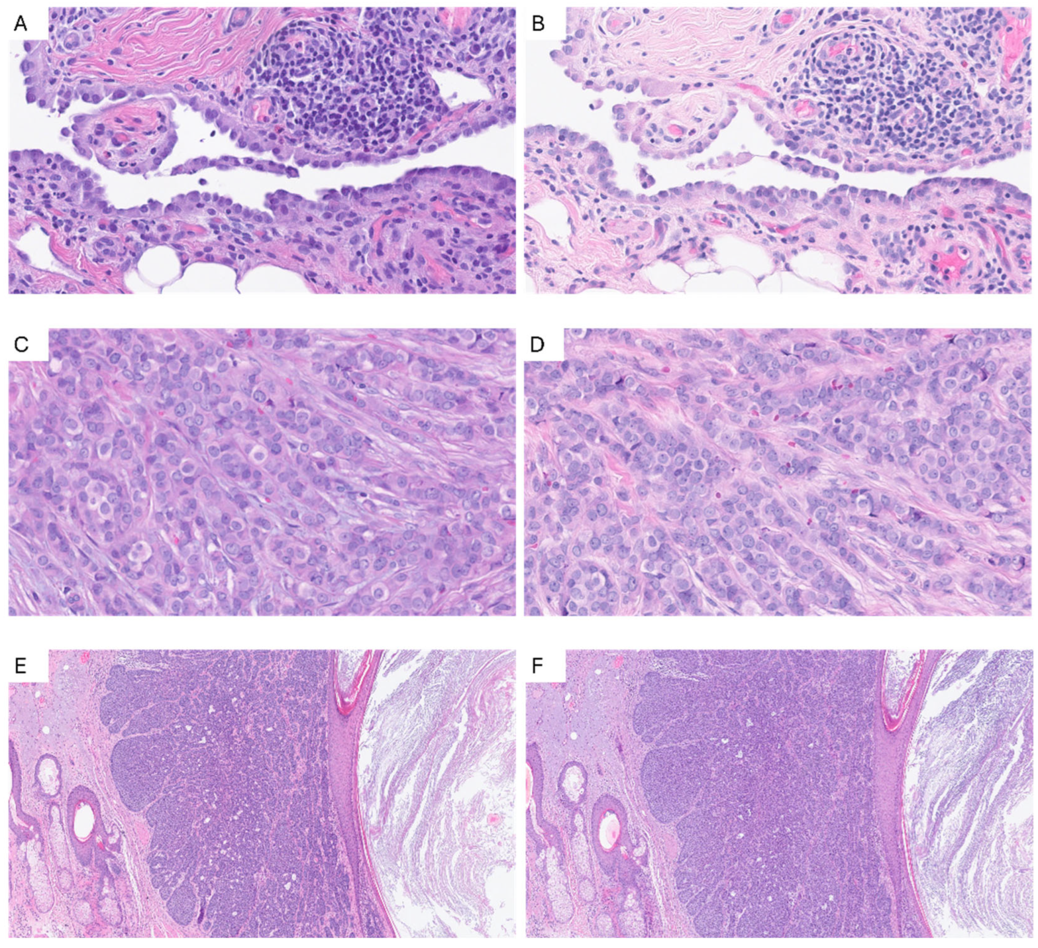

| N. Picture | Tissue | Pathology | Magnification |
|---|---|---|---|
| 1 | Skin | Actinic keratosis | 10× |
| 2 | Skin | Basal cell carcinoma | 5× |
| 20× | |||
| 40× | |||
| 3 | Skin | Dyskeratoma | 20× |
| 40× | |||
| 4 | Prostate | Normal and prostate cancer | 5× |
| 10× | |||
| 20× | |||
| 20× | |||
| 40× | |||
| 5 | Uterus | Leiomyoma | 10× |
| 6 | Uterus | Adenomyosis | 20× |
| 7 | Fallopian tubes | Normal | 20× |
| 8 | Breast | Lobular carcinoma | 20× |
| 9 | Breast | Apocrine metaplasia | 10× |
| 10 | Kidney | Normal and chromophobe renal cell carcinoma | 20× |
| 10× | |||
| 11 | Pleura | Pleural effusion | 20× |
| 40× | |||
| 12 | Gallbladder | Cholecystitis | 10× |
| 50× | |||
| 13 | Colon | Tubular adenoma | 40× |
| 14 | Colon | Mesenteric panniculitis | 20× |
| Monitor | Inches | Resolution (Pixel) | N. Users |
|---|---|---|---|
| LG 32HL512D (LG Electronics, Seoul, Republic of Korea) | 32 | 3840 × 2160 | 7 |
| DELL U3223QE (Dell Technologies, Round Rock, TX, USA) | 32 | 3840 × 2160 | 3 |
| Barco MDPC-8127 (Barco, Kortrijk, Belgium) | 27 | 3840 × 2160 | 1 |
| Apple Studio Display (Apple Inc., Cupertino, CA, USA) | 27 | 5120 × 2880 | 1 |
| Samsung QLED 4K Ultra HD Q7F (Samsung Electronics, Suwon, Republic of Korea) | 75 | 3840 × 2160 | 1 |
| Stroma | Epithelium | Cytoplasm | Nuclei | |||||
|---|---|---|---|---|---|---|---|---|
| TABS n (%) | Standard n (%) | TABS n (%) | Standard n (%) | TABS n (%) | Standard n (%) | TABS n (%) | Standard n (%) | |
| 1 | 13 (4.2) | 16 (5.1) | 14 (4.5) | 20 (6.4) | 17 (5.4) | 22 (7.1) | 22 (7.1) | 33 (10.6) |
| 2 | 89 (28.5) | 126 (40.4) | 102 (32.7) | 95 (30.5) | 103 (33.0) | 110 (35.3) | 103 (33.0) | 102 (32.7) |
| 3 | 206 (66.0) | 170 (54.5) | 196 (62.8) | 197 (63.1) | 191 (61.2) | 179 (57.4) | 187 (59.9) | 176 (56.4) |
| Missing | 4 (1.3) | 0 (0.0) | 0 (0.0) | 0 (0.0) | 1 (0.3) | 1 (0.3) | 0 (0.0) | 1 (0.3) |
Disclaimer/Publisher’s Note: The statements, opinions and data contained in all publications are solely those of the individual author(s) and contributor(s) and not of MDPI and/or the editor(s). MDPI and/or the editor(s) disclaim responsibility for any injury to people or property resulting from any ideas, methods, instructions or products referred to in the content. |
© 2026 by the authors. Licensee MDPI, Basel, Switzerland. This article is an open access article distributed under the terms and conditions of the Creative Commons Attribution (CC BY) license.
Share and Cite
Nibid, L.; Iannaccone, E.; Maffei, E.; Vicomandi, V.; D’Angelo, M.; Bellan, C.; Cerbelli, B.; Cazzaniga, G.; L’imperio, V.; Eccher, A.; et al. Virtual Evaluation of Hematoxylin & Eosin via Digital Pathology Survey (VEED) Project: Results from a Non-Inferiority Study of a Tabs-Based Staining Method. Diagnostics 2026, 16, 1868. https://doi.org/10.3390/diagnostics16121868
Nibid L, Iannaccone E, Maffei E, Vicomandi V, D’Angelo M, Bellan C, Cerbelli B, Cazzaniga G, L’imperio V, Eccher A, et al. Virtual Evaluation of Hematoxylin & Eosin via Digital Pathology Survey (VEED) Project: Results from a Non-Inferiority Study of a Tabs-Based Staining Method. Diagnostics. 2026; 16(12):1868. https://doi.org/10.3390/diagnostics16121868
Chicago/Turabian StyleNibid, Lorenzo, Erica Iannaccone, Elisabetta Maffei, Veronica Vicomandi, Martina D’Angelo, Cristiana Bellan, Bruna Cerbelli, Giorgio Cazzaniga, Vincenzo L’imperio, Albino Eccher, and et al. 2026. "Virtual Evaluation of Hematoxylin & Eosin via Digital Pathology Survey (VEED) Project: Results from a Non-Inferiority Study of a Tabs-Based Staining Method" Diagnostics 16, no. 12: 1868. https://doi.org/10.3390/diagnostics16121868
APA StyleNibid, L., Iannaccone, E., Maffei, E., Vicomandi, V., D’Angelo, M., Bellan, C., Cerbelli, B., Cazzaniga, G., L’imperio, V., Eccher, A., Fanelli, G. N., Gambella, A., Mastracci, L., Ingravallo, G., Marletta, S., Merolla, F., Pisapia, P., Righi, L., Uccella, S., ... Perrone, G. (2026). Virtual Evaluation of Hematoxylin & Eosin via Digital Pathology Survey (VEED) Project: Results from a Non-Inferiority Study of a Tabs-Based Staining Method. Diagnostics, 16(12), 1868. https://doi.org/10.3390/diagnostics16121868