Interesting Images

Primary Pulmonary Artery Sarcoma: Multimodality Imaging of a Rare Intravascular Tumor Mimicking Pulmonary Embolism


1
Clinical School of Thoracic, Tianjin Medical University, Tianjin 300070, China
2
Department of Radiology, Chest Hospital, Tianjin University, Tianjin 300222, China
*
Authors to whom correspondence should be addressed.
These authors contributed equally to this work.
Diagnostics 2026, 16(11), 1687; https://doi.org/10.3390/diagnostics16111687

Submission received: 18 April 2026 / Revised: 28 May 2026 / Accepted: 28 May 2026 / Published: 29 May 2026
(This article belongs to the Section Medical Imaging and Theranostics)

Abstract

Primary pulmonary artery sarcoma (PPAS) is a mesenchymal tumor originating from the pulmonary artery, accounting for approximately 0.001–0.003% of all sarcomas. The early clinical symptoms are atypical, and diagnosis is often delayed, making the management of this disease challenging. The widespread availability of multidetector computed tomography (MDCT), 18F-fluorodeoxyglucose positron emission tomography/computed tomography (18F-FDG PET/CT), and high-resolution echocardiography has significantly improved the diagnostic capability for PPAS. We herein report a 74-year-old female patient who presented with a 3-week history of exertional dyspnea without an apparent trigger. She had received anti-inflammatory therapy at another hospital for one week. Five days before admission, she experienced right-sided chest pain without apparent cause, which was respiratory-related. On the day of admission, laboratory tests revealed a slight elevation in D-dimer levels. Echocardiography showed an irregular, moderately echogenic mass at the origin of the right pulmonary artery. Enhanced computed tomography (CT) of the chest revealed a filling defect in the right pulmonary artery accompanied by bilateral pleural effusion. The patient was given heparin anticoagulation therapy. To confirm the nature of these lesions, a PET/CT scan was conducted five days after admission, which indicated hypermetabolism in the right pulmonary artery, suggesting primary pulmonary artery sarcoma. Due to the poor efficacy of anticoagulation therapy, the patient continued to experience breath-holding after physical activity. Subsequently, catheter-guided interventional angiography was carried out for pulmonary artery thrombectomy and biopsy, and histopathological examination revealed pulmonary artery sarcoma. Given the patient’s respiratory failure and heart failure, as well as the uncertain efficacy of radiotherapy and chemotherapy, interventional pulmonary artery thrombectomy alleviated the chest pain. Currently, the patient’s overall condition is stable.

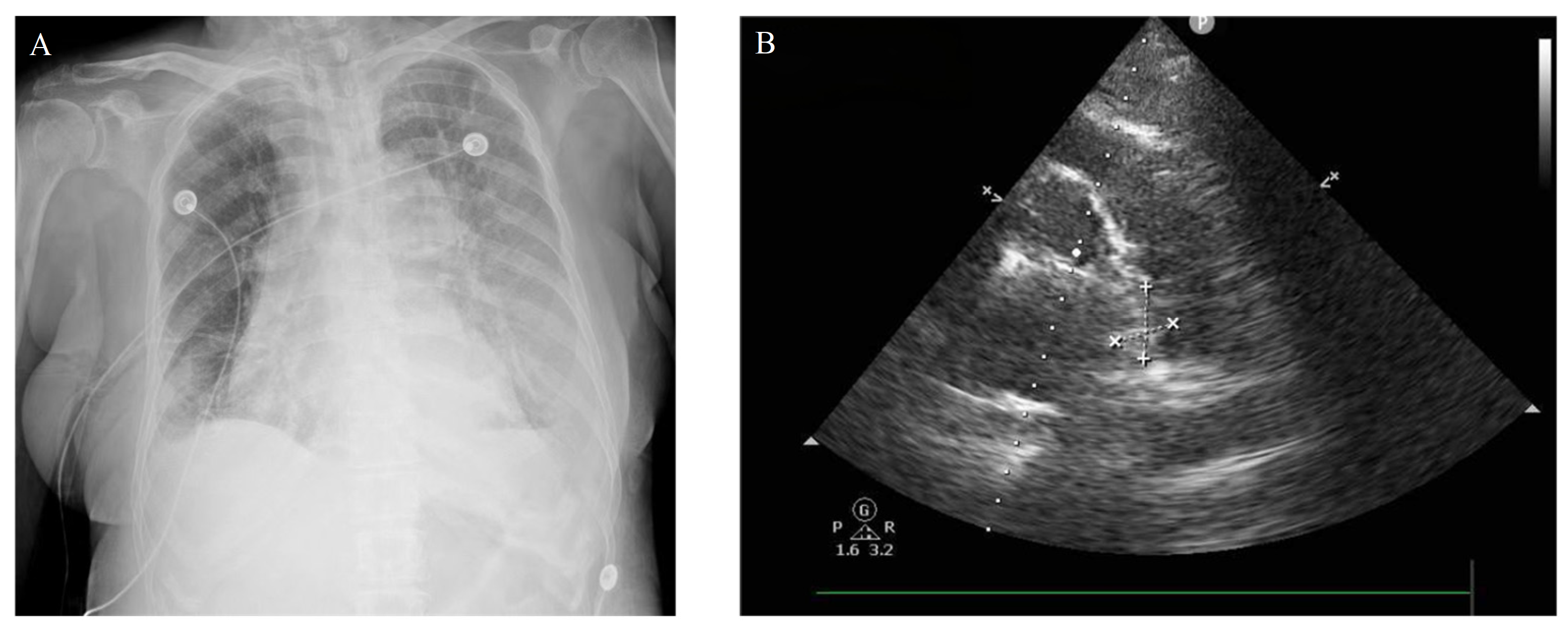
Figure 1. A 74-year-old female patient presented with dyspnea on exertion that started 3 weeks before admission without an apparent trigger. The dyspnea was relieved by rest. She also had intermittent low-grade fever in the afternoon, with a temperature of 37.4 °C, accompanied by chills and night sweats. After one week of anti-infective therapy at an outside hospital, the patient subjectively felt an improvement in dyspnea. Five days before hospital admission, the patient experienced right-sided chest pain without obvious precipitating factors. The pain was characterized as dull and related to respiration. Upon admission, the patient was conscious and alert, showing no symptoms such as palpitations, headache, dizziness, vomiting, or persistent dyspnea. The electrocardiogram (ECG) showed no abnormalities. The blood pressure was normal (115/65 mmHg), with a heart rate of 79 beats per minute and a respiratory rate of approximately 18 breaths per minute. There were no comorbidities such as hypertension, diabetes mellitus, or cerebral infarction, and there was no family history of tumors or genetic disorders. Admission tests indicated slightly elevated D-dimer levels at 0.56 μg/mL (reference range: 0.01–0.55 μg/mL), increased brain natriuretic peptide (BNP) levels at 892.33 pg/mL (reference range: <100 pg/mL), and elevated ultra-sensitive troponin T levels at 0.086 ng/mL (reference range: ≤0.014 ng/mL). The prothrombin time, partial thromboplastin time, and INR values were all within the normal ranges. Deep vein ultrasound of the lower extremities shows normal venous diameter, along with normal right atrial pressure. (A) The bedside anteroposterior chest X-ray shows full hilar structures in both lungs, increased lung markings, and bilateral pleural effusion. (B) Echocardiography reveals an irregular, moderately elevated mass at the origin of the right pulmonary artery, which measures approximately 2.3 × 2.0 cm. The “+” and “×” symbols are markers for measuring lesion length.
Diagnostics 16 01687 g001
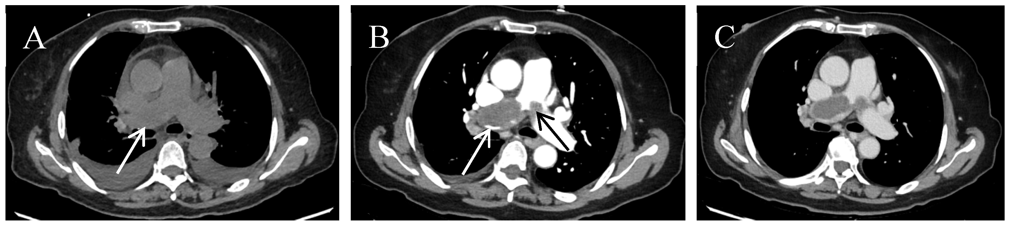
Figure 2. For further diagnosis, the patient underwent an enhanced computed tomography (CT) of the chest, which revealed filling defects in the right pulmonary trunk and its multiple branches, suggesting pulmonary embolism. The possibility of pulmonary artery sarcoma should be considered. (A) Non-contrast chest CT scan shows heterogeneous enlargement of the right pulmonary artery trunk with a slightly low-density shadow visible within (white arrow), with a CT value of 26 HU, bilateral pleural effusion, and minimal pericardial effusion. (B) Arterial phase contrast-enhanced chest CT shows multiple contrast filling defects at the bifurcation of the aortic trunk, right pulmonary artery trunk and its branches (white arrow), and left pulmonary artery trunk (black arrow), with rough edges of the defects and a CT value of 35 HU. (C) Venous phase contrast-enhanced chest CT shows the filling defect area with a CT value of 29 HU; the lesion showed mild enhancement in the arterial phase and no significant enhancement in the delayed phase.
Diagnostics 16 01687 g002
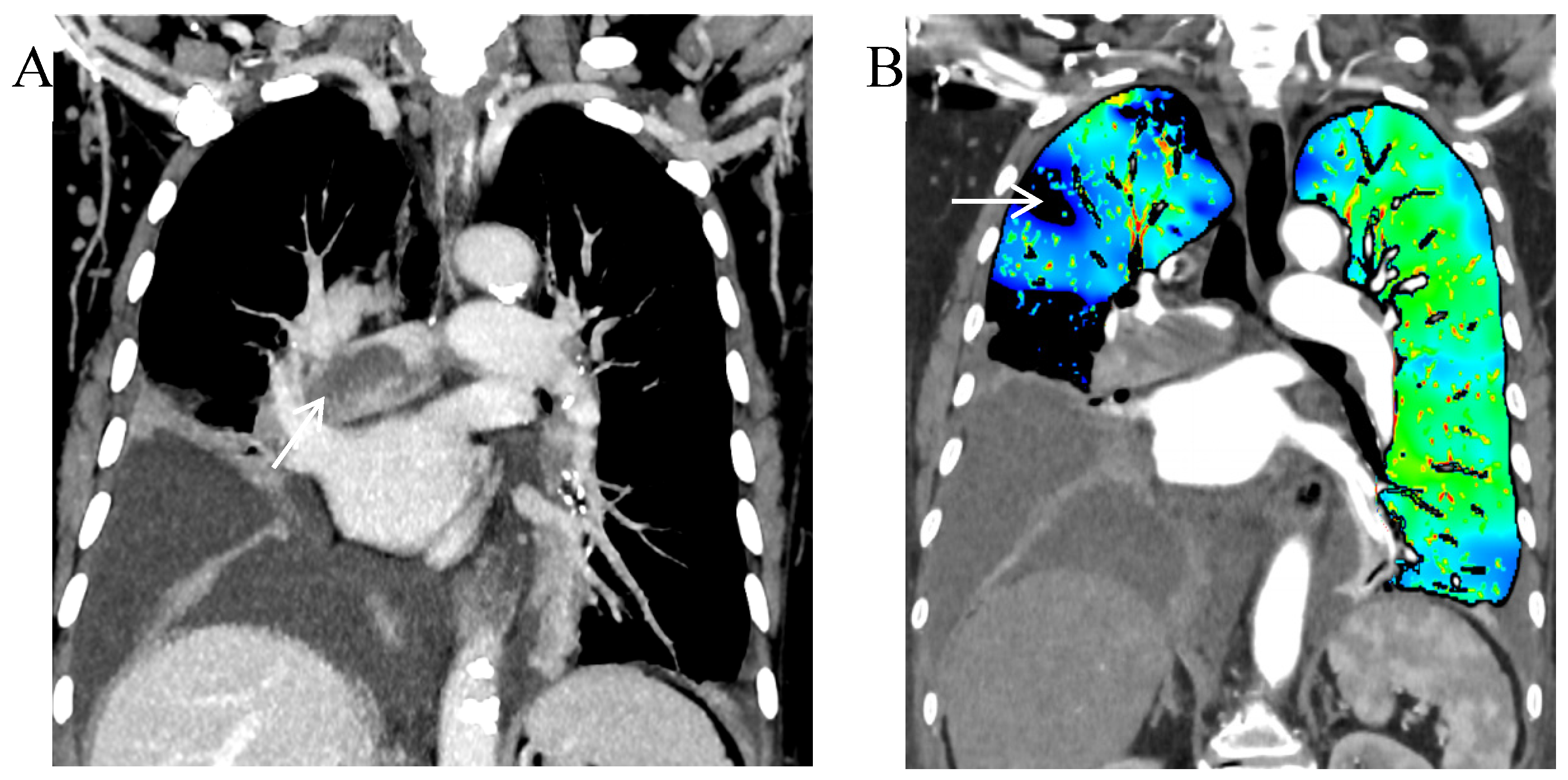
Figure 3. Spectral CT scan with post-processing analysis of patient images. (A) Coronal reconstruction using maximum intensity projection (MIP) highlighting enhanced vessels, showing contrast filling defects in the right pulmonary artery trunk (white arrow). The defect area more clearly shows irregular lesion margins, presenting the “Wall Eclipsing Sign”. (B) Spectral lung perfusion imaging showing irregular perfusion defects in the right lung (white arrow), with normal perfusion in the left lung. The pulmonary perfusion image clearly delineates the actual extent of perfusion defects caused by the tumor. In this case, the pulmonary perfusion image reveals irregular, non-segmental perfusion defects in the right lung, which are distinct from the typical wedge-shaped segmental perfusion defects seen in acute pulmonary embolism. This provides critical clues for identifying neoplastic lesions.
Diagnostics 16 01687 g003
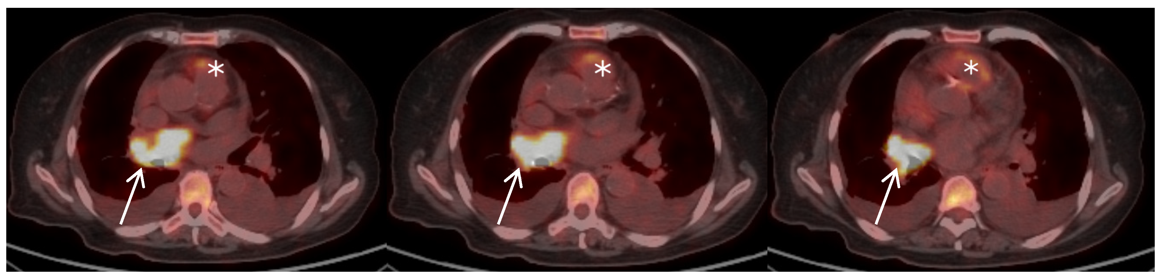
Figure 4. The patient continued to experience dyspnea after exertion despite anticoagulant therapy. As no acute embolic symptoms are currently present, vigilance is warranted to rule out thrombosis at the primary site caused by malignancy. To further evaluate the lesion in the right pulmonary artery, the patient subsequently underwent a PET/CT scan. PET/CT fusion images show heterogeneous enlargement of the right pulmonary artery trunk with uneven thickening of the vessel wall and a soft tissue mass visible locally. The lesion showed heterogeneous intense radiotracer uptake (white arrow) with a maximum standardized uptake value (SUVmax) of 14.7 and an uptake range of approximately 3.1 × 3.2 cm. The pulmonary artery trunk and bilateral pulmonary artery walls were slightly thickened, with radiotracer uptake visible in the pulmonary artery trunk and an SUVmax of 6.6 (white asterisk).
Diagnostics 16 01687 g004
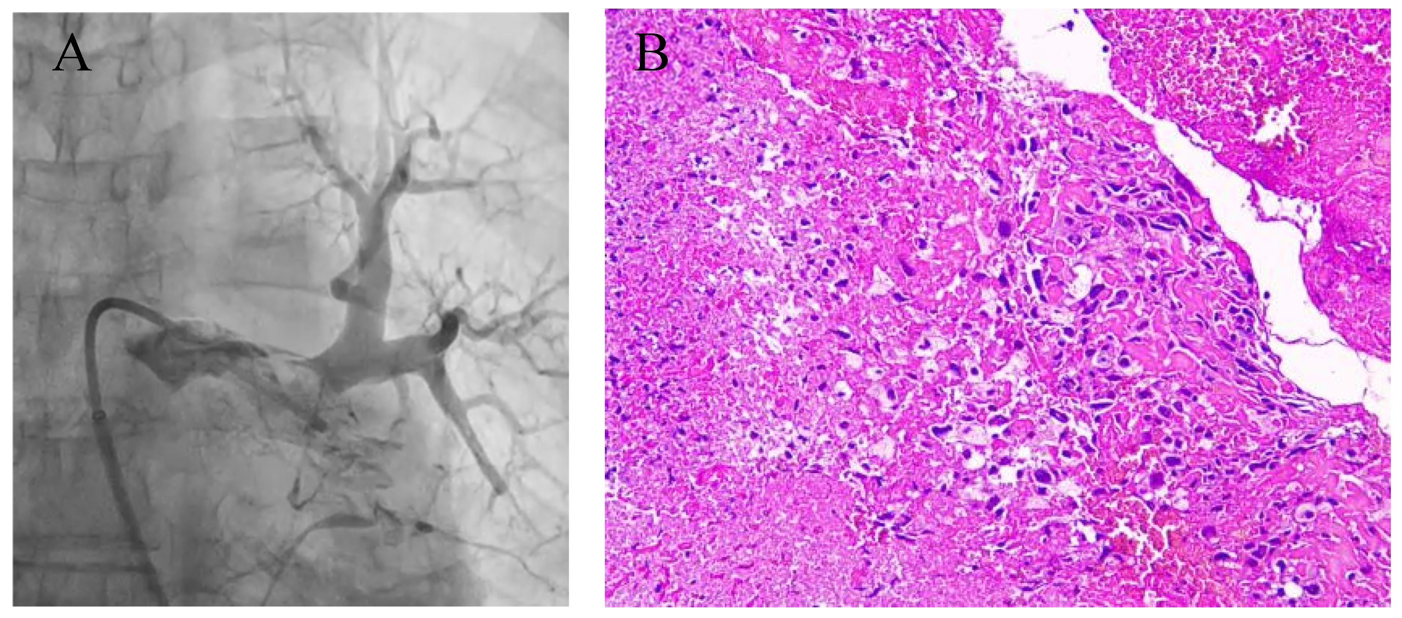
Figure 5. (A) The patient underwent catheter-based interventional embolectomy under angiography. (B) Postoperative pathological results, HE staining, magnification 10 × 10. Short, spindle-shaped atypical cells were identified in the submitted tissue, consistent with myogenic malignant tumor cells, and the diagnosis was pulmonary artery sarcoma. Immunohistochemistry: Vimentin positive, Desmin negative, Actin positive, SMA positive, LCA negative, Ki-67 (>40%), P53 focally weakly positive, CD31 positive, CD34 focally positive, CD117 negative, S-100 negative. Vimentin is a hallmark marker of mesenchymal tumors, indicating their origin from mesenchymal cells. Smooth muscle actin (SMA) positivity indicates smooth muscle differentiation. S-100 negativity excludes melanoma or neuroendocrine tumors. Meanwhile, a high Ki-67 proliferation index (>40%) and abnormal P53 expression suggest a high-grade, aggressive tumor. Primary pulmonary artery sarcoma (PPAS) is a malignant mesenchymal tumor originating from the pulmonary artery, accounting for approximately 0.001–0.003% of all sarcomas [1]. Pathologically, intimal sarcoma is the most common subtype, while other pathological types are extremely rare. The clinical symptoms, laboratory findings, and imaging features of PPAS are non-specific. Its rarity, combined with clinical manifestations similar to those of pulmonary embolism, makes diagnosis challenging [2]. Kashizaki et al. [3] reported that the mean age of PPAS patients was 52.6 years, with approximately equal gender distribution. The time from symptom onset to diagnosis was 12.8 weeks. Common clinical manifestations included chest pain, cough, and dyspnea. Hematological examination revealed mildly elevated D-dimer levels in 41.0% of patients, and elevated brain natriuretic peptide (BNP) or N-terminal pro-BNP (NT-proBNP) levels in 13.8% of patients. This patient was admitted with unexplained chest pain. Laboratory tests upon admission revealed slightly elevated D-dimer levels, making it impossible to distinguish from pulmonary thromboembolism. Advances in medical technology have significantly contributed to the diagnosis of PPAS, particularly computed tomography angiography (CTA) and contrast-enhanced CT, which are crucial for lesion identification and preoperative planning. Pulmonary artery sarcoma presents as an irregular soft-tissue mass within the main trunk or branches of the pulmonary artery. It has ill-defined borders and exhibits polypoid, lobulated, or fungoid-like morphology, often accompanied by vascular wall erosion (the “Wall Eclipsing Sign”). Contrast-enhanced imaging demonstrates heterogeneous enhancement that may extend beyond the vessel, leading to mediastinal and hilar lymphadenopathy or multiple pulmonary nodules. In contrast, pulmonary arterial thromboembolism manifests as a filling defect within the pulmonary artery lumen with smooth margins, appearing cup-shaped or crescent-shaped. It shows no enhancement on contrast-enhanced scans, and chronic pulmonary thromboembolism may be associated with calcification. The “Wall Eclipsing Sign” is a characteristic feature, defined as the following three manifestations: the lumen of the pulmonary trunk, left pulmonary artery, or right pulmonary artery is almost entirely occupied by a low-density mass; the proximal portion of the mass protrudes toward the right ventricular outflow tract; and the lesion obscures one or both walls of the pulmonary trunk, left pulmonary artery, or right pulmonary artery [4]. This sign was observed on contrast-enhanced CT in the present case. A study revealed that among 12 PPAS patients assessed for this sign, all exhibited the “Wall Eclipsing Sign”. In contrast, none of the 156 patients with chronic pulmonary thromboembolism (CPTE) complicated by pulmonary hypertension or the 426 patients with acute pulmonary embolism displayed this sign [4]. However, its sensitivity is limited, and this imaging feature should be interpreted in conjunction with the complete clinical context and other diagnostic clues. Ioakeimidou et al. [5] reported other imaging findings, such as complete lumen obstruction caused by low-attenuation lesions, arterial dilation, and extraluminal tumor extension; these imaging features are equally important for differentiating pulmonary embolism (PE). PPAS demonstrates high metabolic FDG uptake on PET/CT [6], whereas pulmonary artery thromboembolism typically shows photopenic defects. This distinction is critically important for differentiating pulmonary artery sarcoma from pulmonary thromboembolism. The study by Ren et al. [7] demonstrated that the SUVmax of PPAS was significantly higher than that of PTE. The median SUVmax values were 8.9 (range: 3.0–17.2) for PPAS and 1.8 (range: 0.8–3.7) for PTE. When using SUVmax = 2.9 as the threshold, the sensitivity and specificity reached 100% and 93.9%, respectively. In this patient, the lesion SUVmax was 14.7, and the pulmonary trunk SUVmax was 6.6. Both significantly exceeded the aforementioned diagnostic thresholds, strongly supporting the diagnosis of pulmonary artery sarcoma. For the evaluation of masses in the heart, adjacent to the heart, or within major blood vessels, the adoption of standardized diagnostic methods is crucial. Structured echocardiographic assessment is the primary step, allowing for dynamic observation of the tumor’s location, size, shape, mobility, spatial structure, and adjacent relationships, as well as a preliminary evaluation of its benign or malignant nature and degree of infiltration. The DEM scoring system proposed by Paolisso et al. [8], which incorporates six ultrasonographic features, indicates malignancy when ≥3 features are present, providing an evidence-based tool for standardized evaluation of intracardiac masses. Although pulmonary artery sarcoma is located in the main pulmonary artery trunk and has a low direct visualization rate via transthoracic echocardiography, the features of “non-left-sided location” and “wide basal attachment” in this scoring system can still suggest malignancy, prompting further imaging studies and thereby reducing diagnostic delays. Other differential diagnoses for pulmonary artery filling defects include macrovascular vasculitis and tumor embolism originating from extrapulmonary primary tumors. Patients with vasculitis often present with systemic symptoms such as fever, fatigue, and arthralgia, along with a markedly elevated erythrocyte sedimentation rate (ESR) and C-reactive protein levels, as well as vascular wall thickening or circumferential enhancement. For tumor embolism, identification of the primary lesion is essential. Comprehensive PET/CT imaging revealed no presence of malignancies prone to pulmonary arterial embolism, such as lung cancer, renal cell carcinoma, hepatocellular carcinoma, or choriocarcinoma, in this case. These conditions were all excluded based on clinical and imaging findings. While imaging features may suggest pulmonary artery sarcoma, definitive diagnosis depends on histopathological examination. On immunohistochemical staining, Vimentin is strongly expressed in all pulmonary artery sarcomas, with variable positive expression of Desmin and smooth muscle actin (SMA). Endothelial and epithelial markers, as well as S100 protein, are typically negative. MDM2 is expressed in most pulmonary artery sarcomas, and the Ki-67 proliferation index is generally high. Surgery is considered the primary treatment modality for pulmonary artery sarcoma [9]. Surgical treatment was employed in 81.4% of cases, with endarterectomy being the most common approach, accounting for 50.7% of procedures. Although some studies have suggested that a subset of patients receiving chemotherapy and radiotherapy may achieve improved survival compared with surgery alone [10], no standardized treatment approach has been established. In this report, the patient currently suffers from respiratory failure and heart failure, making surgical intervention intolerable, while the efficacy of radiotherapy and chemotherapy remains unclear. After undergoing catheter-guided interventional angiography-assisted pulmonary artery thrombectomy, the patient’s chest pain was alleviated. It is currently believed that alleviative treatment can improve symptoms. The patient is able to independently manage basic daily activities, with a stable overall condition, and continues to undergo follow-up. Our study demonstrates that although primary pulmonary artery sarcoma (PPAS) is rare, considering PPAS in the differential diagnosis of pulmonary embolism (PE) is crucial for raising clinical awareness. Suspect PPAS in patients who present with symptoms mimicking pulmonary embolism but lack typical risk factors, do not respond to anticoagulation, or show atypical imaging features. Once PPAS is clinically suspected, it is recommended to promptly perform examinations such as echocardiography, computed tomography (CT), or positron emission tomography (PET) to achieve early diagnosis and exclusion.
Diagnostics 16 01687 g005

Author Contributions

D.L. and Z.L.: writing original draft preparation; S.L. and H.Z.: editing and supervision. All authors have read and agreed to the published version of the manuscript.

Funding

This research received no external funding.

Institutional Review Board Statement

Ethical review and approval were waived for this study. This waiver applies because it is a single retrospective case report that did not influence the patient’s clinical management and falls outside the definition of regulated human subjects research per institutional and national policies.

Informed Consent Statement

Written informed consent has been obtained from the patients to publish this paper.

Data Availability Statement

No new data were created or analyzed in this study.

Conflicts of Interest

The authors declare no conflicts of interest.

Abbreviations

The following abbreviations are used in this manuscript:
CTComputed tomography
CTAComputed tomography angiography
HUHounsfield unit
PPASPrimary pulmonary artery sarcoma
MDCTMulti-detector row computed tomography
18F-FDG PET/CTFluorine-18 Fluorodeoxyglucose Positron Emission Tomography/Computed Tomography
MIPMaximum intensity projection
BNPBrain natriuretic peptide

References

  1. Cuenca Peris, S.; Bauza, M.; Briones Gómez, A. Primary Pulmonary Artery Sarcoma: A Rare Tumor Diagnosed by EBUS Guided Mediastinal Cryobiopsy. Arch. Bronconeumol. 2024, 60, 531–532. [Google Scholar] [CrossRef] [PubMed]
  2. Hieu, N.L.; Tu, V.N.; Hoan, L.; Hai, H.B.; Luu, D.T.; Cuong, N.N.; Tra My, T.T.; Minh, T.N.; Manh, P.T. A case report of primary pulmonary artery intimal sarcoma. Radiol. Case Rep. 2022, 17, 1986–1990. [Google Scholar] [CrossRef] [PubMed]
  3. Kashizaki, F.; Chen, H.; Tsuchiya, N.; Matsumoto, S.; Osada, R.; Miyasaka, A.; Kaneko, M.; Kikuchi, A.; Kojima, Y.; Yumoto, K.; et al. Clinical Features of Primary Pulmonary Artery Sarcoma: A Systematic Review and Pooled Analysis. Arch. Bronconeumol. 2025, 61, 536–543. [Google Scholar] [CrossRef]
  4. Gan, H.L.; Zhang, J.Q.; Huang, X.Y.; Yu, W. The wall eclipsing sign on pulmonary artery computed tomography angiography is pathognomonic for pulmonary artery sarcoma. PLoS ONE 2013, 8, e83200. [Google Scholar] [CrossRef] [PubMed]
  5. Ioakeimidou, S.; Stephan, F.P.; Pluess, E.; Seeman, M.; Srivastava, D. Pulmonary Artery Intimal Sarcoma Mimics Recurrent Pulmonary Artery Embolism. Eur. J. Case Rep. Intern. Med. 2025, 12, 005150. [Google Scholar] [CrossRef] [PubMed]
  6. Pan, B.; Wang, S.C.; Chen, Z.K.; Chen, X. Primary pulmonary artery sarcoma with intrapulmonary metastases based on PET/CT imaging: A case report and literature review. Ann. Palliat. Med. 2021, 10, 7013–7018. [Google Scholar] [CrossRef] [PubMed]
  7. Ren, J.; Li, H.; Zhang, Q.; Liu, E.; Zeng, B.; Huang, Y.; Wang, L.; Jiang, L. Clinical utility of 18F-FDG PET/CT imaging in patients with pulmonary artery sarcoma. EJNMMI Res. 2022, 12, 18. [Google Scholar] [CrossRef] [PubMed]
  8. Paolisso, P.; Foà, A.; Bergamaschi, L.; Graziosi, M.; Rinaldi, A.; Magnani, I.; Angeli, F.; Stefanizzi, A.; Armillotta, M.; Sansonetti, A.; et al. Echocardiographic Markers in the Diagnosis of Cardiac Masses. J. Am. Soc. Echocardiogr. 2023, 36, 464–473.e2. [Google Scholar] [CrossRef] [PubMed]
  9. Chan, E.Y.; Ravi, V.; Ali, A.; Nguyen, D.T.; Graviss, E.A.; MacGillivray, T.E.; Reardon, M.J. Surgical Management of Primary Pulmonary Artery Sarcoma. Semin. Thorac. Cardiovasc. Surg. 2023, 35, 53–64. [Google Scholar] [CrossRef] [PubMed]
  10. Wong, H.H.; Gounaris, I.; McCormack, A.; Berman, M.; Davidson, D.; Horan, G.; Pepke-Zaba, J.; Jenkins, D.; Earl, H.M.; Hatcher, H.M. Presentation and management of pulmonary artery sarcoma. Clin. Sarcoma Res. 2015, 5, 3. [Google Scholar] [CrossRef] [PubMed]
Disclaimer/Publisher’s Note: The statements, opinions and data contained in all publications are solely those of the individual author(s) and contributor(s) and not of MDPI and/or the editor(s). MDPI and/or the editor(s) disclaim responsibility for any injury to people or property resulting from any ideas, methods, instructions or products referred to in the content.