Impact of Imaging Modality on AI-Based Detection of Incidental Maxillary Sinus Pathology: Comparison of Panoramic Radiography and CBCT

 , , , , and
Abstract
1. Introduction
2. Materials and Methods
2.1. Study Design
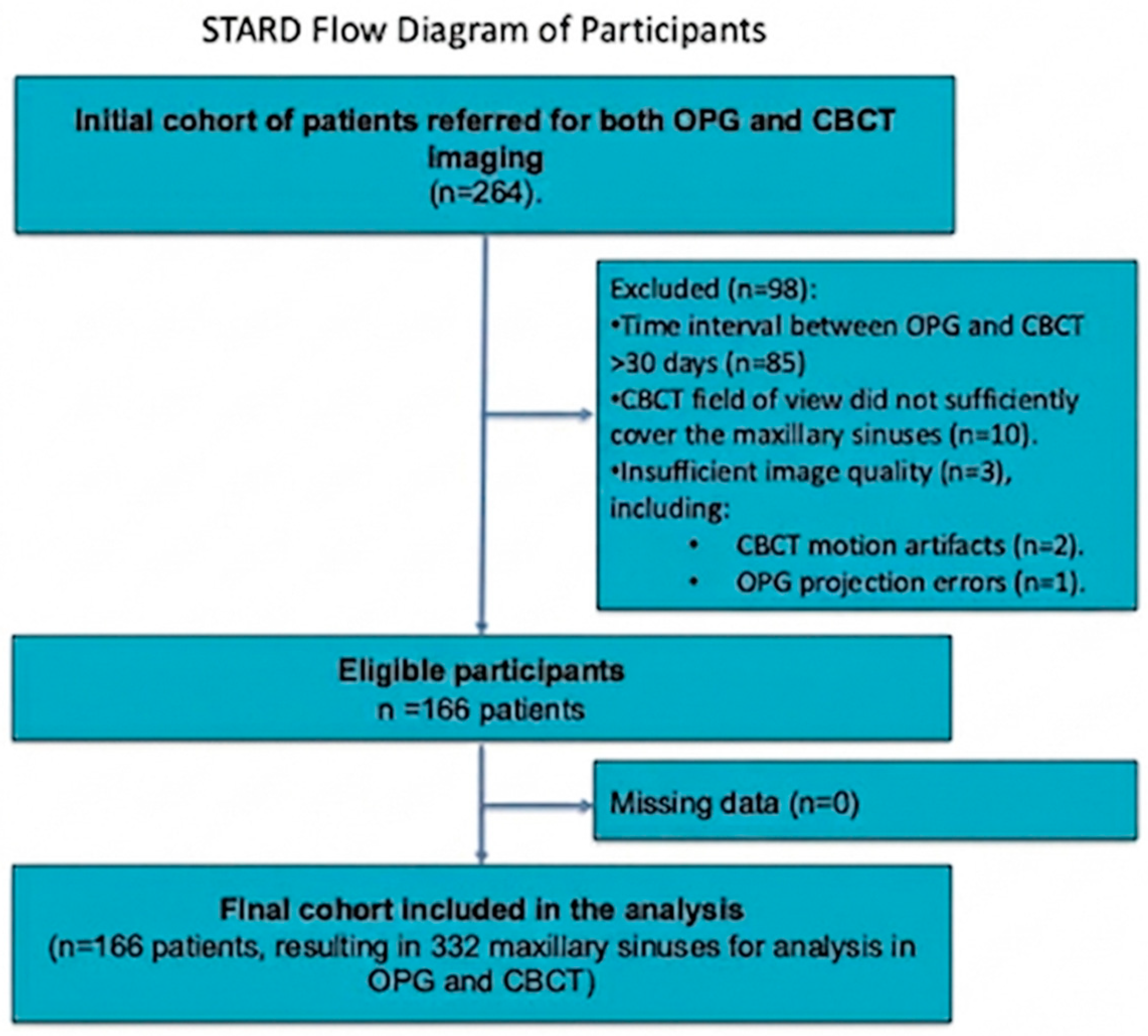
2.2. Patients
2.3. Sample Size, Precision, and a Priori Power
2.4. Image Acquisition
2.5. AI Evaluation
2.6. Human Observer Evaluation (Reference Standard)
2.7. Statistical Evaluation
- •
- A true positive (TP) was recorded if the AI flagged ‘any abnormality’ in a sinus where the reference standard identified a polyp/cyst;
- •
- A false negative (FN) was recorded if the AI did not flag ‘any abnormality’ in a sinus where the reference standard identified a polyp/cyst.
3. Results
3.1. Patients
3.2. Diagnostic Accuracy Parameters
3.3. Relationship Between Lesion Size and AI Platforms’ Diagnostic Accuracy on OPG and CBCT
3.4. Formal Paired Statistical Comparison
3.5. Inter- and Intra-Reader Agreement
4. Discussion
5. Conclusions
Supplementary Materials
Author Contributions
Funding
Institutional Review Board Statement
Informed Consent Statement
Data Availability Statement
Conflicts of Interest
References
- Hsiao, Y.J.; Yang, J.; Resnik, R.R.; Suzuki, J.B. Prevalence of Maxillary Sinus Pathology Based on Cone-Beam Computed Tomography Evaluation of Multiethnicity Dental School Population. Implant. Dent. 2019, 28, 356–366. [Google Scholar] [CrossRef] [PubMed]
- Dogan, M.E.; Uluısık, N.; Yuvarlakbaş, S.D. Author Correction: Retrospective analysis of pathological changes in the maxillary sinus with CBCT. Sci. Rep. 2024, 14, 18236. [Google Scholar] [CrossRef]
- Constantine, S.; Clark, B.; Kiermeier, A.; Anderson, P.P. Panoramic radiography is of limited value in the evaluation of maxillary sinus disease. Oral Surg. Oral Med. Oral Pathol. Oral Radiol. 2019, 127, 237–246. [Google Scholar] [CrossRef]
- Nur, M.; Kayipmaz, S.; Bayram, M.; Celikoglu, M.; Kilkis, D.; Sezgin, O.S. Conventional frontal radiographs compared with frontal radiographs obtained from cone beam computed tomography. Angle Orthod. 2012, 82, 579–584. [Google Scholar] [CrossRef]
- Olmez, H.; Gorgulu, S.; Akin, E.; Bengi, A.O.; Tekdemir, I.; Ors, F. Measurement accuracy of a computer-assisted three-dimensional analysis and a conventional two-dimensional method. Angle Orthod. 2011, 81, 375–382. [Google Scholar] [CrossRef]
- Park, C.S.; Park, J.K.; Kim, H.; Han, S.-S.; Jeong, H.-G.; Park, H. Comparison of conventional lateral cephalograms with corresponding CBCT radiographs. Imaging Sci. Dent. 2012, 42, 201–205. [Google Scholar] [CrossRef]
- Patel, S.; Dawood, A.; Pitt Ford, T.; Whaites, E. The potential applications of cone beam computed tomography in the management of endodontic problems. Int. Endod. J. 2007, 40, 818–830. [Google Scholar] [CrossRef]
- Garlapati, K.; Gandhi Babu, D.B.; Chaitanya, N.C.S.K.; Guduru, H.; Rembers, A.; Soni, P. Evaluation of preference and purpose of utilisation of cone beam computed tomography (CBCT) compared to orthopantomogram (OPG) by dental practitioners—A cross-sectional study. Pol. J. Radiol. 2017, 82, 248–251. [Google Scholar] [CrossRef]
- Vogiatzi, T.; Kloukos, D.; Scarfe, W.; Bornstein, M. Incidence of Anatomical Variations and Disease of the Maxillary Sinuses as Identified by Cone Beam Computed Tomography: A Systematic Review. Int. J. Oral Maxillofac. Implant. 2014, 29, 1301–1314. [Google Scholar] [CrossRef] [PubMed]
- Yeung, A.W.K.; Hung, K.F.; Li, D.T.S.; Leung, Y.Y. The Use of CBCT in Evaluating the Health and Pathology of the Maxillary Sinus. Diagnostics 2022, 12, 2819. [Google Scholar] [CrossRef] [PubMed]
- DGNCT LLC. MFU; DGNCT LLC: Miami, FL, USA, 2025; Available online: https://diagnocat.com/en (accessed on 3 September 2025).
- Issa, J.; Konwinska, M.D.; Kazimierczak, N.; Olszewski, R. Assessing the accuracy of artificial intelligence in mandibular canal segmentation compared to semi-automatic segmentation on cone-beam computed tomography images. Pol. J. Radiol. 2025, 90, 172–179. [Google Scholar] [CrossRef]
- Kazimierczak, W.; Kazimierczak, N.; Issa, J.; Wajer, R.; Wajer, A.; Kalka, S.; Serafin, Z. Endodontic Treatment Outcomes in Cone Beam Computed Tomography Images—Assessment of the Diagnostic Accuracy of AI. J. Clin. Med. 2024, 13, 4116. [Google Scholar] [CrossRef]
- Kwiatek, J.; Leśna, M.; Piskórz, W.; Kaczewiak, J. Comparison of the Diagnostic Accuracy of an AI-Based System for Dental Caries Detection and Clinical Evaluation Conducted by Dentists. J. Clin. Med. 2025, 14, 1566. [Google Scholar] [CrossRef]
- Kazimierczak, W.; Wajer, R.; Wajer, A.; Kalka, K.; Kazimierczak, N.; Serafin, Z. Evaluating the Diagnostic Accuracy of an AI-Driven Platform for Assessing Endodontic Treatment Outcomes Using Panoramic Radiographs: A Preliminary Study. J. Clin. Med. 2024, 13, 3401. [Google Scholar] [CrossRef] [PubMed]
- Futyma-Gąbka, K.; Piskórz, M.; Smala, K.; Miazek, W.; Moskwa, M.; Różyło-Kalinowska, I. Evaluation of effectiveness of a virtual AI-based dental assistant in recognizing mixed dentition on panoramic radiographs. J. Stomatol. 2024, 77, 181–185. [Google Scholar] [CrossRef]
- Çelebi, A.; Imak, A.; Üzen, H.; Budak, Ü.; Türkoğlu, M.; Hanbay, D.; Şengür, A. Maxillary sinus detection on cone beam computed tomography images using ResNet and Swin Transformer-based UNet. Oral. Surg. Oral. Med. Oral. Pathol. Oral. Radiol. 2024, 138, 149–161. [Google Scholar] [CrossRef]
- Zeng, P.; Song, R.; Lin, Y.; Li, H.; Chen, S.; Shi, M.; Cai, G.; Gong, Z.; Huang, K.; Chen, Z. Abnormal maxillary sinus diagnosing on CBCT images via object detection and ‘straight-forward’ classification deep learning strategy. J. Oral. Rehabil. 2023, 50, 1465–1480. [Google Scholar] [CrossRef]
- Hung, K.F.; Ai, Q.Y.H.; King, A.D.; Bornstein, M.M.; Wong, L.M.; Leung, Y.Y. Automatic detection and segmentation of morphological changes of the maxillary sinus mucosa on cone-beam computed tomography images using a three-dimensional convolutional neural network. Clin. Oral. Investig. 2022, 26, 3987–3998. [Google Scholar] [CrossRef] [PubMed]
- Bossuyt, P.M.; Reitsma, J.B.; Bruns, D.E.; Gatsonis, C.A.; Glasziou, P.P.; Irwig, L.; Lijmer, J.G.; Moher, D.; Rennie, D.; de Vet, H.C.; et al. STARD 2015: An Updated List of Essential Items for Reporting Diagnostic Accuracy Studies. Radiology 2015, 277, 826–832. [Google Scholar] [CrossRef]
- Serindere, G.; Bilgili, E.; Yesil, C.; Ozveren, N. Evaluation of maxillary sinusitis from panoramic radiographs and cone-beam computed tomographic images using a convolutional neural network. Imaging Sci. Dent. 2022, 52, 187. [Google Scholar] [CrossRef]
- Lachenbruch, P.A. On the Sample Size for Studies Based upon Mcnemar’s Test. Stat. Med. 1992, 11, 1521–1525. [Google Scholar] [CrossRef] [PubMed]
- Capelli, M.; Gatti, P. Radiological study of maxillary sinus using CBCT: Relationship between mucosal thickening and common anatomic variants in chronic rhinosinusitis. J. Clin. Diagn. Res. 2016, 10, MC07–MC10. [Google Scholar] [CrossRef]
- Ren, S.; Zhao, H.; Liu, J.; Wang, Q.; Pan, Y. Significance of maxillary sinus mucosal thickening in patients with periodontal disease. Int. Dent. J. 2015, 65, 303–310. [Google Scholar] [CrossRef] [PubMed] [PubMed Central]
- Maillet, M.; Bowles, W.R.; McClanahan, S.L.; John, M.T.; Ahmad, M. Cone-beam computed tomography evaluation of maxillary sinusitis. J. Endod. 2011, 37, 753–757. [Google Scholar] [CrossRef] [PubMed]
- Lu, Y.; Liu, Z.; Zhang, L.; Zhou, X.; Zheng, Q.; Duan, X.; Zheng, G.; Wang, H.; Huang, D. Associations between maxillary sinus mucosal thickening and apical periodontitis using cone-beam computed tomography scanning: A retrospective study. J. Endod. 2012, 38, 1069–1074. [Google Scholar] [CrossRef] [PubMed]
- Huang, Y.T.; Hu, S.W.; Huang, J.Y.; Chang, Y.C. Assessment of relationship between maxillary sinus membrane thickening and the adjacent teeth health by cone-beam computed tomography. J. Dent. Sci. 2021, 16, 275–279. [Google Scholar] [CrossRef]
- Breau, R.H.; Clark, E.; Bruner, B.; Cervini, P.; Atwell, T.; Knoll, G.; Leibovich, B.C. A simple method to estimate renal volume from computed tomography. J. Can. Urol. Assoc. 2013, 7, 189–192. [Google Scholar] [CrossRef]
- Shrout, P.E.; Fleiss, J.L. Intraclass correlations: Uses in assessing rater reliability. Psychol. Bull. 1979, 86, 420–428. [Google Scholar] [CrossRef] [PubMed]
- Cohen, J. A Coefficient of Agreement for Nominal Scales. Educ. Psychol. Meas. 1960, 20, 37–46. [Google Scholar] [CrossRef]
- Hicks, S.A.; Strümke, I.; Thambawita, V.; Hammou, M.; Riegler, M.A.; Halvorsen, P.; Parasa, S. On evaluation metrics for medical applications of artificial intelligence. Sci. Rep. 2022, 12, 5979. [Google Scholar] [CrossRef]
- Uthman, A.T.; Abouelenen, H.; Khan, S.; Bseiso, O.; Al-Rawi, N. Diagnostic accuracy of artificial intelligence in the detection of maxillary sinus pathology using computed tomography: A concise systematic review. Imaging Sci. Dent. 2025, 55, 1. [Google Scholar] [CrossRef]
- Kazimierczak, W.; Wajer, R.; Wajer, A.; Kiian, V.; Kloska, A.; Kazimierczak, N.; Janiszewska-Olszowska, J.; Serafin, Z. Periapical Lesions in Panoramic Radiography and CBCT Imaging—Assessment of AI’s Diagnostic Accuracy. J. Clin. Med. 2024, 13, 2709. [Google Scholar] [CrossRef]
- Kuwana, R.; Ariji, Y.; Fukuda, M.; Kise, Y.; Nozawa, M.; Kuwada, C.; Muramatsu, C.; Katsumata, A.; Fujita, H.; Ariji, E. Performance of deep learning object detection technology in the detection and diagnosis of maxillary sinus lesions on panoramic radiographs. Dentomaxillofacial Radiol. 2020, 50, 20200171. [Google Scholar] [CrossRef]
- Alsowey, A.M.; Abdulmonaem, G.; Elsammak, A.; Fouad, Y. Diagnostic performance of multidetector computed tomography (MDCT) in diagnosis of sinus variations. Pol. J. Radiol. 2017, 82, 713–725. [Google Scholar] [CrossRef]
- Xu, J.; Reh, D.D.; Carey, J.P.; Mahesh, M.; Siewerdsen, J.H. Technical assessment of a cone-beam CT scanner for otolaryngology imaging: Image quality, dose, and technique protocols. Med. Phys. 2012, 39, 4932–4942. [Google Scholar] [CrossRef]
- Kuligowski, P.; Jaroń, A.; Preuss, O.; Gabrysz-Trybek, E.; Bladowska, J.; Trybek, G. Association between Odontogenic and Maxillary Sinus Conditions: A Retrospective Cone-Beam Computed Tomographic Study. J. Clin. Med. 2021, 10, 2849. [Google Scholar] [CrossRef]
- Bajoria, A.; Sarkar, S.; Sinha, P. Evaluation of odontogenic maxillary sinusitis with cone beam computed tomography: A retrospective study with review of literature. J. Int. Soc. Prev. Community Dent. 2019, 9, 194. [Google Scholar] [CrossRef]
- Orhan, K.; Aktuna Belgin, C.; Manulis, D.; Golitsyna, M.; Bayrak, S.; Aksoy, S.; Sanders, A.; Önder, M.; Ezhov, M.; Shamshiev, M.; et al. Determining the reliability of diagnosis and treatment using artificial intelligence software with panoramic radiographs. Imaging Sci. Dent. 2023, 53, 199. [Google Scholar] [CrossRef]


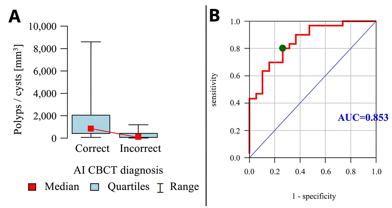
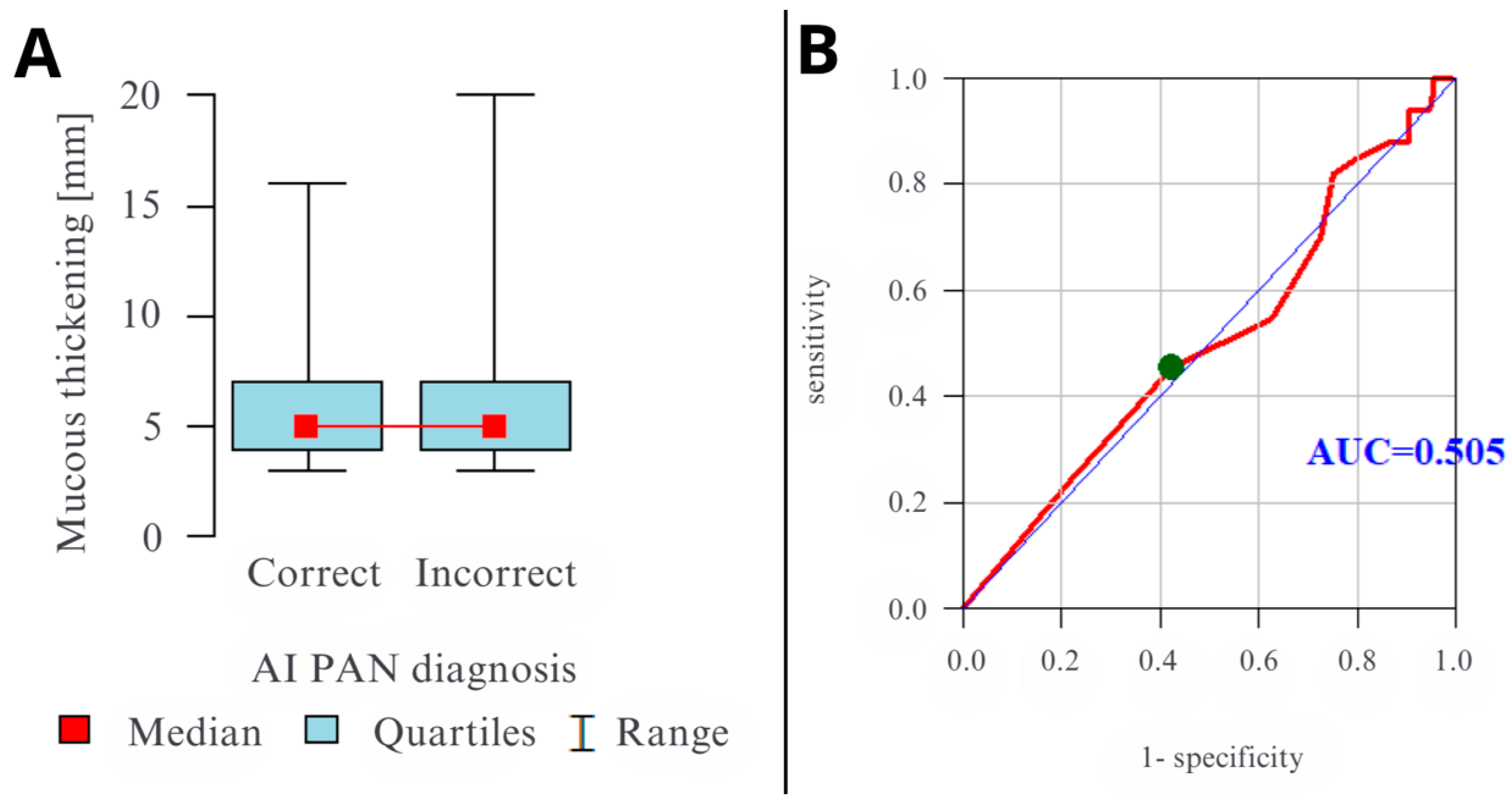
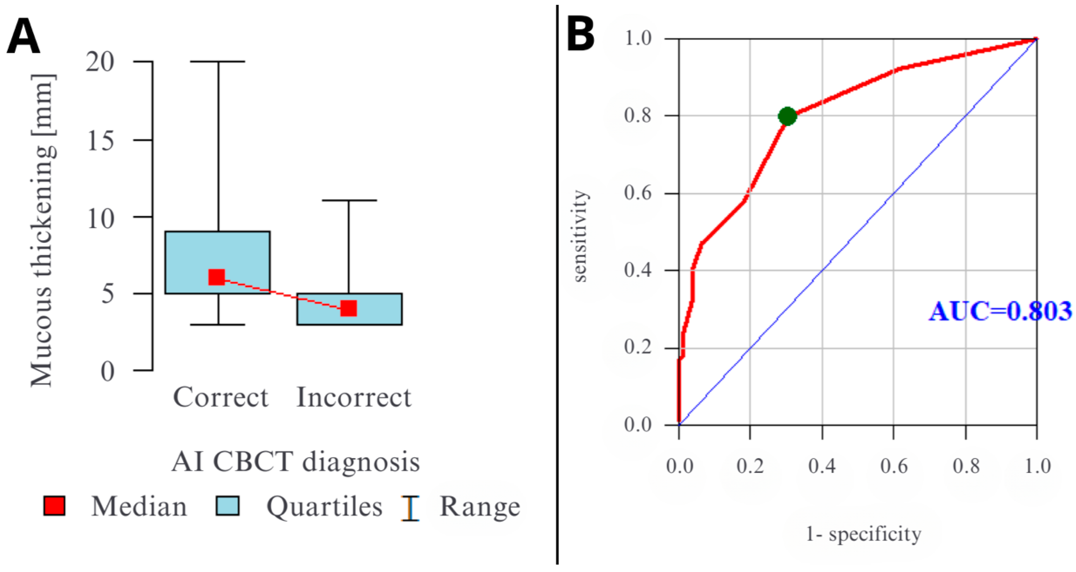

| Characteristic | Value |
|---|---|
| Patients | |
| Total number | 166 |
| Age, years (mean ± SD) | 31.3 ± 15.2 |
| Age, years (range) | 8–70 |
| Sex | |
| Female, n (%) | 113 (68.1) |
| Male, n (%) | 53 (31.9) |
| Sinuses | |
| Total number | 332 |
| Prevalence of Findings by Reference Standard (CBCT Consensus) | |
| Any abnormality, n (%) | 185 (55.7) |
| Mucosal thickening (≥2 mm), n (%) | 167 (50.3) |
| Polyp/Retention cyst, n (%) | 49 (15.2) |
| Free fluid, n (%) | 11 (3.4) |
| Lesion Characteristics (in positive cases) | |
| Mucosal thickening, mm (mean ± SD) | 6.0 ± 3.7 |
| Polyp/Cyst volume, mm3 (median) | 463.3 [131.0–1216.8] |
| Parameter | Modality | Accuracy | Precision | Recall | F1 Score |
|---|---|---|---|---|---|
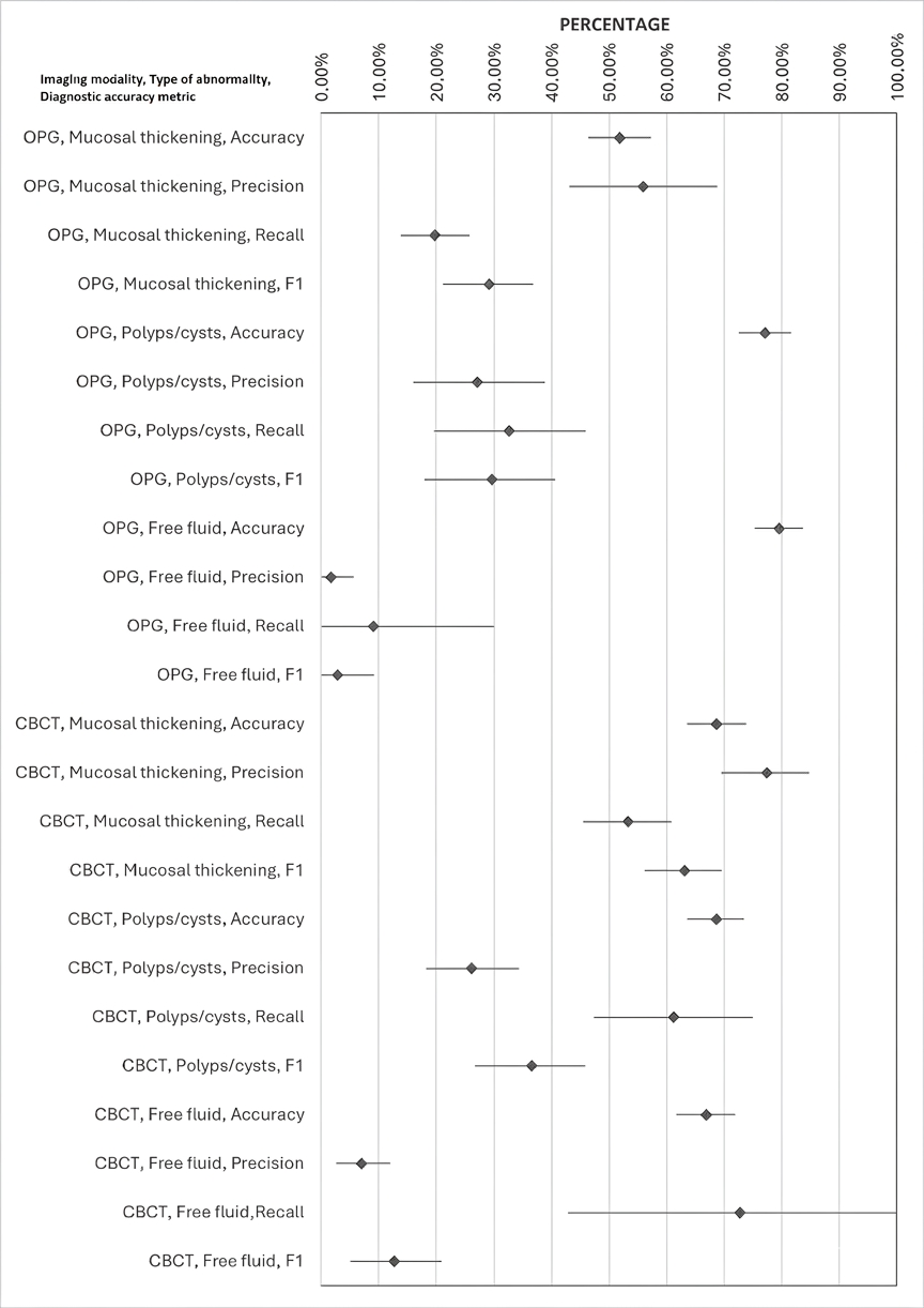
| Mucosal thickening | OPG | 51.81% (46.39–57.23%) | 55.93% (43.14–68.75%) | 19.76% (13.82–25.84%) | 29.20% (21.20–36.75%) |
| Polyps/cysts | 77.11% (72.59–81.63%) | 27.12% (16.00–38.89%) | 32.65% (19.65–46.00%) | 29.63% (18.02–40.65%) | |
| Free fluid | 79.52% (75.30–83.73%) | 1.69% (0.00–5.66%) | 9.09% (0.00–30.00%) | 2.86% (0.00–9.23%) | |
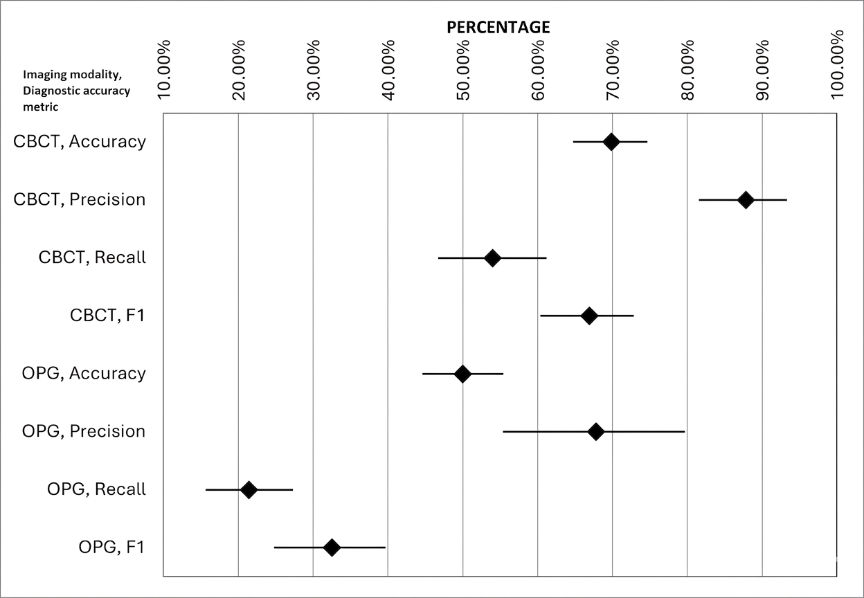
| Any abnormalities | 50.60% (44.58–55.41%) | 67.80% (55.38–79.66%) | 21.39% (15.62–27.32%) | 32.52% (24.79–39.69%) | |
| Mucosal thickening | CBCT | 68.67% (63.55–73.80%) | 77.39% (69.45–84.75%) | 53.29% (45.51–60.87%) | 63.12% (56.18–69.54%) |
| Polyps/cysts | 68.67% (63.55–73.49%) | 26.09% (18.18–34.31%) | 61.22% (47.37–75.00%) | 36.59% (26.75–45.81%) | |
| Free fluid | 66.87% (61.75–71.98%) | 6.96% (2.65–12.00%) | 72.73% (42.86–100.00%) | 12.70% (5.04–20.90%) | |
| Any abnormalities | 69.88% (64.76–74.70%) | 87.83% (81.58–93.33%) | 54.01% (46.74–61.17%) | 66.89% (60.34–72.84%) |
| Modality | TP | FP | TN | FN | Accuracy | Precision | Recall | F1 Score |
|---|---|---|---|---|---|---|---|---|
| CBCT | 100 | 15 | 132 | 85 | 69.9% (64.8–74.7%) | 87.8% (81.6–93.3%) | 54.0% (46.7–61.2%) | 66.9% (60.3–72.8%) |
| OPG | 40 | 19 | 128 | 145 | 50.6% (44.6–55.4%) | 67.8% (55.4–79.7%) | 21.4% (15.6–27.3%) | 32.5% (24.8–39.7%) |
| AI Diagnosis | Modality | N | Polyps/Cysts [mm3] | p | ||||||
|---|---|---|---|---|---|---|---|---|---|---|
| Mean | SD | Median | Min | Max | Q1 | Q3 | ||||
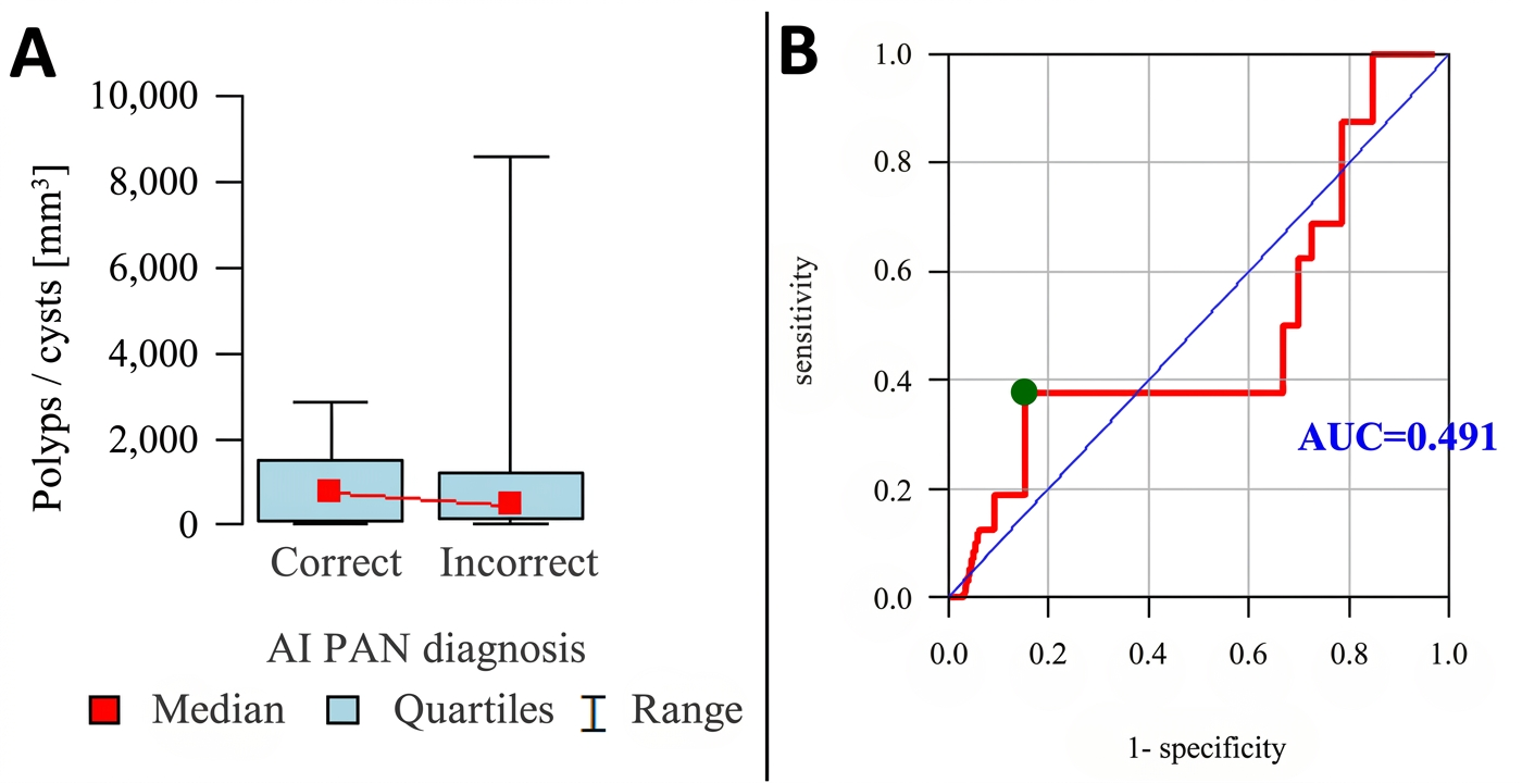
| Correct | OPG | 16 | 924.79 | 909.24 | 759.98 | 46.80 | 2845.44 | 109.20 | 1487.33 | p = 0.924 |
| Incorrect | 33 | 1355.91 | 2278.70 | 463.32 | 24.96 | 8592.48 | 131.04 | 1216.80 | ||
| Correct | CBCT | 30 | 1808.30 | 2284.67 | 837.98 | 62.40 | 8592.48 | 446.16 | 2010.97 | p < 0.001 * |
| Incorrect | 19 | 278.56 | 330.79 | 109.20 | 24.96 | 1216.80 | 71.76 | 436.28 | ||
| AI Diagnosis | Modality | N | Mucosal Thickening [mm] | p | ||||||
|---|---|---|---|---|---|---|---|---|---|---|
| Mean | SD | Median | Min | Max | Q1 | Q3 | ||||
| Correct | OPG | 33 | 5.91 | 3.31 | 5 | 3 | 16 | 4 | 7 | p = 0.933 |
| Incorrect | 134 | 6.13 | 3.75 | 5 | 3 | 20 | 4 | 7 | ||
| Correct | CBCT | 89 | 7.67 | 4.20 | 6 | 3 | 20 | 5 | 9 | p < 0.001 * |
| Incorrect | 78 | 4.27 | 1.56 | 4 | 3 | 11 | 3 | 5 | ||
| Metric | Modality | Count/Total | Value (%) | 95% CI Lower | 95% CI Upper | 95% CI |
|---|---|---|---|---|---|---|
| Accuracy | OPG | 77/166 | 46.39 | 38.97 | 53.97 | 38.97–53.97 |
| CBCT | 109/166 | 65.66 | 58.16 | 72.46 | 58.16–72.46 | |
| Recall | OPG | 43/120 | 35.83 | 27.82 | 44.73 | 27.82–44.73 |
| CBCT | 76/120 | 63.33 | 54.42 | 71.42 | 54.42–71.42 | |
| Precision | OPG | 43/55 | 78.18 | 65.63 | 87.05 | 65.63–87.05 |
| CBCT | 76/89 | 85.39 | 76.60 | 91.26 | 76.60–91.26 |
| Metric | OPG (95% CI) | CBCT (95% CI) | Difference (pp) |
|---|---|---|---|
| Accuracy | 46.39% (38.97–53.97%) | 65.66% (58.16–72.46%) | +19.28 |
| Recall | 35.83% (27.82–44.73%) | 63.33% (54.42–71.42%) | +27.50 |
| Precision | 78.18% (65.63–87.05%) | 85.39% (76.60–91.26%) | +7.21 |
| F1 Score | 0.4914 | 0.7273 | +0.2359 |
Disclaimer/Publisher’s Note: The statements, opinions and data contained in all publications are solely those of the individual author(s) and contributor(s) and not of MDPI and/or the editor(s). MDPI and/or the editor(s) disclaim responsibility for any injury to people or property resulting from any ideas, methods, instructions or products referred to in the content. |
© 2026 by the authors. Licensee MDPI, Basel, Switzerland. This article is an open access article distributed under the terms and conditions of the Creative Commons Attribution (CC BY) license.
Share and Cite
Lackowska, A.; Kazimierczak, N.; Chwarścianek, N.; Sultani, N.; Serafin, Z.; Kazimierczak, W. Impact of Imaging Modality on AI-Based Detection of Incidental Maxillary Sinus Pathology: Comparison of Panoramic Radiography and CBCT. Diagnostics 2026, 16, 1667. https://doi.org/10.3390/diagnostics16111667
Lackowska A, Kazimierczak N, Chwarścianek N, Sultani N, Serafin Z, Kazimierczak W. Impact of Imaging Modality on AI-Based Detection of Incidental Maxillary Sinus Pathology: Comparison of Panoramic Radiography and CBCT. Diagnostics. 2026; 16(11):1667. https://doi.org/10.3390/diagnostics16111667
Chicago/Turabian StyleLackowska, Anna, Natalia Kazimierczak, Natalia Chwarścianek, Nora Sultani, Zbigniew Serafin, and Wojciech Kazimierczak. 2026. "Impact of Imaging Modality on AI-Based Detection of Incidental Maxillary Sinus Pathology: Comparison of Panoramic Radiography and CBCT" Diagnostics 16, no. 11: 1667. https://doi.org/10.3390/diagnostics16111667
APA StyleLackowska, A., Kazimierczak, N., Chwarścianek, N., Sultani, N., Serafin, Z., & Kazimierczak, W. (2026). Impact of Imaging Modality on AI-Based Detection of Incidental Maxillary Sinus Pathology: Comparison of Panoramic Radiography and CBCT. Diagnostics, 16(11), 1667. https://doi.org/10.3390/diagnostics16111667