Endometriosis and Chronic Endometritis: Shared Mechanisms, Diagnostic Challenges, and Clinical Implications in Infertility


Abstract
1. Introduction
2. Methods and Scope of the Narrative Review
3. Epidemiological Association Between Endometriosis and Chronic Endometritis
4. Shared Pathophysiological Mechanisms
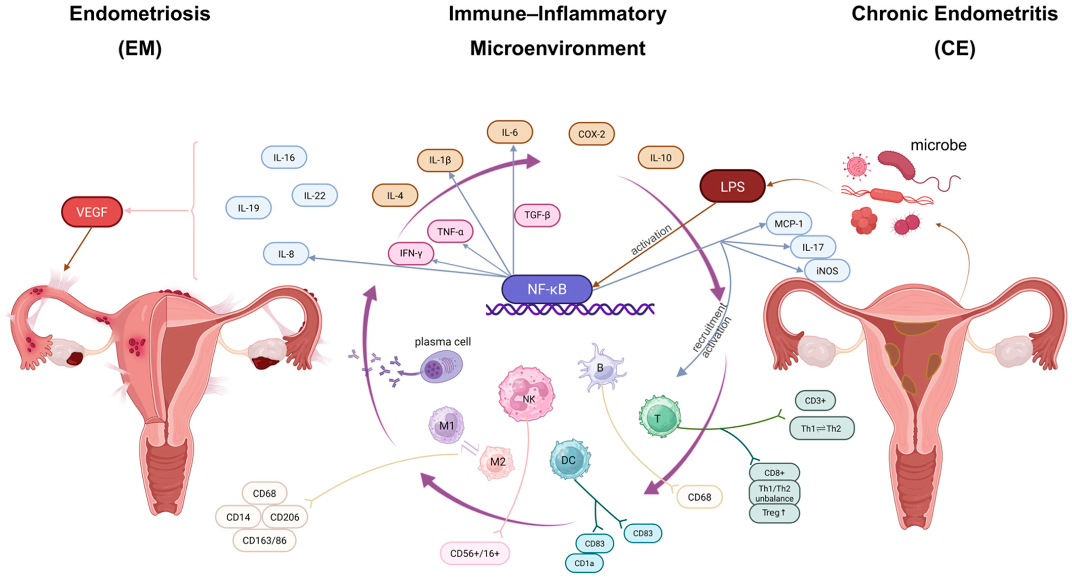
4.1. Chronic Inflammatory Signaling
4.2. Immune-Cell Dysfunction
4.3. Hormonal Dysregulation and Defective Decidualization
4.4. Microbiota and the Mucosal Immune Interface
5. Diagnostic Challenges in Suspected Endometriosis–Chronic Endometritis Comorbidity
5.1. Current Diagnostic Approaches for Endometriosis
5.2. Current Diagnostic Approaches for Chronic Endometritis
5.3. Why Concurrent Disease May Be Overlooked
5.4. Emerging Diagnostic Opportunities
5.5. Clinical Implications for Infertility Evaluation
- ▯
- Unexplained RIF or RPL.
- ▯
- Abnormal uterine bleeding that cannot be adequately explained by EM alone.
- ▯
- Persistent infertility despite appropriate EM-directed treatment, either medical or surgical.
- ▯
- Reproductive failure in which pelvic disease burden does not fully account for the clinical phenotype.
- ▯
- Severe dysmenorrhea, chronic pelvic pain, dyspareunia, or other symptoms suggestive of EM.
- ▯
- Adnexal masses or imaging findings suggestive of ovarian endometrioma or deep infiltrating EM.
- ▯
- Persistent infertility after CE treatment, particularly when symptoms or imaging findings suggest an additional pelvic factor.
- ▯
- In suspected CE, hysteroscopy combined with endometrial biopsy may be considered, particularly in infertility, RIF, or RPL populations. Sampling during the proliferative phase may improve diagnostic interpretation, although practice varies between studies. CD138 immunohistochemistry can improve plasma-cell detection, but diagnostic thresholds remain heterogeneous. A threshold such as ≥5 plasma cells per 10 high-power fields has been proposed in some infertility-focused studies to reduce overdiagnosis, but it should not be regarded as a universally accepted standard.
- ▯
- In suspected EM, transvaginal ultrasound is usually the first-line imaging modality, especially for ovarian endometrioma and some deep infiltrating lesions. MRI may be useful in selected patients with suspected deep disease or complex pelvic anatomy. Laparoscopy should be reserved for selected cases in which confirmation or treatment is expected to influence management.
6. Impact of Coexisting Endometriosis and Chronic Endometritis on Infertility
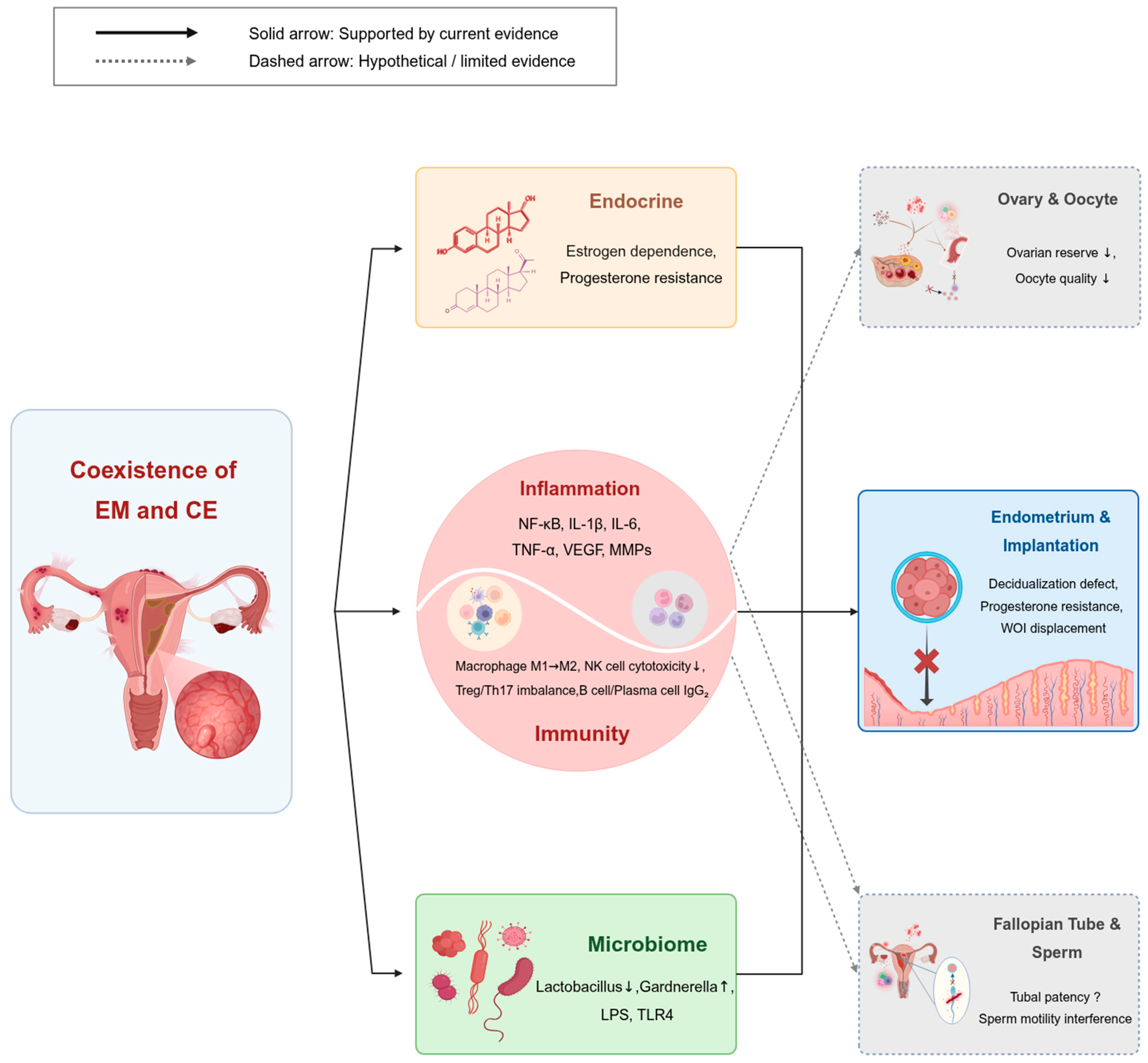
6.1. Endometrial Receptivity and Implantation Failure
6.2. Broader Reproductive Consequences
7. Future Directions
8. Conclusions
Author Contributions
Funding
Institutional Review Board Statement
Informed Consent Statement
Data Availability Statement
Acknowledgments
Conflicts of Interest
Abbreviations
| ART | Assisted reproductive technology |
| CD138 | Syndecan-1 |
| CE | Chronic endometritis |
| CI | Confidence interval |
| COX-2 | Cyclooxygenase 2 |
| CXCL13 | C-X-C motif chemokine ligand 13 |
| DC | Dendritic cell |
| EM | Endometriosis |
| ER | Endoplasmic reticulum |
| HPF | High-power field |
| HR | Hazard ratio |
| IKK | IκB kinase |
| IL | Interleukin |
| IL-1β | Interleukin-1 beta |
| IL-6 | Interleukin-6 |
| IL-8 | Interleukin-8 |
| iNOS | Inducible nitric oxide synthase |
| LBR | Live birth rate |
| LPS | Lipopolysaccharide |
| MMP | Matrix metalloproteinase |
| MRI | Magnetic resonance imaging |
| NF-κB | Nuclear factor kappa-B |
| NK | Natural killer |
| NLRP3 | NLR family pyrin domain-containing 3 |
| OR | Odds ratio |
| PET | Positron emission tomography |
| rASRM | Revised American Society for Reproductive Medicine |
| RIF | Recurrent implantation failure |
| RPL | Recurrent pregnancy loss |
| TLR4 | Toll-like receptor 4 |
| TNF-α | Tumor necrosis factor alpha |
| TVUS | Transvaginal ultrasound |
| VEGF | Vascular endothelial growth factor |
| WOI | Window of implantation |
References
- Kalaitzopoulos, D.R.; Samartzis, N.; Kolovos, G.N.; Mareti, E.; Samartzis, E.P.; Eberhard, M.; Dinas, K.; Daniilidis, A. Treatment of endometriosis: A review with comparison of 8 guidelines. BMC Womens Health 2021, 21, 397. [Google Scholar] [CrossRef] [Scilit]
- As-Sanie, S.; Mackenzie, S.C.; Morrison, L.; Schrepf, A.; Zondervan, K.T.; Horne, A.W.; Missmer, S.A. Endometriosis: A Review. JAMA 2025, 334, 64–78. [Google Scholar] [CrossRef] [Scilit] [PubMed]
- Tomassetti, C.; Johnson, N.P.; Petrozza, J.; Abrao, M.S.; Einarsson, J.I.; Horne, A.W.; Lee, T.T.M.; Missmer, S.; Vermeulen, N.; Zondervan, K.T.; et al. An international terminology for endometriosis, 2021. Hum. Reprod. Open 2021, 2021, hoab029. [Google Scholar] [CrossRef] [Scilit]
- Zondervan, K.T.; Becker, C.M.; Missmer, S.A. Endometriosis. N. Engl. J. Med. 2020, 382, 1244–1256. [Google Scholar] [CrossRef] [Scilit]
- Saunders, P.T.K.; Horne, A.W. Endometriosis: Etiology, pathobiology, and therapeutic prospects. Cell 2021, 184, 2807–2824. [Google Scholar] [CrossRef] [Scilit] [PubMed]
- Guerriero, S.; Ajossa, S.; Pagliuca, M.; Borzacchelli, A.; Deiala, F.; Springer, S.; Pilloni, M.; Taccori, V.; Pascual, M.A.; Graupera, B.; et al. Advances in Imaging for Assessing Pelvic Endometriosis. Diagnostics 2022, 12, 2960. [Google Scholar] [CrossRef] [Scilit]
- De Corte, P.; Klinghardt, M.; von Stockum, S.; Heinemann, K. Time to Diagnose Endometriosis: Current Status, Challenges and Regional Characteristics-A Systematic Literature Review. BJOG 2025, 132, 118–130. [Google Scholar] [CrossRef] [Scilit]
- Ticconi, C.; Inversetti, A.; Marraffa, S.; Campagnolo, L.; Arthur, J.; Zambella, E.; Di Simone, N. Chronic endometritis and recurrent reproductive failure: A systematic review and meta-analysis. Front. Immunol. 2024, 15, 1427454. [Google Scholar] [CrossRef] [Scilit]
- Yasuo, T.; Kitaya, K. Challenges in Clinical Diagnosis and Management of Chronic Endometritis. Diagnostics 2022, 12, 2711. [Google Scholar] [CrossRef] [Scilit]
- Zeng, S.; Liu, X.; Liu, D.; Song, W. Research update for the immune microenvironment of chronic endometritis. J. Reprod. Immunol. 2022, 152, 103637. [Google Scholar] [CrossRef] [Scilit] [PubMed]
- Pirtea, P.; Cicinelli, E.; De Nola, R.; de Ziegler, D.; Ayoubi, J.M. Endometrial causes of recurrent pregnancy losses: Endometriosis, adenomyosis, and chronic endometritis. Fertil. Steril. 2021, 115, 546–560. [Google Scholar] [CrossRef] [Scilit]
- Kitaya, K.; Yasuo, T. Commonalities and Disparities between Endometriosis and Chronic Endometritis: Therapeutic Potential of Novel Antibiotic Treatment Strategy against Ectopic Endometrium. Int. J. Mol. Sci. 2023, 24, 2059. [Google Scholar] [CrossRef] [Scilit]
- Luna Arana, M.; Pereira Sánchez, A.; Vaquero Argüello, G.; Tejerina González, E.; Alonso-Iniesta, M.; Pérez Medina, T. The Role of Chronic Endometritis in Endometriosis: A Personalized Diagnostic Tool? J. Pers. Med. 2026, 16, 73. [Google Scholar] [CrossRef] [Scilit] [PubMed]
- Kalaitzopoulos, D.R.; Catena, U.; Schwartz, A.K.; Schoretsanitis, G.; Leeners, B.; Drakopoulos, P.; Samartzis, N. Chronic Endometritis and Endometriosis: Two Sides of the Same Coin? Reprod. Sci. 2025, 32, 474–487. [Google Scholar] [CrossRef] [Scilit] [PubMed]
- Takebayashi, A.; Kimura, F.; Kishi, Y.; Ishida, M.; Takahashi, A.; Yamanaka, A.; Takahashi, K.; Suginami, H.; Murakami, T. The association between endometriosis and chronic endometritis. PLoS ONE 2014, 9, e88354. [Google Scholar] [CrossRef] [Scilit] [PubMed]
- Cicinelli, E.; Trojano, G.; Mastromauro, M.; Vimercati, A.; Marinaccio, M.; Mitola, P.C.; Resta, L.; de Ziegler, D. Higher prevalence of chronic endometritis in women with endometriosis: A possible etiopathogenetic link. Fertil. Steril. 2017, 108, 289–295.e1. [Google Scholar] [CrossRef] [Scilit]
- Stanford, J.B.; Sanders, J.N.; Simonsen, S.E.; Hammoud, A.; Gibson, M.; Smith, K.R. Methods for a Retrospective Population-based and Clinic-based Subfertility Cohort Study: The Fertility Experiences Study. Paediatr. Perinat. Epidemiol. 2016, 30, 397–407. [Google Scholar] [CrossRef] [Scilit]
- Bell, S.O.; Moreau, C.; Sarnak, D.; Kibira, S.P.S.; Anglewicz, P.; Gichangi, P.; McLain, A.C.; Thoma, M. Measuring non-events: Infertility estimation using cross-sectional, population-based data from four countries in sub-Saharan Africa. Hum. Reprod. 2024, 39, 2848–2860. [Google Scholar] [CrossRef] [Scilit]
- Colgrave, E.M.; Bittinger, S.; Healey, M.; Dior, U.P.; Rogers, P.A.W.; Keast, J.R.; Girling, J.E.; Holdsworth-Carson, S.J. Superficial peritoneal endometriotic lesions are histologically diverse and rarely demonstrate menstrual cycle synchronicity with matched eutopic endometrium. Hum. Reprod. 2020, 35, 2701–2714. [Google Scholar] [CrossRef] [Scilit]
- Trapero, C.; Vidal, A.; Fernández-Montolí, M.E.; Coroleu, B.; Tresserra, F.; Barri, P.; Gómez de Aranda, I.; Sévigny, J.; Ponce, J.; Matias-Guiu, X.; et al. Impaired Expression of Ectonucleotidases in Ectopic and Eutopic Endometrial Tissue Is in Favor of ATP Accumulation in the Tissue Microenvironment in Endometriosis. Int. J. Mol. Sci. 2019, 20, 5532. [Google Scholar] [CrossRef] [Scilit]
- Lin, K.Y.; Chang, C.Y.; Lin, W.C.; Wan, L. Increased risk of endometriosis in patients with endometritis—A nationwide cohort study involving 84,150 individuals. Ginekol. Pol. 2020, 91, 193–200. [Google Scholar] [CrossRef] [Scilit]
- Liu, Y.; Ko, E.Y.; Wong, K.K.; Chen, X.; Cheung, W.C.; Law, T.S.; Chung, J.P.; Tsui, S.K.; Li, T.C.; Chim, S.S. Endometrial microbiota in infertile women with and without chronic endometritis as diagnosed using a quantitative and reference range-based method. Fertil. Steril. 2019, 112, 707–717.e1. [Google Scholar] [CrossRef] [Scilit]
- Qiao, X.; Wu, L.; Liu, D.; Pei, T.; Huang, W. Existence of chronic endometritis and its influence on pregnancy outcomes in infertile women with minimal/mild endometriosis. Int. J. Gynaecol. Obstet. 2023, 160, 628–634. [Google Scholar] [CrossRef] [Scilit] [PubMed]
- Gawron, I.; Derbisz, K.; Jach, R.; Trojnarska, D.; Milian-Ciesielska, K.; Pietrus, M. Pelvic peritoneal endometriosis is linked to the endometrial inflammatory profile: A prospective cohort study. BMC Womens Health 2025, 25, 94. [Google Scholar] [CrossRef] [Scilit]
- Holzer, I.; Ott, J.; Kurz, C.; Hofstetter, G.; Hager, M.; Kuessel, L.; Parry, J.P. Is Chronic Endometritis Associated with Tubal Infertility? A Prospective Cohort Study. J. Minim. Invasive Gynecol. 2021, 28, 1876–1881. [Google Scholar] [CrossRef] [Scilit] [PubMed]
- Kitaya, K.; Takeuchi, T.; Mizuta, S.; Matsubayashi, H.; Ishikawa, T. Endometritis: New time, new concepts. Fertil. Steril. 2018, 110, 344–350. [Google Scholar] [CrossRef] [Scilit] [PubMed]
- Wang, W.; Feng, D.; Ling, B. Biologia Futura: Endometrial microbiome affects endometrial receptivity from the perspective of the endometrial immune microenvironment. Biol. Futur. 2022, 73, 291–300. [Google Scholar] [CrossRef] [Scilit]
- Asghari, S.; Valizadeh, A.; Aghebati-Maleki, L.; Nouri, M.; Yousefi, M. Endometriosis: Perspective, lights, and shadows of etiology. BioMed Pharmacother. 2018, 106, 163–174. [Google Scholar] [CrossRef] [Scilit]
- Buzzaccarini, G.; Vitagliano, A.; Andrisani, A.; Santarsiero, C.M.; Cicinelli, R.; Nardelli, C.; Ambrosini, G.; Cicinelli, E. Chronic endometritis and altered embryo implantation: A unified pathophysiological theory from a literature systematic review. J. Assist. Reprod. Genet. 2020, 37, 2897–2911. [Google Scholar] [CrossRef] [Scilit] [PubMed]
- Horne, A.W.; Missmer, S.A. Pathophysiology, diagnosis, and management of endometriosis. BMJ 2022, 379, e070750. [Google Scholar] [CrossRef] [Scilit]
- Dymanowska-Dyjak, I.; Terpiłowska, B.; Morawska-Michalska, I.; Michalski, A.; Polak, G.; Terpiłowski, M.; Rahnama-Hezavah, M.; Grywalska, E. Immune Dysregulation in Endometriomas: Implications for Inflammation. Int. J. Mol. Sci. 2024, 25, 4802. [Google Scholar] [CrossRef] [Scilit] [PubMed]
- Liu, Y.; Wang, J.; Zhang, X. An Update on the Multifaceted Role of NF-kappaB in Endometriosis. Int. J. Biol. Sci. 2022, 18, 4400–4413. [Google Scholar] [CrossRef] [Scilit]
- Chowdhury, I.; Banerjee, S.; Driss, A.; Xu, W.; Mehrabi, S.; Nezhat, C.; Sidell, N.; Taylor, R.N.; Thompson, W.E. Curcumin attenuates proangiogenic and proinflammatory factors in human eutopic endometrial stromal cells through the NF-κB signaling pathway. J. Cell. Physiol. 2019, 234, 6298–6312. [Google Scholar] [CrossRef] [Scilit]
- Tortorella, C.; Piazzolla, G.; Matteo, M.; Pinto, V.; Tinelli, R.; Sabbà, C.; Fanelli, M.; Cicinelli, E. Interleukin-6, interleukin-1β, and tumor necrosis factor α in menstrual effluents as biomarkers of chronic endometritis. Fertil. Steril. 2014, 101, 242–247. [Google Scholar] [CrossRef] [Scilit] [PubMed]
- Yoneda, E.; Kim, S.; Tomita, K.; Minase, T.; Kayano, M.; Watanabe, H.; Tetsuka, M.; Sasaki, M.; Iwayama, H.; Sanai, H.; et al. Evaluation of Lipopolysaccharide and Interleukin-6 as Useful Screening Tool for Chronic Endometritis. Int. J. Mol. Sci. 2024, 25, 2017. [Google Scholar] [CrossRef] [Scilit] [PubMed]
- Zdrojkowski, Ł.; Jasiński, T.; Ferreira-Dias, G.; Pawliński, B.; Domino, M. The Role of NF-κB in Endometrial Diseases in Humans and Animals: A Review. Int. J. Mol. Sci. 2023, 24, 2901. [Google Scholar] [CrossRef] [Scilit]
- Yoshino, O.; Izumi, G.; Shi, J.; Osuga, Y.; Hirota, Y.; Hirata, T.; Harada, M.; Nishii, O.; Koga, K.; Taketani, Y. Activin-A is induced by interleukin-1β and tumor necrosis factor-α and enhances the mRNA expression of interleukin-6 and protease-activated receptor-2 and proliferation of stromal cells from endometrioma. Fertil. Steril. 2011, 96, 118–121. [Google Scholar] [CrossRef] [Scilit]
- Sikora, J.; Smycz-Kubańska, M.; Mielczarek-Palacz, A.; Kondera-Anasz, Z. Abnormal peritoneal regulation of chemokine activation-The role of IL-8 in pathogenesis of endometriosis. Am. J. Reprod. Immunol. 2017, 77, e12622. [Google Scholar] [CrossRef] [Scilit]
- Bersinger, N.A.; Günthert, A.R.; McKinnon, B.; Johann, S.; Mueller, M.D. Dose-response effect of interleukin (IL)-1β, tumour necrosis factor (TNF)-α, and interferon-γ on the in vitro production of epithelial neutrophil activating peptide-78 (ENA-78), IL-8, and IL-6 by human endometrial stromal cells. Arch. Gynecol. Obstet. 2011, 283, 1291–1296. [Google Scholar] [CrossRef] [Scilit]
- Di Pietro, C.; Caruso, S.; Battaglia, R.; Iraci Sareri, M.; La Ferlita, A.; Strino, F.; Bonaventura, G.; Di Mauro, M.; Barcellona, M.L.; Perciavalle, V.; et al. MiR-27a-3p and miR-124-3p, upregulated in endometrium and serum from women affected by Chronic Endometritis, are new potential molecular markers of endometrial receptivity. Am. J. Reprod. Immunol. 2018, 80, e12858. [Google Scholar] [CrossRef] [Scilit]
- Di Pietro, C.; Cicinelli, E.; Guglielmino, M.R.; Ragusa, M.; Farina, M.; Palumbo, M.A.; Cianci, A. Altered transcriptional regulation of cytokines, growth factors, and apoptotic proteins in the endometrium of infertile women with chronic endometritis. Am. J. Reprod. Immunol. 2013, 69, 509–517. [Google Scholar] [CrossRef] [Scilit]
- Kitaya, K.; Yasuo, T.; Tada, Y.; Hayashi, T.; Iwaki, Y.; Karita, M.; Funabiki, M.; Taguchi, S.; Spillers, D.; Nakamura, Y.; et al. Unusual inflammation in gynecologic pathology associated with defective endometrial receptivity. Histol. Histopathol. 2014, 29, 1113–1127. [Google Scholar] [CrossRef] [Scilit]
- Wira, C.R.; Fahey, J.V.; Sentman, C.L.; Pioli, P.A.; Shen, L. Innate and adaptive immunity in female genital tract: Cellular responses and interactions. Immunol. Rev. 2005, 206, 306–335. [Google Scholar] [CrossRef] [Scilit]
- Moffett, A.; Shreeve, N. Local immune recognition of trophoblast in early human pregnancy: Controversies and questions. Nat. Rev. Immunol. 2023, 23, 222–235. [Google Scholar] [CrossRef] [Scilit]
- Dias Da Silva, I.; Wuidar, V.; Zielonka, M.; Pequeux, C. Unraveling the Dynamics of Estrogen and Progesterone Signaling in the Endometrium: An Overview. Cells 2024, 13, 1236. [Google Scholar] [CrossRef] [Scilit]
- Dai, Y.; Ye, Z.; Lin, X.; Zhang, S. Immunopathological insights into endometriosis: From research advances to future treatments. Semin. Immunopathol. 2025, 47, 31. [Google Scholar] [CrossRef] [Scilit]
- Symons, L.K.; Miller, J.E.; Kay, V.R.; Marks, R.M.; Liblik, K.; Koti, M.; Tayade, C. The Immunopathophysiology of Endometriosis. Trends Mol. Med. 2018, 24, 748–762. [Google Scholar] [CrossRef] [Scilit] [PubMed]
- Berbic, M.; Schulke, L.; Markham, R.; Tokushige, N.; Russell, P.; Fraser, I.S. Macrophage expression in endometrium of women with and without endometriosis. Hum. Reprod. 2009, 24, 325–332. [Google Scholar] [CrossRef] [Scilit] [PubMed]
- Barański, W.; Kaleczyc, J.; Zduńczyk, S.; Podlasz, W.; Długołecka-Malinowska, E.; Janowski, T. Distribution of CD14+ macrophages, CD4+, CD8+ lymphocytes and mRNA expression of inducible nitric oxide synthase in the endometrium of repeat breeding cows. Pol. J. Vet. Sci. 2013, 16, 443–451. [Google Scholar] [CrossRef] [Scilit] [PubMed]
- Nie, M.F.; Xie, Q.; Wu, Y.H.; He, H.; Zou, L.J.; She, X.L.; Wu, X.Q. Serum and Ectopic Endometrium from Women with Endometriosis Modulate Macrophage M1/M2 Polarization via the Smad2/Smad3 Pathway. J. Immunol. Res. 2018, 2018, 6285813. [Google Scholar] [CrossRef] [Scilit]
- Cominelli, A.; Gaide Chevronnay, H.P.; Lemoine, P.; Courtoy, P.J.; Marbaix, E.; Henriet, P. Matrix metalloproteinase-27 is expressed in CD163+/CD206+ M2 macrophages in the cycling human endometrium and in superficial endometriotic lesions. Mol. Hum. Reprod. 2014, 20, 767–775. [Google Scholar] [CrossRef] [Scilit]
- Schulke, L.; Berbic, M.; Manconi, F.; Tokushige, N.; Markham, R.; Fraser, I.S. Dendritic cell populations in the eutopic and ectopic endometrium of women with endometriosis. Hum. Reprod. 2009, 24, 1695–1703. [Google Scholar] [CrossRef] [Scilit] [PubMed]
- Antsiferova, Y.S.; Sotnikova, N.Y.; Posiseeva, L.V.; Shor, A.L. Changes in the T-helper cytokine profile and in lymphocyte activation at the systemic and local levels in women with endometriosis. Fertil. Steril. 2005, 84, 1705–1711. [Google Scholar] [CrossRef] [Scilit]
- Riccio, L.G.C.; Baracat, E.C.; Chapron, C.; Batteux, F.; Abrão, M.S. The role of the B lymphocytes in endometriosis: A systematic review. J. Reprod. Immunol. 2017, 123, 29–34. [Google Scholar] [CrossRef] [Scilit] [PubMed]
- Shih, A.J.; Adelson, R.P.; Vashistha, H.; Khalili, H.; Nayyar, A.; Puran, R.; Herrera, R.; Chatterjee, P.K.; Lee, A.T.; Truskinovsky, A.M.; et al. Single-cell analysis of menstrual endometrial tissues defines phenotypes associated with endometriosis. BMC Med. 2022, 20, 315. [Google Scholar] [CrossRef] [Scilit]
- Yuan, M.; Li, D.; An, M.; Li, Q.; Zhang, L.; Wang, G. Rediscovering peritoneal macrophages in a murine endometriosis model. Hum. Reprod. 2017, 32, 94–102. [Google Scholar] [CrossRef] [Scilit] [PubMed]
- Andreoli, C.G.; Genro, V.K.; Souza, C.A.; Michelon, T.; Bilibio, J.P.; Scheffel, C.; Cunha-Filho, J.S. T helper (Th)1, Th2, and Th17 interleukin pathways in infertile patients with minimal/mild endometriosis. Fertil. Steril. 2011, 95, 2477–2480. [Google Scholar] [CrossRef] [Scilit]
- Kisovar, A.; Becker, C.M.; Granne, I.; Southcombe, J.H. The role of CD8+ T cells in endometriosis: A systematic review. Front. Immunol. 2023, 14, 1225639. [Google Scholar] [CrossRef] [Scilit]
- Králíčková, M.; Fiala, L.; Losan, P.; Tomes, P.; Vetvicka, V. Altered Immunity in Endometriosis: What Came First? Immunol. Investig. 2018, 47, 569–582. [Google Scholar] [CrossRef] [Scilit]
- Kitaya, K.; Tada, Y.; Hayashi, T.; Taguchi, S.; Funabiki, M.; Nakamura, Y. Comprehensive endometrial immunoglobulin subclass analysis in infertile women suffering from repeated implantation failure with or without chronic endometritis. Am. J. Reprod. Immunol. 2014, 72, 386–391. [Google Scholar] [CrossRef] [Scilit]
- Chen, X.; Liu, Y.; Zhao, Y.; Cheung, W.C.; Zhang, T.; Qi, R.; Chung, J.P.W.; Wang, C.C.; Li, T.C. Association between chronic endometritis and uterine natural killer cell density in women with recurrent miscarriage: Clinical implications. J. Obstet. Gynaecol. Res. 2020, 46, 858–863. [Google Scholar] [CrossRef] [Scilit]
- Li, Y.; Yu, S.; Huang, C.; Lian, R.; Chen, C.; Liu, S.; Li, L.; Diao, L.; Markert, U.R.; Zeng, Y. Evaluation of peripheral and uterine immune status of chronic endometritis in patients with recurrent reproductive failure. Fertil. Steril. 2020, 113, 187–196.e1. [Google Scholar] [CrossRef] [Scilit]
- Kitazawa, J.; Kimura, F.; Nakamura, A.; Morimune, A.; Hanada, T.; Amano, T.; Tsuji, S.; Kasahara, K.; Satooka, H.; Hirata, T.; et al. Alteration in endometrial helper T-cell subgroups in chronic endometritis. Am. J. Reprod. Immunol. 2021, 85, e13372. [Google Scholar] [CrossRef] [Scilit]
- Shan, J.; Li, D.J.; Wang, X.Q. Towards a Better Understanding of Endometriosis-Related Infertility: A Review on How Endometriosis Affects Endometrial Receptivity. Biomolecules 2023, 13, 430. [Google Scholar] [CrossRef] [Scilit]
- Osiński, M.; Wirstlein, P.; Wender-Ożegowska, E.; Mikołajczyk, M.; Jagodziński, P.P.; Szczepańska, M. HSD3B2, HSD17B1, HSD17B2, ESR1, ESR2 and AR expression in infertile women with endometriosis. Ginekol. Pol. 2018, 89, 125–134. [Google Scholar] [CrossRef] [Scilit]
- Samadieh, Y.; Favaedi, R.; Ramezanali, F.; Afsharian, P.; Aflatoonian, R.; Shahhoseini, M. Epigenetic Dynamics of HOXA10 Gene in Infertile Women With Endometriosis. Reprod. Sci. 2019, 26, 88–96. [Google Scholar] [CrossRef] [Scilit] [PubMed]
- Tanaka, S.E.; Sakuraba, Y.; Kitaya, K.; Ishikawa, T. Differential Vaginal Microbiota Profiling in Lactic-Acid-Producing Bacteria between Infertile Women with and without Chronic Endometritis. Diagnostics 2022, 12, 878. [Google Scholar] [CrossRef] [Scilit]
- Zizolfi, B.; Foreste, V.; Gallo, A.; Martone, S.; Giampaolino, P.; Di Spiezio Sardo, A. Endometriosis and dysbiosis: State of art. Front. Endocrinol. 2023, 14, 1140774. [Google Scholar] [CrossRef] [Scilit] [PubMed]
- Wei, W.; Zhang, X.; Tang, H.; Zeng, L.; Wu, R. Microbiota composition and distribution along the female reproductive tract of women with endometriosis. Ann. Clin. Microbiol. Antimicrob. 2020, 19, 15. [Google Scholar] [CrossRef] [Scilit] [PubMed]
- Baușic, A.I.G.; Scurtu, F.; Manu, A.; Matasariu, D.R.; Brătilă, E. Gut Microbiota Dysbiosis in Endometriosis: A Potential Link to Inflammation and Disease Progression. Int. J. Mol. Sci. 2025, 26, 5144. [Google Scholar] [CrossRef] [Scilit]
- Singh, S.S.; Allaire, C.; Al-Nourhji, O.; Bougie, O.; Bridge-Cook, P.; Duigenan, S.; Kroft, J.; Lemyre, M.; Leonardi, M.; Leyland, N.; et al. Guideline No. 449: Diagnosis and Impact of Endometriosis—A Canadian Guideline. J. Obstet. Gynaecol. Can. 2024, 46, 102450. [Google Scholar] [CrossRef] [Scilit] [PubMed]
- Horne, A.W.; Daniels, J.; Hummelshoj, L.; Cox, E.; Cooper, K.G. Surgical removal of superficial peritoneal endometriosis for managing women with chronic pelvic pain: Time for a rethink? BJOG 2019, 126, 1414–1416. [Google Scholar] [CrossRef] [Scilit]
- Tsonis, O.; Gkrozou, F.; Dimitriou, E.; Barmpalia, Z.; Tsonis, K.; Vatopoulou, A.; Paschopoulos, M. Hysteroscopic features suggestive of chronic endometritis: A systematic review. Hum. Fertil. 2023, 26, 1530–1543. [Google Scholar] [CrossRef] [Scilit] [PubMed]
- Cicinelli, E.; Vitagliano, A.; Kumar, A.; Lasmar, R.B.; Bettocchi, S.; Haimovich, S. Unified diagnostic criteria for chronic endometritis at fluid hysteroscopy: Proposal and reliability evaluation through an international randomized-controlled observer study. Fertil. Steril. 2019, 112, 162–173.e2. [Google Scholar] [CrossRef] [Scilit]
- Klimaszyk, K.; Svarre Nielsen, H.; Wender-Ozegowska, E.; Kedzia, M. Chronic endometritis—Is it time to clarify diagnostic criteria? Ginekol. Pol. 2023, 94, 152–157. [Google Scholar] [CrossRef] [Scilit]
- Dang, J.; Xiao, X.; Li, M. Correlation of hysteroscopic findings of chronic endometritis with CD138 immunohistochemistry and their correlation with pregnancy outcomes. J. Assist. Reprod. Genet. 2024, 41, 2477–2483. [Google Scholar] [CrossRef] [Scilit] [PubMed]
- Li, Y.; Xu, S.; Yu, S.; Huang, C.; Lin, S.; Chen, W.; Mo, M.; Lian, R.; Diao, L.; Ding, L.; et al. Diagnosis of chronic endometritis: How many CD138(+) cells/HPF in endometrial stroma affect pregnancy outcome of infertile women? Am. J. Reprod. Immunol. 2021, 85, e13369. [Google Scholar] [CrossRef] [Scilit]
- Ryan, E.; Tolani, A.T.; Zhang, J.; Cruz, G.I.; Folkins, A.K.; Lathi, R.B. The menstrual cycle phase impacts the detection of plasma cells and the diagnosis of chronic endometritis in endometrial biopsy specimens. Fertil. Steril. 2022, 118, 787–794. [Google Scholar] [CrossRef] [Scilit]
- Nisenblat, V.; Bossuyt, P.M.; Shaikh, R.; Farquhar, C.; Jordan, V.; Scheffers, C.S.; Mol, B.W.; Johnson, N.; Hull, M.L. Blood biomarkers for the non-invasive diagnosis of endometriosis. Cochrane Database Syst. Rev. 2016, 2016, Cd012179. [Google Scholar] [CrossRef] [Scilit]
- Bendifallah, S.; Suisse, S.; Puchar, A.; Delbos, L.; Poilblanc, M.; Descamps, P.; Golfier, F.; Jornea, L.; Bouteiller, D.; Touboul, C.; et al. Salivary MicroRNA Signature for Diagnosis of Endometriosis. J. Clin. Med. 2022, 11, 612. [Google Scholar] [CrossRef] [Scilit]
- Griffiths, M.J.; Horne, A.W.; Gibson, D.A.; Roberts, N.; Saunders, P.T.K. Endometriosis: Recent advances that could accelerate diagnosis and improve care. Trends Mol. Med. 2024, 30, 875–889. [Google Scholar] [CrossRef] [Scilit]
- Ding, D.; Chen, Y.; Liu, X.; Jiang, Z.; Cai, X.; Guo, S.W. Diagnosing Deep Endometriosis Using Transvaginal Elastosonography. Reprod. Sci. 2020, 27, 1411–1422. [Google Scholar] [CrossRef] [Scilit]
- Balogova, S.; Daraï, E.; Noskovicova, L.; Lukac, L.; Talbot, J.N.; Montravers, F. Interference of Known or Suspected Endometriosis in Reporting FDG PET/CT Performed in Another Indication. Clin. Nucl. Med. 2022, 47, 305–313. [Google Scholar] [CrossRef] [Scilit]
- Brulport, A.; Bourdon, M.; Vaiman, D.; Drouet, C.; Pocate-Cheriet, K.; Bouzid, K.; Marcellin, L.; Santulli, P.; Abo, C.; Jeljeli, M.; et al. An integrated multi-tissue approach for endometriosis candidate biomarkers: A systematic review. Reprod. Biol. Endocrinol. 2024, 22, 21. [Google Scholar] [CrossRef] [Scilit] [PubMed]
- Taranovska, O.; Likhachov, V.; Dobrovolska, L.; Makarov, O.G.; Shymanska, Y.V. Possibility for non-invasive diagnosis of chronic endometritis in women at risk during pregravid preparation. Wiad. Lek. 2019, 72, 64–67. [Google Scholar] [CrossRef] [Scilit]
- Guleken, Z.; Bulut, H.; Depciuch, J.; Tarhan, N. Diagnosis of endometriosis using endometrioma volume and vibrational spectroscopy with multivariate methods as a noninvasive method. Spectrochim. Acta A Mol. Biomol. Spectrosc. 2022, 264, 120246. [Google Scholar] [CrossRef] [Scilit] [PubMed]
- Li, B.; Chen, H.; Lin, X.; Duan, H. Multimodal learning system integrating electronic medical records and hysteroscopic images for reproductive outcome prediction and risk stratification of endometrial injury: A multicenter diagnostic study. Int. J. Surg. 2024, 110, 3237–3248. [Google Scholar] [CrossRef] [Scilit]
- Mittal, S.; Tong, A.; Young, S.; Jha, P. Artificial intelligence applications in endometriosis imaging. Abdom. Radiol. 2025, 50, 4901–4913. [Google Scholar] [CrossRef] [Scilit]
- Snyder, D.L.; Sidhom, S.; Chatham, C.E.; Tillotson, S.G.; Zapata, R.D.; Modave, F.; Solly, M.; Quevedo, A.; Moawad, N.S. Utilizing Artificial Intelligence: Machine Learning Algorithms to Develop a Preoperative Endometriosis Prediction Model. J. Minim. Invasive Gynecol. 2025, 32, 784–792. [Google Scholar] [CrossRef] [Scilit]
- Cicinelli, E.; Bettocchi, S.; de Ziegler, D.; Loizzi, V.; Cormio, G.; Marinaccio, M.; Trojano, G.; Crupano, F.M.; Francescato, R.; Vitagliano, A.; et al. Chronic Endometritis, a Common Disease Hidden Behind Endometrial Polyps in Premenopausal Women: First Evidence From a Case-Control Study. J. Minim. Invasive Gynecol. 2019, 26, 1346–1350. [Google Scholar] [CrossRef] [Scilit] [PubMed]
- Bouet, P.E.; El Hachem, H.; Monceau, E.; Gariépy, G.; Kadoch, I.J.; Sylvestre, C. Chronic endometritis in women with recurrent pregnancy loss and recurrent implantation failure: Prevalence and role of office hysteroscopy and immunohistochemistry in diagnosis. Fertil. Steril. 2016, 105, 106–110. [Google Scholar] [CrossRef] [Scilit] [PubMed]
- Santulli, P.; Lamau, M.C.; Marcellin, L.; Gayet, V.; Marzouk, P.; Borghese, B.; Lafay Pillet, M.C.; Chapron, C. Endometriosis-related infertility: Ovarian endometrioma per se is not associated with presentation for infertility. Hum. Reprod. 2016, 31, 1765–1775. [Google Scholar] [CrossRef] [Scilit] [PubMed]
- Cicinelli, E.; Matteo, M.; Trojano, G.; Mitola, P.C.; Tinelli, R.; Vitagliano, A.; Crupano, F.M.; Lepera, A.; Miragliotta, G.; Resta, L. Chronic endometritis in patients with unexplained infertility: Prevalence and effects of antibiotic treatment on spontaneous conception. Am. J. Reprod. Immunol. 2018, 79, e12782. [Google Scholar] [CrossRef] [Scilit]
- Yang, S.; Wang, H.; Li, D.; Li, M. An Estrogen-NK Cells Regulatory Axis in Endometriosis, Related Infertility, and Miscarriage. Int. J. Mol. Sci. 2024, 25, 3362. [Google Scholar] [CrossRef] [Scilit]
- Lim, H.J.; Sun, J.; Min, B.; Song, M.; Kim, T.H.; Kim, B.J.; Hwang, K.R.; Lee, T.S.; Jeon, H.W.; Kim, S.M. Endometriosis and Adverse Pregnancy Outcomes: A Nationwide Population-Based Study. J. Clin. Med. 2023, 12, 5392. [Google Scholar] [CrossRef] [Scilit]
- Tan, Z.; Gong, X.; Li, Y.; Hung, S.W.; Huang, J.; Wang, C.C.; Chung, J.P.W. Impacts of endometrioma on ovarian aging from basic science to clinical management. Front. Endocrinol. 2022, 13, 1073261. [Google Scholar] [CrossRef] [Scilit]
- Park, W.; Lim, W.; Kim, M.; Jang, H.; Park, S.J.; Song, G.; Park, S. Female reproductive disease, endometriosis: From inflammation to infertility. Mol. Cells 2025, 48, 100164. [Google Scholar] [CrossRef] [Scilit] [PubMed]


| Category | Study | Year | Study Design | Population/Sample | Main Finding | Main Limitation | Ref. |
|---|---|---|---|---|---|---|---|
| Direct coexistence and association studies | Takebayashi et al. | 2014 | Observational | Women undergoing gynecologic evaluation, including EM cases | Reported a significant association between EM and CE. CE in EM 52.9% vs. non-EM 27.0% | Early study; diagnostic criteria for CE differ from later reports | [15] |
| Cicinelli et al. | 2017 | Case–control | Hysterectomy patients with/without EM (stage IV) | Found a higher prevalence of CE in women with EM, CE in EM 38.5% vs. 14.1%; OR 3.8. | Referral bias and heterogeneity in CE diagnosis may affect the strength of association | [16] | |
| Lin KY et al. | 2020 | Cohort (nationwide) | 84,150 women (EM vs. non-EM) | Endometritis → EM HR 1.58 (95% CI 1.48–1.68) | ICD-9 based; no histologic confirmation of CE | [21] | |
| Liu Y et al. | 2019 | Case–control | Infertile women with/without CE | CE associated with reduced Lactobacillus; EM mentioned as comorbidity. | Main focus on microbiota, not specifically designed for EM-CE coexistence | [22] | |
| Supportive studies | Qiao X et al. | 2023 | Cohort | Infertile women with minimal/mild EM (n = 201) | CE in 24.4%; CE independently reduced pregnancy (HR 0.58) and live birth rates | Infertility cohort only; not designed for prevalence estimation | [23] |
| Gawron et al. | 2025 | Prospective cohort study | Women with peritoneal EM (n = 64) | Peritoneal EM associated with endometrial inflammatory profile (OR 3.43) | Small sample; supports biological overlap, not direct coexistence | [24] | |
| Holzer I et al. | 2021 | Prospective cohort | Infertile women undergoing hysteroscopy/laparoscopy (n = 100) | CE associated with tubal occlusion (OR 5.27); positive correlation with rASRM score | Pilot study; indirectly supports EM-CE link | [25] | |
| Reviews | Kalaitzopoulos DR et al. | 2025 | Meta-analysis | 6 studies (n = 900) | CE in EM pooled 28%; OR 2.07 (95% CI 1.11–3.84) | High heterogeneity (I2 = 43%); different diagnostic criteria | [14] |
| Kitaya K & Yasuo T | 2023 | Narrative review | Summarizes commonalities in immunity, microbiota, treatment | EM and CE share immune and inflammatory features | No quantitative synthesis | [12] | |
| Pirtea P et al. | 2021 | Systematic review | Focus on RPL | CE in RPL 29.7%; treatment improves LBR | Not specifically designed for EM-CE coexistence | [11] |
| Mechanistic Domain | Evidence in Endometriosis | Evidence in Chronic Endometritis | Shared Consequence | Reproductive Relevance |
|---|---|---|---|---|
| Chronic inflammatory signaling | Persistent inflammatory activation with increased IL-1β, IL-6, IL-8, TNF-α, and NF-κB-related signaling contributes to lesion survival, stromal-cell proliferation, angiogenesis, and tissue remodeling [28,30,31,32,33,37,38,39]. | Sustained low-grade endometrial inflammation with increased IL-1β, IL-6, TNF-α, and NF-κB-related activation has been described in CE [29,34,35,36,40,41]. | Persistent inflammatory amplification and chronic tissue injury | Impaired implantation, reduced endometrial receptivity, and infertility |
| Immune-cell dysfunction | Reduced NK-cell cytotoxicity, increased macrophage infiltration, and abnormalities in dendritic cells, B cells, and T-cell subsets support a dysregulated immune microenvironment in EM [46,47,48,49,50,51,52,53,54,55,56,57,58,59]. | CE is associated with increased local immunoglobulin expression, altered NK-cell subsets, elevated macrophage and dendritic-cell populations, and shifts in T-helper-cell balance [10,60,61,62,63]. | Disturbed immune homeostasis and persistent local inflammation | Abnormal embryo–endometrium interaction and impaired receptivity |
| Hormonal imbalance and progesterone resistance | Progesterone resistance and altered estrogen-dependent signaling are central features of EM-related endometrial dysfunction [64,65]. | Persistent inflammation in CE may interfere with hormone-dependent endometrial maturation, and CE has been associated with delayed progesterone-dependent transformation and impaired endometrial receptivity [11,40,41]. | Abnormal endometrial transformation and impaired hormonal responsiveness | Reduced receptivity and defective implantation timing |
| Defective decidualization and endometrial maturation | EM is associated with altered endometrial gene regulation and impaired decidualization [64,65,66]. | CE has been associated with displacement of the window of implantation, defective decidualization, and delayed endometrial maturation [11,40,41]. | Embryo–endometrium asynchrony | Implantation failure and early reproductive loss |
| Microbiota-related mucosal immune activation | Reproductive tract dysbiosis and mucosal immune disturbance have been proposed in EM, although current evidence remains limited and less consistent [27,68,69,70]. | CE is more directly linked to microbial persistence, LPS-related inflammation, and altered vaginal microbiota, including reduced lactic-acid-producing bacteria [35,36,67]. | Chronic mucosal inflammatory priming and endometrial immune disturbance | Persistent endometrial dysfunction and reduced implantation potential |
| Abnormal tissue repair and remodeling | EM is associated with lesion persistence, fibrosis, pelvic remodeling, and chronic inflammatory repair responses [28,30,31]. | Repeated inflammatory injury in CE is associated with altered transcription of cytokines, growth factors, and apoptosis-related mediators involved in endometrial repair [40,41]. | Altered tissue homeostasis and incomplete resolution of inflammation | Suboptimal reproductive microenvironment |
| Diagnostic Modality | Target Disease | Current Status | Main Assessment and Clinical Use | Main Limitation | Role in Suspected EM–CE coexistence | Ref. |
|---|---|---|---|---|---|---|
| Clinical symptoms and reproductive history | Both | First-line triage | Assesses pelvic pain, dysmenorrhea, abnormal uterine bleeding, infertility, recurrent implantation failure, or recurrent pregnancy loss; readily available for initial clinical triage. | Manifestations are nonspecific and may overlap across diseases | Helps identify patients who may warrant selective evaluation for concurrent disease | [12,16,90,91] |
| TVUS | EM | Standard first-line | First-line imaging for ovarian endometrioma and some deep infiltrating lesions. | Limited sensitivity for superficial peritoneal EM; does not assess CE | Useful for pelvic disease burden, but cannot exclude concomitant endometrial inflammation | [71] |
| Pelvic MRI | EM | Selected adjunct | Assesses deep infiltrating disease and anatomical extent with high soft-tissue resolution. | Less useful for subtle or superficial disease; not a routine tool for all patients | Complements TVUS in selected women with suspected complex EM | [71,81] |
| Laparoscopy with histological confirmation | EM | Reference standard in selected cases | Allows direct pelvic visualization and histological confirmation in selected cases. | Invasive; costly; not suitable for universal infertility screening | Confirms EM but does not assess the uterine cavity unless additional procedures are performed | [72] |
| Hysteroscopy | CE | Selected CE assessment | Visualizes the uterine cavity and findings suggestive of CE, including micropolyps, hyperemia, and stromal edema. | Findings are not fully specific and remain operator-dependent | Raises suspicion of CE when pelvic findings alone do not explain reproductive dysfunction | [73,74,75,76] |
| Endometrial biopsy with CD138 immunohistochemistry | CE | Core CE confirmatory tool | Detects endometrial stromal plasma cells and is widely used to support CE diagnosis. | Diagnostic thresholds are inconsistent; focal disease and biopsy timing may affect detection | Core tool for targeted CE evaluation in women suspected of having concurrent disease | [74,75,76,77,78] |
| Histopathology without immunostaining | CE | Supportive only | Assesses routine inflammatory changes and possible plasma-cell-related abnormalities; broadly available. | Lower sensitivity and greater interpretive variability than CD138-based assessment | May provide supportive information but is less robust for standardized CE diagnosis | [75,77,78] |
| PET-based imaging and elastography | EM | Investigational/specialized adjunct | May assess selected deep lesions or tissue characteristics beyond routine imaging in specialized settings. | Limited clinical validation and small study populations | Promising adjuncts for difficult or selected cases, but not suitable for routine concurrent-disease screening | [81,82,83] |
| Circulating and microRNA-based biomarkers | EM | Early-validation/experimental | Assesses candidate blood-, tissue-, or saliva-based signatures as potentially less invasive diagnostic adjuncts. | Most candidates remain in the discovery or early validation phase | Promising adjuncts, but not yet ready for routine use in infertility-related comorbidity assessment | [79,80,84] |
| Microbiota-based testing and inflammatory biomarkers | CE | Exploratory adjunct | Assesses dysbiosis patterns, LPS, IL-6, or menstrual blood markers as possible adjunctive indicators of inflammation. | Lack of standardization and uncertain diagnostic thresholds | May support future recognition of persistent intrauterine inflammation in selected patients | [35,67,85] |
| Multimodal diagnostic models | Both | Early-validation integrated approach | Integrates symptoms, imaging, hysteroscopy, histopathology, and molecular data to improve diagnostic precision. | Still investigational; requires external validation | Relevant future direction for concurrent EM–CE assessment | [86,87] |
| Artificial intelligence-assisted imaging and prediction models | Both | Early-development future adjunct | Uses imaging or preoperative data for risk stratification in selected settings. | Evidence remains early and disease-specific; broader validation is required | Potential future adjunct for integrated assessment, but not ready for routine implementation | [88,89] |
| Knowledge Gap | Why It Matters | Current Limitation | Research Priority |
|---|---|---|---|
| Lack of standardized diagnostic criteria for CE | Prevents reliable prevalence estimates and valid comparison across studies | Variable CD138 thresholds, biopsy timing, sampling methods, and pathologic interpretation remain unresolved | Establish consensus diagnostic criteria for CE, including standardized histologic thresholds and specimen assessment |
| Limited prospective evidence on EM–CE coexistence | Prevents clear assessment of temporal sequence and causal direction | Most available data are retrospective or cross-sectional and largely derived from infertility-centered cohorts | Conduct prospective cohort studies specifically designed to evaluate EM–CE comorbidity |
| Inadequate stratification by disease phenotype and severity | Different subgroups may have different biological and clinical relevance | EM is often treated as a single disease entity, and CE is frequently analyzed as a binary diagnosis | Perform phenotype- and severity-stratified analyses according to EM subtype, disease stage, and CE severity |
| Limited integrated diagnostic models | Concurrent disease may be missed when diagnostic pathways remain compartmentalized | Imaging, hysteroscopy, pathology, and biomarkers are rarely evaluated together in the same framework | Develop and validate multimodal diagnostic approaches combining clinical, imaging, hysteroscopic, histopathologic, and molecular data |
| Uncertain reproductive impact of coexistence | Clinical relevance depends on reproductive outcomes rather than coexistence alone | Few studies directly evaluate implantation, miscarriage, ART outcomes, or live birth in women with confirmed EM–CE comorbidity | Prioritize outcome-oriented studies using clinically meaningful reproductive endpoints |
| Uncertain value of integrated management | It remains unclear whether identifying and treating CE in women with EM improves reproductive outcomes | Evidence on intervention, treatment sequencing, and subgroup benefit remains limited | Conduct prospective interventional studies in selected infertility populations to evaluate integrated diagnostic and treatment strategies |
Disclaimer/Publisher’s Note: The statements, opinions and data contained in all publications are solely those of the individual author(s) and contributor(s) and not of MDPI and/or the editor(s). MDPI and/or the editor(s) disclaim responsibility for any injury to people or property resulting from any ideas, methods, instructions or products referred to in the content. |
© 2026 by the authors. Licensee MDPI, Basel, Switzerland. This article is an open access article distributed under the terms and conditions of the Creative Commons Attribution (CC BY) license.
Share and Cite
Bai, S.; Zhou, Z.; Zhan, H. Endometriosis and Chronic Endometritis: Shared Mechanisms, Diagnostic Challenges, and Clinical Implications in Infertility. Diagnostics 2026, 16, 1648. https://doi.org/10.3390/diagnostics16111648
Bai S, Zhou Z, Zhan H. Endometriosis and Chronic Endometritis: Shared Mechanisms, Diagnostic Challenges, and Clinical Implications in Infertility. Diagnostics. 2026; 16(11):1648. https://doi.org/10.3390/diagnostics16111648
Chicago/Turabian StyleBai, Siqi, Zihan Zhou, and Hong Zhan. 2026. "Endometriosis and Chronic Endometritis: Shared Mechanisms, Diagnostic Challenges, and Clinical Implications in Infertility" Diagnostics 16, no. 11: 1648. https://doi.org/10.3390/diagnostics16111648
APA StyleBai, S., Zhou, Z., & Zhan, H. (2026). Endometriosis and Chronic Endometritis: Shared Mechanisms, Diagnostic Challenges, and Clinical Implications in Infertility. Diagnostics, 16(11), 1648. https://doi.org/10.3390/diagnostics16111648