1. Introduction
Radiofrequency ablation is a minimally invasive thermal therapy widely employed for the local treatment of primary and metastatic solid tumors [
1,
2,
3,
4,
5,
6]. The technique relies on delivering alternating electrical current through an inserted electrode, inducing resistive (Joule) heating that triggers coagulative necrosis [
7,
8,
9,
10]. Due to its reduced invasiveness and compatibility with image-guided interventions, RFA has emerged as a cornerstone therapeutic modality for patients unsuitable for surgical resection [
11,
12,
13,
14].
The therapeutic efficacy of RFA is governed by the spatiotemporal distribution of deposited electromagnetic (EM) energy and the resulting thermal field, which collectively dictate the extent of tissue necrosis [
15,
16,
17]. In contrast to microwave ablation (MWA), where dielectric heating via EM wave propagation predominates, or cryoablation, which relies on freeze–thaw cycles to induce cellular injury through ice-crystal formation and vascular stasis, RFA operates in the quasi-static regime. Consequently, local power deposition is predominantly determined by tissue electrical conductivity [
18,
19,
20,
21]. Because conductivity exhibits a strong nonlinear dependence on temperature and tissue state that initially increases with heating before collapsing due to dehydration and protein denaturation, accurate prediction of its dynamic evolution is critical for estimating ablation margins, avoiding premature impedance roll-off, and optimizing treatment outcomes.
Computational modeling has emerged as a vital tool for understanding the biophysical mechanisms of thermal ablation and for optimizing patient-specific treatment planning [
22,
23,
24,
25,
26,
27]. Nevertheless, many existing RFA models rely on the assumption of homogeneous tissue properties or employ oversimplified representations of biological heterogeneity [
28,
29,
30]. In reality, tumor and peritumoral microarchitecture exhibits substantial spatial variability, particularly in vascular density, extracellular matrix composition, and interstitial fluid content, which is macroscopically characterized by tissue porosity [
31,
32,
33,
34]. Moreover, prior studies frequently adopt axisymmetric or idealized geometries, which inherently constrain the representation of three-dimensional heterogeneity present in real tumors.
During ablation, dehydration, protein coagulation, and carbonization dynamically remodel the tissue microstructure, generating localized zones of markedly reduced electrical conductivity. These regions perturb current pathways and redistribute Joule heating, thereby influencing lesion growth and morphology. Crucially, the bidirectional coupling between evolving tissue properties and electromagnetic energy deposition remains inadequately captured in conventional modeling frameworks, limiting their ability to predict impedance dynamics and ablation margins with clinical fidelity.
Modeling heat transfer in biological tissue poses additional challenges [
35,
36,
37,
38,
39]. Classical bioheat models based on local thermal equilibrium (LTE) approximate perfusion as an isotropic source term, neglecting directional advective transport. Although modified LTE formulations include advective contributions, the effective velocity field is typically prescribed empirically without rigorous physical justification [
31]. To address these limitations, we propose a modified LTE formulation in which advective transport is computed via an auxiliary finite-element equation evaluating the scalar product of an effective radial velocity vector and the temperature gradient. This velocity field represents fluid motion radiating outward from the electrode, consistent with the cylindrical symmetry of current distribution, providing a physically grounded representation of volume-averaged perfusion [
40,
41].
This study integrates the above thermal model into a patient-specific, three-dimensional multiphysics framework to investigate the coupled electromagnetic–thermal dynamics of RFA, explicitly accounting for structural tissue heterogeneity through spatially varying porosity. Coupled with a quasi-static electromagnetic solver and an Arrhenius tissue damage model for dynamic property updates, the framework simulates conductivity evolution across varying applied voltages and ablation durations. The primary objective is to delineate regions of conductivity collapse driven by peri-electrode desiccation, thereby linking microstructural heterogeneity to macroscopic impedance dynamics and enhancing the quantitative interpretation of image-guided ablation.
2. Materials and Methods
2.1. Mathematical Model
RFA is modeled as a coupled electromagnetic–thermal process in which electrical energy delivered via an inserted electrode is converted into heat through Joule heating. The computational framework integrates quasi-static electromagnetic conduction, advective heat transfer in porous tissue, and temperature-dependent coagulative necrosis into a unified multiphysics formulation. Macroscopic volume-averaging is employed to capture spatially heterogeneous tissue properties while preserving computational efficiency at clinically relevant scales. At typical RFA operating frequencies (≈500 kHz), displacement currents are negligible compared to conduction currents. Under this quasi-static approximation, the electric potential distribution
V is governed by the charge conservation equation [
31,
42]:
The electric field is derived from the potential gradient as
=
, yielding the volumetric Joule heating source
in the bioheat equation. Electrical conductivity
is a state-dependent property governed by local temperature
T, accumulated thermal damage
, and spatially varying porosity
. Because
scales directly with tissue water content, it initially increases with moderate heating but undergoes a sharp decline near the vaporization threshold (≈100 °C) due to dehydration and phase change [
31,
42]:
where
is the electrical conductivity at the reference body temperature (37 °C), and
is a piecewise function capturing thermal enhancement and vaporization-induced drop.
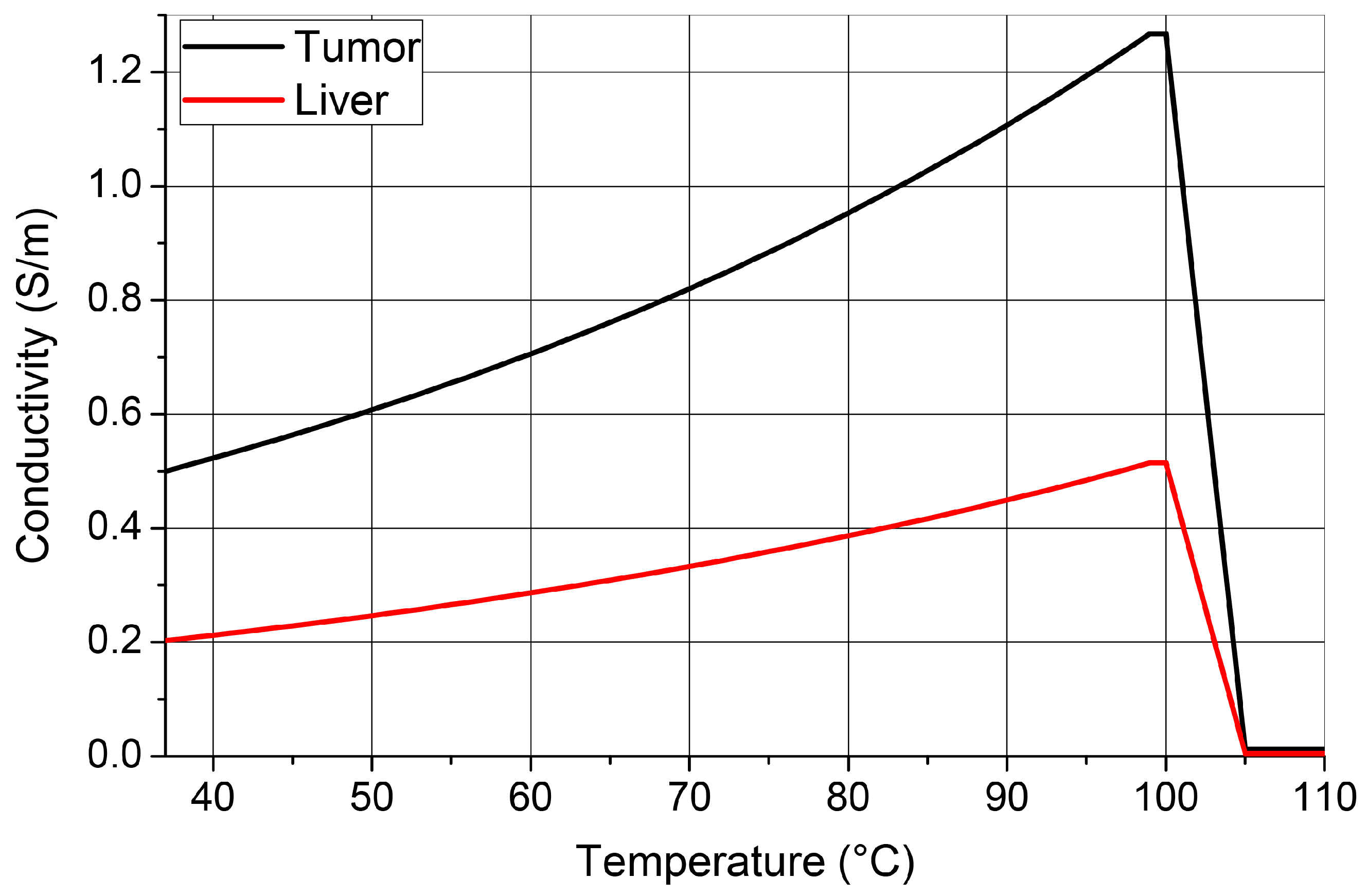
As illustrated in
Figure 1 (computed using Equation (
2) and parameters from
Table 1), electrical conductivity differs markedly between healthy liver parenchyma and tumor tissue, with healthy tissue generally exhibiting higher baseline values. Both tissues follow a characteristic nonlinear trend: conductivity increases with temperature up to ≈100 °C due to enhanced ionic mobility, then undergoes a sharp decline driven by dehydration and vaporization, reaching a residual plateau in desiccated, coagulated tissue. Because conductivity directly governs resistive power deposition
, maintaining tissue temperature in the vicinity of 100 °C, while avoiding premature desiccation, it optimizes electromagnetic energy coupling and ablation efficiency.
The tumor tissue properties adopted in this study correspond to hepatocellular carcinoma (HCC), the most prevalent primary liver malignancy treated with RFA [
2,
6]. While metastatic lesions (e.g., colorectal) exhibit distinct thermophysical characteristics, the present framework is readily adaptable through substitution of tissue-specific parameters from
Table 1. Regarding underlying parenchymal disease, the baseline model assumes non-cirrhotic liver tissue; however, the porosity field
can be calibrated to reflect fibrotic or steatotic conditions by adjusting baseline porosity and perfusion parameters, as outlined in recent porous-media formulations [
31,
33].
Thermal transport is governed by a modified LTE formulation [
31] derived from volume-averaged porous-media theory. Beyond conduction, tissue perfusion is modeled as an advective heat transfer mechanism driven by effective microvascular motion. Unlike classical LTE approaches that approximate perfusion via isotropic volumetric source terms, the present framework explicitly resolves directional heat transport through the convective term
, thereby capturing macroscopic advective effects inherent to anisotropic microvascular architecture.
The effective velocity field is prescribed as a radially outward vector centered on the ablation electrode, aligning with the cylindrical symmetry of the current density and thermal gradients. Rather than resolving discrete vasculature, represents a homogenized transport mechanism consistent with porous-media averaging, capturing preferential heat dissipation away from the heat source. The advective contribution is evaluated through an auxiliary finite-element equation, replacing empirical perfusion coefficients with a physically consistent closure for the volume-averaged energy balance.
Irreversible tissue damage is quantified using the Arrhenius kinetic model [
42,
43], with electromagnetic and thermal properties updated dynamically as functions of temperature and accumulated necrosis. Following established protocols [
41], the necrotic fraction is defined as
, where complete coagulation is assumed at
(corresponding to
).
2.2. Modified Local Thermal Equilibrium Formulation
Heat transfer during radiofrequency ablation involves conductive diffusion, advective transport due to blood perfusion, and thermally induced tissue alterations. At the macroscopic scale, tissue is treated as a porous medium in LTE, governed by a single temperature field for both solid and fluid phases. The classical Pennes’ bioheat equation is expressed as [
31,
44]:
where
,
c, and
k denote effective density, heat capacity, and thermal conductivity, respectively, and
represents Joule heating, while
denotes the perfusion-induced heat transfer. Conventional formulations approximate perfusion via isotropic volumetric source terms, inherently neglecting directional advective heat transport driven by microvascular blood flow. Accordingly, the present framework adopts a modified LTE formulation that incorporates an explicit advective term derived from volume-averaged blood velocity:
with
. Here,
37 °C is the reference temperature. The piecewise definitions for
and
reflect the thermophysical shifts due to vaporization and tissue desiccation [
42].
is the coefficient which is 0 for
, and 1 in other cases.
For liver tissue and blood, density
and specific heat capacity
exhibit discontinuous changes above 100 °C to account for interstitial water vaporization and tissue desiccation [
42]. Thermal conductivity
k and the reference electrical conductivity
are treated as temperature-independent in the baseline model, while the strong nonlinear dependence of electrical conductivity is captured by the multiplicative scaling factor in Equation (
2). Tumor tissue properties are assumed temperature-invariant across the simulated range, consistent with prior modeling frameworks [
42]. All adopted thermophysical and electrical material properties used in the numerical simulations are summarized in
Table 1.
Table 1.
Thermophysical and electrical properties of materials used in simulations. Parameter values are taken from [
42]. Temperature-dependent properties for liver tissue and blood are specified for regimes below and above 100 °C.
Table 1.
Thermophysical and electrical properties of materials used in simulations. Parameter values are taken from [
42]. Temperature-dependent properties for liver tissue and blood are specified for regimes below and above 100 °C.
| Material | Regime | (kg/m3) | c (J/(kg · K)) | (W/(m · K)) | (S/m) |
|---|
| Liver Tissue | 100 °C | 1080 * | 3455 * | 0.515 | 0.203 *** |
| Liver Tissue | 100 °C | 370 ** | 2156 ** | 0.515 | 0.203 *** |
| Liver tumor | – | 1045 | 3760 | 0.6 | 0.5 |
| Blood | 100 °C | 1000 * | 3639 * | 0.49 | 0.667 |
| Blood | 100 °C | 370 ** | 2156 ** | 0.49 | 0.667 |
| Electrode | – | 8000 | 480 | 15 | |
| Plastic (insulator) | – | 70 | 1045 | 0.026 | |
In contrast to conventional approaches where the advective term is explicitly prescribed, the scalar product is computed via an auxiliary finite-element equation solved concurrently with the thermal field. This ensures numerical consistency between the discretized temperature gradient and advective transport, eliminating phenomenological averaging at the element level. The effective velocity is oriented radially from the electrode, capturing volume-averaged perfusion without resolving discrete vasculature, while and denote blood density and specific heat capacity. Coupled with dynamic property updates driven by temperature and accumulated damage, this formulation establishes a tightly coupled feedback loop between heat deposition, tissue necrosis, and electrical conductivity.
2.3. Spatial Porosity Distribution
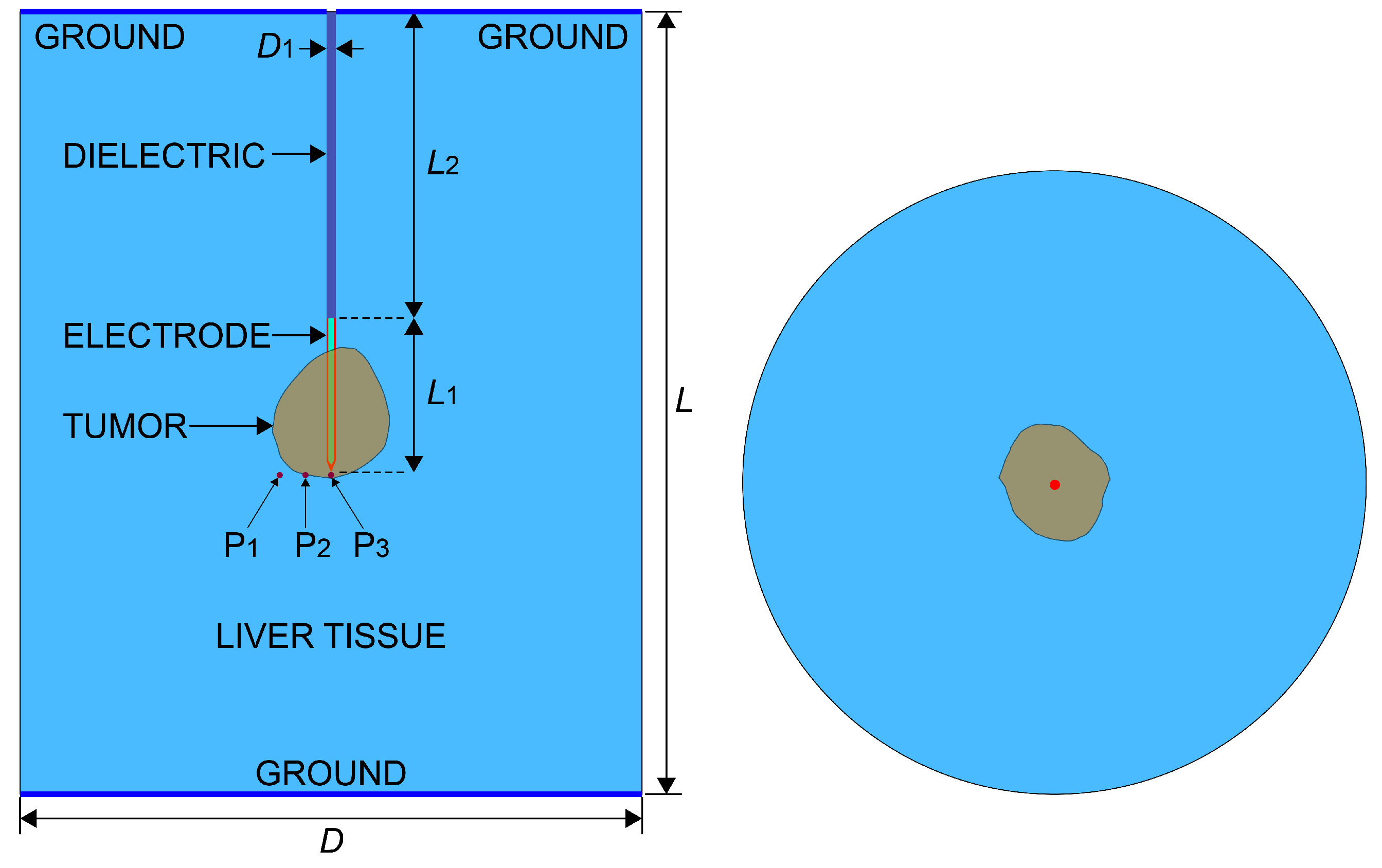
The patient-specific tumor geometry defines the computational domain, as illustrated in
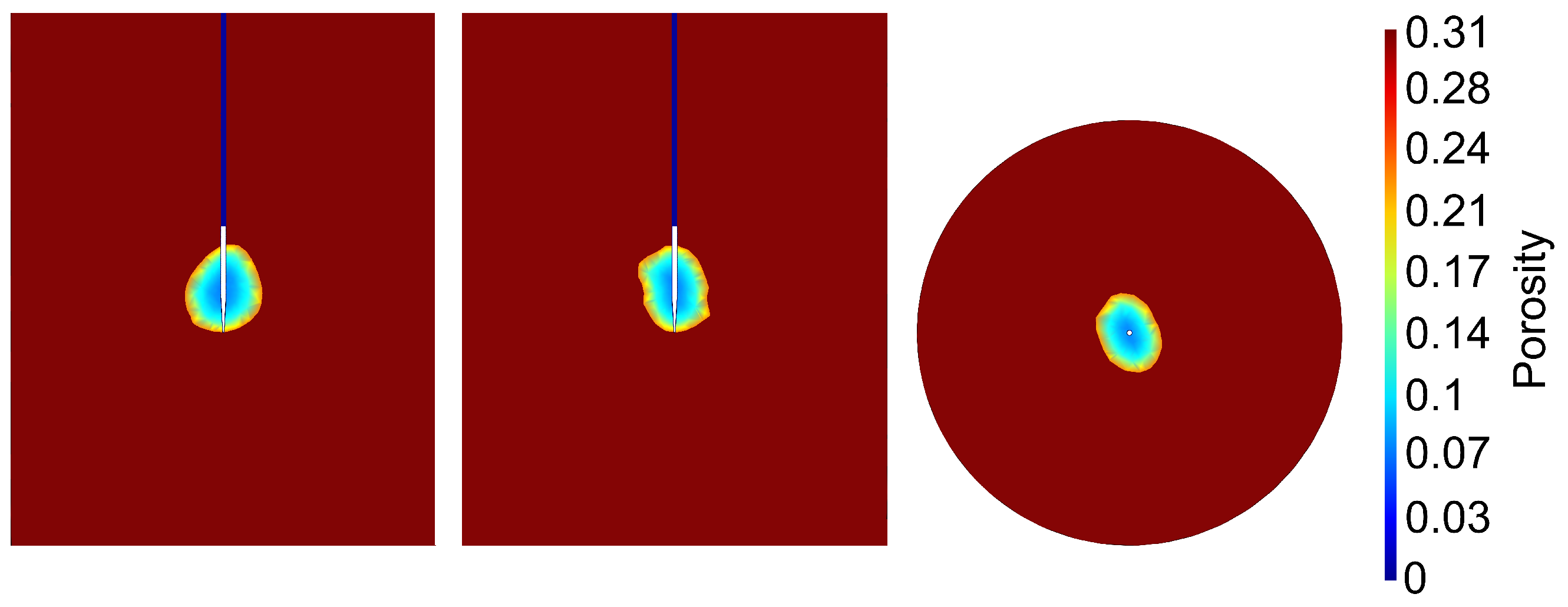
Figure 2. Within this domain, tissue porosity is implemented as a spatially varying scalar field assigned to each finite element via the Gmsh meshing framework. Instead of imposing idealized radial profiles, this geometry-informed approach captures continuous porosity gradients across the tumor volume, transitioning from low-porosity central regions (
) to highly vascularized peripheral tumor (
), as visualized in
Figure 3. By mapping porosity directly onto the computational mesh, structural heterogeneity is preserved without geometric simplifications, allowing electromagnetic field distributions and advective heat transport to be directly governed by the local tissue architecture.
3. Results
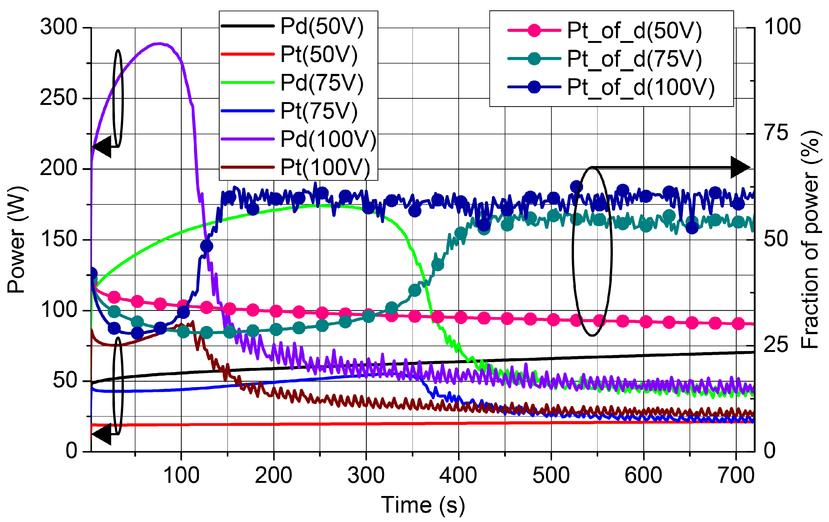
Total absorbed power exhibited strong voltage-dependent nonlinearity, as can be observed from
Figure 4. At 100 V, power peaked at ≈280 W (75 s) before declining to 40–80 W, reflecting initial conductivity enhancement followed by dehydration-induced collapse. The tumor power fraction increased from ≈
to ≈
, indicating progressive current confinement as surrounding tissue impedance rose. At 75 V, peak power (≈175 W) occurred later (300 s) with smoother transition to steady state. At 50 V, power remained low (50–70 W) with minimal conductivity changes, yielding weak energy focusing. Higher relative tumor power reflects spatial confinement rather than improved efficacy; effective ablation requires balancing total power delivery with controlled localization.
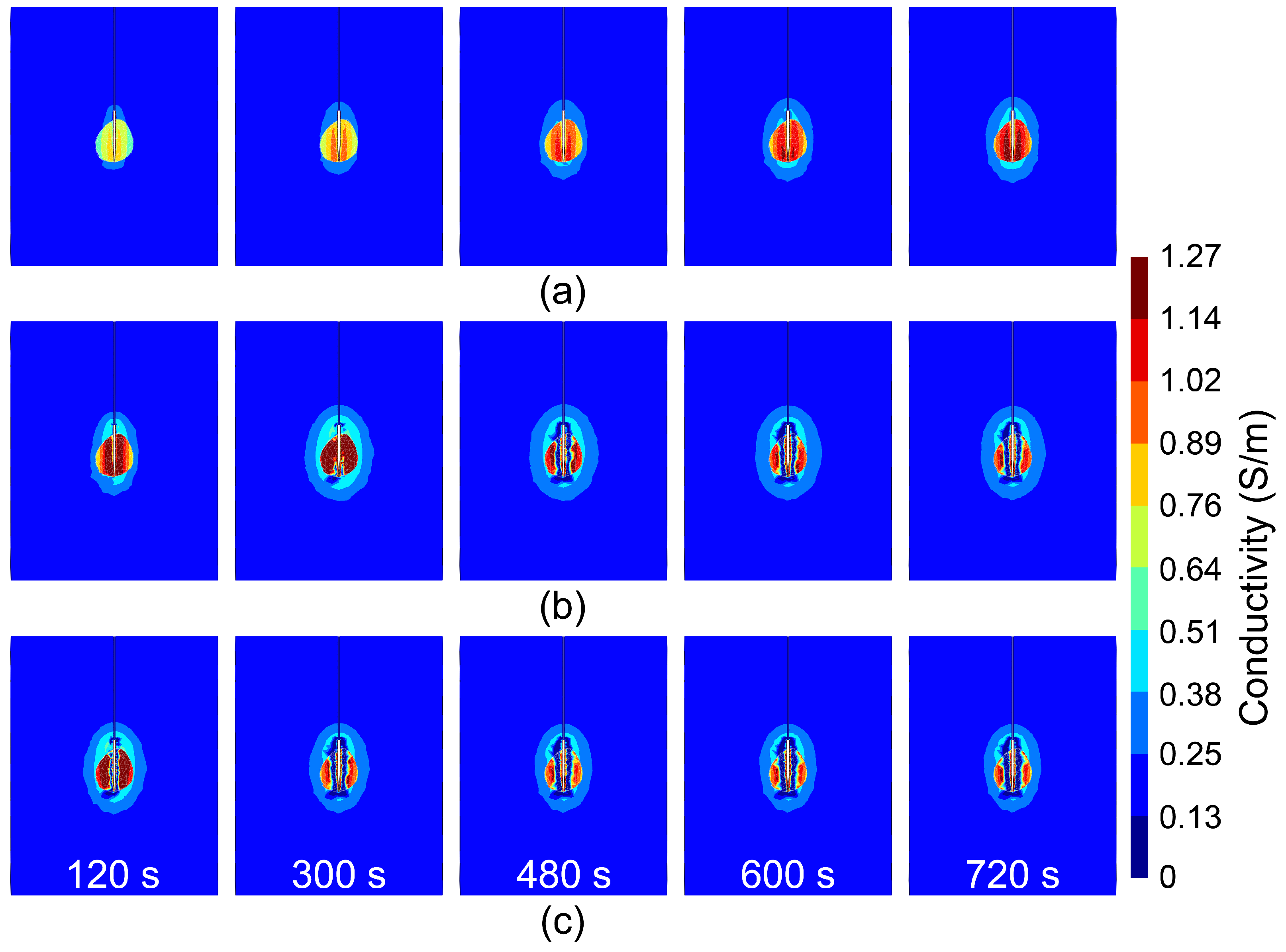
Electrical conductivity evolution revealed three distinct electro-thermal regimes (
Figure 5). At 50 V, conductivity increased monotonically throughout the simulation (720 s), remaining spatially confined to the tumor without evidence of conductivity collapse (
Figure 5a). At 75 V, localized conductivity reduction emerged near the electrode at ≈300 s and progressed to dominate the 480–720 s interval as dehydration established a low-conductivity peri-electrode layer (
Figure 5b). At 100 V, conductivity collapse initiated by 120 s and was fully developed by 300 s, accounting for the early power peak and subsequent rapid decay (
Figure 5c). These results demonstrate a voltage-dependent transition from thermally enhanced conductivity (low voltage) to damage-driven conductivity collapse (high voltage), mediated by dehydration and coagulative necrosis.
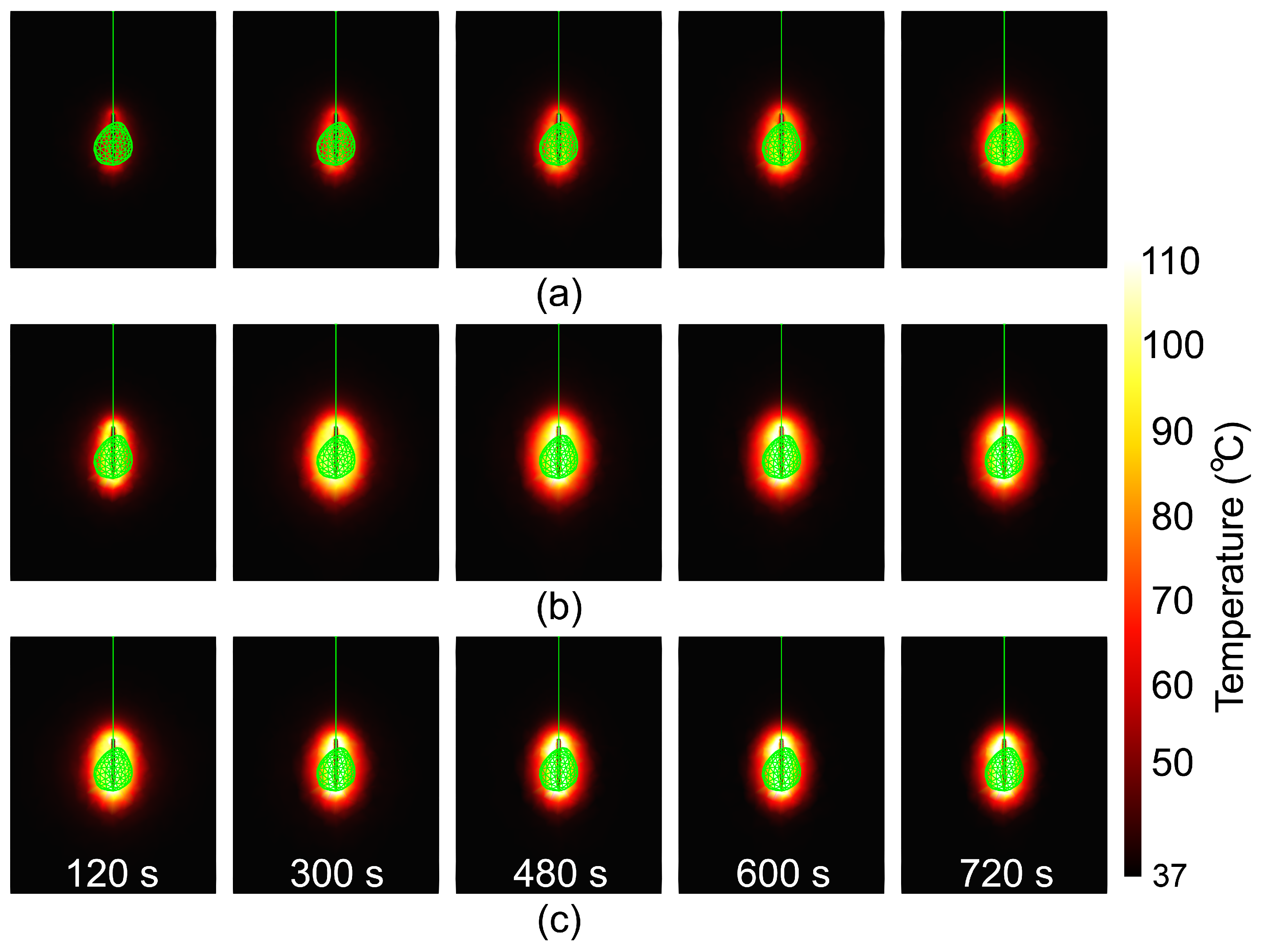
Figure 6 presents spatiotemporal temperature distributions for input voltages of (a) 50 V, (b) 75 V, and (c) 100 V. At 50 V (
Figure 6a), temperature rises gradually, reaching ≈100 °C only after 720 s; this moderate heating preserves electrical coupling and sustains steady radial heat propagation. At 75 V (
Figure 6b), temperature surpasses 100 °C within 120 s and achieves optimal spatial uniformity by ≈300 s. At 100 V (
Figure 6c), intense initial heating induces a rapid drop in electrical conductivity near the electrode, limiting further energy transfer despite elevated local temperatures.
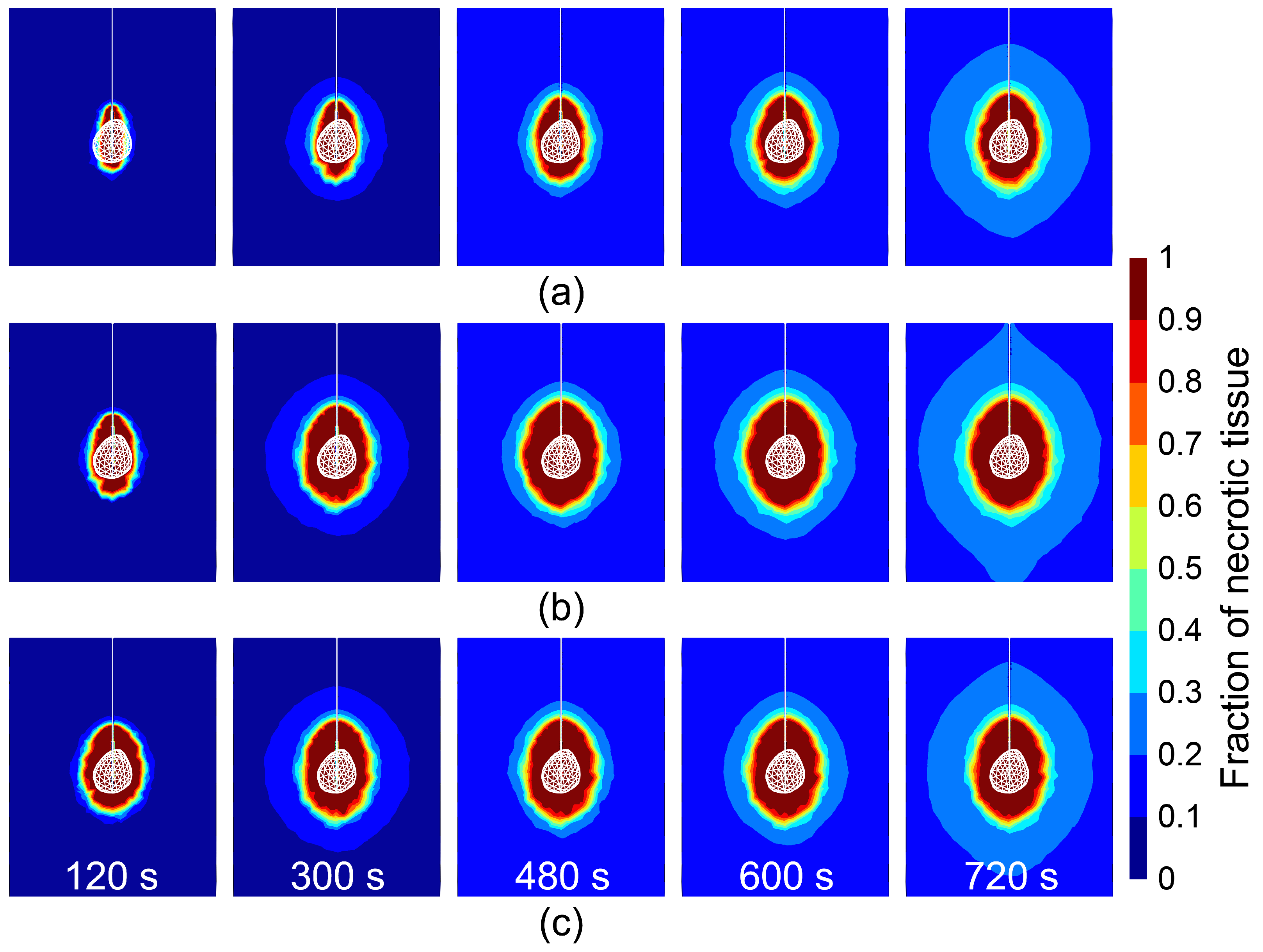
Figure 7 shows the evolution of the tissue damage fraction corresponding to the temperature fields in
Figure 6. At 50 V (
Figure 7a), necrosis develops gradually, ultimately encompassing the tumor with moderate safety margins. At 75 V (
Figure 7b), complete tumor necrosis is achieved ≈2× faster than at 50 V, though damage expansion decelerates after 300 s owing to reduced electrical conductivity near the electrode. At 100 V (
Figure 7c), necrosis stabilizes after 300 s at a spatial extent comparable to the 75 V case, with negligible subsequent progression.
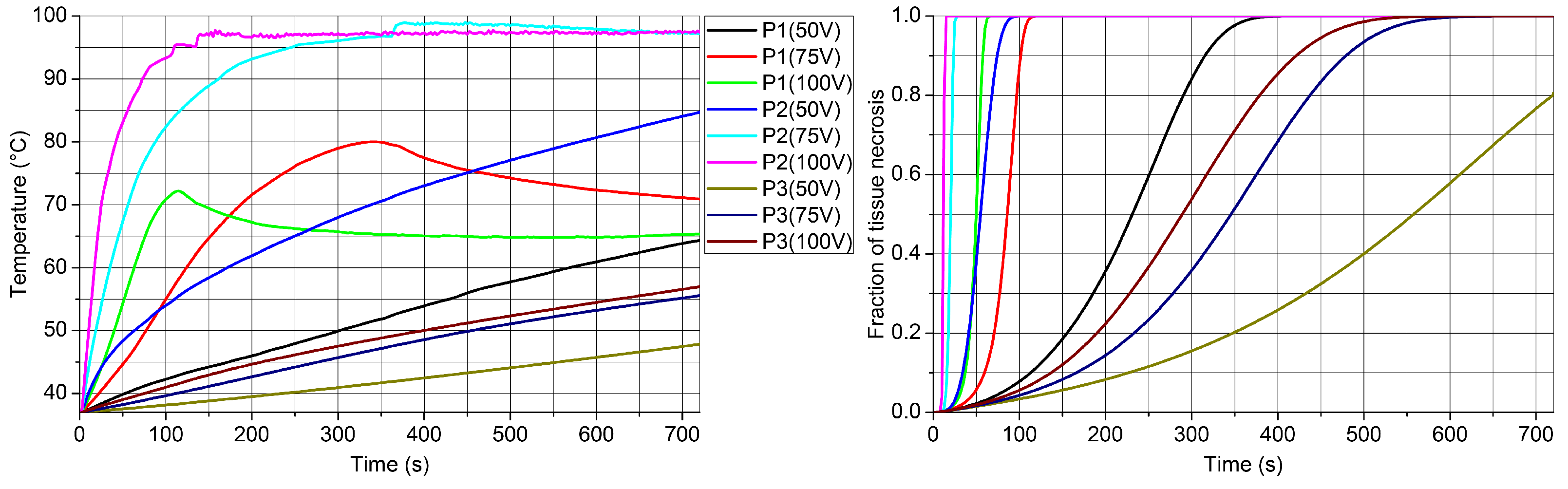
Figure 8 illustrates the temperature and necrosis dynamics at three control points: P3 (electrode tip), P2 (5 mm lateral), and P1 (10 mm lateral). At P3, high voltages induce a rapid temperature rise but delay necrosis onset owing to early dehydration near the electrode. At P2, complete tissue damage is achieved within 15 s at 100 V and 28 s at 75 V, identifying this region as the zone of optimal energy transfer. At P1, temperature progression decelerates at higher voltages, reflecting current redistribution driven by conductivity gradients. Collectively, these results demonstrate that ablation efficacy is governed by spatially distributed electro-thermal feedback rather than peak temperature near the electrode alone.
4. Discussion
This study demonstrates that dynamic electrical conductivity evolution, driven by spatially varying tissue porosity and thermal damage, governs energy deposition and ablation efficacy in radiofrequency ablation (RFA). By coupling patient-specific 3D geometry with a modified local thermal equilibrium formulation that explicitly resolves advective heat transport, the framework captures electro-thermal feedback mechanisms absent in conventional homogeneous models. The simulated temporal heating profiles, peri-electrode temperatures (100–105 °C at 75 V), and radial thermal gradients (≈15 °C mm
−1) closely match experimental miniature-sensor measurements [
15]. Furthermore, predicted necrotic diameters (2.8 ± 0.4 cm at 720 s) align with MRI-validated clinical benchmarks [
21], while the reproduced 60–80% conductivity decline accurately reflects the impedance rise signaling peri-electrode desiccation and self-limiting energy transfer.
While clinical RFA protocols employ a continuous spectrum of voltage settings, the three representative levels examined here (50, 75, and 100 V) were deliberately selected to delineate the key electro-thermal regimes governing ablation dynamics: (i) stable conductivity and gradual thermal propagation at low voltage, (ii) optimal energy coupling with controlled tissue desiccation at intermediate voltage, and (iii) premature conductivity collapse that confines current to peripheral viable tissue at high voltage. The underlying governing equations vary continuously with applied potential, indicating that intermediate settings (e.g., 60–90 V) exhibit smoothly interpolating behavior. This expectation is supported by preliminary simulations at 70, 80, and 90 V, which confirmed monotonic transitions in power deposition, conductivity evolution, and necrosis progression without introducing qualitatively new regimes. Consequently, while exhaustive parameter sweeps may refine optimal voltage boundaries for specific anatomical contexts, they are not expected to alter the fundamental conclusion that ablation efficacy is governed by spatially distributed electro-thermal feedback rather than peak applied voltage alone.
From a clinical perspective, these findings have direct implications for intraoperative protocol selection and monitoring. The early conductivity collapse observed at 100 V illustrates why aggressive power settings may paradoxically limit lesion growth by inducing rapid peri-electrode desiccation—a phenomenon clinically recognized as impedance roll-off. Conversely, the sustained coupling at 75 V supports moderate, prolonged energy delivery to achieve complete tumor coverage while preserving current pathways. This reinforces the value of impedance trend analysis, rather than absolute thresholds, as a real-time diagnostic indicator of ablation progression. Furthermore, the geometry-informed porosity mapping employed here is explicitly designed for future integration with contrast-enhanced CT or multiparametric MRI. Once derived from radiomic texture analysis or machine-learning segmentation, patient-specific porosity fields could enable pre-procedural simulation of impedance trajectories, allowing tailored voltage titration that accounts for individual tumor histology and adjacent vasculature.
The robustness of the identified regimes was evaluated through localized sensitivity analysis, varying Arrhenius constants, porosity gradients, and effective perfusion velocity independently by . Across all perturbations, the qualitative hierarchy of operating regimes remained unchanged, and the ablation efficacy ranking (75 V > 50 V > 100 V) was preserved, with only absolute necrosis times exhibiting moderate variability (). This confirms that the observed electro-thermal transitions are inherent structural features of the coupled system rather than artifacts of specific parameter choices. Nevertheless, several limitations warrant acknowledgment. The assumption of radially symmetric effective perfusion, while computationally efficient, does not fully capture patient-specific vascular architectures. Additionally, validation against aggregated literature benchmarks cannot substitute for prospective patient-matched imaging correlation, and the piecewise approximations for temperature-dependent properties may not capture the full continuum of tissue transformation during ablation. The current baseline also assumes non-cirrhotic parenchyma; however, the porosity field can be calibrated to reflect fibrotic or steatotic conditions by adjusting baseline transport parameters.
Regarding clinical translation, the geometry-informed porosity mapping employed here is explicitly designed for future integration with imaging-derived patient-specific fields. In practice, spatial porosity distributions can be approximated from contrast-enhanced CT or multiparametric MRI using radiomic texture analysis or machine-learning-based segmentation of vascular and extracellular matrix patterns. Once integrated, these maps would enable pre-procedural simulation of patient-specific impedance trajectories, allowing tailored voltage and duration protocols that account for underlying parenchymal disease (e.g., fibrosis, steatosis) and tumor histology. This pathway positions the model as a decision-support tool for personalized RFA planning, reducing empirical guesswork and improving procedural safety.
5. Conclusions
This work establishes spatially varying tissue porosity as a critical determinant of electro-thermal feedback in radiofrequency ablation. By resolving the coupling between microstructural heterogeneity and dynamic conductivity evolution, the proposed framework addresses a fundamental limitation of conventional homogeneous models. The key advance lies not merely in improved geometric fidelity, but in enabling mechanistic interpretation of intraoperative impedance dynamics as a window into the underlying tissue state.
Several limitations warrant acknowledgment. The assumption of radially symmetric effective perfusion, while computationally efficient, does not capture patient-specific vascular architectures that may influence heat dissipation patterns. Additionally, validation against aggregated literature benchmarks, while demonstrating consistency with established ranges, cannot substitute for prospective patient-matched imaging correlation. The temperature-dependent property updates, though physically motivated, rely on piecewise approximations that may not fully capture the continuum of tissue transformation during ablation.
Future research directions should prioritize three areas: First, integration of contrast-enhanced pre-procedural imaging to derive patient-specific porosity maps rather than geometry-informed estimates; second, global sensitivity analysis to quantify the relative influence of Arrhenius parameters, porosity gradients, and perfusion assumptions on predicted ablation margins; and third, prospective clinical validation comparing model-predicted impedance trajectories against intraoperative measurements and post-ablation MRI confirmation.
Author Contributions
Conceptualization, B.R. and N.B.; methodology, N.B. and M.R.-R.; software, N.B. and B.R.; validation, M.R.-R., Š.M. and B.R.; formal analysis, M.R.-R. and Š.M.; investigation, N.B.; writing—original draft preparation, N.B.; writing—review and editing, M.R.-R. and Š.M.; visualization, N.B.; supervision, B.R. All authors have read and agreed to the published version of the manuscript.
Funding
Š.M. acknowledges that this research was supported by the Slovak Research and Development Agency under projects Nr. APVV-23-0522 and Nr. APVV-22-0133.
Institutional Review Board Statement
Not applicable.
Informed Consent Statement
Not applicable.
Data Availability Statement
The raw data supporting the conclusions of this article will be made available by the authors on request.
Acknowledgments
N.B. and M.R.-R. acknowledge that this research was supported by the Institute of Physics Belgrade, University of Belgrade, through a grant from the Ministry of Science, Technological Development and Innovation of the Republic of Serbia.
Conflicts of Interest
The authors declare no conflicts of interest.
References
- Hanna, N.N. Radiofrequency ablation of primary and metastatic hepatic malignancies. Clin. Color. Cancer 2004, 4, 92–100. [Google Scholar] [CrossRef]
- Izzo, F.; Granata, V.; Grassi, R.; Fusco, R.; Palaia, R.; Delrio, P.; Carrafiello, G.; Azoulay, D.; Petrillo, A.; Curley, S.A. Radiofrequency Ablation and Microwave Ablation in Liver Tumors: An Update. Oncologist 2019, 24, e990–e1005. [Google Scholar] [CrossRef] [PubMed]
- Yousaf, M.N.; Ehsan, H.; Muneeb, A.; Wahab, A.; Sana, M.K.; Neupane, K.; Chaudhary, F.S. Role of Radiofrequency Ablation in the Management of Unresectable Pancreatic Cancer. Front. Med. 2021, 7, 624997. [Google Scholar] [CrossRef] [PubMed]
- Yang, G.; Wang, G.; Sun, J.; Xiong, Y.; Li, W.; Tang, T.; Li, J. The prognosis of radiofrequency ablation versus hepatic resection for patients with colorectal liver metastases: A systematic review and meta-analysis based on 22 studies. Int. J. Surg. 2021, 87, 105896. [Google Scholar] [CrossRef]
- Goduguchinta, V.; Ebrahim, M.; Patel, R.; Randhawa, N.; Khalyfa, A.; Inamullah, M.; Desai, R.; Ayub, K. Safety and Efficacy of Radiofrequency Ablation in Management of Various Pancreatic Neoplasms. J. Clin. Med. 2025, 14, 3958. [Google Scholar] [CrossRef]
- Xie, G.L.; Zhong, Z.H.; Ye, T.W.; Xiao, Z.Q. Radiofrequency ablation combined with immunotherapy to treat hepatocellular carcinoma: A comprehensive review. BMC Surg. 2025, 25, 47. [Google Scholar] [CrossRef]
- Dewhirst, M.W.; Viglianti, B.L.; Lora-Michiels, M.; Hanson, M.; Hoopes, P.J. Basic principles of thermal dosimetry and thermal thresholds for tissue damage from hyperthermia. Int. J. Hyperth. 2003, 19, 267–294. [Google Scholar] [CrossRef]
- Wu, M.S.; Kao, W.Y.; Chiang, M.F.; Chuang, P.H.; Hsu, S.J.; Wang, S.Y.; Chang, C.W.; Lin, C.C.; Hung, C.H.; Wu, C.H.; et al. Consensus Guideline of Ablation for Metastatic Liver Tumors by Taiwan Academy of Tumor Ablation. Liver Cancer 2025, 15, 149–164. [Google Scholar] [CrossRef]
- Won, D.-S.; An, J.; Kim, J.W.; Park, Y.; Lee, S.S.; Kim, H.-S.; Park, J.-H. Radiofrequency ablation with sine and square electrical waveforms to enhance ablation range. Front. Bioeng. Biotechnol. 2024, 12, 1450331. [Google Scholar] [CrossRef] [PubMed]
- Bates, A.P.; Ullah, W. The Biophysics of Radiofrequency Ablation and Factors Affecting Lesion Size. Arrhythm. Electrophysiol. Rev. 2026, 15, e08. [Google Scholar] [CrossRef]
- Hiraoka, A.; Horiike, N.; Yamashita, Y.; Koizumi, Y.; Doi, K.; Yamamoto, Y.; Hasebe, A.; Ichikawa, S.; Yano, M.; Miyamoto, Y.; et al. Efficacy of radiofrequency ablation therapy compared to surgical resection in 164 patients in Japan with single hepatocellular carcinoma smaller than 3 cm, along with report of complications. Hepatogastroenterology 2008, 55, 2171–2174. [Google Scholar] [PubMed]
- Conticchio, M.; Inchingolo, R.; Delvecchio, A.; Laera, L.; Ratti, F.; Gelli, M.; Anelli, F.; Laurent, A.; Vitali, G.; Magistri, P.; et al. Radiofrequency ablation vs. surgical resection in elderly patients with hepatocellular carcinoma in Milan criteria. World J. Gastroenterol. 2021, 27, 2205–2218. [Google Scholar] [CrossRef]
- Komorowski, A.L. The Emerging Role of Ablation in the Treatment of Primary and Metastatic Cancer of the Liver. J. Clin. Med. 2025, 14, 8016. [Google Scholar] [CrossRef]
- Todeschini, L.; Caimano, M.; Mattia, A.; Cristin, L.; Martinino, A.; Bianco, G.; Spoletini, G.; Giovinazzo, F. Radiofrequency ablation versus surgical resection in colorectal liver metastasis: Insight from an umbrella review. Front. Oncol. 2025, 15, 1494996. [Google Scholar] [CrossRef] [PubMed]
- Tseng, H.; Lin, S.E.; Chang, Y.L.; Chen, M.H.; Hung, S.H. Determining the critical effective temperature and heat dispersal pattern in monopolar radiofrequency ablation using temperature-time integration. Exp. Ther. Med. 2016, 11, 763–768. [Google Scholar] [CrossRef]
- Lobascio, F.; Di Modugno, R.; Fiore, M.; Di Modugno, N.; Bruno, C.; De Nicolo, T.; Barberis, R.V.; Cabiale, K.; Radoiu, M. Microwave and Radiofrequency Ablation: A Comparative Study between Technologies in Ex Vivo Tissues. Drugs Drug Candidates 2024, 3, 550–565. [Google Scholar] [CrossRef]
- Meng, L.; Zhang, X.; Zhang, X.; Wei, Y.; Xiao, Y. Application of different energy ablations in the treatment of solid tumors. Med-X 2026, 4, 10. [Google Scholar] [CrossRef]
- Ekstrand, V.; Wiksell, H.; Schultz, I.; Sandstedt, B.; Rotstein, S.; Eriksson, A. Influence of electrical and thermal properties on RF ablation of breast cancer: Is the tumour preferentially heated? BioMed Eng. OnLine 2005, 4, 41. [Google Scholar] [CrossRef] [PubMed] [PubMed Central]
- Rossmann, C.; Haemmerich, D. Review of temperature dependence of thermal properties, dielectric properties, and perfusion of biological tissues at hyperthermic and ablation temperatures. Crit. Rev. Biomed. Eng. 2014, 42, 10. [Google Scholar] [CrossRef]
- Wu, J.; Zhou, Z.; Huang, Y.; Deng, X.; Zheng, S.; He, S.; Huang, G.; Hu, B.; Shi, M.; Liao, W.; et al. Radiofrequency ablation: Mechanisms and clinical applications. MedComm 2024, 5, e746. [Google Scholar] [CrossRef] [PubMed]
- Lee, C.-M.; Seo, J.-Y.; Kim, J.-C.; Chon, M.-K. Gradual Temperature Rise in Radiofrequency Ablation: Enhancing Lesion Quality and Safety in Porcine Myocardial Tissue. Bioengineering 2025, 12, 360. [Google Scholar] [CrossRef] [PubMed]
- González-Suárez, A.; Pérez, J.J.; Irastorza, R.M.; D’Avila, A.; Berjano, E. Computer modeling of radiofrequency cardiac ablation: 30 years of bioengineering research. Comput. Methods Programs Biomed. 2022, 214, 106546. [Google Scholar] [CrossRef]
- Irastorza, R.; Trujillo, M.; Villagrán, J.; Berjano, E. Computer modelling of RF ablation in cortical osteoid osteoma: Assessment of the insulating effect of the reactive zone. Int. J. Hyperth. 2016, 32, 221–230. [Google Scholar] [CrossRef][Green Version]
- Audigier, C.; Kim, Y.; Dillow, A.; Boctor, E.M. Computational modeling of radiofrequency ablation: Evaluation on ex vivo data using ultrasound monitoring. In Medical Imaging 2017: Image-Guided Procedures, Robotic Interventions, and Modeling; Webster, R.J., Fei, B., Eds.; SPIE: Bellingham, WA, USA, 2017; p. 1013525. [Google Scholar] [CrossRef]
- Loya, R.R.; Jutte, P.C.; Kwee, T.C.; van Ooijen, P.M.A. Computer 3D modeling of radiofrequency ablation of atypical cartilaginous tumours in long bones using finite element methods and real patient anatomy. Eur. Radiol. Exp. 2022, 6, 21. [Google Scholar] [CrossRef]
- Radmilović-Radjenović, M.; Radjenović, D.; Radjenović, B. Finite element analysis of the effect of microwave ablation on the liver, lung, kidney, and bone malignant tissues. Europhys. Lett. 2022, 136, 28001. [Google Scholar] [CrossRef]
- Pérez, J.J.; Ewertowska, E.; Berjano, E. Computer Modeling for Radiofrequency Bipolar Ablation Inside Ducts and Vessels: Relation Between Pullback Speed and Impedance Progress. Lasers Surg. Med. 2020, 52, 897. [Google Scholar] [CrossRef] [PubMed]
- Chang, I.A.; Nguyen, U.D. Thermal modeling of lesion growth with radiofrequency ablation devices. BioMed Eng. OnLine 2004, 3, 27. [Google Scholar] [CrossRef] [PubMed]
- Gough, A.; Stern, A.M.; Maier, J.; Lezon, T.; Shun, T.Y.; Chennubhotla, C.; Schurdak, M.E.; Haney, S.A.; Taylor, D.L. Biologically Relevant Heterogeneity: Metrics and Practical Insights. SLAS Discov. 2017, 22, 213–237. [Google Scholar] [CrossRef]
- Voglreiter, P.; Mariappan, P.; Pollari, M.; Flanagan, R.; Blanco Sequeiros, R.; Portugaller, R.H.; Fütterer, J.; Schmalstieg, D.; Kolesnik, M.; Moche, M. RFA Guardian: Comprehensive Simulation of Radiofrequency Ablation Treatment of Liver Tumors. Sci. Rep. 2018, 8, 787. [Google Scholar] [CrossRef]
- Tucci, C.; Trujillo, M.; Berjano, E.; Iasiello, M.; Andreozzi, A.; Vanoli, G.P. Pennes’ bioheat equation vs. porous media approach in computer modeling of radiofrequency tumor ablation. Sci. Rep. 2021, 11, 5272. [Google Scholar] [CrossRef]
- Tucci, C.; Trujillo Guillen, M.; Berjano, E.; Iasiello, M.; Andreozzi, A.; Vanoli, G.P. Mathematical modeling of microwave liver ablation with a variable-porosity medium approach. Comput. Methods Programs Biomed. 2022, 214, 106569. [Google Scholar] [CrossRef]
- Andreozzi, A.; Brunese, L.; Iasiello, M.; Tucci, C.; Vanoli, G.P. Numerical Investigation of a Thermal Ablation Porous Media-Based Model for Tumoral Tissue with Variable Porosity. Computation 2021, 9, 50. [Google Scholar] [CrossRef]
- Andreozzi, A.; Brunese, L.; Iasiello, M.; Tucci, C.; Vanoli, G.P. A New Thermal Damage-Controlled Protocol for Thermal Ablation Modeled with Modified Porous Media-Based Bioheat Equation with Variable Porosity. Processes 2022, 10, 236. [Google Scholar] [CrossRef]
- Ahmed, M.; Liu, Z.; Humphries, S.; Goldberg, S.N. Computer modeling of the combined effects of perfusion, electrical conductivity, and thermal conductivity on tissue heating patterns in radiofrequency tumor ablation. Int. J. Hyperth. 2008, 24, 577–588. [Google Scholar] [CrossRef]
- Liu, Z.; Ahmed, M.; Gervais, D.; Humphries, S.; Goldberg, S.N. Computer modeling of factors that affect the minimum safety distance required for radiofrequency ablation near adjacent nontarget structures. J. Vasc. Interv. Radiol. 2008, 19, 1079–1086. [Google Scholar] [CrossRef] [PubMed]
- Duan, B.; Wen, R.; Fu, Y.; Chua, K.J.; Chui, C.K. Probabilistic finite element method for large tumor radiofrequency ablation simulation and planning. Med. Eng. Phys. 2016, 38, 1360–1368. [Google Scholar] [CrossRef]
- Singh, S.; Melnik, R. Thermal ablation of biological tissues in disease treatment: A review of computational models and future directions. Electromagn. Biol. Med. 2020, 39, 49–88. [Google Scholar] [CrossRef]
- Lodato, F.; De Vita, E.; Patrone, R.; Izzo, F.; Iadicicco, A.; Stefania, C.; Rita, M.; Riccio, D.; Ruello, G. Radio Frequency Ablation of Liver Tissue Assisted by Saline Infusion: Computational Modeling and Experimental Assessment. IEEE Trans. Instrum. Meas. 2025, 74, 4008711. [Google Scholar] [CrossRef]
- Guo, Z.Y.; Tao, W.Q.; Shah, R.K. The field synergy (coordination) principle and its applications in enhancing single phase convective heat transfer. Int. J. Heat. Mass. Transf. 2005, 48, 1797–1807. [Google Scholar] [CrossRef]
- Tao, W.Q.; He, Y.L.; Wang, Q.W.; Qu, Z.G.; Song, F.Q. A unified analysis on enhancing single phase convective heat transfer with field synergy principle. Int. J. Heat. Mass. Transf. 2002, 45, 5005–5011. [Google Scholar] [CrossRef]
- Fang, Z.; Wei, H.; Zhang, H.; Moser, M.A.J.; Zhang, W.; Qian, Z.; Zhang, B. Radiofrequency ablation for liver tumors abutting complex blood vessel structures: Treatment protocol optimization using response surface method and computer modeling. Int. J. Hyperth. 2022, 39, 733–742. [Google Scholar] [CrossRef]
- Bošković, N.; Radjenović, B.; Nikolić, S.; Radmilović-Radjenović, M. Effectiveness of microwave ablation using two simultaneous antennas for liver malignancy treatment. Open Phys. 2024, 22, 20240079. [Google Scholar] [CrossRef]
- Pennes, H.H. Analysis of tissue and arterial blood temperatures in the resting human forearm. J. Appl. Physiol. 1948, 1, 93–122. [Google Scholar] [CrossRef]
| Disclaimer/Publisher’s Note: The statements, opinions and data contained in all publications are solely those of the individual author(s) and contributor(s) and not of MDPI and/or the editor(s). MDPI and/or the editor(s) disclaim responsibility for any injury to people or property resulting from any ideas, methods, instructions or products referred to in the content. |



 ,
,