Computed Tomography Patterns of Pneumocystis jirovecii Pneumonia According to Immune Status

 , and
, and
Abstract
1. Introduction
2. Material and Methods
2.1. Study Design and Population
2.2. Clinical and Laboratory Data
2.3. Diagnosis of Pneumocystis jirovecii Pneumonia
2.4. Chest Radiography
2.5. Computed Tomography
2.6. Image Analysis
2.7. Respiratory Coinfections
2.8. Statistical Analysis
2.9. Ethics Approval
3. Results
3.1. Patient Characteristics
3.2. Chest Radiography Findings
3.3. Computed Tomography Findings
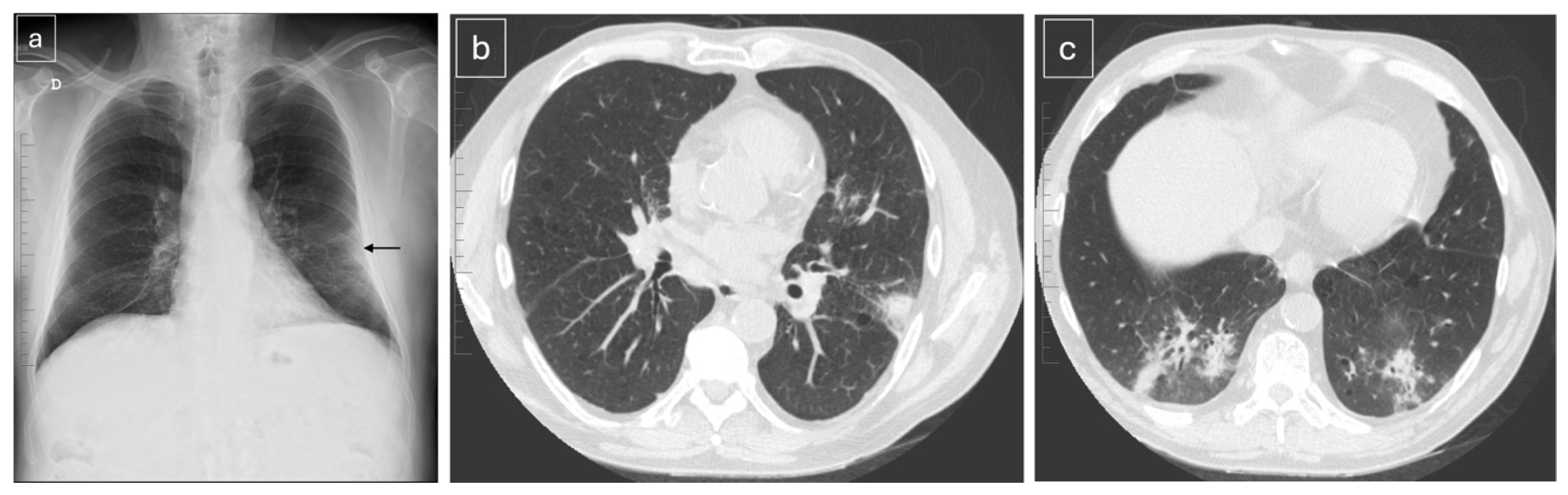
3.4. Relationship Between Chest Radiography and CT
3.5. Imaging Patterns According to Type of Immunosuppression in Non-HIV Patients
3.6. Respiratory Coinfections
4. Discussion
5. Conclusions
Supplementary Materials
Author Contributions
Funding
Institutional Review Board Statement
Informed Consent Statement
Data Availability Statement
Acknowledgments
Conflicts of Interest
References
- Ibrahim, A.; Chattaraj, A. Pneumocystis jirovecii pneumonia: A review of management in human immunodeficiency virus (HIV) and non-HIV immunocompromised patients. Avicenna J. Med. 2023, 13, 23–34. [Google Scholar] [PubMed]
- McDonald, E.G.; Afshar, A. Pneumocystis jirovecii pneumonia in people living with HIV: A review. Clin. Microbiol. Rev. 2024, 37, e00101–e00122. [Google Scholar] [CrossRef] [PubMed]
- Vicente, P.; Parra Fariñas, R. Impact of pneumocystosis on the Spanish healthcare system, 1997–2020: Profile of HIV and non-HIV immunocompromised patients. J. Infect. Public Health 2023, 1, 831–840. [Google Scholar] [CrossRef] [PubMed]
- Apostolopoulou, A.; Fishman, J.A. The pathogenesis and diagnosis of Pneumocystis jirovecii pneumonia. J. Fungi 2022, 8, 1167. [Google Scholar] [CrossRef] [PubMed]
- Wang, W.; Mo, Q. Clinical characteristics of Pneumocystis jirovecii pneumonia in non-HIV-infected patients. J. Thorac. Dis. 2025, 17, 8904–8914. [Google Scholar] [CrossRef] [PubMed]
- Rego de Figueiredo, I.; Vieira Alves, R. Pneumocystis jirovecii pneumonia: A comparison study between HIV and non-HIV immunocompromised patients. Pulmonology 2019, 25, 271–274. [Google Scholar] [CrossRef] [PubMed]
- Trubin, P.A.; Azar, M.M. Current concepts in the diagnosis and management of Pneumocystis jirovecii pneumonia in solid organ transplantation. Infect. Dis. Clin. N. Am. 2023, 37, 617–640. [Google Scholar] [CrossRef] [PubMed]
- Asai, N.; Motojima, S. Pathophysiological mechanism of non-HIV Pneumocystis jirovecii pneumonia. Respir. Investig. 2022, 60, 522–530. [Google Scholar] [CrossRef] [PubMed]
- Tasaka, S.; Tokuda, H. Comparison of clinical and radiological features of Pneumocystis jirovecii pneumonia between malignancy cases and AIDS cases: A multicenter study. Intern. Med. 2010, 49, 273–281. [Google Scholar] [CrossRef] [PubMed]
- Wills, N.K.; Adriaanse, M. Chest X-ray features of HIV-associated Pneumocystis pneumonia in adults: A systematic review and meta-analysis. Open Forum Infect. Dis. 2024, 11, ofae146. [Google Scholar] [CrossRef] [PubMed]
- Kanne, J.P.; Yandow, D.R. Pneumocystis jirovecii pneumonia: High-resolution CT findings in patients with and without HIV infection. AJR Am. J. Roentgenol. 2012, 198, W555–W561. [Google Scholar] [CrossRef] [PubMed]
- Cereser, L.; Dallorto, A. Pneumocystis jirovecii pneumonia at high-resolution CT in non-HIV immunocompromised patients: Spectrum of findings and mimickers. Eur. J. Radiol. 2019, 116, 116–127. [Google Scholar] [CrossRef] [PubMed]
- Kumagai, S.; Arita, M. Prognostic significance of crazy-paving ground-glass opacities in non-HIV Pneumocystis jirovecii pneumonia: An observational cohort study. BMC Pulm. Med. 2019, 19, 47. [Google Scholar] [CrossRef] [PubMed]
- Arora, S.; Walker, S.M. Pneumonia in immunocompromised patients. Radiographics 2025, 45, e250021. [Google Scholar] [CrossRef] [PubMed]
- Wu, H.Y.; Wu, K.S. Identifying predictors for bacterial and fungal coinfection on chest computed tomography in patients with Pneumocystis pneumonia. J. Microbiol. Immunol. Infect. 2021, 54, 701–709. [Google Scholar] [CrossRef] [PubMed]
- Lécuyer, R.; Issa, N. Epidemiology and clinical impact of respiratory coinfections at diagnosis of Pneumocystis jirovecii pneumonia. J. Infect. Dis. 2022, 225, 868–880. [Google Scholar] [CrossRef] [PubMed]
- Maschmeyer, G.; Helweg-Larsen, J. ECIL guidelines for treatment of Pneumocystis jirovecii pneumonia in non-HIV-infected haematology patients. J. Antimicrob. Chemother. 2016, 71, 2405–2413. [Google Scholar] [CrossRef] [PubMed]
- Hardak, E.; Brook, O. Radiological features of Pneumocystis jirovecii pneumonia in immunocompromised patients with and without AIDS. Lung 2010, 188, 159–163. [Google Scholar] [CrossRef] [PubMed]
- Albasata, H.; Gioia, F. Outcome of Pneumocystis pneumonia in transplant and non-transplant HIV-negative immunocompromised patients. Transpl. Infect. Dis. 2024, 26, e14321. [Google Scholar] [CrossRef] [PubMed]


| Total n = 72 (%) | HIV n = 27 (%) | Non-HIV n = 45 (%) | p Value | |
|---|---|---|---|---|
| Age. years (mean ± SD) | 58.5 ± 13.6 | 49.3 ± 10.0 | 64.4 ± 12.3 | 0.004 |
| Male sex | 51 (70.8) | 24 (88.9) | 27 (60.0) | 0.009 |
| Immunosuppression | ||||
| CD4+ T-cell count < 200 cells/µL; (mean ± SD) | 25 (96.2); (51.2 ± 46.9) | |||
| Hematologic malignancy | 10 (22.2) | |||
| HSCT | 5 (11.1) | |||
| SOT | 10 (22.2) | |||
| Solid tumor | 8 (17.8) | |||
| Other * | 12 (26.7) | |||
| Any coinfection | 46 (63.9) | 19 (70.4) | 27 (60.0) | 0.506 |
| Symptom onset to hospital admission (days) mean ± SD | 11.87 ± 15.128 | 19.12 ± 14.9 | 7.69 ± 13.744 | 0.002 |
| Symptom onset to sample collection (days) mean ± SD | 18.51 ± 13.89 | 24.69 ± 13.04 | 14.93 ± 13.214 | 0.004 |
| Total n = 71 (%) | HIV n = 27 (%) | Non-HIV n = 44 (%) | p Value | |
|---|---|---|---|---|
| Normal | 23 (32.4) | 6 (22.2) | 17 (38.6) | 0.242 |
| Abnormal | 48 (67.6) | 21 (77.8) | 27 (61.4) | |
| Interstitial pattern | 31 (64.6) | 17 (81.0) | 14 (51.9) | 0.69 |
| Consolidation | 16 (33.3) | 4 (19.0) | 12 (44.4) | |
| Alveolo-interstitial | 1 (2.1) | 0 (0.0) | 1 (3.7) | |
| Lung distribution | ||||
| Interstitial pattern | 31 (64.6) | 17 (81.0) | 14 (51.9) | 0.075 |
| Field distribution | 1.000 | |||
| Upper | 2 (6.5) | 1 (5.9) | 1 (7.1) | |
| Middle | 6 (19.4) | 3 (17.6) | 3 (21.4 | |
| Upper and middle | 3 (9.7) | 2 (11.8) | 1 (7.1) | |
| Middle and lower | 6 (19.4) | 3 (17.6) | 3 (21.4) | |
| Lower | 2 (6.5) | 1 (5.9) | 1 (7.1) | |
| All lung zones | 12 (38.7) | 7 (41.2) | 5 (35.7) | |
| Unilateral/bilateral | 0.18 | |||
| Unilateral | 10 (32.3) | 2 (11.8) | 8 (57.1) | |
| Bilateral | 21 (67.7) | 15 (71.4) | 6 (42.9) | |
| Intrapulmonary distribution | 0.502 | |||
| Central | 10 (32.3) | 7 (41.2) | 3 (21.4) | |
| Peripheral | 5 (16.1) | 2 (11.8) | 3 (21.4) | |
| Diffuse | 16 (51.6) | 8 (47.1) | 8 (57.1) | |
| Consolidation | 16 (33.3) | 4 (19.0) | 12 (46.2) | 0.051 |
| Field distribution | 0.430 | |||
| Upper | 3 (18.8) | 0 (0.0) | 3 (25.0) | |
| Middle | 4 (25.0) | 1 (25.0) | 3 (25.0) | |
| Upper and middle | 1 (6.2) | 1 (25.0) | 0 (0.0) | |
| Lower | 6 (37.5) | 2 (50) | 4 (33.3) | |
| All lung zones | 2 (12.5) | 0 (0.0) | 2 (16.7) | |
| Unilateral/bilateral | 0.020 | |||
| Unilateral | 11 (68.8) | 1 (25.0) | 10 (83.3) | |
| Bilateral | 5 (31.3) | 3 (75.0) | 2 (16.7) | |
| Intrapulmonary distribution | 0.791 | |||
| Central | 3 (18.8) | 1 (25.0) | 2 (32.1) | |
| Peripheral | 7 (43.8) | 1 (25.0) | 6 (50.0) | |
| Diffuse | 6 (37.5) | 2 (50.0) | 4 (33.3) | |
| Alveolo-interstitial | 1 (2.1) | 0 (0.0) | 1 (3.7) | |
| All lung zones | 1 (100.0) | 0 (0.0) | 1 (100.0) | |
| Bilateral | 1 (100.0) | 0 (0.0) | 1 (100.0) | |
| Diffuse | 1 (100.0) | 0 (0.0) | 1 (100.0) | |
| Pleural effusion | 2 (4.9) | 0 (0.0) | 2 (16.7) | 0.232 |
| Total n = 62 (%) | HIV n = 20 (%) | Non-HIV n = 42 (%) | p Value | |
|---|---|---|---|---|
| Normal | 7 (11.3) | 1 (5.0) | 6 (14.3) | 0.687 |
| Abnormal | 55 (88.7) | 19 (95.0) | 36 (85.7) | |
| Ground-glass opacity | 34 (61.8) | 10 (52.6) | 24 (66.7) | 0.029 |
| Alveolo-interstitial | 11 (20.0) | 7 (36.8) | 4 (11.1) | |
| Consolidation | 9 (16.4) | 1 (5.3) | 8 (22.2) | |
| Micronodular | 1 (1.8) | 1 (5.3) | 0 (0.0) | |
| Lung distribution | ||||
| Ground-glass opacity | 34 (63.6) | 10 (57.9) | 24 (66.7) | |
| Field distribution | 0.912 | |||
| Upper | 2 (5.9) | 0 (0.0) | 2 (8.3) | |
| Upper and middle | 15 (26.6) | 4 (40.0) | 11 (45.8) | |
| Middle and lower | 4 (11.8) | 1 (10.0) | 3 (12.5) | |
| Lower | 2 (5.9) | 1 (10.0) | 1 (4.2) | |
| All lung zones | 11 (32.4 | 4 (40.0) | 7 (29.2) | |
| Unilateral/bilateral | 1.000 | |||
| Unilateral | 2 (5.9) | 0 (0.0) | 2 (8.3) | |
| Bilateral | 32 (94.1) | 10 (100.0) | 22 (91.7) | |
| Intrapulmonary distribution | 1.000 | |||
| Central | 1 (2.9) | 0 (0.0) | 1 (4.2) | |
| Peripheral | 3 (8.8) | 1 (10.0) | 2 (8.3) | |
| Diffuse | 30 (88.2) | 9 (90.0) | 21 (87.5) | |
| Alveolo-interstitial pattern | 11 (20.0) | 7 (36.8) | 4 (11.1) | |
| Field distribution | 0.716 | |||
| Upper and middle | 4 (36.4) | 3 (42.9) | 1 (25.0) | |
| Middle and lower | 1 (9.1) | 1 (14.3) | 0 (0.0) | |
| All lung zones | 6 (54.5) | 3 (42.9) | 3 (75.0) | |
| Unilateral/bilateral | ||||
| Bilateral | 11 (100.0) | 7 (100.0) | 4 (100.0) | |
| Intrapulmonary distribution | ||||
| Diffuse | 11 (100.0) | 7 (100.0) | 4 (100.0) | |
| Consolidation | 9 (16.4) | 1 (5.3) | 8 (22.2) | |
| Field distribution | 1.000 | |||
| Upper | 2 (22.2) | 0 (0.0) | 2 (25.0) | |
| Upper and middle | 2 (22.2) | 0 (0.0) | 2 (25.0) | |
| Middle and lower | 2 (22.2) | 0 (0.0) | 2 (25.0) | |
| Lower | 2 (22.2) | 1 (100.0) | 1 (12.5) | |
| All lung zones | 1 (11.1) | 0 (0.0) | 1 (12.5) | |
| Unilateral/bilateral | 1.000 | |||
| Unilateral | 5 (55.6) | 1 (100.0) | 4 (50.0) | |
| Bilateral | 4 (44.4) | 0 (0.0) | 4 (50.0) | |
| Intrapulmonary distribution | 1.000 | |||
| Central | 1 (11.1) | 0 (0.0) | 1 (12.5) | |
| Peripheral | 2 (22.2) | 0 (0.0) | 2 (25.0) | |
| Diffuse | 6 (66.7) | 1 (100.0) | 5 (62.5) | |
| Micronodular | 1 (2.4) | 1 (9.1) | 0 (0.0) | |
| Field distribution | ||||
| All lung zones | 1 (100.0) | 1 (100.0) | 0 (0.0) | |
| Unilateral/bilateral | ||||
| Bilateral | 1 (100.0) | 1 (100.0) | 0 (0.0) | |
| Intrapulmonary distribution | ||||
| Diffuse | 1 (100.0) | 1 (100.0) | 0 (0.0) | |
| Mosaic pattern | 24 (51.1) | 12 (66.7) | 12 (41.4) | 0.034 |
| Pulmonary cysts | 5 (9.1) | 5 (26.3) | 0 (0.0) | 0.001 |
| Crazy paving | 22 (43.6) | 8 (42.1) | 14 (38.9) | 0.817 |
| Mediastinal lymphadenopathy | 3 (5.5) | 2 (10.5) | 1 (2.8) | 0.272 |
| Pleural effusion | 13 (23.6) | 1 (5.3) | 12 (33.3) | 0.022 |
| Unilateral | 5 (38.5) | 0 (0.0) | 5 (41.7) | |
| Bilateral | 8 (61.5) | 1 (100.0) | 7 (58.3) | |
| Pneumothorax | 2 (5.7) | 0 (0.0) | 2 (7.7) | 0.539 |
| Fibrosis | 1 (2.8) | 0 (0.0) | 1 (4.2) | 1.000 |
| Bronchiectasis | 9 (23.7) | 3 (23.1) | 6 (24.0) | 1.000 |
| Hematologic Malignancy n = 10 (%) | HSCT n = 5 (%) | SOT n = 10 (%) | Solid Tumor n = 8 (%) | Other n = 12 (%) | |
|---|---|---|---|---|---|
| Chest radiography | n = 10 (100.0) | n = 5 (100.0) | n = 10 (100.0) | n = 8 (100.0) | n = 11 (91.7) |
| Normal | 5 (50.0) | 2 (40.0) | 4 (40.0) | 1 (12.5) | 5 (45.4) |
| Interstitial pattern | 4 (40.0) | 3 (60.0) | 1 (10.0) | 4 (50.0) | 3 (27.3) |
| Consolidation | 1 (10.0) | 0 (0.0) | 5 (50.0) | 3 (37.5) | 3 (27.3) |
| CT | n = 9 (90.0) | n = 4 (80.0) | n = 10 (100.0) | n = 7 (100.0) | n = 12 (100.0) |
| Normal | 0 (0.0) | 0 (0.0) | 2 (20.0) | 0 (0.0) | 4 (33.3) |
| Ground-glass opacity | 8 (89.0) | 3 (75.0) | 3 (30.0) | 5 (71.4) | 5 (41.7) |
| Alveolo-interstitial | 1 (11.0) | 0 (0.0) | 0 (0.0) | 2 (28.6) | 1 (8.3) |
| Consolidation | 0 (0.0) | 1 (25.0) | 5 (50.0) | 0 (0.0) | 2 (16.7) |
| Mosaic attenuation | 3 (33.3) | 2 (50.0) | 0 (0.0) | 3 (42.9) | 4 (33.3) |
| Crazy paving | 6 (66.7) | 1 (25.0) | 1 (20.0) | 5 (71.4) | 1 (8.3) |
| Pleural effusion | 4 (44.4) | 0 (0.0) | 3 (30.0) | 4 (57.1) | 1 (8.3) |
| No Coinfection | Any Coinfection | |||||
|---|---|---|---|---|---|---|
| Chest Radiography | HIV n = 8 (%) | Non-HIV n = 18 (%) | p Value | VIH n = 19 (%) | Non-VIH n = 27 (%) | p Value |
| Normal | 3 (37.5) | 4 (22.2) | 0.354 | 3 (15.8) | 13 (48.1) | 0.109 |
| Abnormal | 5 (62.5) | 13 (76.5) | 16 (84.2) | 14 (51.9) | ||
| Interstitial pattern | 4 (80.0) | 5 (38.5) | 0.498 | 13 (81.2) | 9 (64.3) | 0.417 |
| Consolidation | 1 (20.0) | 7 (53.8) | 3 (18.8) | 5 (35.7) | ||
| Alveolo-interstitial | 0 (0.0) | 1 (7.7) | 0 (0.0) | 0 (0.0) | ||
| CT | HIV n = 5 (%) | Non-HIV n = 17 (%) | p value | HIV n = 15 (%) | Non-HIV n = 25 (%) | p value |
| Normal | 1 (20.0) | 4 (23.5) | 0 (0.0) | 2 (8.0) | ||
| Abnormal | 4 (80.0) | 13 (76.5) | 15 (100.0) | 23 (92.0) | ||
| Ground Glass opacity | 2 (50.0) | 9 (69.2) | 0.238 | 9 (60.0) | 15 (65.2) | 0.178 |
| Alveolo-interstitial | 2 (50.0) | 1 (7.7) | 5 (33.3) | 3 (13.0) | ||
| Consolidation | 0 (0.0) | 3 (23.1) | 1 (6.7) | 5 (21.7) | ||
| Cysts | 1 (25.0) | 0 (0.0) | 0.235 | 4 (26.7) | 0 (0.0) | 0.018 |
| Mosaic pattern | 3 (75.0) | 7 (53.8) | 0.452 | 9 (60.0) | 5 (21.7) | 0.017 |
| Crazy paving | 1 (25.0) | 7 (53.8) | 0.312 | 7 (46.7) | 7 (30.4) | 0.311 |
Disclaimer/Publisher’s Note: The statements, opinions and data contained in all publications are solely those of the individual author(s) and contributor(s) and not of MDPI and/or the editor(s). MDPI and/or the editor(s) disclaim responsibility for any injury to people or property resulting from any ideas, methods, instructions or products referred to in the content. |
© 2026 by the authors. Licensee MDPI, Basel, Switzerland. This article is an open access article distributed under the terms and conditions of the Creative Commons Attribution (CC BY) license.
Share and Cite
Parra-Fariñas, R.; Infante-Armisen, J.; Cifrián-Casuso, P.; Belhassen-García, M.; Pardo-Lledías, J.; Parra-Blanco, J.A. Computed Tomography Patterns of Pneumocystis jirovecii Pneumonia According to Immune Status. Diagnostics 2026, 16, 1593. https://doi.org/10.3390/diagnostics16111593
Parra-Fariñas R, Infante-Armisen J, Cifrián-Casuso P, Belhassen-García M, Pardo-Lledías J, Parra-Blanco JA. Computed Tomography Patterns of Pneumocystis jirovecii Pneumonia According to Immune Status. Diagnostics. 2026; 16(11):1593. https://doi.org/10.3390/diagnostics16111593
Chicago/Turabian StyleParra-Fariñas, Raúl, Javier Infante-Armisen, Pilar Cifrián-Casuso, Moncef Belhassen-García, Javier Pardo-Lledías, and José Antonio Parra-Blanco. 2026. "Computed Tomography Patterns of Pneumocystis jirovecii Pneumonia According to Immune Status" Diagnostics 16, no. 11: 1593. https://doi.org/10.3390/diagnostics16111593
APA StyleParra-Fariñas, R., Infante-Armisen, J., Cifrián-Casuso, P., Belhassen-García, M., Pardo-Lledías, J., & Parra-Blanco, J. A. (2026). Computed Tomography Patterns of Pneumocystis jirovecii Pneumonia According to Immune Status. Diagnostics, 16(11), 1593. https://doi.org/10.3390/diagnostics16111593