Article

Accuracy and Agreement of a 45 mg Versus a 75 mg 13C-Urea Breath Test for the Diagnosis of Helicobacter pylori Infection in Adults: A Randomized Crossover Trial

1
Guangdong Provincial Key Laboratory of Gastroenterology, Department of Gastroenterology, Nanfang Hospital, Southern Medical University, Guangzhou 510000, China
2
Integrative Microecology Clinical Center, Shenzhen Key Laboratory of Gastrointestinal Microbiota and Disease, Shenzhen Clinical Research Center for Digestive Disease, Shenzhen Technology Research Center of Gut Microbiota Transplantation, Department of Gastroenterology, Shenzhen Hospital, Southern Medical University, Shenzhen 518000, China
*
Authors to whom correspondence should be addressed.
These authors contributed equally to this work.
Diagnostics 2026, 16(10), 1567; https://doi.org/10.3390/diagnostics16101567
Submission received: 20 March 2026 / Revised: 12 May 2026 / Accepted: 18 May 2026 / Published: 21 May 2026
(This article belongs to the Section Clinical Diagnosis and Prognosis)

Abstract

Background: The optimal 13C-urea breath test (UBT) dosage for diagnosing Helicobacter pylori remains debated. We compared the accuracy and agreement of a low-dose 45 mg tablet (without citric acid) versus the standard 75 mg granule (with citric acid) protocol. Methods: In this prospective, randomized crossover trial, adults underwent both a 45 mg and a 75 mg 13C-UBT on the same day. Breath samples were collected at 15 and 30 min. The primary outcome was diagnostic agreement at 30 min. Secondary outcomes included diagnostic performance against a composite reference standard, wherein concordant 45 mg and 75 mg UBT results were presumed to represent true H. pylori status, while discordant cases underwent endoscopic reference testing (rapid urease test and histology, with immunohistochemistry where required), stratified by age and BMI. Results: For the 431 participants included, the 45 mg and 75 mg tests showed substantial agreement at 30 min (90.3%; κ = 0.766). The 30 min sampling time yielded significantly better accuracy than 15 min for both doses. The 45 mg protocol achieved excellent accuracy (AUC 0.953), statistically non-inferior to the 75 mg protocol (AUC 0.966; p = 0.50). Notably, in participants aged <40 years or with BMI < 25.0 kg/m2, the 45 mg protocol demonstrated robust performance (AUC 0.977 and 0.966, respectively), comparable to the 75 mg standard (p > 0.05). No adverse events occurred. Conclusions: The low-dose 45 mg 13C-UBT provides high diagnostic agreement and comparable clinical performance to the standard 75 mg protocol without requiring citric acid acidification. Its robust performance in younger, lower-BMI individuals shows clinical promise. However, because diagnostic accuracy analyses partly relied on a composite reference assumption, further externally validated studies are required before broad screening-policy claims can be made.

1. Introduction

Helicobacter pylori (H. pylori) infection represents a significant global public health challenge, with a prevalence exceeding 50% worldwide and reaching as high as 70–90% in developing nations [1,2]. This spiral-shaped, Gram-negative bacterium colonizes the human gastric mucosa and is a primary etiological agent for various gastrointestinal diseases, including chronic gastritis, peptic ulcer disease, gastric mucosa-associated lymphoid tissue (MALT) lymphoma, and gastric adenocarcinoma [3]. Recognizing its carcinogenic potential, the International Agency for Research on Cancer (IARC) of the World Health Organization classified H. pylori as a Class I carcinogen in 1994 [4,5]. Consequently, the accurate and timely diagnosis of H. pylori is crucial for effective clinical management, eradication therapy, and the prevention of long-term complications [6,7].
Among the available diagnostic modalities, the 13C-Urea Breath Test (13C-UBT) has emerged as the leading non-invasive method [3,8]. Owing to its high accuracy, specificity, and convenience, the 13C-UBT is widely recommended by major international and national consensus guidelines—including the Kyoto Global Consensus, Maastricht V/VI Consensus, and American College of Gastroenterology (ACG) guidelines—as a preferred choice for both initial diagnosis and confirmation of eradication post-treatment [9,10,11,12]. This test relies on the principle that H. pylori’s urease enzyme hydrolyzes orally administered 13C-labeled urea, releasing 13C-labeled carbon dioxide (13CO2) that can be detected in the patient’s exhaled breath [13].
Despite its widespread clinical application, the optimal dosage of 13C-urea remains a subject of considerable debate. Historically, doses have progressively decreased from as high as 350 mg, with current clinical practice commonly employing either a 75 mg or a 45 mg dose [14]. While some practitioners believe that a higher dose (75 mg) may yield a stronger signal and thus greater accuracy, emerging evidence suggests that lower doses may be equally effective [15,16]. In fact, several studies have demonstrated that 13C-UBT doses as low as 15–50 mg can achieve diagnostic sensitivity and specificity exceeding 95% [17,18,19]. Lowering the dose offers significant clinical and economic advantages. Specifically, reducing the dosage directly lowers test costs, a critical factor for large-scale population screening programs and in resource-limited settings. Additionally, it minimizes patient exposure to stable isotopes, aligning with the principles of diagnostic stewardship. From a workflow perspective, demonstrating the efficacy of a lower-dose tablet could simplify supply chain logistics compared to bulkier granule formulations. While lower doses may potentially enhance specificity by minimizing false-positive results from oral urease-producing bacteria, the influence of the drug formulation—such as tablets versus granules—on dissolution, gastric distribution, and ultimately, diagnostic performance has not been fully elucidated [20].
Therefore, clarifying the non-inferiority of the 45 mg dose has substantial policy implications for updating testing guidelines. To address this knowledge gap, we designed this prospective, randomized, crossover trial. The primary objective of our study was to directly compare the diagnostic accuracy and agreement between a low-dose 45 mg 13C-urea tablet and a standard-dose 75 mg 13C-urea granule formulation for the diagnosis of H. pylori infection in a cohort of adult participants.

2. Methods

2.1. Trial Design and Oversight

We conducted a prospective, single-center, randomized, open-label, two-period crossover trial at Shenzhen Hospital, Southern Medical University, in Shenzhen, China [21]. The trial was designed to evaluate the diagnostic agreement between a low-dose (45 mg) and a standard-dose (75 mg) 13C-urea breath test (UBT). In this design, each participant served as their own control.
The study was conducted in accordance with the principles of the Declaration of Helsinki and Good Clinical Practice guidelines [22]. The protocol (version 1.2) was approved by the Medical Ethics Committee of Shenzhen Hospital, Southern Medical University (approval no. NYSZYYEC20230016). All participants provided written informed consent before enrollment. The trial was prospectively registered at the Chinese Clinical Trial Registry (ChiCTR2400090596; registration date: 9 October 2024). Patients and the public were not involved in the design, conduct, or reporting of this study. The funders had no role in the study design; in the collection, analysis, or interpretation of the data; or in the decision to submit the manuscript for publication. The authors vouch for the accuracy and completeness of the data and for the fidelity of the trial to the protocol.

2.2. Participants

We enrolled participants from October 2023 through June 2024 from the gastroenterology outpatient clinic. Eligible participants were adults between 18 and 75 years of age scheduled to undergo a 13C-UBT for the diagnosis of H. pylori infection.
Exclusion criteria included the use of proton-pump inhibitors or H2-receptor antagonists within the previous 2 weeks or the use of antibiotics or bismuth-containing compounds within the previous 4 weeks, in line with current clinical guidelines [10,23]. Additional criteria were a history of H. pylori eradication therapy within 4 weeks, participation in another interventional clinical trial, severe comorbidities that would preclude completion of the procedures, and pregnancy or breastfeeding.

2.3. Randomization and Blinding

Participants were randomly assigned in a 1:1 ratio to one of two sequences: the 45 mg test followed by the 75 mg test (Sequence A), or the 75 mg test followed by the 45 mg test (Sequence B). The randomization sequence was generated with the use of SAS software (version 9.4, SAS Institute Inc., Cary, NC, USA) with permuted blocks of eight by a statistician who was not involved in patient enrollment. The allocation sequence was concealed in a secure, central, password-protected database and was revealed sequentially to the clinical research coordinator only after a participant had been confirmed as eligible and had provided consent.
The trial was open-label because of the explicitly labeled dosages on the commercial test kits. However, personnel analyzing the automated breath-sample results, pathologists evaluating histologic specimens, and the data analysts were unaware of the sequence assignments until the analysis was complete.

2.4. Study Procedures

All procedures were performed in the morning after an overnight fast. Participants underwent both the 45 mg and 75 mg 13C-UBT interventions on the same day, separated by a washout period of at least two hours. For each intervention, a baseline (0 min) breath sample was collected before the participant ingested the assigned dose: either a 45 mg tablet (without citric acid) or 75 mg of granules (containing citric acid) dissolved in water. Post-dose breath samples were collected at 15 and 30 min. All breath samples were analyzed for 13CO2 enrichment with the use of a single infrared spectrometer (Breath ID, Exalenz Bioscience Ltd., Modiin, Israel) [24].
For participants with discordant results between the two 13C-UBT interventions, a definitive diagnostic classification was determined by an endoscopic reference standard. H. pylori infection status was defined based on the concordance of the rapid urease test (RUT) and histology [25]. Participants were classified as H. pylori-positive if both the RUT and histology were positive, or if histology alone was positive (given its higher specificity). Conversely, participants were classified as negative only if both tests yielded negative results. Rapid urease testing and histology were performed for all patients undergoing endoscopy. In cases of discordance where RUT was positive but routine histology was negative, additional immunohistochemical staining was performed to confirm the presence of H. pylori; if immunohistochemistry was negative, the patient was classified as uninfected.

2.5. Outcomes

The pre-specified primary outcome was the diagnostic agreement between the 45 mg and 75 mg 13C-UBTs. Agreement was selected as the primary endpoint because performing invasive endoscopy (the reference standard) on all participants, particularly asymptomatic individuals in a screening context, was considered ethically unjustified and logistically challenging. Therefore, the study was powered to primarily demonstrate that the 45 mg protocol yields results that are statistically concordant with the standard 75 mg clinical protocol.
Pre-specified secondary outcomes included the diagnostic performance (sensitivity, specificity, predictive values, and area under the receiver operating characteristic curve [AUC]) of four test protocols (45 mg and 75 mg doses, each with 15 and 30 min samples). Additional secondary outcomes were subgroup analyses of the primary outcome and an analysis of discordant cases against the endoscopic reference standard [26,27]. For the diagnostic accuracy analyses (sensitivity, specificity, and AUC) of the full per-protocol population, a composite reference standard was applied: participants with concordant results between the 45 mg and 75 mg tests were presumed to represent their true H. pylori status, while the definitive H. pylori status for those with discordant results was determined by the endoscopic reference standard. The safety outcome was the incidence of any adverse events, systematically collected via a telephone follow-up one week after the procedure.

2.6. Statistical Analysis

The sample size was calculated to provide 80% power for a non-inferiority analysis of the sensitivity and specificity of the 45 mg test as compared with the 75 mg test, with a non-inferiority margin of −10 percentage points and a one-sided alpha of 0.025 [28]. All other statistical analyses were pre-specified and performed with the use of SPSS software (version 26.0, IBM Corp., Armonk, NY, USA). A two-sided p value of less than 0.05 was considered to indicate statistical significance.
The primary efficacy analysis was conducted on all randomized participants who had an evaluable result for both 13C-UBT interventions (the per-protocol population). We assessed diagnostic agreement by calculating the overall percent agreement and Cohen’s kappa coefficient, each with their corresponding 95% confidence intervals (CIs). The McNemar test was used to assess systematic disagreement. For secondary outcomes, we calculated sensitivity, specificity, and predictive values with 95% CIs. We compared AUCs with the use of the method of DeLong et al. [29]. Pre-specified subgroup analyses were conducted to assess the consistency of the primary outcome. Age was selected as a stratification factor because aging is associated with an increased prevalence of atrophic gastritis and intestinal metaplasia, which can reduce urease activity and potentially affect UBT sensitivity [30,31]. The cutoff of 40 years was chosen based on epidemiological data from East Asian populations showing a significant increase in the prevalence of atrophic gastritis and intestinal metaplasia in individuals aged 40 years and older compared to those under 40 [32]. All analyses were performed on a complete-case basis; no imputation was used for missing data.

3. Results

3.1. Participant Flow and Baseline Characteristics

From October 2023 through June 2024, a total of 500 participants were assessed for eligibility. Of these, 20 were excluded (15 did not meet inclusion criteria primarily due to recent proton-pump inhibitor or antibiotic use, 3 declined to participate, and 2 were excluded for other reasons), and 480 underwent randomization to one of two crossover sequences. A total of 431 participants completed both study interventions and were included in the per-protocol analysis for the primary outcome (Figure 1, File S1).
The two randomized sequence groups were well balanced with respect to all pre-specified baseline demographic and clinical characteristics (Table 1). The mean age of the participants was 42.1 years, and 255 (58.9%) were female. Regarding the clinical indication for testing, the majority of participants (336 [78.0%]) underwent the UBT for initial diagnosis, while 95 (22.0%) were tested for post-eradication confirmation. There were no significant differences between the groups in any of the baseline variables.

3.2. Intervention Delivery

Of the 480 randomized participants, 469 (97.7%) received both the 45 mg and 75 mg 13C-UBT interventions as allocated in their assigned crossover sequence. Protocol deviations related to the administration of the interventions were recorded in 11 participants (2.3%). In Sequence A (45 mg → 75 mg), 8 of 240 participants (3.3%) did not receive the interventions as planned; in Sequence B (75 mg → 45 mg), 3 of 240 participants (1.3%) did not. These deviations were due to the participants’ inability to provide breath samples at the pre-specified 15 and 30 min intervals. Adherence to the single-dose administration for each period was otherwise complete. The actual mean time interval between the two 13C-UBT interventions was 153.17 min (153.19 min for Sequence A and 153.15 min for Sequence B). This interval of over 2.5 h was implemented to minimize potential carryover effects from the initial isotope administration. Furthermore, consistent with our rigorous exclusion criteria, no concomitant medications known to interfere with 13C-UBT results (such as proton-pump inhibitors, H2-receptor antagonists, bismuth, or antibiotics) were reported during the testing period.

3.3. Outcomes

3.3.1. Primary Outcome: Diagnostic Agreement

In the per-protocol population of 431 participants, the primary outcome was the diagnostic agreement between the 45 mg and 75 mg tests at 30 min. The overall agreement was 90.3%, with a Cohen’s kappa coefficient of 0.766 (95% CI, 0.698 to 0.834), indicating substantial agreement (Table 2). The McNemar test showed no significant systematic disagreement between the two tests (p = 0.088). Agreement was consistent when analyzed by randomization sequence. At the 15 min time point, the overall agreement was 87.0% (κ = 0.686).

3.3.2. Secondary Outcomes: Diagnostic Performance

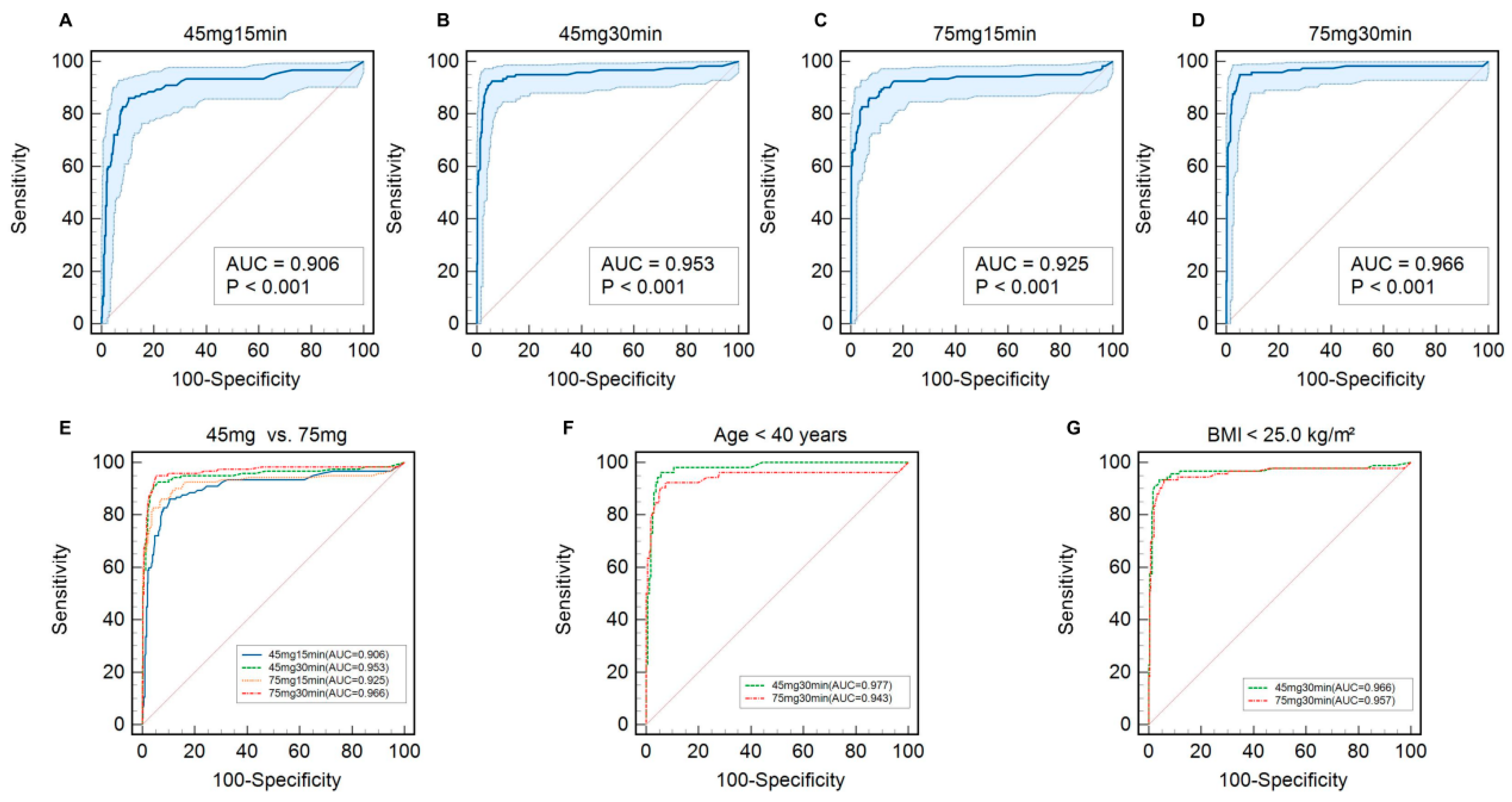
The diagnostic performance of all four test protocols is shown in Table 3 and Figure 2. At the 30 min time point, the 45 mg test demonstrated excellent diagnostic accuracy, with an area under the receiver operating characteristic curve (AUC) of 0.953 (95% CI, 0.929 to 0.971). The 75 mg, 30 min protocol yielded a numerically higher AUC of 0.966 (95% CI, 0.945 to 0.981); however, this difference was not statistically significant (p = 0.50). Specifically, the 30 min sampling time yielded a significantly greater AUC compared with the 15 min time point for both the 45 mg dose (0.953 [95% CI, 0.929 to 0.971] vs. 0.906 [95% CI, 0.874 to 0.932]; p = 0.026) and the 75 mg dose (0.966 [95% CI, 0.945 to 0.981] vs. 0.925 [95% CI, 0.895 to 0.948]; p = 0.037). To comprehensively elucidate the temporal testing dynamics, we further evaluated the categorical agreement and continuous correlation between the two time points. For the 45 mg dose, the diagnostic agreement between the 15 and 30 min samples was 89.3% (Cohen’s kappa = 0.737), with a strong positive correlation in continuous delta-over-baseline values (Pearson’s r = 0.840; p < 0.001). Parallel findings were observed for the 75 mg dose, which demonstrated 91.2% diagnostic agreement (Cohen’s kappa = 0.791) and a robust linear correlation (Pearson’s r = 0.914; p < 0.001). Despite this substantial intra-dose concordance, the 30 min sampling protocol consistently provided superior diagnostic accuracy, establishing it as the optimal sampling threshold.
In key subgroup analyses, the comparable performance of the two 30 min protocols was consistent. Among participants younger than 40 years of age (n = 212), the AUC for the 45 mg protocol was 0.977, as compared with 0.943 for the 75 mg protocol, a difference that was not significant (p = 0.24) (Figure 2F). Similarly, in participants with a body-mass index of less than 25.0 kg/m2 (n = 334), the 45 mg protocol yielded an AUC of 0.966, as compared with 0.957 for the 75 mg protocol, a difference that was also not significant (p = 0.70) (Figure 2G).
To resolve the diagnostic discrepancies, the 42 participants with discordant UBT results at 30 min underwent the definitive endoscopic reference standard. Based on rapid urease testing and histology, 15 were confirmed as H. pylori-positive and 27 as H. pylori-negative. Among these 42 cases, the 75 mg protocol correctly identified significantly more H. pylori-positive participants than the 45 mg protocol (11 of 15 [73.3%] vs. 4 of 15 [26.7%]; p = 0.001). Conversely, the 45 mg protocol correctly identified significantly more H. pylori-negative participants (16 of 27 [59.3%] vs. 11 of 27 [40.7%]; p < 0.001) (Table 4).

3.4. Safety Outcomes

No adverse events of any grade were reported by any participant during the one-week follow-up period (Table 5).

3.5. Ancillary Analyses

In pre-specified subgroup analyses, the diagnostic agreement (as measured by Cohen’s kappa) between the 45 mg and 75 mg tests at 30 min was broadly consistent across most participant subgroups, including those defined by sex, alcohol use, smoking status, and family history of gastric cancer (Table 6). The agreement was substantial in both female participants (Cohen’s κ, 0.744) and male participants (κ, 0.676).

4. Discussion

In this prospective, randomized crossover trial, we demonstrated that a low-dose 45 mg 13C-UBT has substantial agreement and comparable diagnostic accuracy to the standard 75 mg test for the diagnosis of H. pylori infection in adults. Our findings support a more nuanced, potentially stratified, approach to test selection.
Our primary analysis revealed substantial agreement between the two protocols at the 30 min time point (κ, 0.766). This is consistent with previous research that has validated the efficacy of lower 13C-urea doses [18,33,34,35]. The consistency of this agreement across both randomization sequences confirms that the crossover design did not introduce significant bias. Furthermore, our confirmation that the 30 min sampling time is superior to the 15 min time point for both doses aligns with established guidelines, which emphasize adequate incubation for accurate results. This finding justifies focusing subsequent head-to-head comparisons on this optimal timing.
The central finding of our study was the statistical non-inferiority of the 45 mg protocol compared to the 75 mg standard at 30 min (AUC, 0.953 vs. 0.966; p = 0.50). While some earlier studies and international consensus reports have favored a 75 mg dose to maximize signal strength and sensitivity, our data contributes to a growing body of evidence suggesting that lower doses can achieve excellent diagnostic performance.
While our study focused on the 45 mg vs. 75 mg comparison, the quest for an “optimal” dose requires viewing our results within the broader evidence base. Prior studies have evaluated doses as low as 15–20 mg, often necessitating the addition of citric acid test meals to delay gastric emptying and enhance urease-substrate interaction. In our study, notably, the 45 mg tablet achieved excellent diagnostic accuracy without the inclusion of citric acid, a component present in the standard 75 mg granule formulation. This suggests that 45 mg represents an operational “sweet spot”: it provides sufficient substrate to yield a stable signal, statistically comparable to the acidified high-dose standard, while avoiding the complexity and palatability issues associated with citric acid, thus offering a simplified and patient-friendly low-dose option.
Our study’s most novel contribution, however, is the stratification by age and body-mass index. The robust performance of the 45 mg protocol in younger (<40 years) and lower-BMI (<25.0 kg/m2) individuals provides crucial new context. Physiologically, younger adults typically have higher gastric acid output and less atrophic gastritis compared to older populations, providing an optimal environment for urease activity. This supports the biological plausibility of using a lower 45 mg dose in this specific demographic. The lack of a significant difference compared to the 75 mg test in these large demographic groups strongly implies that the low-dose protocol is a highly effective tool for these individuals [36,37]. This has important practical implications, offering an opportunity to reduce medication dosage, lower costs, and minimize isotope exposure without compromising diagnostic yield, making it an attractive option for large-scale screening programs in younger populations.
Our analysis of discordant cases offers a nuanced perspective on the operational characteristics of each dose. The finding that the 75 mg protocol was more accurate for identifying true positive cases is in line with the rationale for its use in post-treatment testing, where maximizing sensitivity is critical [38,39]. Conversely, the superior accuracy of the 45 mg protocol in correctly identifying true negative cases suggests a higher specificity. This is consistent with systematic reviews proposing that lowering the urea dose may reduce the risk of false-positive results, possibly by minimizing urease activity from non-H. pylori urease-producing organisms. This suggests a potential strategy for tailored test selection: the 45 mg protocol may serve as an ideal, cost-effective tool for initial diagnosis or screening, while the 75 mg protocol remains valuable for high-stakes scenarios like confirming eradication. It is worth noting that our cohort included a proportion of patients undergoing post-eradication testing (approximately 22%). While the overall agreement between the two protocols remained high, future studies stratified strictly by clinical indication are warranted to evaluate if prior treatment alters the diagnostic dynamics of the low-dose formulation.
The safety profile of both protocols was excellent, with no adverse events reported. The diagnostic agreement was also found to be broadly consistent across other subgroups defined by sex, smoking, and alcohol use, further strengthening the generalizability of our findings [40]. This study has several strengths, including its prospective, randomized crossover design which minimizes inter-individual variability. Limitations include the single-center design, which may limit external validity. Second, while we reported the clinical indications for testing, we did not perform separate diagnostic accuracy analyses stratified by initial diagnosis versus post-eradication confirmation, nor did we grade the histologic severity of gastric inflammation. Third, detailed anthropometric data such as specific body weight and waist circumference were not collected, precluding a more granular correlation between patient body habitus and UBT values. Finally, Chinese patients typically have a lower mean stature and body weight compared to Western populations, which may influence gastric volume and isotope distribution. Therefore, while the 45 mg dose is highly effective in our cohort, these findings warrant reproduction in diverse ethnic populations.
In conclusion, the 45 mg, 30 min 13C-UBT provides substantial diagnostic agreement and comparable clinical performance to the 75 mg standard. While its robust performance in younger, lower-BMI individuals shows clinical promise, our diagnostic accuracy estimates rely partly on a composite reference assumption rather than independent endoscopic verification in all participants. Therefore, further externally validated studies using independent reference standards are required before broad screening-policy claims are made.

Supplementary Materials

The following supporting information can be downloaded at https://www.mdpi.com/article/10.3390/diagnostics16101567/s1: File S1 CONSORT 2025 checklist.

Author Contributions

J.H. (Jun Huang): Conceptualization, Formal analysis, Investigation, Data curation, Writing—original draft, Visualization. Y.X.: Investigation, Resources, Writing—review & editing. J.H. (Jian Huang): Participant follow-up. B.L.: Methodology, Validation, Writing—review & editing. Y.C.: Conceptualization, Methodology, Supervision, Funding acquisition, Project administration, Writing—review & editing. All authors have read and agreed to the published version of the manuscript.

Funding

This study was supported by the Shenzhen Science and Technology Program (KCXFZ20211020163558024), Shenzhen Medical Research Fund (C2401027), Shenzhen Key Laboratory of Gastrointestinal Microbiota and Disease (ZDSYS20220606100800002), Shenzhen Science and Technology Program (LCYSSQ20220823091405012), and Research Foundation of Shenzhen Hospital of Southern Medical University (PY2022YM06).

Institutional Review Board Statement

This study was conducted in accordance with the Declaration of Helsinki and approved by the Medical Ethics Committee of Shenzhen Hospital of Southern Medical University approval code No. NYSZYYEC20230016; approval date: 7 April 2023.

Informed Consent Statement

Informed consent was obtained from all subjects involved in the study.

Data Availability Statement

The data that support the findings of this study are available from the corresponding author upon reasonable request.

Acknowledgments

We thank Chenghai Yang and Anjiang Wang for their invaluable support in patient recruitment. We also gratefully acknowledge the support of the funding agencies.

Conflicts of Interest

The authors declare no conflicts of interest.

References

  1. Ye, L.; Yan, K.; Tian, Z.; Xiao, Z.-H.; Xie, R.-Y.; Xie, Z.-Y.; Tao, L. Helicobacter Pylori Infection Is Linked to Metabolic Dysfunction and Associated Steatotic Liver Disease: A Large Cross-Sectional Study. World J. Gastroenterol. 2025, 31, 102563. [Google Scholar] [CrossRef]
  2. Hooi, J.K.Y.; Lai, W.Y.; Ng, W.K.; Suen, M.M.Y.; Underwood, F.E.; Tanyingoh, D.; Malfertheiner, P.; Graham, D.Y.; Wong, V.W.S.; Wu, J.C.Y.; et al. Global Prevalence of Helicobacter Pylori Infection: Systematic Review and Meta-Analysis. Gastroenterology 2017, 153, 420–429. [Google Scholar] [CrossRef] [PubMed]
  3. Di Rienzo, T.A.; D’Angelo, G.; Ojetti, V.; Campanale, M.C.; Tortora, A.; Cesario, V.; Zuccalà, G.; Franceschi, F. 13C-Urea Breath Test for the Diagnosis of Helicobacter Pylori Infection. Eur. Rev. Med. Pharmacol. Sci. 2013, 17, 51–58. [Google Scholar]
  4. Pathak, C.M.; Bhasin, D.K.; Khanduja, K.L. Urea Breath Test for Helicobacter Pylori Detection: Present Status. Trop. Gastroenterol. Off. J. Dig. Dis. Found. 2004, 25, 156–161. [Google Scholar]
  5. IARC Working Group on the Evaluation of Carcinogenic Risks to Humans. Schistosomes, Liver Flukes and Helicobacter pylori; International Agency for Research on Cancer: Lyon, France, 1994. [Google Scholar]
  6. Yin, Z.; Xiao, S.; Tian, X.; Yuan, Z.; Zhou, L. The Necessity and Appropriate Range of the Diagnostic “Gray Zone” of 13C-Urea Breath Test. Saudi J. Gastroenterol. Off. J. Saudi Gastroenterol. Assoc. 2022, 28, 385–392. [Google Scholar] [CrossRef]
  7. Ford, A.C.; Tsipotis, E.; Yuan, Y.; Leontiadis, G.I.; Moayyedi, P. Efficacy of Helicobacter Pylori Eradication Therapy for Functional Dyspepsia: Updated Systematic Review and Meta-Analysis. Gut 2022, 71, 1967–1975. [Google Scholar] [CrossRef]
  8. Jambi, L.K. Systematic Review and Meta-Analysis on the Sensitivity and Specificity of 13C/14C-Urea Breath Tests in the Diagnosis of Helicobacter Pylori Infection. Diagnostics 2022, 12, 2428. [Google Scholar] [CrossRef]
  9. Abd Rahim, M.A.; Johani, F.H.; Shah, S.A.; Hassan, M.R.; Abdul Manaf, M.R. (13)C-Urea Breath Test Accuracy for Helicobacter Pylori Infection in the Asian Population: A Meta-Analysis. Ann. Glob. Health 2019, 85, 110. [Google Scholar] [CrossRef]
  10. Malfertheiner, P.; Megraud, F.; O’Morain, C.A.; Gisbert, J.P.; Kuipers, E.J.; Axon, A.T.; Bazzoli, F.; Gasbarrini, A.; Atherton, J.; Graham, D.Y.; et al. Management of Helicobacter Pylori Infection-the Maastricht V/Florence Consensus Report. Gut 2017, 66, 6–30. [Google Scholar] [CrossRef]
  11. Katz, P.O.; Dunbar, K.B.; Schnoll-Sussman, F.H.; Greer, K.B.; Yadlapati, R.; Spechler, S.J. ACG Clinical Guideline for the Diagnosis and Management of Gastroesophageal Reflux Disease. Am. J. Gastroenterol. 2022, 117, 27–56. [Google Scholar] [CrossRef]
  12. Sugano, K.; Tack, J.; Kuipers, E.J.; Graham, D.Y.; El-Omar, E.M.; Miura, S.; Haruma, K.; Asaka, M.; Uemura, N.; Malfertheiner, P.; et al. Kyoto Global Consensus Report on Helicobacter Pylori Gastritis. Gut 2015, 64, 1353–1367. [Google Scholar] [CrossRef]
  13. Said, Z.N.A.; El-Nasser, A.M. Evaluation of Urea Breath Test as a Diagnostic Tool for Helicobacter Pylori Infection in Adult Dyspeptic Patients. World J. Gastroenterol. 2024, 30, 2302–2307. [Google Scholar] [CrossRef]
  14. Leal, Y.A.; Flores, L.L.; Fuentes-Pananá, E.M.; Cedillo-Rivera, R.; Torres, J. 13C-Urea Breath Test for the Diagnosis of Helicobacter Pylori Infection in Children: A Systematic Review and Meta-Analysis. Helicobacter 2011, 16, 327–337. [Google Scholar] [CrossRef]
  15. Kwon, Y.H.; Kim, N.; Lee, J.Y.; Choi, Y.J.; Yoon, K.; Hwang, J.J.; Lee, H.J.; Lee, A.; Jeong, Y.S.; Oh, S.; et al. The Diagnostic Validity of Citric Acid-Free, High Dose (13)C-Urea Breath Test after Helicobacter Pylori Eradication in Korea. Helicobacter 2015, 20, 159–168. [Google Scholar] [CrossRef]
  16. Mattar, R.; Villares, C.A.; Marostegam, P.F.F.; Chaves, C.E.; Pinto, V.B.; Carrilho, F.J. Low Dose Capsule Based 13c-Urea Breath Test Compared with the Conventional 13c-Urea Breath Test and Invasive Tests. Arq. Gastroenterol. 2014, 51, 133–138. [Google Scholar] [CrossRef]
  17. Ferwana, M.; Abdulmajeed, I.; Alhajiahmed, A.; Madani, W.; Firwana, B.; Hasan, R.; Altayar, O.; Limburg, P.J.; Murad, M.H.; Knawy, B. Accuracy of Urea Breath Test in Helicobacter Pylori Infection: Meta-Analysis. World J. Gastroenterol. 2015, 21, 1305–1314. [Google Scholar] [CrossRef]
  18. Wong, W.M.; Lam, S.K.; Lai, K.C.; Chu, K.M.; Xia, H.H.X.; Wong, K.W.; Cheung, K.L.; Lin, S.K.; Wong, B.C.Y. A Rapid-Release 50-Mg Tablet-Based 13C-Urea Breath Test for the Diagnosis of Helicobacter Pylori Infection. Aliment. Pharmacol. Ther. 2003, 17, 253–257. [Google Scholar] [CrossRef]
  19. Gatta, L.; Vakil, N.; Ricci, C.; Osborn, J.F.; Tampieri, A.; Perna, F.; Miglioli, M.; Vaira, D. A Rapid, Low-Dose, 13C-Urea Tablet for the Detection of Helicobacter Pylori Infection before and after Treatment. Aliment. Pharmacol. Ther. 2003, 17, 793–798. [Google Scholar] [CrossRef]
  20. Ohara, S.; Kato, M.; Saito, M.; Fukuda, S.; Kato, C.; Hamada, S.; Nagashima, R.; Obara, K.; Suzuki, M.; Honda, H.; et al. Comparison between a New 13C-Urea Breath Test, Using a Film-Coated Tablet, and the Conventional 13C-Urea Breath Test for the Detection of Helicobacter Pylori Infection. J. Gastroenterol. 2004, 39, 621–628. [Google Scholar] [CrossRef]
  21. World Medical Association. World Medical Association Declaration of Helsinki: Ethical Principles for Medical Research Involving Human Subjects. JAMA 2013, 310, 2191–2194. [Google Scholar] [CrossRef]
  22. Hopewell, S.; Chan, A.-W.; Collins, G.S.; Hróbjartsson, A.; Moher, D.; Schulz, K.F.; Tunn, R.; Aggarwal, R.; Berkwits, M.; Berlin, J.A.; et al. CONSORT 2025 Statement: Updated Guideline for Reporting Randomized Trials. JAMA 2025, 333, 1998–2005. [Google Scholar] [CrossRef]
  23. Malfertheiner, P.; Megraud, F.; Rokkas, T.; Gisbert, J.P.; Liou, J.-M.; Schulz, C.; Gasbarrini, A.; Hunt, R.H.; Leja, M.; O’Morain, C.; et al. Management of Helicobacter Pylori Infection: The Maastricht VI/Florence Consensus Report. Gut 2022, 71, 1724–1762. [Google Scholar] [CrossRef]
  24. Sheu, B.S.; Lee, S.C.; Yang, H.B.; Wu, H.W.; Wu, C.S.; Lin, X.Z.; Wu, J.J. Lower-Dose 13C-Urea Breath Test to Detect Helicobacter Pylori Infection-Comparison between Infrared Spectrometer and Mass Spectrometry Analysis. Aliment. Pharmacol. Ther. 2000, 14, 1359–1363. [Google Scholar] [CrossRef]
  25. Panek, P.; Rymarowicz, J.; Czeliński, M.; Pośpiech, J.; Dembiński, M.; Winiarski, M.; Major, P.; Kukla, M. Concordance of the Rapid Urease Test and the Histopathology Report in Detecting Helicobacter Pylori Infection in a Bariatric Population. A Retrospective Cohort Study. Prz. Gastroenterol. 2025, 20, 395–399. [Google Scholar] [CrossRef]
  26. Ansari, S.; Yamaoka, Y. Helicobacter Pylori Infection, Its Laboratory Diagnosis, and Antimicrobial Resistance: A Perspective of Clinical Relevance. Clin. Microbiol. Rev. 2022, 35, e0025821. [Google Scholar] [CrossRef]
  27. Wang, Y.-K.; Kuo, F.-C.; Liu, C.-J.; Wu, M.-C.; Shih, H.-Y.; Wang, S.S.W.; Wu, J.-Y.; Kuo, C.-H.; Huang, Y.-K.; Wu, D.-C. Diagnosis of Helicobacter Pylori Infection: Current Options and Developments. World J. Gastroenterol. 2015, 21, 11221–11235. [Google Scholar] [CrossRef]
  28. Wang, X.; Ji, X. Sample Size Estimation in Clinical Research: From Randomized Controlled Trials to Observational Studies. Chest 2020, 158, S12–S20. [Google Scholar] [CrossRef]
  29. DeLong, E.R.; DeLong, D.M.; Clarke-Pearson, D.L. Comparing the Areas Under Two or More Correlated Receiver Operating Characteristic Curves: A Nonparametric Approach. Biometrics 1988, 44, 837–845. [Google Scholar] [CrossRef] [PubMed]
  30. Vara-Luiz, F.; Mendes, I.; Palma, C.; Mascarenhas, P.; Teles, A.E.; Santos, I.C.; Nunes, G.; Patita, M.; Mocanu, I.; Pires, S.; et al. Clinical, Endoscopic and Histologic Differences in Gastric Mucosa between Younger and Older Adults: An Observational Study on the Aging Stomach. Med. Sci. 2025, 13, 224. [Google Scholar] [CrossRef]
  31. Wang, S.; Ding, H.; Li, L.; Zhao, R.; Chai, N. Targeted Nanomedicines for the Treatment of Helicobacter Pylori Infection. Mater. Today Bio 2025, 32, 101820. [Google Scholar] [CrossRef] [PubMed]
  32. Joo, Y.-E.; Park, H.-K.; Myung, D.-S.; Baik, G.-H.; Shin, J.-E.; Seo, G.-S.; Kim, G.H.; Kim, H.U.; Kim, H.Y.; Cho, S.-I.; et al. Prevalence and Risk Factors of Atrophic Gastritis and Intestinal Metaplasia: A Nationwide Multicenter Prospective Study in Korea. Gut Liver 2013, 7, 303–310. [Google Scholar] [CrossRef]
  33. Graham, D.Y.; Malaty, H.M.; Cole, R.A.; Martin, R.F.; Klein, P.D. Simplified 13C-Urea Breath Test for Detection of Helicobacter Pylori Infection. Am. J. Gastroenterol. 2001, 96, 1741–1745. [Google Scholar] [CrossRef]
  34. Gisbert, J.P.; Pajares, J.M. Review Article: 13C-Urea Breath Test in the Diagnosis of Helicobacter Pylori Infection—A Critical Review. Aliment. Pharmacol. Ther. 2004, 20, 1001–1017. [Google Scholar] [CrossRef]
  35. Yang, H.R.; Ko, J.S.; Seo, J.K. Does the Diagnostic Accuracy of the 13C-Urea Breath Test Vary with Age Even after the Application of Urea Hydrolysis Rate? Helicobacter 2008, 13, 239–244. [Google Scholar] [CrossRef]
  36. Li, C.; Yue, J.; Ding, Z.; Zhang, Q.; Xu, Y.; Wei, Q.; Wang, J.; Ning, X.; Zeng, H.; Cao, J. Prevalence and Predictors of Helicobacter Pylori Infection in Asymptomatic Individuals: A Hospital-Based Cross-Sectional Study in Shenzhen, China. Postgrad. Med. 2022, 134, 686–692. [Google Scholar] [CrossRef]
  37. Kopacova, M.; Koupil, I.; Seifert, B.; Fendrichova, M.S.; Spirkova, J.; Vorisek, V.; Rejchrt, S.; Douda, T.; Tacheci, I.; Bures, J. Blood Pressure and Stature in Helicobacter Pylori Positive and Negative Persons. World J. Gastroenterol. 2014, 20, 5625–5631. [Google Scholar] [CrossRef]
  38. Hu, Y.; Zhang, Z.-Y.; Wang, F.; Zhuang, K.; Xu, X.; Liu, D.-S.; Fan, H.-Z.; Yang, L.; Jiang, K.; Zhang, D.-K.; et al. Effects of Amoxicillin Dosage on Cure Rate, Gut Microbiota, and Antibiotic Resistome in Vonoprazan and Amoxicillin Dual Therapy for Helicobacter Pylori: A Multicentre, Open-Label, Non-Inferiority Randomised Controlled Trial. Lancet Microbe 2025, 6, 100975. [Google Scholar] [CrossRef]
  39. Boltin, D.; Levi, Z.; Perets, T.T.; Schmilovitz-Weiss, H.; Gingold-Belfer, R.; Dickman, R.; Dotan, I. Correlation between Quantitative 13C-Urea Breath Test and Helicobacter Pylori Treatment Success in a Population-Based Cohort. Gastroenterol. Res. Pract. 2018, 2018, 5439539. [Google Scholar] [CrossRef]
  40. Graham, D.Y.; Malaty, H.M.; Evans, D.G.; Evans, D.J.J.; Klein, P.D.; Adam, E. Epidemiology of Helicobacter Pylori in an Asymptomatic Population in the United States. Effect of Age, Race, and Socioeconomic Status. Gastroenterology 1991, 100, 1495–1501. [Google Scholar] [CrossRef]
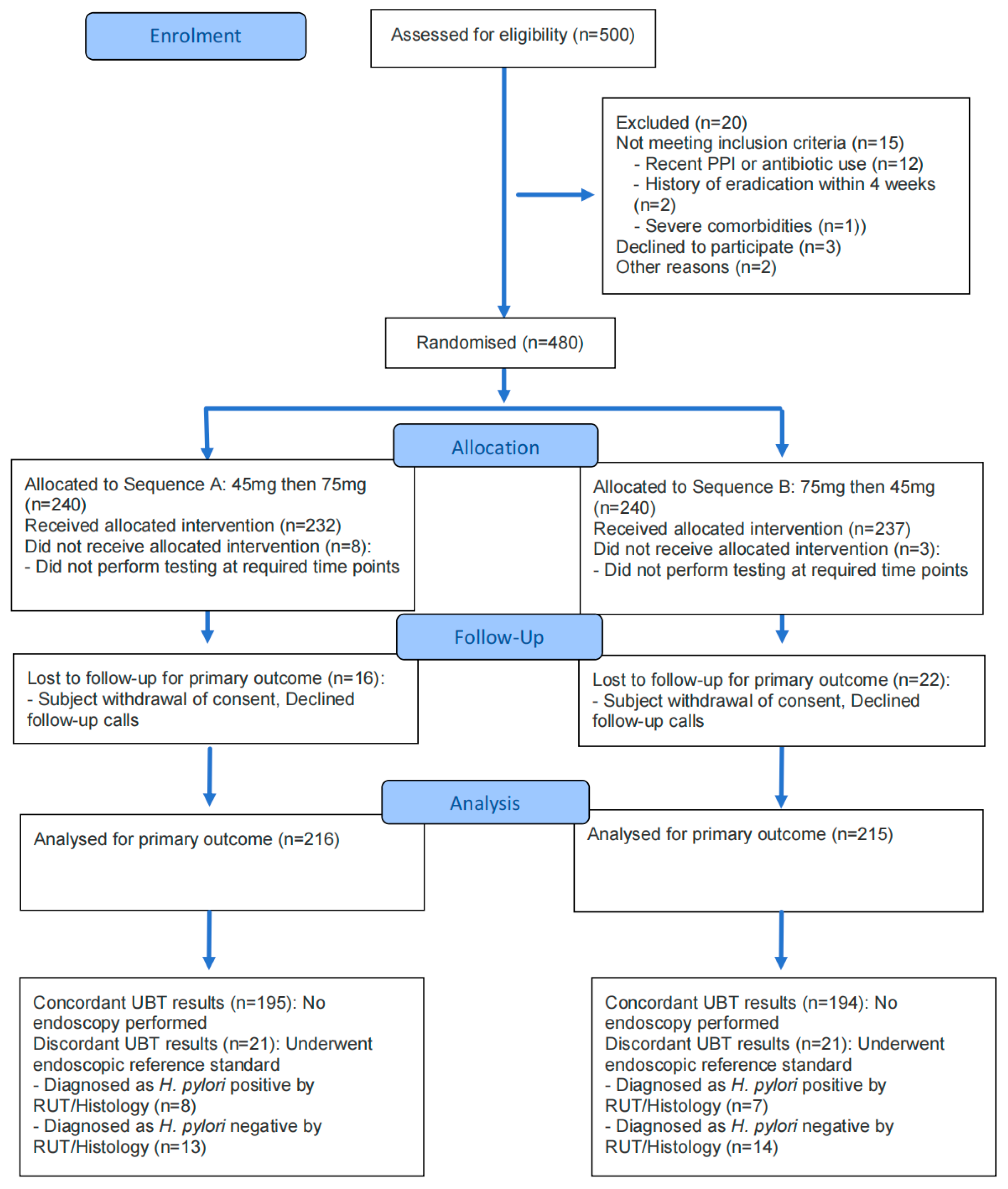
Figure 1. Enrollment, randomization, and follow-up of trial participants. Of the 500 participants assessed for eligibility, 480 underwent randomization to one of two crossover sequences. A total of 431 participants completed both study interventions and were included in the per-protocol analysis for the primary outcome. To establish the composite reference standard for secondary diagnostic accuracy analyses, participants with concordant 30 min UBT results (n = 389) were presumed to reflect their true H. pylori status without invasive testing. The 42 participants with discordant UBT results underwent the definitive endoscopic reference standard (RUT and histology) to determine their final infection status.
Diagnostics 16 01567 g001
Figure 2. Diagnostic performance of four 13C-urea breath test protocols overall and in key subgroups. Shown are the receiver operating characteristic (ROC) curves for the diagnostic performance of the four test protocols. The performance in the total per-protocol population (n = 431) is shown for the 45 mg, 15 min test (Panel A); the 45 mg, 30 min test (Panel B); the 75 mg, 15 min test (Panel C); and the 75 mg, 30 min test (Panel D). A composite overlay of these four protocols is shown in (Panel E). Head-to-head comparisons of the two 30 min protocols are shown for participants younger than 40 years of age (Panel F) and for those with a body-mass index of less than 25.0 kg/m2 (Panel G). AUC denotes the area under the curve. The shaded area around each curve represents the 95% confidence interval.
Diagnostics 16 01567 g002
Table 1. Baseline characteristics of the participants *.
CharacteristicSequence A: 45 mg then 75 mg
(n = 216)
Sequence B: 75 mg then 45 mg
(n = 215)
p-Value
Age (years)43.38 ± 15.6240.62 ± 14.770.061
Sex (%)0.224
Female134 (62.04)121 (56.28)
Male82 (37.96)94 (43.72)
BMI (kg/m2) 22.54 ± 3.2422.64 ± 3.180.734
Smoking Status (%)0.573
Current or Former16 (7.41)13 (6.05)
 Never200 (92.59)202 (93.95)
Alcohol Consumption (%)0.831
 Current or Former11 (5.09)10 (4.65)
 Never205 (94.91)205 (95.35)
Family History of Gastric Cancer (%)0.993
 Yes7 (3.24)7 (3.26)
 No209 (96.76)208 (96.74)
Indication for testing0.210
Initial diagnosis163 (75.46)173 (80.47)
Post-eradication confirmation53 (24.54)42 (19.53)
* Plus–minus values are means ± SD. Data for categorical variables are presented as number (percentage). There were no significant differences in baseline characteristics between the two randomized sequence groups. BMI: The body-mass index is the weight in kilograms divided by the square of the height in meters.
Table 2. Diagnostic agreement between 45 mg and 75 mg 13C-urea breath test protocols *.
Characteristic75 mg Test ResultOverall Agreement, % (No./Total No.)Cohen’s Kappa (95% CI) McNemar p Value §
PositiveNegative
30 min Time Point Comparison
Total Population (n = 431) 90.3 (389/431)0.766 (0.698 to 0.834)0.088
 45 mg Test, Positive10615
 45 mg Test, Negative27283
Sequence A (n = 216) 90.3 (195/216)0.776 (0.684 to 0.868)0.189
 45 mg Test, Positive587
 45 mg Test, Negative14137
Sequence B (n = 215) 90.2 (194/215)0.754 (0.652 to 0.856)0.383
 45 mg Test, Positive488
 45 mg Test, Negative13146
15 min Time Point Comparison
Total Population (n = 431) 87.0 (375/431)0.686 (0.610 to 0.762)0.504
 45 mg Test, Positive9825
 45 mg Test, Negative31277
* Data are for participants included in the per-protocol analysis. Sequence A: 45 mg test followed by 75 mg test; Sequence B: 75 mg test followed by 45 mg test. CI denotes confidence interval. Cohen’s kappa values are interpreted as follows: 0.61–0.80 as substantial and 0.81–1.00 as almost perfect agreement. § p value was calculated via the McNemar test to assess systematic disagreement between the paired tests.
Table 3. Diagnostic performance of 13C-urea breath test protocols at optimal cutoffs in the per-protocol population *.
Test Protocol and Time PointOptimal Cutoff (δ/L)Sensitivity, % (95% CI)Specificity, % (95% CI)PPV, % (95% CI)NPV, % (95% CI)Youden’s Index (J)Area Under ROC Curve (95% CI)
45 mg Dose
 15 min Sample>3.986.1 (78.6 to 91.7)89.6 (85.7 to 92.8)77.2 (69.4 to 83.8)93.9 (90.5 to 96.3)0.7570.906 (0.874 to 0.932)
 30 min Sample>3.992.6 (86.5 to 96.6)94.2 (90.9 to 96.5)86.3 (79.5 to 91.5)97.0 (94.4 to 98.6)0.8680.953 (0.929 to 0.971)
75 mg Dose
 15 min Sample>5.186.1 (78.6 to 91.7)92.9 (89.4 to 95.5)82.7 (75.2 to 88.6)94.4 (91.3 to 96.6)0.7900.925 (0.895 to 0.948) §¶
* The diagnostic performance was evaluated against the composite reference standard(concordant UBT results presumed as true status; discordant cases verified by endoscopy). Analyses were based on the per-protocol population with a definitive H. pylori status (n = 431), which included 122 positive and 309 negative cases. CI, confidence interval; NPV, negative predictive value; PPV, positive predictive value; ROC, receiver-operating characteristic. Confidence intervals were calculated with the use of the Wilson score method. The optimal cutoff value for the delta-over-baseline (DOB) value was determined by maximizing the Youden index (J).  p = 0.03 for the comparison with the 15 min sample in the 45 mg dose group. § p = 0.04 for the comparison with the 15 min sample in the 75 mg dose group.  p = 0.50 for the comparison with the 45 mg, 30 min test. All p values for AUC comparisons were calculated with the use of the DeLong method.
Table 4. Analysis of discordant results between the 45 mg and 75 mg protocols at 30 min *.
Reference Standard StatusNo. of Discordant CasesCorrect Classifications by 45 mg Protocol, No. (%)Correct Classifications by 75 mg Protocol, No. (%)p Value
Positive for H. pylori154 (26.7)11 (73.3)0.001
Negative for H. pylori2716 (59.3)11 (40.7)<0.001
* Data are for the 42 participants with discordant results between the 45 mg and 75 mg protocols at the 30 min time point. A correct classification for a positive case is a true positive; a correct classification for a negative case is a true negative.  p values were calculated with the use of Fisher’s exact test to compare the proportions of correct classifications between the two protocols within each stratum of the reference standard status.
Table 5. Adverse events in the safety population *.
Event CategorySequence A (45 mg → 75 mg) (n = 216)Sequence B (75 mg → 45 mg) (n = 215)Total (n = 432)
Any adverse event—no. of participants (%)0 (0.0)0 (0.0)0 (0.0)
Serious adverse event—no. (%)0 (0.0)0 (0.0)0 (0.0)
Adverse event leading to withdrawal—no. (%)0 (0.0)0 (0.0)0 (0.0)
* The safety population included all 432 randomized participants. Data are presented as the number of participants with an event (percentage). No adverse events of any grade were reported by any participant during the one-week follow-up period.
Table 6. Subgroup analyses of diagnostic agreement between 45 mg and 75 mg tests at 30 min *.
SubgroupCategoryNo. of ParticipantsCohen’s Kappa (95% CI)
OverallTotal Population4310.766 (0.700 to 0.832)
Age Group<40 years2120.689 (0.578 to 0.799)
≥40 years2190.832 (0.753 to 0.911)
SexFemale2550.744 (0.648 to 0.840)
Male1760.676 (0.564 to 0.788)
Body-Mass Index<25.0 kg/m23340.786 (0.713 to 0.860)
≥25.0 kg/m2970.699 (0.548 to 0.850)
Smoking StatusNever4020.785 (0.718 to 0.852)
Current or former290.494 (0.161 to 0.827)
Alcohol UseNever4100.769 (0.700 to 0.838)
Current or former210.704 (0.396 to 1.000)
Family History of Gastric CancerNo4170.760 (0.691 to 0.829)
Yes141.000
* Analyses were based on the per-protocol population, comparing the results of the 45 mg and 75 mg 13C-urea breath tests at the 30 min time point. CI denotes confidence interval. Confidence intervals were calculated using the standard error from the statistical output (CI = κ ± 1.96 × SE). The kappa coefficient is based on a very small number of participants and should be interpreted with caution.
Disclaimer/Publisher’s Note: The statements, opinions and data contained in all publications are solely those of the individual author(s) and contributor(s) and not of MDPI and/or the editor(s). MDPI and/or the editor(s) disclaim responsibility for any injury to people or property resulting from any ideas, methods, instructions or products referred to in the content.