Article

Performance Comparison of Two In-House PCR Methods for Detecting Neisseria meningitidis in Asymptomatic Carriers and Antimicrobial Resistance Profiling

by
Mekonnen Atimew
1,2,*,
Melaku Yidenekachew
1,
Marchegn Yimer
1,
Ashenafi Alemu
1,
Dawit Hailu Alemayehu
1,
Tadelo Wondimagegn
3,
Fitsumbiran Tajebe
3,
Gashaw Adane
3,
Tesfaye Gelanew
1,† and
Getachew Tesfaye Beyene
1,*,†
1
Armauer Hansen Research Institute (AHRI), Addis Ababa P.O. Box 1005, Ethiopia
2
Department of Medical Laboratory Sciences, College of Medicine & Health Sciences, Wolaita Sodo University, Wolaita Sodo P.O. Box 138, Ethiopia
3
Department of Immunology and Molecular Biology, College of Medicine & Health Sciences, University of Gondar, Gondar P.O. Box 196, Ethiopia
*
Authors to whom correspondence should be addressed.
These authors contributed equally to this work.
Diagnostics 2025, 15(5), 637; https://doi.org/10.3390/diagnostics15050637
Submission received: 16 December 2024 / Revised: 5 February 2025 / Accepted: 6 February 2025 / Published: 6 March 2025
(This article belongs to the Section Diagnostic Microbiology and Infectious Disease)

Abstract

:
Background/Objective: Bacteriological culture has been a widely used method for the detection of meningococcus, but it has low sensitivity and a long turnaround time. Molecular detection targeting capsule transport A (ctrA) gene has been used, but over 16% of meningococcal carriage isolates lack ctrA, resulting in false-negative reports. The Cu-Zn superoxide dismutase gene (sodC) is specific to N. meningitidis, and is not found in other Neisseria species, making it a useful target for improved detection of non-groupable meningococci without intact ctrA. The primary objective of this study was to evaluate the performance compassion of two in-house PCR methods, sodC gene- and ctrA gene-based PCR assays, for detecting N. meningitidis in asymptomatic carriers. The secondary objective was to assess antimicrobial resistance profiling of N. meningitidis isolates. Methods: The performance of sodC gene-based PCR assay compared to ctrA gene-based PCR for detection of N. meningitidis was evaluated using clinical samples (pharyngeal swabs; n = 137) collected from suspected asymptomatic carriers and culture-confirmed meningococci isolates (n = 49). Additionally, the antimicrobial sensitivity of the 49 isolates against antimicrobial drugs was determined using a disk diffusion test. Result: Of 49 DNA samples from culture-positive N. meningitidis isolates, the sodC gene-based PCR accurately identified all 49, whereas the ctrA gene-based PCR identified only 33 out of 49. Of 137 pharyngeal swabs, the sodC gene-based assay detected N. meningitidis DNA in 105 (76.6%), while the ctrA-based assay detected N. meningitidis DNA in 64 (46.7%). Out of the 49 N. meningitidis isolates, 43 (87.8%) were resistant to amoxicillin, 42 (83.7%) to ampicillin, 32 (65.3%) to trimethoprim–sulfamethoxazole, 22 (44.9%) to ceftazidime, 18 (36.7%) to ceftriaxone, and 7 (15.2%) to meropenem. Additionally, the majority of the isolates, 36 (73.5%), were sensitive to cefepime, 31 (63.3%) to ceftriaxone and meropenem, and 26 (53.1%) to ceftazidime. Conclusions: The findings of this study highlight the necessity of adopting non-capsular sodC-based PCR to replace ctrA in resource-constrained laboratories to improve N. meningitidis detection, and underscore the importance of periodic antimicrobial resistance surveillance to inform and adapt treatment strategies.

1. Introduction

Neisseria meningitidis, often referred to as meningococcus, is a Gram-negative bacterium that can cause a spectrum of diseases ranging from mild sepsis with rapid recovery, to fulminant meningococcemia [1]. It is carried by around 5–10% of healthy individuals in the nasopharynx [2]. Every year, more than 1.2 million cases of bacterial meningitis are estimated to occur globally, causing about 14.25% of deaths [3]. In Ethiopia, bacterial meningitis is an important cause of premature death and disability, being the 9th most common cause of years of life lost and disability-adjusted life years. Early treatment is essential in the clinical management of meningitis. A delay in therapy negatively affects the prognosis for patients with meningitis [4]. The incidence of meningitis varies depending on age, geographic location, species, genotype, strain, and serotype of the causative agents [5].
Accurate diagnosis and early treatment of meningococcal infections are essential due to their worldwide distribution, increased case fatality and morbidity rate, epidemic potential, and the serious complications that can occur [6]. For confirming the etiology, CSF and/or blood culture have been used as the gold standard for the diagnosis of meningococcal infection [7]. Identification and detection of bacterial pathogens in cases of suspected meningitis are important in guiding appropriate treatment and prophylaxis. However, diagnosis of bacterial meningitis is often difficult [8]. Traditional laboratory diagnosis of meningococcal disease (MD) has relied heavily on bacteriologic culture methods, but the bacterial growth rates, particularly in patients who have received pre-admission antibiotic treatment, are very low and the culture methods have low sensitivity due to the frequent initiation of antimicrobial therapy before clinical sample collection [9]. Many studies have shown that the high rates of morbidity and mortality linked to MD, particularly in children, are partly caused by delayed detection and diagnosis [10,11,12,13].
Molecular assays can be used to diagnose invasive meningococcal infections when previous antibiotic therapy may inhibit bacterial growth [7]. The capsule transport operon A (ctrA) gene is the most commonly targeted gene for molecular detection of N. meningitidis because ctrA is thought to be present in all invasive strains of N. meningitidis due to the importance of the capsule in preventing complement-mediated killing [14]. However, the high genetic diversity of N. meningitidis and rearrangements in the ctrA gene make the detection of this important pathogen by ctrA-targeted PCR testing difficult [15,16]. Studies show that ctrA is absent in 16% or more of carriage isolates [17,18]. ctrA-based assays have been shown to produce false-negative results due to ctrA sequence variations, which commonly result from multiple nucleotide substitutions [19,20,21]. Several cases of invasive and sometimes fatal disease caused by capsule-null (cnl) strains have been reported to lack ctrA [22,23,24].
Taken together, these findings question the utility of the ctrA-based PCR assay for the detection of N. meningitis in carriage specimens and warrant the need to develop reliable molecular approaches for the detection and characterization of this pathogen. Multiplex PCR targeting non-capsular genes such as the Cu-Zn superoxide dismutase gene (sodC), the phenol metabolism gene (metA), and the sulfite exporter (tauE) could be alternative or complementary targets to ctrA to improve the detection of N. meningitidis in carriage specimens [25]. Specifically, molecular methods targeting only the sodC gene-based PCR assay may be cost-effective diagnostic methods in resource-constrained settings, and have several advantages over culture-based biochemical assays, including increased sensitivity, specificity, speed, and efficiency. In support of its specificity to N. meningitidis, sodC is not found in other Neisseria spp. Furthermore, there are no reports of meningococci lacking the sodC gene [26].
Antimicrobial resistance in Neisseria meningitidis remains a concern and varies across studies and countries. A global meta-analysis found low levels of antimicrobial resistance (1–3.4%) to ceftriaxone, cefotaxime, ciprofloxacin, and rifampin, but not to penicillin (27.2%) [27]. However, studies in Ethiopia have shown high levels of resistance to ciprofloxacin (50–60%), cotrimoxazole (62–100%), ceftriaxone (13–69.4%), and penicillin (95.8%) among asymptomatic carriers, with multidrug resistance in 14.3–60.4% of isolates [28,29,30]. These findings highlight the need for continued surveillance and appropriate antibiotic stewardship to effectively manage N. meningitidis infections.
The primary objective of this study was to develop and validate an in-house molecular method with improved sensitivity for the detection of N. meningitidis infection. To this end, an in-house sodC-targeted PCR assay was developed and validated in clinical (pharyngeal swabs) samples and culture-positive N. meningitidis isolates. The results from this study demonstrated that our in-house sodC-targeted PCR assay exhibited enhanced sensitivity compared to the ctrA-targeted assay in detecting N. meningitidis infections in carriers, supporting its adoption as a valuable diagnostic tool in resource-constrained microbiology laboratories. The ultimate goal of introducing diagnostic tests with improved sensitivity and specificity is to provide appropriate chemoprophylaxis and treatment strategies. Consequently, we set a secondary objective to assess the antimicrobial resistance profiles of N. meningitidis isolates from asymptomatic carriers between 2010 and 2012. These were also used to validate our in-house sodC-targeted PCR assay.

2. Materials and Methods

2.1. Ethical Consideration

This study was conducted on Ethiopian archived clinical (pharyngeal swabs) and culture samples collected from the MenAfriCar study, a multi-country cross-sectional survey conducted in seven African meningitis belt countries (Chad, Ethiopia, Ghana, Mali, Niger, Nigeria, and Senegal) in 2010, 2011, and 2012 [31]. An ethical waiver for the use of these archived isolates and clinical specimens was obtained from the All African Leprosy Rehabilitation Center (ALERT)/Armauer Hansen Research Institute (AHRI) Ethics Committee with approval number: PO-63-22.

2.2. Carriage Specimens

As mentioned above, we used stored samples collected from asymptomatic Neisseria spp. carriers as part of the MenAfriCar project [31]. The study samples consisted of randomly selected pharyngeal swabs (n = 137) and culture-positive N. meningitidis isolates (n = 49). These samples were stored at −80 °C in 1 mL STGG medium (containing skim milk, tryptisoya, glucose, and glycerol) at the AHRI Microbiology Laboratory. The 49 N meningitidis isolates were used to validate the in-house sodC gene-based PCR assay, while its performance comparison was evaluated in 137 clinical samples archived in 1 mL STGG.

2.3. Characterization and Confirmation of N. meningitidis Isolates

N. meningitidis was identified by microscopic examination and biochemical tests. Briefly, cryopreserved bacterial isolates were thawed at room temperature. A loopful of these samples was then sub-cultured onto fresh chocolate (CHO) and modified Thayer Martin (MTM) agar plates, which were supplemented with vancomycin, colistin, nystatin, trimethoprim (VCNT) (OxoidTM Hampshire, UK), and IsoVitaleX enrichment (OxoidTM Hampshire, UK). Colonies were further confirmed using Gram stain and oxidase tests. After confirming the presence of Gram-negative diplococci and oxidase-positive isolates, a pure colony was sub-cultured again on a blood agar plate (BAP) with 5–10% CO2 for 24 to 48 h to ensure the purity of the culture for further biochemical testing.
A carbohydrate utilization test (glucose, maltose, lactose, and sucrose) was performed using cystine trypticase agar (CTA) to differentiate N. meningitidis from Moraxella species and other nonpathogenic Neisseria species. Isolates that were Gram-negative diplococci, oxidase-positive, glucose fermenters, maltose fermenters, and non-fermenters of lactose and sucrose were confirmed as N. meningitidis, a total of 49 isolates.

2.4. Antimicrobial Susceptibility Testing (AST)

AST was performed on 49 isolates of N. meningitidis, all of which exhibited the standard characteristics of N. meningitidis colonies using Mueller−Hinton agar (MHA) with 5% sheep blood [32]. Briefly, 3–5 colonies from a blood agar plate were suspended, and the turbidity was adjusted to match a 0.5 McFarland standard. The surface of the MHA with 5% sheep blood agar was then thoroughly coated with the bacterial suspension using a sterile swab. After allowing the plate to dry for 3–5 min, antibiotic discs were evenly placed on the inoculated plate with sterile forceps. The plate was incubated in 5% CO2 at 37 °C for 24 h. The antibiotics selected for AST profiling were based on the Ministry of Health Ethiopia guidelines, fourth edition 2021 (https://www.slideshare.net/TesfayeWorkie/stg-2021pdf#1, accessed on 10 January 2025). The tested antibiotics included ceftriaxone (30 μg), meropenem (30 µg), cefepime (30 µg), trimethoprim−sulfamethoxazole (1.25/23.75 µg), ampicillin (10 µg), amoxicillin (10 µg), and ceftazidime (30 µg). The diameters of the inhibition zones around the discs were measured to the nearest millimeter using a graduated caliper, and these measurements were compared to standard charts to determine the susceptibility, intermediate resistance, or resistance of the bacteria to the tested antibiotics, according to the USA clinical and laboratory standard institute 2022 [33].

2.5. DNA Preparation and Quantification

DNA was isolated from 137 clinical samples (pharyngeal swabs) preserved in STGG broth, as well as from the 49 isolates. Briefly, samples stored in 1 mL of STGG broth at −80 °C were thawed at room temperature. Then, 200 μL of the sample was mixed with 200 μL of 1x TE buffer; in the case of the cultured sample, 3–5 colonies were picked up from the agar plate, then vortexed vigorously, and incubated in a water bath at 95 °C for 15 min. The tubes were then transferred to −20 °C for 10 min to release bacterial DNA through heat shock, followed by a 2 min incubation at room temperature. The tubes were centrifuged at 14,000 rpm for 5 min, and the genomic DNA remained in the upper aqueous phase (supernatant). The supernatant containing the DNA was transferred to a separate 2 mL sterile tube, and the DNA concentration was measured using a NanoDrop 2000/2000c spectrophotometer (Thermo Fisher Scientific, Waltham, MA, USA). The DNA was then stored at −80 °C until used as a template for the PCR assay.

2.6. In-House Development of sodC-Based PCR Assay

We selected the sodC gene as the target for developing a PCR assay aimed at detecting N. meningitidis in clinical samples. The sodC gene was chosen for several reasons: (i) it offers high specificity for detecting N. meningitidis as it is absent in other Neisseria species; (ii) the sodC gene in N. meningitidis encodes virulence factors and a periplasmic enzyme, making it less prone to antigenic variation due to selective pressure; and (iii) there are no known strains of meningococci that lack the sodC gene. These factors collectively suggest that a PCR assay based on the sodC gene can detect all N. meningitidis strains from various geographical regions without cross-reacting with other Neisseria species [19].

2.7. Primer Design

Primers (both forward and reverse) were designed using SnapGene Viewer (Table 1). The sodC sequences of N. meningitidis were sourced from Gene Bank (accession number >NZ_CP021520.1:999157-999717). The specificity of these primers to N. meningitidis was verified using the NCBI nucleotide BLAST tool.

2.8. sodC PCR Amplification Conditions

The sodC PCR reaction was performed in a 25 μL mixture, which included 0.625 μL of each primer (10 μM), 2.5 μL of dNTPs (2.5 μM), 0.5 μL of DNA polymerase, 2.5 μL of polymerase buffer with magnesium acetate (5 μL of 10x), 15.25 μL of nuclease-free water, and 3 μL of genomic DNA as the template. Each run incorporated both positive and negative controls. The optimized amplification protocol started with an initial denaturation at 95 °C for 3 min, followed by 35 cycles of denaturation at 94 °C for 10 s, annealing at 56 °C for 30 s, extension at 72 °C for 30 s, and a final extension at 72 °C for 10 min. The amplified product was then analyzed on a 2% agarose gel using 1x Tris-acetate EDTA (TAE) buffer and visualized with Bio-Rad UV scanning (a chemi PRO UV scanner, Hercules, CA, USA). The optimal conditions for the sodC PCR reaction and the most effective primer pairs were identified. This assay was then validated using the culture technique as the gold standard and then compared with the ctrA PCR assay.

2.9. ctrA Gene-Based PCR Assay

The ctrA gene-based PCR assay, which includes the paired oligonucleotide primer: forward primer (ctrA-F): 5′-GCTGCGGTAGGTGGTTCAA-3′ and reverse primer (ctrA-R): 5′-TTGTCGCGGATTTGCAACTA-3, was initially developed by Lansac et al. in 2000 [34], and later used at AHRI [35]. Briefly, the PCR reactions comprised 0.625 μL of both forward and reverse primers (10 μM each), 2.5 μL of dNTPs (2.5 μM), 0.5 μL of DNA polymerase, 2.5 μL of polymerase buffer with magnesium acetate (5 μL of 10x), 15.25 μL of nuclease-free water, and 3 μL of genomic DNA as the template, making a final volume of 25 μL. Each run included positive and negative controls. The amplification protocol was optimized with an initial denaturation at 95 °C for 3 min, followed by 30 s at 94 °C for denaturation, 45 s at 52 °C for annealing, and 1 min at 72 °C for extension, with a final extension at 72 °C for 10 min over 35 cycles. The amplified product was analyzed on a 2% agarose gel using 1x TAE buffer and visualized with Bio-Rad UV scanning (a chemi PRO UV scanner, Hercules, CA, USA). Representative gel images of the PCR products from both the sodC and ctrA genes, obtained from control strains ATCC (serogroup A: Z2491; W: A22; X: 860060; Y: 71/94) and suspected samples, are shown in Figure 1.

2.10. Data Management and Analysis

The data obtained were entered into Epi Info version 7 and exported into STATA version 14 for data cleaning and analysis. Results are presented in frequency tables and charts. Bivariate and multivariate logistic regression models were used to analyze data, and p-values less than 0.05 were considered statistically significant.

3. Results

3.1. Optimal Conditions for the sodC Gene-Based PCR Assay

To determine the optimal conditions for sodC gene-based PCR assay, a set of primer pairs targeting the sodC gene was optimized by using the temperature gradient of the annealing temperature (Figure 1) and concentration of oligonucleotide forward and reverse primer pairs (Table 1). The sodC gene target was amplified using a selected optimized concentration of pair of primers at optimized PCR conditions: initial denaturation at 95 °C for 3 min, followed by 35 cycles; denaturation at 94 °C for 10 s; annealing at 56 °C for 30 s; extension at 72 °C for 30 s; and a final extension at 72 °C for 10 min.

3.2. Performance Comparison Between sodC Gene-Based Detection of N. meningitidis and ctrA-Based Detection

To validate our in-house-developed sodC gene-based assay for detecting N. meningitidis, we utilized culture-positive N. meningitidis isolates and compared the results with those from the ctrA-based PCR detection method. Among the 49 DNA samples from culture-positive N. meningitidis isolates used for validation, the sodC gene-based PCR accurately identified all 49 culture-confirmed isolates. In contrast, the ctrA gene-based PCR detected only 33 of these isolates. This demonstrates a 100% concordance between the culture method and the sodC gene-based PCR assay (Table 2). After validating our in-house PCR assay targeting the sodC gene with culture-positive isolates, we assessed its concordance with the PCR assay targeting the ctrA gene using DNAs obtained from 137 pharyngeal swabs. The sodC gene-based method detected N. meningitidis DNA in 76.64% of the samples, whereas the ctrA gene-based method identified it in only 46.72% (Table 2).

3.3. Antimicrobial Susceptibility Pattern of N. meningitidis Isolates

The antimicrobial susceptibility patterns of the 49 N. meningitidis isolates are summarized in Table 3. Among these isolates, resistance was found in 43 (87.8%) to amoxicillin, 42 (83.7%) to ampicillin, 32 (65.3%) to trimethoprim–sulfamethoxazole, 22 (44.9%) to ceftazidime, and 18 (36.7%) to both ceftriaxone and meropenem. Additionally, seven isolates (15.2%) showed resistance to cefepime. On the other hand, a notable number of isolates were sensitive to cefepime (36 isolates, 73.5%), ceftriaxone and meropenem (31 isolates, 63.3%), and ceftazidime (26 isolates, 53.1%).

3.4. Antimicrobial Susceptibility Patterns of N. meningitidis Isolates by Age and Sex of Asymptomatic Carriers

The antimicrobial susceptibility patterns of the 49 N. meningitidis isolates, based on the age and sex of the asymptomatic carriers from whom they were isolated, are summarized in Table 4 and Table 5. No significant differences in antimicrobial susceptibility patterns were observed based on the sex and age of carriers, although slight variations in resistance percentiles were noted across different age groups for some antibiotics. For example, isolates from those aged 24–29 showed the lowest percentile of resistance to ampicillin and amoxicillin, while isolates from those aged 10–14 showed the highest percentile of resistance to these same antibiotics (Table 5).

4. Discussion

The results of this study offer important insights into the diagnostic techniques for the detection of N. meningitidis, particularly focusing on comparing PCR-targeted sodC and ctrA genes. The findings clearly show that, compared to the ctrA gene, the sodC-based PCR was a more sensitive and accurate method of detecting N. meningitidis.
The higher detection rate of N. meningitidis using PCR targeting the sodC gene highlights the potential of this method as an effective tool for the diagnosis of meningococcal disease [19,25]. In contrast, the lower detection rates of N. meningitidis using the ctrA gene methods suggest that this method may be less sensitive and reliable for detecting bacteria in clinical samples. This has important implications for clinical diagnosis, as timely and accurate detection of N. meningitidis is essential for initiating appropriate treatment and implementing public health measures to prevent the spread of the disease. In a similar study that tested pharyngeal swabs for the presence of N. meningitidis by PCR with sodC and ctrA as target genes, 75.8% (491/647) of clinical samples tested positive for the sodC gene [36].
Furthermore, sodC-based real-time PCR identified a higher detection rate of N. meningitidis isolates; 518 of 520 (99.6%) isolates of N. meningitidis were positive for sodC, whereas ctrA detection occurred only in 368 of 520 (70.8%) isolates. Similar to our report, gene-based PCR showed higher sensitivity to the sodC gene than to the ctrA gene [19].
Contrary to our finding, in another molecular assay for the detection of meningococci in normally sterile sites using sodC and ctrA genes, sodC-based RT-PCR was found to be 7.5% less sensitive than ctrA-based RT-PCR [37].
Resistance to trimethoprim–sulphamethoxazole was high among meningococcal isolates. The widespread resistance to this antibiotic is possibly due to the early introduction of sulphonamides [38]. In contrast, an investigation in children in Greece in 2004 revealed that ceftriaxone sensitivity was present in all isolates [39]. Additionally, we observed no significant differences in antimicrobial susceptibility patterns based on the sex and age of carriers, although slight variations in resistance percentiles were noted across different age groups for some antibiotics. This observation aligns with a previous study conducted in the Meskan and Mareko Districts, Gurage Zone, Ethiopia [28].
N. meningitidis is developing a resistance to antibiotics that are recommended for the management of meningococcal meningitis for epidemic response. These findings have important implications for clinical practice and public health. Healthcare providers should be aware of the drug susceptibility profile of N. meningitidis in their region and consider these factors when selecting antimicrobial therapy for patients with suspected or confirmed meningococcal infections. Additionally, public health efforts should focus on surveillance of antimicrobial resistance in N. meningitidis and the development of new treatment strategies to combat resistant strains. Furthermore, the ability of PCR targeting the sodC gene to detect a larger proportion of N. meningitidis isolates has implications for epidemiological studies and surveillance of meningococcal carriers [19,25]. Accurate and comprehensive surveillance data are essential for understanding the epidemiology and dynamics of N. meningitidis transmission, as well as for informing public health interventions such as vaccination strategies. The higher detection rate of N. meningitidis using PCR targeting the sodC gene may also be valuable for identifying asymptomatic carriers of the bacteria, which is critical for implementing targeted control measures to prevent outbreaks.
This study was not without its limitations. First, the sample size utilized for the performance evaluations was modest, though it was adequate. Second, while sodC is specific to N. meningitidis, this study did not assess the specificity of the in-house sodC-targeted PCR assay using a panel of DNAs from non-N. meningitidis isolates, including N. sicca, N. gonorrhoeae, Streptococcus pneumoniae, and Haemophilus influenzae.
Although the present study demonstrated that SodC gene-based assay is a promising method for the detection of non-groupable N. meningitidis, especially in carriage, its use for the diagnosis of meningococcal meningitis warrants caution because of the potential for false results and public health implications [19,37]. The sodC gene-based PCR assay has been shown to be less sensitive in sterile fluids (e.g., cerebrospinal fluid) than the ctrA gene-based assay [19]. In addition, it is critical to differentiate cases of bacterial meningitis (N. meningitidis, S. pneumoniae, H. influenzae, and Streptococcus agalactiae) to ensure effective treatment. However, some bacterial species, such as H. influenzae, have homologous sodC genes [40], which can lead to false-positive results. Therefore, combining the sodC gene-based assay with ctrA gene-based detection could help improve the accurate diagnosis of meningococcal meningitis, especially in cases of suspected bacterial meningitis, thereby improving patient management.

5. Conclusions

The sodC gene-based PCR assay was found to be superior in sensitivity in detecting N. meningitidis in carriage specimens compared with ctrA gene-based PCR. The observed high prevalence of antibiotic resistance warrants the need to continue to monitor antibiotic resistance that might influence treatment and for future implementation of chemoprophylaxis for carriers and those household members who have contacts with confirmed meningitis cases.

Author Contributions

Conceptualization, T.G. and G.T.B.; methodology, M.A., G.T.B. and T.G.; validation, MA., T.G. and G.T.B.; formal analysis; M.A., T.G. and G.T.B.; investigation, M.A., M.Y. (Melaku Yidenekachew), M.Y. (Marchegn Yimer), A.A., D.H.A., T.W., F.T., G.A., T.G. and G.T.B.; resources, T.G. and G.T.B.; data curation, M.A., T.G. and G.T.B.; writing—original draft preparation, M.A., T.G. and G.T.B.; writing—review and editing, T.G. and G.T.B.; visualization, M.A. and G.T.B.; supervision, T.W., F.T., G.A., T.G. and G.T.B.; project administration, G.T.B.; funding acquisition, T.G. and G.T.B. All authors have read and agreed to the published version of the manuscript.

Funding

This work was funded by the Grand Challenge Ethiopia [AH0/0/006/0028/20]-(T. G. & G.T.B) and The European Union through the African Academy of Sciences (ARISE-PP-FA-050)-GTB.

Institutional Review Board Statement

The study was conducted in accordance with the Declaration of Helsinki, and approved by the Institutional Review Board (or Ethics Committee) of The AHRI/ALFRT Ethics Review Committee (protocol code PO-63-22 and date of approval: 9 January 2023).

Informed Consent Statement

Not applicable.

Data Availability Statement

The data supporting the findings of this study are available upon reasonable request from the corresponding author.

Acknowledgments

The authors are grateful to the University of Gondar, the College of Medicine and Health Science, the School of Biomedical and Laboratory Sciences, and the Armauer Hansen Research Institute for their guidance and willingness to provide us with the opportunity to conduct research.

Conflicts of Interest

The authors declare no conflicts of interest. The funders had no role in the design of this study; in the collection, analyses, or interpretation of data; in the writing of the manuscript; or in the decision to publish the results.

Abbreviations

AHRI, Armauer Hansen Research Institute; AST, Antimicrobial susceptibility testing; ATCC, American Type Culture Collection; CSF, Cerebrospinal fluid; ctrA, Capsule transport to cell surface gene; DNA, Deoxyribonucleic acid; MD, Meningococcal disease; MHA, Muller Hinton Agar; PCR, Polymerase chain reaction; sodC, Cu-Zn superoxide dismutase gene

References

  1. Manchanda, V.; Gupta, S.; Bhalla, P. Meningococcal Disease: History, Epidemiology, Pathogenesis, Clinical Manifestations, Diagnosis, Antimicrobial Susceptibility and Prevention. Indian J. Med. Microbiol. 2006, 24, 7–19. [Google Scholar] [CrossRef] [PubMed]
  2. Bårnes, G.K.; Kristiansen, P.A.; Beyene, D.; Workalemahu, B.; Fissiha, P.; Merdekios, B.; Bohlin, J.; Préziosi, M.-P.; Aseffa, A.; Caugant, D.A. Prevalence and Epidemiology of Meningococcal Carriage in Southern Ethiopia Prior to Implementation of MenAfriVac, a Conjugate Vaccine. BMC Infect. Dis. 2016, 16, 639. [Google Scholar] [CrossRef] [PubMed]
  3. Bianchi, L.; Napoli, Z.; Donati, S.; Lencioni, P.; Santoni, F.; Lari, R. Emergency Management in Bacterial Meningitis and Sepsis: Application of Real Time-Polymerase Chain Reaction and FilmArray Technology Performed Directly on Cerebrospinal Fluid and Blood Samples. Microbiol. Medica 2015, 30, 4–14. [Google Scholar] [CrossRef]
  4. Bårnes, G.K.; Gudina, E.K.; Berhane, M.; Abdissa, A.; Tesfaw, G.; Abebe, G.; Feruglio, S.L.; Caugant, D.A.; Jørgensen, H.J. New Molecular Tools for Meningitis Diagnostics in Ethiopia—A Necessary Step towards Improving Antimicrobial Prescription. BMC Infect. Dis. 2018, 18, 684. [Google Scholar] [CrossRef] [PubMed]
  5. Başpınar, E.Ö.; Dayan, S.; Bekçibaşı, M.; Tekin, R.; Ayaz, C.; Deveci, Ö.; Hoşoğlu, S. Comparison of Culture and PCR Methods in the Diagnosis of Bacterial Meningitis. Braz. J. Microbiol. 2017, 48, 232–236. [Google Scholar] [CrossRef] [PubMed]
  6. Nemescu, R.E.; Ursu, R.G.; Dorobăț, C.M.; Iancu, L.S. The Efficiency of sodC Gene/N. Meningitidis Detection in Comparison with the Classical Methods for the Diagnosis of Meningococcal Infection/Evaluarea Eficienţei Real Time PCR TaqMan Utilizând Gena sodC/N. Meningitidis În Comparaţie Cu Metodele Clasice Utilizate În Diagnosticul Infecţiei Meningococice. Rom. Rev. Lab. Med. 2015, 23, 21–30. [Google Scholar] [CrossRef]
  7. Qurbanalizadegan, M.; Ranjbar, R.; Ataee, R.; Hajia, M.; Goodarzi, Z.; Farshad, S.; Jafari, N.J.; Panahi, Y.; Kohanzad, H.; Rahbar, M.; et al. Specific PCR Assay for Rapid and Direct Detection of Neisseria meningitidis in Cerebrospinal Fluid Specimens. Iran. J. Public Health 2010, 39, 45–50. [Google Scholar]
  8. Seward, R.J.; Towner, K.J. Evaluation of a PCR-Immunoassay Technique for Detection of Neisseria meningitidis in Cerebrospinal Fluid and Peripheral Blood. J. Med. Microbiol. 2000, 49, 451–456. [Google Scholar] [CrossRef]
  9. Wylie, P.A.; Stevens, D.; Drake, W.; Stuart, J.; Cartwright, K. Epidemiology and Clinical Management of Meningococcal Disease in West Gloucestershire: Retrospective, Population Based Study. BMJ 1997, 315, 774–779. [Google Scholar] [CrossRef]
  10. Kuppermann, N.; Malley, R.; Inkelis, S.H.; Fleisher, G.R. Clinical and Hematologic Features Do Not Reliably Identify Children With Unsuspected Meningococcal Disease. Pediatrics 1999, 103, e20. [Google Scholar] [CrossRef]
  11. Diallo, K.; Feteh, V.F.; Ibe, L.; Antonio, M.; Caugant, D.A.; Du Plessis, M.; Deghmane, A.-E.; Feavers, I.M.; Fernandez, K.; Fox, L.M.; et al. Molecular Diagnostic Assays for the Detection of Common Bacterial Meningitis Pathogens: A Narrative Review. EBioMedicine 2021, 65, 103274. [Google Scholar] [CrossRef] [PubMed]
  12. Ferguson, L.E.; Hormann, M.D.; Parks, D.K.; Yetman, R.J. Neisseria meningitidis: Presentation, Treatment, and Prevention. J. Pediatr. Health Care 2002, 16, 119–124. [Google Scholar] [CrossRef]
  13. Wunrow, H.Y.; Bender, R.G.; Vongpradith, A.; Sirota, S.B.; Swetschinski, L.R.; Novotney, A.; Gray, A.P.; Ikuta, K.S.; Sharara, F.; Wool, E.E.; et al. Global, Regional, and National Burden of Meningitis and Its Aetiologies, 1990–2019: A Systematic Analysis for the Global Burden of Disease Study 2019. Lancet Neurol. 2023, 22, 685–711. [Google Scholar] [CrossRef] [PubMed]
  14. Nolfi-Donegan, D.; Konar, M.; Vianzon, V.; MacNeil, J.; Cooper, J.; Lurie, P.; Sedivy, J.; Wang, X.; Granoff, D.M.; McNamara, L. Fatal Nongroupable Neisseria meningitidis Disease in Vaccinated Patient Receiving Eculizumab. Emerg. Infect. Dis. 2018, 24, 1561–1564. [Google Scholar] [CrossRef]
  15. Schoen, C.; Tettelin, H.; Parkhill, J.; Frosch, M. Genome Flexibility in Neisseria meningitidis. Vaccine 2009, 27, B103–B111. [Google Scholar] [CrossRef]
  16. Unalan-Altintop, T.; Karagoz, A.; Hazirolan, G. A Diagnostic Challenge in Clinical Laboratory: Misidentification of Neisseria Subflava as Neisseria meningitidis by MALDI-TOF MS. Acta Microbiol. Et Immunol. Hung. 2020, 67, 258–260. [Google Scholar] [CrossRef] [PubMed]
  17. Claus, H.; Maiden, M.C.; Maag, R.; Frosch, M.; Vogel, U. Many carried meningococci lack the genes required for capsule synthesis and transport. Microbiology 2002, 148, 1813–1819. [Google Scholar] [CrossRef] [PubMed]
  18. Dolan-Livengood, J.M.; Miller, Y.K.; Martin, L.E.; Urwin, R.; Stephens, D.S. Genetic basis for nongroupable Neisseria meningitidis. J. Infect. Dis. 2003, 187, 1616–1628. [Google Scholar] [CrossRef] [PubMed]
  19. Dolan Thomas, J.; Hatcher, C.P.; Satterfield, D.A.; Theodore, M.J.; Bach, M.C.; Linscott, K.B.; Zhao, X.; Wang, X.; Mair, R.; Schmink, S.; et al. sodC-Based Real-Time PCR for Detection of Neisseria meningitidis. PLoS ONE 2011, 6, e19361. [Google Scholar] [CrossRef]
  20. Jaton, K.; Ninet, B.; Bille, J.; Greub, G. False-Negative PCR Result Due to Gene Polymorphism: The Example of Neisseria meningitidis. J. Clin. Microbiol. 2010, 48, 4590–4591. [Google Scholar] [CrossRef]
  21. Cavrini, F.; Liguori, G.; Andreoli, A.; Sambri, V. Multiple Nucleotide Substitutions in the Neisseria meningitidis Serogroup C ctrA Gene Cause False-Negative Detection by Real-Time PCR. J. Clin. Microbiol. 2010, 48, 3016–3018. [Google Scholar] [CrossRef] [PubMed]
  22. Ganesh, K.; Allam, M.; Wolter, N.; Bratcher, H.B.; Harrison, O.B.; Lucidarme, J.; Borrow, R.; de Gouveia, L.; Meiring, S.; Birkhead, M.; et al. Molecular Characterization of Invasive Capsule Null Neisseria meningitidis in South Africa. BMC Microbiol. 2017, 17, 40. [Google Scholar] [CrossRef] [PubMed]
  23. Johswich, K.O.; Zhou, J.; Law, D.K.S.; St Michael, F.; McCaw, S.E.; Jamieson, F.B.; Cox, A.D.; Tsang, R.S.W.; Gray-Owen, S.D. Invasive Potential of Nonencapsulated Disease Isolates of Neisseria meningitidis. Infect. Immun. 2012, 80, 2346–2353. [Google Scholar] [CrossRef] [PubMed]
  24. Hoang, L.M.N.; Thomas, E.; Tyler, S.; Pollard, A.J.; Stephens, G.; Gustafson, L.; McNabb, A.; Pocock, I.; Tsang, R.; Tan, R. Rapid and Fatal Meningococcal Disease Due to a Strain of Neisseria meningitidis Containing the Capsule Null Locus. Clin. Infect. Dis. 2005, 40, e38–e42. [Google Scholar] [CrossRef]
  25. Sirluck-Schroeder, I.; Al-Rawahi, G.N.; Gadkar, V.; Hoang, L.; Tsang, R.; Tilley, P. Limitation of ctrA as a Target for Neisseria meningitidis Identification and Potential Alternative Targets. J. Clin. Microbiol. 2022, 60, e0015222. [Google Scholar] [CrossRef] [PubMed]
  26. Joshi, R.; Saroj, S.D. Survival and Evasion of Neisseria meningitidis from Macrophages. Med. Microecol. 2023, 17, 100087. [Google Scholar] [CrossRef]
  27. Rostamian, M.; Chegene Lorestani, R.; Jafari, S.; Mansouri, R.; Rezaeian, S.; Ghadiri, K.; Akya, A. A Systematic Review and Meta-Analysis on the Antibiotic Resistance of Neisseria meningitidis in the Last 20 Years in the World. Indian J. Med. Microbiol. 2022, 40, 323–329. [Google Scholar] [CrossRef] [PubMed]
  28. Mulatu, F.; Mekonnen, Z.; Yeshitela, B.; Tilahun, H.; Yidnekachew, M.; Yimer, M.; Lema, T.; Mhiret, W.; Desta, K.; Ali, O.; et al. Antibiotic Susceptibility Patterns of Neisseria Meningitides Isolates from Asymptomatic Carriers in Gurage Zone, Southern Ethiopia. Am. J. Health Res. 2019, 7, 12–18. [Google Scholar] [CrossRef]
  29. Alemayehu, T.; Mekasha, A.; Abebe, T. Nasal Carriage Rate and Antibiotic Susceptibility Pattern of Neisseria meningitidis in Healthy Ethiopian Children and Adolescents: A Cross-Sectional Study. PLoS ONE 2017, 12, e0187207. [Google Scholar] [CrossRef] [PubMed]
  30. Tefera, Z.; Mekonnen, F.; Tiruneh, M.; Belachew, T. Carriage Rate of Neisseria meningitidis, Antibiotic Susceptibility Pattern and Associated Risk Factors among Primary School Children in Gondar Town, Northwest Ethiopia. BMC Infect. Dis. 2020, 20, 358. [Google Scholar] [CrossRef]
  31. Cooper, L.V.; Robson, A.; Trotter, C.L.; Aseffa, A.; Collard, J.-M.; Daugla, D.M.; Diallo, A.; Hodgson, A.; Jusot, J.-F.; Omotara, B.; et al. Risk Factors for Acquisition of Meningococcal Carriage in the African Meningitis Belt. Trop. Med. Int. Health 2019, 24, 392–400. [Google Scholar] [CrossRef] [PubMed]
  32. Taha, M.-K. Molecular Detection and Characterization of Neisseria meningitidis. Expert Rev. Mol. Diagn. 2002, 2, 143–150. [Google Scholar] [CrossRef]
  33. Prudhomme, C.; Joannard, B.; Lina, G.; De Launay, E.; Dumitrescu, O.; Hodille, E. Drug Susceptibility Testing of Nocardia Spp. Using the Disk Diffusion Method. Ann. Clin. Microbiol. Antimicrob. 2024, 23, 105. [Google Scholar] [CrossRef] [PubMed]
  34. Lansac, N.; Picard, F.J.; Ménard, C.; Boissinot, M.; Ouellette, M.; Roy, P.H.; Bergeron, M.G. Novel Genus-Specific PCR-Based Assays for Rapid Identification of Neisseria Species and Neisseria meningitidis. Eur. J. Clin. Microbiol. Infect. Dis. 2000, 19, 443–451. [Google Scholar] [CrossRef] [PubMed]
  35. Diallo, K.; Coulibaly, M.D.; Rebbetts, L.S.; Harrison, O.B.; Lucidarme, J.; Gamougam, K.; Tekletsion, Y.; Bugri, A.; Toure, A.; Issaka, B.; et al. Development of a PCR Algorithm to Detect and Characterize Neisseria meningitidis Carriage Isolates in the African Meningitis Belt. PLoS ONE 2018, 13, e0206453. [Google Scholar] [CrossRef] [PubMed]
  36. Morselli, S.; Gaspari, V.; Cantiani, A.; Salvo, M.; Foschi, C.; Lazzarotto, T.; Marangoni, A. Meningococcal Carriage in “Men Having Sex With Men” With Pharyngeal Gonorrhoea. Front. Cell. Infect. Microbiol. 2021, 11, 798575. [Google Scholar] [CrossRef]
  37. Higa, F.T.; Fukasawa, L.O.; Gonçalves, M.G.; Salgado, M.M.; de Lemos, A.P.S.; Harrison, L.H.; de Oliveira, P.L.; da Silva, C.N.; Sacchi, C.T. Use of sodC versus ctrA for Real-Time Polymerase Chain Reaction-Based Detection of Neisseria meningitidis in Sterile Body Fluids. Mem. Inst. Oswaldo Cruz 2013, 108, 246–247. [Google Scholar] [CrossRef]
  38. Rohani, M.Y.; Ahmad Afkhar, F.; Amir, M.A.L.; Muhd Amir, K.; Sahura, H.; Fairuz, A.; Norazah, A.; Monalisa, A.R.; Tay, A.W.; Azizah, M.; et al. Serogroups and Antibiotic Susceptibility Patterns of Neisseria meningitidis Isolated from Army Recruits in a Training Camp. Malays. J. Pathol. 2007, 29, 91–94. [Google Scholar]
  39. Pavlopoulou, I.D.; Daikos, G.L.; Alexandrou, H.; Petridou, E.; Pangalis, A.; Theodoridou, M.; Syriopoulou, V.P. Carriage of Neisseria meningitidis by Greek Children: Risk Factors and Strain Characteristics. Clin. Microbiol. Infect. 2004, 10, 137–142. [Google Scholar] [CrossRef]
  40. Tsang, R.S.W. A Narrative Review of the Molecular Epidemiology and Laboratory Surveillance of Vaccine Preventable Bacterial Meningitis Agents: Streptococcus Pneumoniae, Neisseria meningitidis, Haemophilus Influenzae and Streptococcus Agalactiae. Microorganisms 2021, 9, 449. [Google Scholar] [CrossRef] [PubMed]
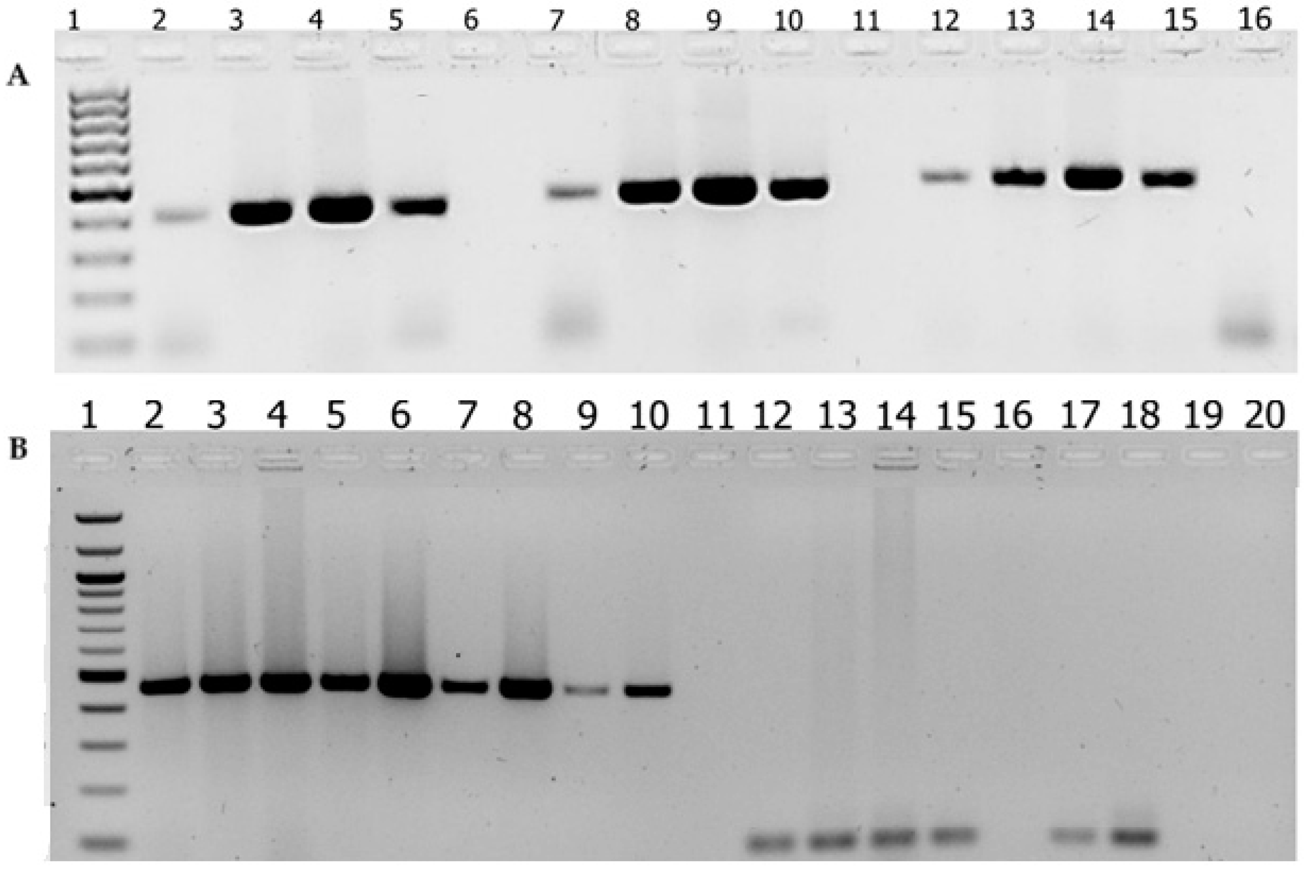
Figure 1. Agarose gel electrophoresis of PCR-amplified products using in-house sodC- and ctrA-targeted PCR assays. (A) Agarose gel electrophoresis of the PCR products obtained from optimization of PCR targeting the sodC gene. Lane 1 is 1 kb+ DNA ladder marker; Lanes 2, 7, and 12 are N. meningitidis ATCC control strain; and Lanes 3, 8, and 13, Lanes 4, 9, and 14, and Lanes 5, 10, and 15 are N. meningitidis isolates of serogroups X, Y, and W, respectively, amplified by three different versions of sodC primers. Lanes 6, 11, and 16 are negative controls. (B) Agarose gel electrophoresis of the PCR products obtained from PCR targeting the sodC gene and the ctrA gene. Lane 1, 1 kb+ DNA ladder marker; Lane 2, N. meningitidis ATCC control strain, Lanes 3–10, clinical samples amplified by primers targeting the sodC; Lane 11, negative control; and Lanes 12–20, clinical samples amplified by primers targeting the ctrA gene.
Diagnostics 15 00637 g001
Table 1. List of oligonucleotide primers designed for the N. meningitidis sodC gene target.
S. NoOligonucleotide5′-3′ Nucleotide Sequences
1.sodC Fw1-PCR ATGAATATGAAAACCTTATTAGCACTAGCGGTTAGTGCAG
2.sodC Fw14-48 CCTTATTAGCACTAGCGGTTAGTGCAGTATGTTC
3.sodC Fw14-PCR CCTTATTAGCACTAGCGGTTAG
4.sodC Fw64 GCACACGAGCATAATACGATACCTAAAGGTGCTTC
5.sodC Fw118 CAACTTGATCCAGCAAACGGTAACAAAGATGTGGG
6.sodC Fw361 GCACACTTAGGTGATTTACCTGCATTAACTG
7.sodC Rv478-PCR GGATCATAATAGAGTGACCGCGAAC
8.sodC Rv520-PCR CAAGTGGAGCTGGATGATCGGAGTG
9.sodC Rv561-PCR TTATTTAATCACGCCACATGCCATACGTGG
Table 2. Performance comparison between two in-house PCR methods for detection of Neisseria meningitidis in carriage specimens: N. meningitidis isolates (n = 49) and pharyngeal swabs (n = 137).
In-House PCR Assay TotalPositive Negative
DNA from culture-confirmed isolates
sodC49490
ctrA493316
DNA from clinical samples (pharyngeal swabs)
sodC13710532
ctrA1376473
Table 3. Drug susceptibility profile of 49 N. meningitidis isolates.
Antimicrobials with Disk ContentSusceptibility ProfileNo (49)%
Ceftriaxone/CRO (30 µg)I a--
R b1836.7
S c3163.3
Ampicillin (10 µg)I--
R4283.7
S816.3
Amoxicillin (10 µg)I--
R4387.8
S612.2
Trimethoprim–sulfamethoxazole1/SXT
(1.25/23.75 µg)
I612.2
R3265.3
S1122.5
Cefepime (30 µg)I311.3
R715.2
S3673.5
Meropenem (30 µg)I--
R1836.7
S3163.3
Ceftazidime (30 µg)I12
R2244.9
S2653.1
a I = intermediate. b R = resistance. c S = sensitivity.
Table 4. Bivariable logistic regression analysis for association between sex and antibiotic susceptibility pattern of 49 N. meningitidis isolates.
AntibioticsSexOR (95% CI)p-Value
Male
(n = 26)
Female
(n = 23)
Ceftriaxone S18131.73 (0.54, 5.58)0.36
R810
AmpicillinS531.58 (0.33, 7.53)0.56
R2120
AmoxicillinS330.87 (0.15, 4.80)0.87
R2320
Trimethoprim–sulfamethoxazole/SXTS651.08 (0.28, 4.15)0.91
R2018
CefepimeS20190.70 (0.17, 2.88)0.62
R64
MeropenemS18131.73 (0.54, 5.59)0.36
R810
CeftazidimeS15111.49 (0.48, 4.60)0.49
R1112
Table 5. Bivariable logistic regression analysis for the association between age and antibiotics susceptibility pattern of 49 N. meningitidis isolates.
AntibioticsAgeOR (95% CI)p-Value
≤1011–20≥21
Ceftriaxone S516101.00
2.0 (0.54, 5.59)
1.28 (0.54, 5.59)

0.44
0.75
R4104
AmpicillinS2421.00
0.64 (0.09, 4.24)
0.58 (0.06, 5.11)

0.64
0.63
R72212
AmoxicillinS1321.00
1.04 (0.09, 11.52)
1.33 (0.10, 17.27)

0.97
0.82
R82312
Trimethoprim–sulfamethoxazole/SXTS2631.00
0.95 (0.12, 7.23)
1.05 (0.17, 6.46)

0.96
0.95
R72011
CefepimeS819121.00
0.75 (0.06, 9.72)
0.34 (0.03, 3.23)

0.82
0.34
R172
MeropenemS516101.00
2.0 (0.34, 11.54)
1.28 (0.27, 5.93)

0.44
0.75
R4104
CeftazidimeS31491.00
3.6 (0.62, 21.03)
2.3 (0.47, 11.34)

0.15
0.23
R6125
Disclaimer/Publisher’s Note: The statements, opinions and data contained in all publications are solely those of the individual author(s) and contributor(s) and not of MDPI and/or the editor(s). MDPI and/or the editor(s) disclaim responsibility for any injury to people or property resulting from any ideas, methods, instructions or products referred to in the content.