Article

Interplay Between Type 2 Diabetes Susceptibility and Prostate Cancer Progression: Functional Insights into C2CD4A

1
Department of Life Sciences and Institute of Genome Sciences, National Yang Ming Chiao Tung University, Taipei 112, Taiwan
2
Department of Anatomy, School of Medicine, China Medical University, Taichung 406, Taiwan
3
Department of Medicine, Mackay Medical College, New Taipei City 252, Taiwan
4
Department of Urology, National Taiwan University Hospital, College of Medicine, National Taiwan University, Taipei 100, Taiwan
5
Division of Urology, Department of Surgery, Kaohsiung Veterans General Hospital, Kaohsiung 813, Taiwan
6
Department of Urology, School of Medicine, National Yang-Ming University, Taipei 112, Taiwan
7
Department of Pharmacy, College of Pharmacy and Health Care, Tajen University, Pingtung 907, Taiwan
8
Department of Urology, E-Da Hospital, Kaohsiung 824, Taiwan
9
School of Medicine for International Students, I-Shou University, Kaohsiung 840, Taiwan
10
Department of Pharmacy, China Medical University, Taichung 404, Taiwan
11
Graduate Institute of Clinical Medicine, College of Medicine, Kaohsiung Medical University, Kaohsiung 807, Taiwan
12
Department of Urology, Kaohsiung Medical University Hospital, Kaohsiung 807, Taiwan
13
Institute of Medical Science and Technology, College of Medicine, National Sun Yat-Sen University, Kaohsiung 804, Taiwan
*
Authors to whom correspondence should be addressed.
These authors contributed equally to this work.
Diagnostics 2025, 15(21), 2767; https://doi.org/10.3390/diagnostics15212767
Submission received: 20 August 2025 / Revised: 28 October 2025 / Accepted: 29 October 2025 / Published: 31 October 2025

Abstract

Background/Objective: Biochemical recurrence (BCR) after radical prostatectomy (RP) for prostate cancer indicates disease progression. Although type 2 diabetes mellitus (T2D) shows a paradoxical association with prostate cancer risk, the prognostic role of T2D-related genetic variants remains unclear. Methods: We analyzed 113 common T2D susceptibility-related single-nucleotide polymorphisms (SNPs) in 644 Taiwanese men with localized prostate cancer (D’Amico risk classification: 12% low, 34% intermediate, and 54% high) treated with RP. Associations between SNPs and BCR were assessed using Cox regression, adjusting for key clinicopathological factors. Functional annotation was performed using HaploReg and FIVEx, while The Cancer Genome Atlas transcriptomic data were analyzed for C2 calcium-dependent domain-containing 4A (C2CD4A) expression. Gene set enrichment analysis (GSEA) and gene set variation analysis (GSVA) were applied to explore related biological pathways. Results: C2CD4A SNP rs4502156 was independently associated with a reduced risk of BCR (hazard ratio = 0.80, p = 0.035). The protective C allele correlated with higher C2CD4A expression. Low C2CD4A expression is associated with advanced pathological stages, higher Gleason scores, and disease progression. GSEA revealed negative enrichment of mitotic and chromatid segregation pathways in high-C2CD4A-expressing tumors, with E2F targets being the most suppressed. GSVA confirmed an inverse correlation between C2CD4A expression and E2F pathway activity, with CDKN2C as a co-expressed functional gene. Conclusions: The T2D-related variant rs4502156 in C2CD4A independently predicts a lower risk of BCR, potentially via suppression of the E2F pathway, and may serve as a germline biomarker for postoperative risk stratification.

1. Introduction

Prostate cancer is one of the most commonly diagnosed malignancies among men worldwide [1]. Although radical prostatectomy (RP) and radiotherapy achieve excellent oncological control of localized disease, biochemical recurrence (BCR) remains a major clinical challenge [2]. Current risk stratification models based on prostate-specific antigen (PSA) level, Gleason grade, pathological stage, and validated genomic classifiers such as Decipher or Oncotype DX [3,4] have improved prognostic accuracy but still misclassify a subset of patients, resulting in both overtreatment of indolent tumors and undertreatment of aggressive disease. Therefore, the identification of additional germline or molecular biomarkers that can refine postoperative risk assessment is of considerable clinical importance [5].
Epidemiological evidence suggests that type 2 diabetes mellitus (T2D) has a complex and paradoxical relationship with prostate cancer. Multiple large-scale studies and meta-analyses have indicated that T2D is associated with a modest reduction in the incidence of prostate cancer, particularly early-stage disease [6,7]. However, patients with T2D who develop prostate cancer often present with more aggressive tumors and experience poorer outcomes, including a higher risk of cancer progression and related death [8,9]. These observations may be explained by the metabolic and hormonal alterations characteristic of T2D, such as insulin resistance, hyperinsulinemia, chronic inflammation, and reduced testosterone levels, which can influence tumor development and progression in different ways [10]. Recent studies have elucidated several key molecular pathways linking T2D to prostate cancer progression. Hyperinsulinemia in T2D increases circulating insulin-like growth factor (IGF)-1 levels, activating downstream signaling cascades that enhance prostate cancer cell proliferation, survival, and metastasis via augmented glucose uptake and suppression of apoptosis [11,12]. Chronic inflammation, driven by T2D-associated hyperglycemia and insulin resistance, induces pro-inflammatory cytokines such as tumor necrosis factor (TNF)-α and interleukin (IL)-6, activates nuclear factor (NF)-κB, and generates reactive oxygen species. These processes collectively promote a tumor microenvironment conducive to invasiveness, epithelial–mesenchymal transition, and therapy resistance [13,14]. Moreover, phosphatidylinositol 3-kinase/protein kinase B/mammalian target of rapamycin signaling, often hyperactivated by insulin and IGF-1 in the diabetic state, contributes to the metabolic reprogramming of prostate cancer cells, inhibition of autophagy, and progression to castration-resistant disease by coordinating nutrient sensing with unchecked growth and anti-apoptotic responses [15,16]. Genome-wide association studies (GWASs) have identified hundreds of common variants that contribute to T2D susceptibility, highlighting the highly polygenic nature of this metabolic condition [17]. The genotypic risk scores of multiple single-nucleotide polymorphisms (SNPs), which aggregate the effects of these loci, have proven useful in predicting T2D risk but have shown minimal direct association with prostate cancer incidence [18,19]. Currently, there is limited evidence regarding the impact of T2D-related germline variants on cancer progression. Understanding whether such variants modulate disease recurrence could provide novel insights into the metabolic–genetic interplay between these two conditions and reveal new biomarkers for precise risk stratification.
This study investigated the association between established T2D susceptibility loci and BCR in men with localized prostate cancer treated with RP. By integrating genetic and clinical data, we aimed to identify metabolism-related genetic markers that may enhance postoperative prognostic assessments and guide individualized management strategies.

2. Patients and Methods

2.1. Patient and Response Assessment

In total, 644 men with histologically confirmed prostate cancer who underwent RP at National Taiwan University Hospital, Kaohsiung Medical University Hospital, and Kaohsiung Veterans General Hospital were enrolled in this study [20,21]. Institutional Review Board approval was obtained from Kaohsiung Medical University Hospital (KMU-HIRB-2013132), and all participants provided written informed consent. This study complied with the Declaration of Helsinki and the Good Clinical Practice guidelines. All participants were unrelated Han Taiwanese men diagnosed by systematic prostate biopsy, prompted by elevated PSA levels (>4 ng/mL) or abnormal findings on digital rectal examination during evaluation for lower urinary tract symptoms. Disease stage was defined according to the American Joint Committee on Cancer TNM classification based on pathological examination, pelvic computed tomography or magnetic resonance imaging, and radionuclide bone scans—only patients with clinically localized or locally advanced prostate cancer who underwent RP as primary treatment were included. Exclusion criteria included the receipt of neoadjuvant or adjuvant androgen deprivation therapy or radiotherapy before PSA recurrence, incomplete clinicopathological data, or insufficient postoperative follow-up (less than two months). After applying these criteria, 644 patients were included in the final analyses. Clinical and pathological data, including age, PSA level at diagnosis, pathological stage, Gleason score, surgical margin status, and lymph node metastasis, were retrieved from medical records and pathology reports. BCR was defined as two consecutive PSA measurements of 0.2 ng/mL or more after RP [22,23]. BCR-free survival was calculated from the date of surgery until BCR or the last follow-up. During the median follow-up period of 51 months, 229 (35.6%) patients achieved BCR (Table 1). Elevated PSA levels at diagnosis, advanced pathological stage, higher Gleason scores, higher International Society of Urological Pathology grades, high D’Amico risk classification, and positive surgical margins were significantly associated with BCR (p < 0.05) [24].

2.2. SNP Selection and Genotyping

In total, 139 common T2D-associated SNPs previously identified in a large-scale GWAS meta-analysis (62,892 patients with T2D and 596,424 healthy controls) were initially selected [25]. Genomic DNA was extracted from peripheral blood samples using the QIAamp DNA Blood Kit (Qiagen, Taipei City 100, Taiwan) and genotyped at the National Center for Genome Medicine using the Affymetrix Axiom platform (Thermo Fisher Scientific, Taichung 401, Taiwan) [26]. Variants with a minor allele frequency (MAF) of less than 0.03 and call rate of less than 0.95 or deviating from Hardy–Weinberg equilibrium (p < 1 × 10⁻⁴) were excluded, leaving 113 SNPs for final analysis.

2.3. Bioinformatic Analyses

To assess the regulatory potential of rs4502156, an SNP in C2 calcium-dependent domain-containing 4A (C2CD4A), HaploReg v4.2 was used to evaluate its effects on chromatin states, transcription factor-binding motifs, and evolutionary conservation [27]. Expression quantitative trait locus (eQTL) associations between the rs4502156 genotype and C2CD4A expression were examined using the FIVEx database and linear regression [28]. To determine the clinical relevance of C2CD4A and cyclin-dependent kinase 4 inhibitor C (CDKN2C), their expression profiles were analyzed using The Cancer Genome Atlas prostate adenocarcinoma (TCGA-PRAD) dataset (497 primary tumors and 52 normal tissues, with no overlap with our cohort) via the Genomic Data Commons Data Portal.

2.4. Differential Gene Expression and Gene Enrichment Analyses

To uncover the molecular mechanisms linked to C2CD4A, Gene Ontology (GO) and Hallmark pathway assessments were performed using gene set enrichment analysis (GSEA) [29]. Patients in the TCGA-PRAD cohort were first categorized into high- and low-expression groups based on the median C2CD4A expression. Differential gene expression between groups was computed using the R limma package (v3.64.1). The resulting log-fold change rankings were analyzed via GSEA for GO biological processes and Hallmark sets using clusterProfiler (v4.16.0) in R. The five leading pathways were visualized using enrichplot (v1.28.2).

2.5. Gene Set Variation Analysis

Gene set variation analysis (GSVA) converts gene expression profiles across samples into pathway enrichment metrics, enabling the detection of subtle variations in extensive gene collections [30]. The GSVA package (v2.2.0) in R was used to derive the E2F target scores for each TCGA-PRAD specimen.

2.6. Statistical Analyses

All statistical analyses were performed using R (v4.5.1) and Statistical Product and Service Solutions (v19.0.0; IBM), with a two-tailed p-value of < 0.05 indicating statistical significance. Survival differences were assessed using Kaplan–Meier curves and log-rank tests. Cox proportional hazards models (univariate and multivariate) were used to evaluate the links between clinical features and outcomes, yielding hazard ratios (HRs) and 95% confidence intervals (CIs). Relationships between C2CD4A and CDKN2C expression levels, E2F GSVA scores, and cancer attributes were determined using Spearman and Pearson correlation tests.

3. Results

To investigate the relationship between T2D-related genetic variants and prostate cancer progression, we evaluated the association of 113 established T2D susceptibility-related SNPs with BCR following RP in patients with prostate cancer. Two SNPs, rs11098676 in nudix hydrolase 6 (NUDT6) and rs4502156 in C2CD4A, were associated with BCR (p < 0.05; Table 2). In the multivariate Cox regression analysis adjusted for age, PSA level at diagnosis, pathological stage, Gleason score, surgical margin status, and lymph node metastasis, rs4502156 remained independently significant (Figure 1). Moreover, each additional minor C allele of rs4502156 conferred a 20% reduction in BCR risk (HR = 0.80; 95% CI = 0.65–0.98; p = 0.035).
HaploReg analysis of rs4502156 annotated the variant and its linkage disequilibrium proxies as eQTLs that could potentially disrupt several transcription factor-binding motifs (Figure 2A). The FIVEx database analysis corroborated this finding, indicating that the protective C allele was strongly associated with increased C2CD4A expression in multiple immune cell types, particularly monocytes and macrophages (p < 5 × 10⁻⁸; Figure 2B). However, this association was not statistically significant in prostate tissue (p = 0.245).
To determine the clinical significance of C2CD4A expression in prostate cancer, we analyzed transcriptomic data from 497 tumors and 52 normal tissues in TCGA-PRAD. C2CD4A mRNA levels did not differ significantly between tumorous and normal tissues (Figure 3). Nevertheless, lower C2CD4A expression was significantly associated with a more advanced pathological stage, a higher Gleason score, and disease progression (p ≤ 0.046). C2CD4A expression showed modest discriminative ability in predicting disease progression, with an area under the receiver operating characteristic curve (AUC) of 0.57. This association was attenuated after adjustment for age, PSA level, pathological stage, and Gleason score (HR = 0.95; 95% CI = 0.85–1.06; p = 0.357).
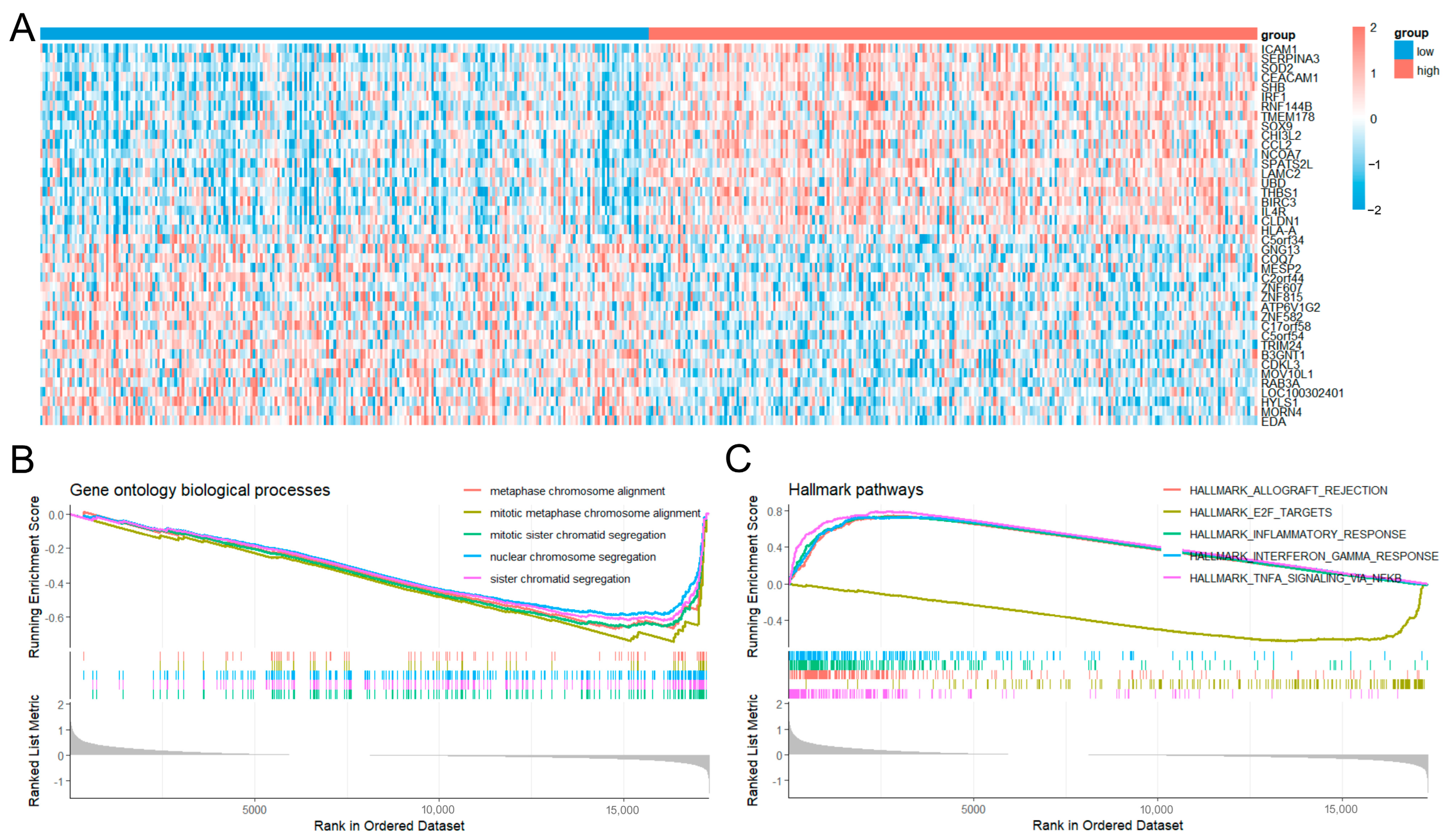
We further explored the biological role of C2CD4A by stratifying patients from TCGA-PRAD into high- and low-expression groups based on median expression levels (Figure 4A). GSEA of the ranked differential expression profiles revealed significant negative enrichment of genes involved in mitotic sister chromatid segregation, sister chromatid segregation, and nuclear chromosome segregation in the high expression group (Figure 4B). Consistent with this, Hallmark pathway analysis identified E2F targets as the most significantly downregulated pathway in the high-C2CD4A-expresion group (normalized enrichment score = −2.58, false discovery rate = 3.33 × 10⁻¹⁰; Figure 4C).
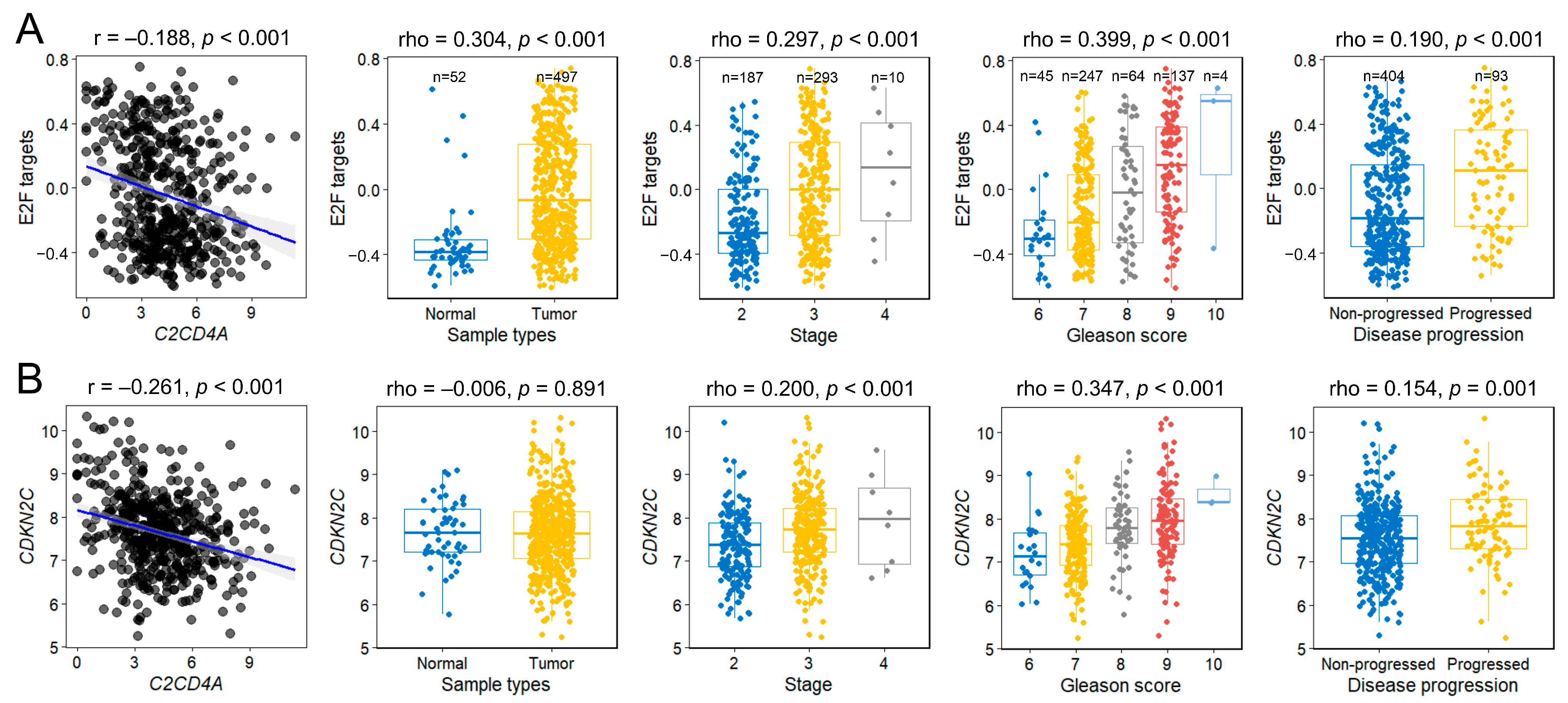
Given this association, we quantified E2F pathway activity in each sample using GSVA. C2CD4A expression levels were inversely correlated with E2F GSVA scores (Figure 5A). E2F pathway activity was elevated in tumor tissues compared with normal tissues and was much higher in tumors at the advanced stage, with a high Gleason score and progressive disease (p < 0.001). The E2F GSVA scores showed discriminative ability in predicting disease progression, with an AUC of 0.64, and remained significant after adjustment for age, PSA, pathological stage, and Gleason score (HR = 3.01; 95% CI = 1.53–5.90; p = 0.01). To further elucidate this relationship, we examined the clinical relevance of CDKN2C (the most significantly differentially expressed E2F target gene between the high- and low-C2CD4A-expression groups). C2CD4A expression was inversely correlated with CDKN2C levels (Figure 5B). Although CDKN2C levels did not differ significantly between normal and tumorous tissues, its expression was higher in more aggressive tumors (p ≤ 0.001). The CDKN2C expression level also showed discriminative ability in predicting disease progression, with an AUC of 0.61, but the association was no longer significant after multivariable adjustment (HR = 1.20; 95% CI = 0.95–1.51; p = 0.134).

4. Discussion

In this multicenter cohort of Taiwanese men with localized prostate cancer, we identified a significant association between rs4502156 (an SNP in the T2D-related C2CD4A gene) and a reduced risk of BCR following RP, independent of established clinicopathological factors. The protective C allele, which is linked to increased C2CD4A expression, is associated with more favorable tumor characteristics, including a lower pathological stage and Gleason score. In contrast, a T2D polygenic risk score constructed from 113 SNPs showed no significant association with BCR (HR = 0.94, 95% CI = 0.68–1.29, p = 0.681), suggesting that the aggregate genetic risk for T2D does not broadly influence prostate cancer recurrence. In contrast, specific loci such as rs4502156 may exert context-dependent effects. Bioinformatics analyses further suggested that elevated C2CD4A expression attenuates E2F target gene activity, which is critical for cell cycle regulation, potentially restraining tumor progression. These findings highlight a novel genetic–metabolic link in prostate cancer prognosis, expanding prior evidence of the paradoxical role of T2D in cancer incidence and aggressiveness. Although validation in an independent cohort would be ideal, these results should be interpreted as exploratory and promising, providing a preliminary framework for future mechanistic and clinical investigations.
The SNP rs4502156, which resides in the 3′ region of C2CD4A, was annotated as an eQTL and predicted to disrupt several transcription factor-binding motifs, including heterochromatin protein 1 (HP1), nuclear factor erythroid 2–related factor 2 (NRF2), transcription factor 11 (TCF11, also known as NRF1), and MAF bZIP transcription factor G (MafG). These disruptions may alter C2CD4A expression and its tumor-suppressive function in prostate cancer, which is potentially amplified by diabetes-related mechanisms. Specifically, disruption of the HP1-binding motif can impair heterochromatin maintenance, thereby modulating the epigenetic regulation of C2CD4A, as HP1 downregulation has been linked to genome instability in diabetes and cancer progression [31,32]. Similarly, alterations in the NRF2 motif may weaken antioxidant and detoxification responses, exacerbating oxidative stress—a hallmark of T2D—where NRF2 dysregulation promotes cancer survival via metabolic reprogramming [33,34]. Additionally, interference with the TCF11 and MafG motifs could disrupt heterodimeric regulation of proteasomal and stress-response genes, contributing to proteotoxic stress in T2D, as TCF11 plays a tumor-suppressive role in proteostasis and MafG, via its lncRNA axis, influences glucose metabolism [35,36]. eQTL analysis showed that the protective C allele was associated with increased C2CD4A expression across multiple immune cell subsets. In contrast, only a non-significant trend was observed in prostate tissue, likely due to the limited sample size. C2CD4A encodes a nuclear C2 domain-containing protein that is best known for its role in metabolic regulation, particularly within pancreatic β-cells where it functions as a transcriptional cofactor that enhances glycolytic gene expression while suppressing “disallowed” genes, with C2cd4a-knockout mice showing impaired glucose tolerance [37]. In cancer, the role of C2CD4A appears to be context-dependent. Elevated C2CD4A transcript levels have been documented in colon cancer, where higher gene expression correlates with an advanced disease stage [38,39]. Moreover, functional studies have shown that C2CD4A promotes tumor growth by binding to p53 and enhancing its ubiquitination and degradation, thereby suppressing apoptosis [40]. Conversely, in bladder cancer, C2CD4A upregulation has been linked to treatment with the anticancer compound isorhapontigenin, which induces cell cycle arrest and activates interferon signaling [41]. Although germline variants of C2CD4A have been well established to be associated with T2D susceptibility, their contribution to cancer risk remains poorly defined; only one case–control study reported a protective association between C2CD4A/B region variants and lung cancer risk [42].
GSEA of C2CD4A-associated expression networks identified a negative correlation between C2CD4A expression and that of E2F target genes, including CDKN2C. E2F transcription factors modulate the expression of genes critical for proliferation, DNA replication, and apoptosis, and their dysregulation is a hallmark of oncogenesis across tumor types [43]. CDKN2C (p18INK4C), a cyclin-dependent kinase (CDK) inhibitor belonging to the INK4 family, suppresses CDK4/6 activity and induces G1 arrest [44]. CDKN2C is frequently functionally inactivated in multiple malignancies; for instance, pan-cancer analyses have described decreased expression or deletion in diverse tumors [45]. Paradoxically, CDKN2C is overexpressed in certain cancer types, such as small-cell lung cancer, where elevated levels are associated with poor survival [45]. In prostate cancer, E2F-related gene signatures encompassing CDKN2C can be used to stratify recurrence risk, with higher signature expression predicting worse recurrence-free survival, which is consistent with net pathway activation despite the nominal inhibitory role of CDKN2C [46].
Furthermore, inflammation- and TNF-α–related pathways were also enriched in our C2CD4A GSEA. These pathways are known to converge on the cell cycle machinery and modulate E2F transcriptional activity via CDKs. TNF-α activates NF-κB, which transcriptionally induces cyclin D and CDK6; these cyclin D/CDK4/6 complexes phosphorylate the retinoblastoma (RB) protein, releasing E2F and promoting G1–S phase transition. Elevated E2F activity in tumors frequently arises from CDK overexpression and RB inactivation [47]. E2F factors also reciprocally modulate inflammatory signaling: in response to TNF-α, E2F1 physically interacts with the NF-κB subunit RelA, competing with NF-κB inhibitor-α for RelA binding, thereby influencing NF-κB nuclear translocation and fine-tuning the amplitude and timing of inflammatory gene expression. NF-κB targets include cyclin D and the CDK inhibitor p21, highlighting the intricate feedback between inflammation and cell-cycle control [48]. Beyond their canonical cell-cycle roles, CDK4/6 kinases exert immune-modulatory functions. In CD8⁺ T cells, CDK6 deficiency does not impair TNF-α or interferon-γ production but enhances IL-2 secretion, while CDK4/6 inhibition augments antigen presentation and cytokine production, demonstrating the dual role of these kinases in both proliferation and immune regulation [49]. Collectively, these observations underscore the complexity of the C2CD4A–E2F axis and warrant further investigation into its mechanistic role in prostate cancer progression and its potential as a prognostic or therapeutic target.
This study has several strengths. To our knowledge, this is the first study to evaluate T2D-associated genetic variants in relation to BCR in prostate cancer, with comprehensive clinicopathological adjustment. Second, a combination of genomic, transcriptomic, and pathway analyses allowed us to establish statistical associations and mechanistic insights, thereby providing a biologically coherent link between C2CD4A variants and disease progression. Third, the use of a well-defined homogenous Han Taiwanese cohort minimized the potential confounding effects of population stratification. Nevertheless, this study had several significant limitations that should be considered when interpreting the findings. First, the false discovery rate (FDR) for the identified SNP was relatively high (p = 0.011; Benjamini–Hochberg FDR = 0.760), suggesting a potential risk of type I error. Therefore, the association between rs4502156 and BCR should be viewed as exploratory until confirmed in independent cohorts. Second, the retrospective design may have introduced selection bias, and the modest sample size (n = 644) may have limited the power to detect small effect sizes or rare-variant associations. Power estimation suggested adequate detection for HRs ≤ 0.75 or ≥ 1.33 at α = 0.05, but the study was underpowered for weaker genetic effects. Third, the study population consisted exclusively of Taiwanese men of Han ethnicity, limiting the generalizability of the findings to other ethnic groups. Differences in allele frequencies and linkage disequilibrium patterns across populations may influence the strength or direction of genetic associations. Thus, external validation in multi-ethnic cohorts is required to confirm the clinical relevance of these findings. Fourth, the functional effects of rs4502156 were inferred primarily from bioinformatic annotations and expression correlation analyses rather than from direct laboratory validation. Although in silico analyses provided supportive evidence, the absence of tumor sequencing data in our cohort precluded direct genotype-expression integration, and TCGA data may not fully reflect post-radical prostatectomy disease biology. Finally, the follow-up duration, although sufficient for BCR assessment, did not allow the evaluation of long-term outcomes, such as cancer-specific survival. Consequently, the prognostic implications of rs4502156, beyond early recurrence, remain unclear. Collectively, these limitations underscore the need for larger, prospectively designed, multicenter studies that incorporate experimental validation and extended clinical follow-up to substantiate the current findings and clarify their translational potential.

5. Conclusions

In conclusion, we have elucidated a mechanistic link between genetic susceptibility to T2D and the progression of prostate cancer, mediated by C2CD4A modulation of the E2F pathways, offering a potential biomarker for refined risk stratification post-RP. Future prospective studies on diverse populations are warranted to validate the prognostic utility of rs4502156 and to explore the therapeutic targeting of C2CD4A–E2F interactions. Although the present findings await validation in an independent cohort, they offer an exploratory yet compelling signal that supports further research into the metabolic–genomic interplay underlying prostate cancer progression. Importantly, the clinical implications of rs4502156 and C2CD4A should be considered complementary to, rather than a replacement for, established genomic classifiers, offering an additional layer of prognostic information. Integrating such germline variants into existing nomograms could enhance personalized disease management and reduce overtreatment, while identifying high-risk patients for intensified surveillance or adjuvant therapies, ultimately improving outcomes in patients with prostate cancer.

Author Contributions

Conceptualization and methodology, Y.-T.C., C.-F.C., S.-P.H. and B.-Y.B.; resources and funding acquisition, C.-Y.H., C.-C.Y., V.C.-H.L., S.-P.H. and B.-Y.B.; formal analysis and investigation, Y.-T.C., C.-F.C., S.-P.H. and B.-Y.B.; data curation and visualization, Y.-T.C., C.-F.C., L.-C.C. and T.-L.L. All authors have read and agreed to the published version of the manuscript.

Funding

This work was supported by the (grant nos: 110-2320-B-A49A-515, 110-2314-B-002-113, 111-2314-B-002-240-MY3, 111-2320-B-039-021-MY3, 111-2218-E-037-001, 112-2218-E-037-001, 113-2218-E-037-001, 112-2314-B-037-127, 113-2314-B-037-016, and 114-2320-B-039-024-MY3), the National Health Research Institute (grant no: NHRI-EX114-11313SI), the Kaohsiung Medical University (grant nos: KMUH110-0R54, KMUH111-1R58, KMUH112-2R59, and KMUH113-3R52), and the China Medical University (grant nos: CMU111-MF-09, CMU112-MF-10, CMU113-TC-04, and CMU113-MF-11). The funders had no role in study design, data collection and analysis, decision to publish, or preparation of the manuscript.

Institutional Review Board Statement

The study was conducted in accordance with the Declaration of Helsinki and approved by the Research Ethics Committee of the Kaohsiung Medical University Hospital (approval no. KMUHIRB-2013132, 21 January 2014).

Informed Consent Statement

Written informed consent was obtained from all participants prior to the interviews and specimen collection.

Data Availability Statement

Data will be available on reasonable request.

Acknowledgments

The Authors thank Chao-Shih Chen for data analysis, and the National Centre for Genome Medicine, Taiwan, for technical support. The results published here are based in part on data generated by the HaploReg and TCGA projects.

Conflicts of Interest

The authors declare no conflicts of interest.

References

  1. Sung, H.; Ferlay, J.; Siegel, R.L.; Laversanne, M.; Soerjomataram, I.; Jemal, A.; Bray, F. Global Cancer Statistics 2020: GLOBOCAN Estimates of Incidence and Mortality Worldwide for 36 Cancers in 185 Countries. CA Cancer J. Clin. 2021, 71, 209–249. [Google Scholar] [CrossRef]
  2. Eastham, J.A.; Auffenberg, G.B.; Barocas, D.A.; Chou, R.; Crispino, T.; Davis, J.W.; Eggener, S.; Horwitz, E.M.; Kane, C.J.; Kirkby, E.; et al. Clinically Localized Prostate Cancer: AUA/ASTRO Guideline, Part I: Introduction, Risk Assessment, Staging, and Risk-Based Management. J. Urol. 2022, 208, 10–18. [Google Scholar] [CrossRef] [PubMed]
  3. Morgan, T.M.; Daignault-Newton, S.; Spratt, D.E.; Dunn, R.L.; Singhal, U.; Okoth, L.A.; Feng, F.Y.; Johnson, A.M.; Lane, B.R.; Linsell, S.; et al. Impact of Gene Expression Classifier Testing on Adjuvant Treatment Following Radical Prostatectomy: The G-MINOR Prospective Randomized Cluster-crossover Trial. Eur. Urol. 2025, 87, 228–237. [Google Scholar] [CrossRef] [PubMed]
  4. Pritchard, C.C.; Mateo, J.; Walsh, M.F.; De Sarkar, N.; Abida, W.; Beltran, H.; Garofalo, A.; Gulati, R.; Carreira, S.; Eeles, R.; et al. Inherited DNA-Repair Gene Mutations in Men with Metastatic Prostate Cancer. N. Engl. J. Med. 2016, 375, 443–453. [Google Scholar] [CrossRef] [PubMed]
  5. Liu, J.C.; Shen, W.C.; Shih, T.C.; Tsai, C.W.; Chang, W.S.; Cho, Y.; Tsai, C.H.; Bau, D.T. The current progress and future prospects of personalized radiogenomic cancer study. Biomedicine 2015, 5, 2. [Google Scholar] [CrossRef]
  6. Bansal, D.; Bhansali, A.; Kapil, G.; Undela, K.; Tiwari, P. Type 2 diabetes and risk of prostate cancer: A meta-analysis of observational studies. Prostate Cancer Prostatic Dis. 2013, 16, 151–158. [Google Scholar] [CrossRef]
  7. Pagano, A.P.; da Silva, B.R.; Vieira, F.T.; Meira Filho, L.F.; Purcell, S.A.; Lewis, J.D.; Mackenzie, M.L.; Robson, P.J.; Vena, J.E.; Silva, F.M.; et al. Association Between Diabetes and Risk of Prostate Cancer: A Systematic Review and Meta-Analysis of Observational Studies. World J. Mens. Health 2025, 43, 304–320. [Google Scholar] [CrossRef]
  8. Cai, H.; Xu, Z.; Xu, T.; Yu, B.; Zou, Q. Diabetes mellitus is associated with elevated risk of mortality amongst patients with prostate cancer: A meta-analysis of 11 cohort studies. Diabetes Metab. Res. Rev. 2015, 31, 336–343. [Google Scholar] [CrossRef]
  9. Kelkar, S.; Oyekunle, T.; Eisenberg, A.; Howard, L.; Aronson, W.J.; Kane, C.J.; Amling, C.L.; Cooperberg, M.R.; Klaassen, Z.; Terris, M.K.; et al. Diabetes and Prostate Cancer Outcomes in Obese and Nonobese Men After Radical Prostatectomy. JNCI Cancer Spectr. 2021, 5, pkab023. [Google Scholar] [CrossRef]
  10. Gallagher, E.J.; LeRoith, D. Obesity and Diabetes: The Increased Risk of Cancer and Cancer-Related Mortality. Physiol. Rev. 2015, 95, 727–748. [Google Scholar] [CrossRef]
  11. Elemam, N.M.; Hotait, H.Y.; Saleh, M.A.; El-Huneidi, W.; Talaat, I.M. Insulin-like growth factor family and prostate cancer: New insights and emerging opportunities. Front. Endocrinol. 2024, 15, 1396192. [Google Scholar] [CrossRef]
  12. Janssen, J. Hyperinsulinemia and Its Pivotal Role in Aging, Obesity, Type 2 Diabetes, Cardiovascular Disease and Cancer. Int. J. Mol. Sci. 2021, 22, 7797. [Google Scholar] [CrossRef]
  13. Sharma, U.; Sahu, A.; Shekhar, H.; Sharma, B.; Haque, S.; Kaur, D.; Tuli, H.S.; Mishra, A.; Ahmad, F. The heat of the battle: Inflammation’s role in prostate cancer development and inflammation-targeted therapies. Discov. Oncol. 2025, 16, 108. [Google Scholar] [CrossRef]
  14. Tsalamandris, S.; Antonopoulos, A.S.; Oikonomou, E.; Papamikroulis, G.A.; Vogiatzi, G.; Papaioannou, S.; Deftereos, S.; Tousoulis, D. The Role of Inflammation in Diabetes: Current Concepts and Future Perspectives. Eur. Cardiol. 2019, 14, 50–59. [Google Scholar] [CrossRef]
  15. Pungsrinont, T.; Kallenbach, J.; Baniahmad, A. Role of PI3K-AKT-mTOR Pathway as a Pro-Survival Signaling and Resistance-Mediating Mechanism to Therapy of Prostate Cancer. Int. J. Mol. Sci. 2021, 22, 11088. [Google Scholar] [CrossRef]
  16. Yoon, M.S. The Role of Mammalian Target of Rapamycin (mTOR) in Insulin Signaling. Nutrients 2017, 9, 1176. [Google Scholar] [CrossRef] [PubMed]
  17. Imamura, M.; Maeda, S. Perspectives on genetic studies of type 2 diabetes from the genome-wide association studies era to precision medicine. J. Diabetes Investig. 2024, 15, 410–422. [Google Scholar] [CrossRef] [PubMed]
  18. Raynor, L.A.; Pankow, J.S.; Rasmussen-Torvik, L.J.; Tang, W.; Prizment, A.; Couper, D.J. Pleiotropy and pathway analyses of genetic variants associated with both type 2 diabetes and prostate cancer. Int. J. Mol. Epidemiol. Genet. 2013, 4, 49–60. [Google Scholar] [PubMed]
  19. Yu, O.H.; Foulkes, W.D.; Dastani, Z.; Martin, R.M.; Eeles, R.; Consortium, P.; Investigators, C.G.; Richards, J.B. An assessment of the shared allelic architecture between type II diabetes and prostate cancer. Cancer Epidemiol. Biomark. Prev. 2013, 22, 1473–1475. [Google Scholar] [CrossRef]
  20. significance of prostate cancer susceptibility variants on prostate-specific antigen recurrence after radical prostatectomy. Cancer Epidemiol. Biomark. Prev. 2009, 18, 3068–3074. [CrossRef]
  21. Huang, S.P.; Lan, Y.H.; Lu, T.L.; Pao, J.B.; Chang, T.Y.; Lee, H.Z.; Yang, W.H.; Hsieh, C.J.; Chen, L.M.; Huang, L.C.; et al. Clinical significance of runt-related transcription factor 1 polymorphism in prostate cancer. BJU Int. 2011, 107, 486–492. [Google Scholar] [CrossRef]
  22. Bao, B.Y.; Pao, J.B.; Lin, V.C.; Huang, C.N.; Chang, T.Y.; Lan, Y.H.; Lu, T.L.; Lee, H.Z.; Chen, L.M.; Ting, W.C.; et al. Individual and cumulative association of prostate cancer susceptibility variants with clinicopathologic characteristics of the disease. Clin. Chim. Acta 2010, 411, 1232–1237. [Google Scholar] [CrossRef]
  23. Freedland, S.J.; Sutter, M.E.; Dorey, F.; Aronson, W.J. Defining the ideal cutpoint for determining PSA recurrence after radical prostatectomy. Prostate-specific antigen. Urology 2003, 61, 365–369. [Google Scholar] [CrossRef] [PubMed]
  24. Li, C.Y.; Huang, S.P.; Chen, Y.T.; Wu, H.E.; Cheng, W.C.; Huang, C.Y.; Yu, C.C.; Lin, V.C.; Geng, J.H.; Lu, T.L.; et al. TNFRSF13B is a potential contributor to prostate cancer. Cancer Cell Int. 2022, 22, 180. [Google Scholar] [CrossRef]
  25. Xue, A.; Wu, Y.; Zhu, Z.; Zhang, F.; Kemper, K.E.; Zheng, Z.; Yengo, L.; Lloyd-Jones, L.R.; Sidorenko, J.; Wu, Y.; et al. Genome-wide association analyses identify 143 risk variants and putative regulatory mechanisms for type 2 diabetes. Nat. Commun. 2018, 9, 2941. [Google Scholar] [CrossRef]
  26. Huang, S.P.; Chen, L.C.; Chen, Y.T.; Lee, C.H.; Huang, C.Y.; Yu, C.C.; Lin, V.C.; Lu, T.L.; Bao, B.Y. PTBP1 Genetic Variants Affect the Clinical Response to Androgen-deprivation Therapy in Patients With Prostate Cancer. Cancer Genom. Proteom. 2021, 18, 325–334. [Google Scholar] [CrossRef]
  27. Ward, L.D.; Kellis, M. HaploReg v4: Systematic mining of putative causal variants, cell types, regulators and target genes for human complex traits and disease. Nucleic Acids Res. 2016, 44, D877–D881. [Google Scholar] [CrossRef] [PubMed]
  28. Kwong, A.; Boughton, A.P.; Wang, M.; VandeHaar, P.; Boehnke, M.; Abecasis, G.; Kang, H.M. FIVEx: An interactive eQTL browser across public datasets. Bioinformatics 2022, 38, 559–561. [Google Scholar] [CrossRef]
  29. Subramanian, A.; Tamayo, P.; Mootha, V.K.; Mukherjee, S.; Ebert, B.L.; Gillette, M.A.; Paulovich, A.; Pomeroy, S.L.; Golub, T.R.; Lander, E.S.; et al. Gene set enrichment analysis: A knowledge-based approach for interpreting genome-wide expression profiles. Proc. Natl. Acad. Sci. USA 2005, 102, 15545–15550. [Google Scholar] [CrossRef] [PubMed]
  30. Hanzelmann, S.; Castelo, R.; Guinney, J. GSVA: Gene set variation analysis for microarray and RNA-seq data. BMC Bioinform. 2013, 14, 7. [Google Scholar] [CrossRef]
  31. Chang, C.W.; Shen, Y.C.; Yan, S.J. HP1a-mediated heterochromatin formation inhibits high dietary sugar-induced tumor progression. Cell Death Dis. 2021, 12, 1130. [Google Scholar] [CrossRef]
  32. Jeon, Y.H.; Kim, G.W.; Kim, S.Y.; Yi, S.A.; Yoo, J.; Kim, J.Y.; Lee, S.W.; Kwon, S.H. Heterochromatin Protein 1: A Multiplayer in Cancer Progression. Cancers 2022, 14, 763. [Google Scholar] [CrossRef] [PubMed]
  33. Oh, Y.S.; Jun, H.S. Effects of Glucagon-Like Peptide-1 on Oxidative Stress and Nrf2 Signaling. Int. J. Mol. Sci. 2017, 19, 26. [Google Scholar] [CrossRef] [PubMed]
  34. Rojo de la Vega, M.; Chapman, E.; Zhang, D.D. NRF2 and the Hallmarks of Cancer. Cancer Cell 2018, 34, 21–43. [Google Scholar] [CrossRef]
  35. Pradas-Juni, M.; Hansmeier, N.R.; Link, J.C.; Schmidt, E.; Larsen, B.D.; Klemm, P.; Meola, N.; Topel, H.; Loureiro, R.; Dhaouadi, I.; et al. A MAFG-lncRNA axis links systemic nutrient abundance to hepatic glucose metabolism. Nat. Commun. 2020, 11, 644. [Google Scholar] [CrossRef]
  36. Wang, M.; Ren, Y.; Hu, S.; Liu, K.; Qiu, L.; Zhang, Y. TCF11 Has a Potent Tumor-Repressing Effect Than Its Prototypic Nrf1alpha by Definition of Both Similar Yet Different Regulatory Profiles, With a Striking Disparity from Nrf2. Front. Oncol. 2021, 11, 707032. [Google Scholar] [CrossRef]
  37. Kuo, T.; Kraakman, M.J.; Damle, M.; Gill, R.; Lazar, M.A.; Accili, D. Identification of C2CD4A as a human diabetes susceptibility gene with a role in beta cell insulin secretion. Proc. Natl. Acad. Sci. USA 2019, 116, 20033–20042. [Google Scholar] [CrossRef]
  38. Lu, J.; Chen, Q. Transcriptome-based identification of molecular markers related to the development and prognosis of Colon cancer. Nucleosides Nucleotides Nucleic Acids 2021, 40, 1114–1124. [Google Scholar] [CrossRef]
  39. Yuan, Q.; Zhang, W.; Shang, W. Identification and validation of a prognostic risk-scoring model based on sphingolipid metabolism-associated cluster in colon adenocarcinoma. Front. Endocrinol. 2022, 13, 1045167. [Google Scholar] [CrossRef]
  40. Rong, Z.; Luo, Z.; Fu, Z.; Zhang, P.; Li, T.; Zhang, J.; Zhu, Z.; Yu, Z.; Li, Q.; Qiu, Z.; et al. The novel circSLC6A6/miR-1265/C2CD4A axis promotes colorectal cancer growth by suppressing p53 signaling pathway. J. Exp. Clin. Cancer Res. 2021, 40, 324. [Google Scholar] [CrossRef] [PubMed]
  41. Li, A.H.; Park, S.Y.; Li, P.; Zhou, C.; Kluz, T.; Li, J.; Costa, M.; Sun, H. Transcriptome Analysis Reveals Anti-Cancer Effects of Isorhapontigenin (ISO) on Highly Invasive Human T24 Bladder Cancer Cells. Int. J. Mol. Sci. 2024, 25, 1783. [Google Scholar] [CrossRef]
  42. Han, F.; Qian, L.; Zhang, Y.; Liu, P.; Li, R.; Chen, M. C2CD4A/B variants in the predisposition of lung cancer in the Chinese Han population. Funct. Integr. Genom. 2022, 22, 331–340. [Google Scholar] [CrossRef]
  43. Kent, L.N.; Leone, G. The broken cycle: E2F dysfunction in cancer. Nat. Rev. Cancer 2019, 19, 326–338. [Google Scholar] [CrossRef] [PubMed]
  44. Li, Q.; Jiang, B.; Guo, J.; Shao, H.; Del Priore, I.S.; Chang, Q.; Kudo, R.; Li, Z.; Razavi, P.; Liu, B.; et al. INK4 Tumor Suppressor Proteins Mediate Resistance to CDK4/6 Kinase Inhibitors. Cancer Discov. 2022, 12, 356–371. [Google Scholar] [CrossRef] [PubMed]
  45. Li, G.S.; Chen, G.; Liu, J.; Tang, D.; Zheng, J.H.; Luo, J.; Jin, M.H.; Lu, H.S.; Bao, C.X.; Tian, J.; et al. Clinical significance of cyclin-dependent kinase inhibitor 2C expression in cancers: From small cell lung carcinoma to pan-cancers. BMC Pulm. Med. 2022, 22, 246. [Google Scholar] [CrossRef] [PubMed]
  46. Yang, C.; Chen, L.; Niu, Q.; Ge, Q.; Zhang, J.; Tao, J.; Zhou, J.; Liang, C. Identification and validation of an E2F-related gene signature for predicting recurrence-free survival in human prostate cancer. Cancer Cell Int. 2022, 22, 382. [Google Scholar] [CrossRef]
  47. Vishnoi, K.; Viswakarma, N.; Rana, A.; Rana, B. Transcription Factors in Cancer Development and Therapy. Cancers 2020, 12, 2296. [Google Scholar] [CrossRef]
  48. Ankers, J.M.; Awais, R.; Jones, N.A.; Boyd, J.; Ryan, S.; Adamson, A.D.; Harper, C.V.; Bridge, L.; Spiller, D.G.; Jackson, D.A.; et al. Dynamic NF-kappaB and E2F interactions control the priority and timing of inflammatory signalling and cell proliferation. eLife 2016, 5, e10473. [Google Scholar] [CrossRef]
  49. Klein, K.; Witalisz-Siepracka, A.; Gotthardt, D.; Agerer, B.; Locker, F.; Grausenburger, R.; Knab, V.M.; Bergthaler, A.; Sexl, V. T Cell-Intrinsic CDK6 Is Dispensable for Anti-Viral and Anti-Tumor Responses In Vivo. Front. Immunol. 2021, 12, 650977. [Google Scholar] [CrossRef]
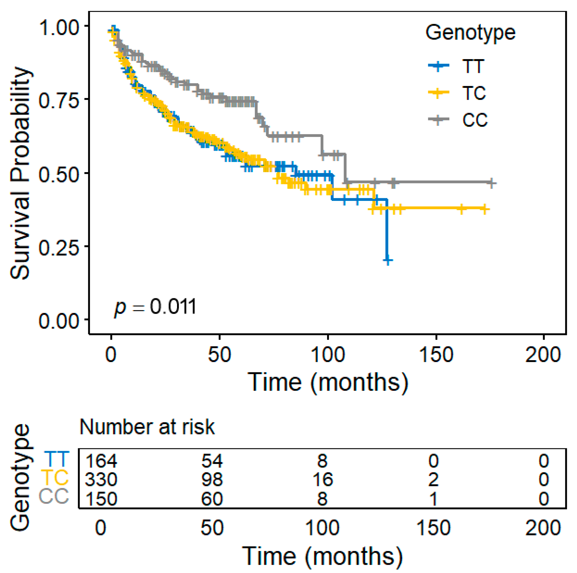
Figure 1. Kaplan–Meier curves of biochemical recurrence-free survival following radical prostatectomy, stratified by rs4502156 genotypes in C2CD4A.
Diagnostics 15 02767 g001
Figure 2. Functional characterization of the C2CD4A rs4502156. (A) Regulatory annotation from HaploReg showing that rs4502156 and its linkage disequilibrium proxies are expression quantitative trait loci (eQTLs) that could potentially disrupt multiple transcription factor-binding motifs. (B) Tissue-wide eQTL identified from the FIVEx database. The dashed horizontal line indicates the genome-wide significance threshold (p = 5 × 10⁻⁸). Data points indicate the direction and significance of the association: positive (triangles), negative (inverted triangles), and nonsignificant (circles).
Diagnostics 15 02767 g002
Figure 3. Association between C2CD4A mRNA expression and clinicopathological features of prostate cancer. Expression values (log2(x+1)-transformed RNA-Seq by expectation-maximization counts) were analyzed against the tumor stage, Gleason score, and disease progression status using data from The Cancer Genome Atlas prostate adenocarcinoma cohort. Lower C2CD4A expression was significantly associated with more aggressive disease.
Diagnostics 15 02767 g003
Figure 4. Gene Ontology annotation and pathway enrichment of genes associated with C2CD4A expression in prostate cancer. (A) Heatmap of the top 20 upregulated and downregulated differentially expressed genes between the high- and low-C2CD4A-expression groups in The Cancer Genome Atlas prostate adenocarcinoma cohort. (B) GSEA of Gene Ontology biological processes, highlighting the top five pathways enriched in the high-expression group. (C) GSEA of Hallmark pathways showing the top five enriched pathways, with E2F targets being the most significantly enriched gene set.
Diagnostics 15 02767 g004
Figure 5. Clinical relevance of E2F pathway activity and CDKN2C expression in prostate cancer. (A) GSVA scores for E2F targets and (B) CDKN2C expression levels according to tumor stage, Gleason score, and disease progression status in The Cancer Genome Atlas prostate adenocarcinoma cohort. Both the E2F GSVA scores and CDKN2C expression levels were inversely correlated with the C2CD4A expression levels and elevated in more aggressive diseases. The blue regression line in scatter plots represents the best-fit linear model, and the shaded area indicates the 95% confidence interval.
Diagnostics 15 02767 g005
Table 1. Clinicopathologic characteristics of the study population.
CharacteristicsNo BCRBCRp
No. of patients, n (%)415 (64.4)229 (35.6)
Median age at diagnosis, years (IQR)66 (62–70)66 (61–71)0.299
Median PSA at diagnosis, ng/mL (IQR)9.3 (6.2–15.0)14.7 (8.4–25.9)<0.001
Pathologic stage, n (%)
 T1/T2275 (66.4)89 (39.6)<0.001
 T3/T4/N1139 (33.6)136 (60.4)
Pathologic Gleason score, n (%)
 2–7369 (88.4)164 (71.6)<0.001
 8–1048 (11.6)65 (28.4)
ISUP grade group
 1117 (28.2)36 (15.7)<0.001
 2 and 3250 (60.2)128 (55.9)
 422 (5.3)20 (8.7)
 526 (6.3)45 (19.7)
D’Amico risk classification, n (%)
 Low65 (15.7)12 (5.3)<0.001
 Intermediate166 (40.1)50 (21.9)
 High183 (44.2)166 (72.8)
Surgical margin, n (%)
 Negative320 (77.1)140 (61.1)<0.001
 Positive95 (22.9)89 (38.9)
Lymph node metastasis, n (%)
 Negative323 (95.8)182 (95.3)0.763
 Positive14 (4.2)9 (4.7)
Abbreviations: BCR, biochemical recurrence; IQR, interquartile range; PSA, prostate-specific antigen; ISUP, International Society of Urological Pathology. The median follow-up duration was 51 months. Subtotals may not sum to the total number of patients due to missing data.
Table 2. Association of type 2 diabetes-related genetic variants with biochemical recurrence after radical prostatectomy.
SNP IDChromosomePositionMapped GeneAlleleMAFHWERisk Allele for T2DRisk Allele for BCRHRp
rs2296173139913351MACF1A>G0.1661.000GA0.8760.317
rs12088739151506886MIR4421A>G0.1000.664AA0.8040.197
rs11276551117530507PTGFRNC>T0.4380.080CT1.1610.093
rs3408741214159256PROX1-AS1T>C0.3870.282CC1.0180.859
rs28204261219660535LOC102723886 (LYPLAL1)A>G0.3300.426GA0.8980.299
rs3483301229672955ABCB10G>A0.4350.130GG0.8830.212
rs28671252622827TMEM18C>T0.0780.165CT1.0280.875
rs780094227741237GCKRT>C0.4920.814CT0.9690.737
rs243019260585806MIR4432HGC>T0.3450.603CC0.8530.109
rs1009358265276452CEP68T>C0.2840.441TT0.9760.822
rs101696132111934977BCL2L11C>T0.4550.692CT1.0380.685
rs126176592121309759LOC105373585 (GLI2)C>T0.1840.192CC0.9180.480
rs75729702161136656RBMS1G>A0.1850.605GG0.9950.968
rs133892192165528876COBLL1C>T0.0980.501CC0.8300.286
rs29721442227101411MIR5702G>A0.0641.000GA1.1290.518
rs75617982228973660SPHKAPG>A0.3250.858GA1.0750.480
rs1899951312394840PPARGC>T0.0530.701CT1.0810.710
rs1496653323454790UBE2E2A>G0.2131.000AG1.1320.276
rs11926707346925539PTH1RC>T0.3590.932CC0.9610.684
rs2292662363897215ATXN7C>T0.4240.520CT1.0910.354
rs6795735364705365ADAMTS9-AS2T>C0.2420.915CT0.8120.067
rs44720283152053250MBNL1C>T0.4381.000TT1.1070.285
rs119252273170766618TNIKG>A0.1690.576GG0.9010.423
rs76510903185513392IGF2BP2A>G0.2490.599GA1.0000.999
rs38879253186665645ST6GAL1C>T0.4750.157TC0.9520.591
rs180121446303022WFS1T>C0.0770.045TC1.1130.490
rs17086692453134293SPATA18G>T0.2570.918GT1.1450.196
rs993380483584496SCD5G>A0.3640.735AA1.1200.233
rs76742124103988899SLC9B2G>T0.3820.868GT1.1300.209
rs110986764123833154NUDT6C>T0.0391.000CC0.5580.047
rs76852964153254121TMEM154C>T0.4421.000CT1.0350.713
rs1061813514847331ANKHA>G0.1820.012GA0.8540.225
rs4865796553272664ARL15A>G0.1170.124AG1.0240.867
rs459193555806751C5orf67G>A0.4990.387GA1.1220.235
rs2307111575003678POC5C>T0.4310.576TT1.1750.089
rs6878122576427311ZBED3-AS1A>G0.0600.499GA0.9750.898
rs100774315112927686YTHDC2C>A0.1010.829CA1.2960.064
rs105022667281654RREB1A>G0.4140.808AG1.0460.623
rs7756992620679709CDKAL1A>G0.4820.432GA0.9920.934
rs2857605631524851NFKBIL1T>C0.1560.072TC1.2500.066
rs1063355632627714HLA-DQB1G>T0.3290.656GG0.8400.089
rs9369425643810974LOC107986598 (VEGFA)A>G0.1440.265GG1.1910.177
rs72892910650816887TFAP2BG>T0.1340.173TT1.2520.074
rs8539746127068983RPS4XP9C>T0.4681.000TT1.0140.883
rs22460126131898208ARG1, MED23T>C0.4020.327CT0.9670.722
rs6222176160766770SLC22A3T>C0.2960.258TT0.9180.410
rs17168486714898282DGKBC>T0.4880.529TC0.9400.526
rs2191348715064255AGMOT>G0.3110.000TT0.8360.107
rs2908282744248828YKT6G>A0.1941.000AG0.9330.569
rs22993837103418846RELNC>T0.4040.807TC0.9420.524
rs132391867117510621CTTNBP2C>T0.1880.438TT1.0600.617
rs132342697130429186LOC105375508 (KLF14)T>A0.3270.789TA1.0330.750
rs1177491589188762LOC157273(TNKS)C>T0.3160.364TT1.0400.684
rs10100265810633159PINX1A>C0.3570.494AA0.9000.296
rs17411031819852310LPLC>G0.1871.000CC0.8480.198
rs10087241830863722PURGA>G0.0331.000GG1.1080.691
rs12681990836859186KCNU1T>C0.3430.029CT0.9610.675
rs516946841519248ANK1C>T0.1300.484CT1.2150.139
rs7845219895937502TP53INP1C>T0.2810.923TC0.9600.699
rs38021778118185025SLC30A8G>A0.4610.134GA1.0130.890
rs22941208146003567ZNF34G>A0.2860.923AA1.1380.198
rs1097443894291928GLIS3A>C0.3780.616CA0.9500.596
rs1063192922003367CDKN2B-AS1/CDKN2BA>G0.1800.351AG1.0460.715
rs10811661922134094CDKN2B-AS1T>C0.4080.626TC1.0760.432
rs1758632934025640UBAP2G>C0.1560.100GC1.0380.783
rs17791483981898980LOC101927450 (TLE1)A>G0.0521.000AG1.1800.424
rs2796441984308948LOC101927502 (TLE1)A>G0.3840.803GA0.9270.433
rs10114341996919182LOC107987099 (PTPDC1)T>C0.0900.338TT0.7670.148
rs6876219136137065ABOA>G0.3400.541GG1.0280.787
rs112576551012307894CDC123T>C0.4490.000TT0.9940.949
rs26161321071469514FAM241BG>A0.4840.814AA1.0750.436
rs7532701080964975ZMIZ1T>C0.4550.812CC1.0820.389
rs1159174110101976501CHUKG>C0.0630.100GG0.9740.897
rs242101610124167512PLEKHA1C>T0.4330.066CC0.9340.457
rs2237892112839751KCNQ1C>T0.3400.485CT1.0030.974
rs52151117408630KCNJ11T>C0.3740.276CC1.0840.414
rs79295431149351026TYRLA>C0.1480.061CA0.8430.223
rs15522241172433098ARAP1A>C0.0641.000AC1.3250.095
rs108309631192708710MTNR1BC>G0.4310.690GC0.9190.366
rs793130211128236058ETS1A>C0.1540.228CA0.8350.201
rs6723254611128398938ETS1C>T0.1720.679TC0.8110.114
rs12299509124406281CCND2G>A0.4720.070GA1.1500.116
rs110484561226463082ITPR2C>T0.3070.581CC0.9550.647
rs108429941227965150LOC105369709 (KLHL42)C>T0.2050.811CT1.1110.367
rs22611811266212318RPSAP52C>T0.1150.334TT1.2770.064
rs14804741266326943HMGA2G>A0.0850.610GG0.9270.671
rs71383001271439589TSPAN8C>T0.3700.556CT1.0550.592
rs111071161293978504SOCS2G>T0.3300.248TG0.9950.956
rs94090412123491572PITPNM2A>G0.1380.136AA0.9530.722
rs82547612124568456ZNF664-FAM101AT>C0.3700.736TT0.9020.288
rs5766741333554302KLA>G0.2120.906GA0.9530.681
rs9637401351096095DLEU1T>A0.3440.543AA1.0960.339
rs13597901380717156LOC105370275 (SPRY2)G>A0.2890.848GA1.0520.621
rs45021561562383155C2CD4AT>C0.4880.637TT0.7880.011
rs9820771563823301USP3A>G0.0560.713AG1.3770.087
rs71770551577832762LOC101929457 (HMG20A)G>A0.3330.002AG0.9130.343
rs994014916300641FAM234AG>A0.4290.689GG0.9510.598
rs71857351653822651FTOA>G0.1130.116GG1.1130.471
rs2444151669666683NFAT5G>A0.1051.000GG0.9250.603
rs29259791681534790CMIPC>T0.4260.378TC0.9530.606
rs8068804173985864ZZEF1G>A0.1600.306AG0.8950.386
rs129456011717653411RAI1T>C0.0950.822TC1.1140.487
rs116517551736099840HNF1BT>C0.1730.335CT0.9240.524
rs99119831745885756OSBPL7C>T0.1570.767TT1.0560.665
rs98942201746989154UBE2ZA>G0.2510.834AA0.8500.137
rs7240767187070642LAMA1C>T0.3060.927CC0.9950.958
rs129701341857884750MC4RG>A0.1560.765AA1.2530.068
rs104019691919407718SUGP1T>C0.1050.529CC1.2210.172
rs81082691946158513GIPRT>G0.4380.750GT0.9450.550
rs65152362022435749LOC105372562 (FOXA2)C>A0.3330.185AC0.8830.232
rs60596622032675727EIF2S2G>A0.1560.882GG0.9900.936
rs48104262043001721HNF4AC>T0.4130.686TT1.1280.199
rs48231822244377442SAMM50G>A0.4920.388GG0.9740.772
Abbreviations: SNP, single nucleotide polymorphism; MAF, minor allele frequency; HWE, Hardy–Weinberg equilibrium; T2D, type 2 diabetes mellitus; BCR, biochemical recurrence; HR, hazard ratio.
Disclaimer/Publisher’s Note: The statements, opinions and data contained in all publications are solely those of the individual author(s) and contributor(s) and not of MDPI and/or the editor(s). MDPI and/or the editor(s) disclaim responsibility for any injury to people or property resulting from any ideas, methods, instructions or products referred to in the content.