Abstract
Background/Objectives: We have previously reported the engineering of a point-of-care (POC) system that fully automates the procedures for nucleic acid extraction and multiplexed real-time RT-PCR, with a major advantage of high-level multiplexing. In this study, we applied and validated the system in a respiratory tract infection setting. Methods: An automatic nested real-time RT-PCR assay was developed (POCm). It was a 40-plex assay that simultaneously detected 39 epidemiologically important respiratory pathogens in 1.5 h in the POC system. The analytical and clinical performance was evaluated. Results: The analytical sensitivities of the POCm assay were comparable to those of its single-plex counterparts performed manually on a bench-top. The minimum detectable concentrations ranged from 53 copies/mL to 5.3 × 10³ copies/mL for all pathogen targets except hCoV-NL63 (5.3 × 10⁴ copies/mL). The quantitative performance was demonstrated by the linear correlations between Ct values and input concentrations for all pathogen targets, with 24 of them demonstrating coefficients of correlation (r) greater than 0.9. The POCm assay was subsequently evaluated in 283 clinical samples. A high level of agreement (98.2–100%) was achieved for pathogen detection results between POCm and standard diagnostic methods. The POCm result was also fully concordant with the result of another commercial POC multiplex platform. For positive clinical samples, pairwise Ct values measured by POCm closely correlated with those of the bench-top reference method (r = 0.70). The feasibility of mutation genotyping of the viral subtype was further demonstrated. Conclusions: This study demonstrated the practicality of POCm for routine testing in clinical laboratories. Further clinical trials are being conducted to evaluate the clinical performance of the system.
1. Introduction
Pathogen genetic testing is an essential tool to realize a near-to-real-time respiratory infection control, including patient care and epidemic surveillance. Point-of-care (POC) systems can dramatically increase the accessibility of genetic testing in critical low-infrastructure sites such as doctors’ offices, community clinics, and borders [1,2,3]. Lab-on-chip nucleic acid analysis is one of the most promising POC testing methods. By using microfluidic technologies, the entire testing procedure can be miniaturized into a micro-chip, which permits ease of use by minimally trained operators [4]. However, one of the challenges in POC development is the capability to detect a diverse number of potential causative agents in one assay. Acute respiratory tract infection, for example, can be caused by over 20 different strains of viral pathogens [5]. In addition to causative viruses, extra multiplexing capacity is required to discriminate bacteria and fungi that can cause indistinguishable clinical symptoms [5]. The growth of biomarkers developed by next-generation sequencing further necessitates high-order multiplex testing.
To maximize the value of lab-on-chip POC testing, we previously developed an in-house POC system, which was designed to (i) conduct high-order multiplexing with 120 physically separated mini-compartments for real-time PCR; (ii) ensure ease of use with less than 5 min of manual handling; and (iii) incur relatively low manufacturing cost. The whole POC system consists of an enclosed cartridge, where all reactions are carried out, and a microfluidic analyzer for controlling microfluidics and reaction conditions. The development of the automation system and its mechanics has been reported previously [6]. The detection chemistry is based on a multiplexed nested-RT-PCR, followed by a panel of single-plex TaqMan real-time PCRs in individual nano-compartments. The use of PCR methodology provides high flexibility for customizing and updating assay panels from time to time [7,8,9]. It enables rapid, sensitive, and specific detection and characterization of pathogens directly from clinical specimens. It also offers potential for nucleic acid quantification and mutation genotyping.
Nucleic acid amplification tests (NAATs), including PCR, have become an integral part of molecular diagnostics and are used as “standard-of-care” tests for many infectious diseases [10]. Their use in the diagnosis of respiratory infections [11,12], sexually transmitted infections [13], and infectious diarrhea [14] has been recommended in the corresponding guidelines. The ability of NAATs to detect pathogens not recovered by standard culture methods is significant and may aid the diagnosis of pneumonia [15] and bloodstream infections [16]. Miniaturized PCR-based systems are being developed for rapid diagnostics applications. A few of these systems are designed for multiplex detection of up to three or four respiratory pathogen targets [17,18], while the majority focus on single-target detection [19,20]. Some require sample pre-processing, which limits their use outside laboratory settings [21]. Our POCm system surpasses the previous innovations, demonstrating the ability of conducting a 40-plex assay with full automation.
In this study, we evaluated this high-order multiplexing POC system (POCm) in a laboratory setting. Respiratory infection was selected as a model since it is the most common infection globally and is associated with high morbidity and mortality [5,22]. Here, we describe the laboratory assay development, analytical validation, and clinical application of a 40-plex POCm test, which targets 39 pathogen strains that are most relevant to Hong Kong and nearby Asian regions. The test was fully automated with a sample-to-result turnaround time of 90 min. By achieving genetic testing capacity that is simple, rapid, comprehensive, and inexpensive, this POCm platform could fill the unmet needs of patients, healthcare professionals, and epidemiologists in infectious disease control.
2. Materials and Methods
2.1. POCm Cartridge and Analyzer
All testing procedures are fully automated in the POCm system, which comprises a single-use reaction cartridge and a microfluidic analyzer (Supplementary Figure S1). The cartridge consists of a panel of reaction chambers for nucleic acid extraction, reverse-transcription polymerase-chain reaction (RT-PCR) and real-time PCRs. Primers and/or probes (Integrated DNA Technologies, Coralville, IA, USA) are pre-spotted and dried into relevant chambers during manufacturing. The cartridge further includes multiple reagent reservoirs pre-packaged with buffers and reaction master mixes. The ready-to-use cartridge is stable for a minimum of 6 months when stored at 4 °C. All fluidics within the cartridge are controlled by microchannels and valves interconnecting the reservoirs and chambers. The whole testing procedures are enclosed inside the cartridge. The sealed cartridge is disposed after each test, hence minimizing the risk of contamination by infectious samples and PCR products. The cartridge is manufactured by the injection-molding method, which allows mass production at relatively low cost.
The microfluidic analyzer serves the functions of (i) manipulating the transfer of samples and reagents within the microfluidic network in the cartridge; (ii) controlling reaction temperatures and thermal cycles; and (iii) measuring fluorescent intensities during real-time PCR. It also monitors all reaction parameters and reports any failure during the run. A schematic representation of the validation of the POCm system is shown in Supplementary Figure S2.
2.2. POCm Testing Procedures
Three hundred microliters of nasopharyngeal aspirate, endotracheal aspirate, or viral transport medium (VTM) containing swab samples was added to the sample chamber of the cartridge. For deep throat saliva, 0.5 to 1 mL of sample was mixed with 2 mL of VTM, and 300 μL of the diluted sample was added. The cartridge was then loaded into the microfluidic analyzer, and the entire testing workflow was automated in the analyzer. After the run was completed, the analyzer automatically performed data analysis and reported results.
2.3. Assay Panel Design
The multiplexed detection panel consisted of 40 targets for 39 respiratory tract disease pathogens, including 26 viruses, 11 bacteria, and 2 fungi (Supplementary Table S1). The pathogens were selected by Department of Microbiology, The University of Hong Kong, based on their epidemiological significance in Hong Kong SAR and nearby regions. Since the assay was designed during the early outbreak of COVID, two detection targets were designed for SARS-CoV-2 in order to enhance the detection confidence. Enterovirus and rhinovirus were not included in this evaluation study since the POCm assay cannot confidently distinguish between them. Further assay optimization is being carried out, and the two viruses will be included in the next version of the POCm assay.
Three quality control assays were further included in the panel. They were glyceraldehyde 3-phosphate dehydrogenase (GAPDH) as the sample quality control, schizosaccharomyces pombe RNA (SUC1) (ATCC, Manassas, VA, USA; Cat# 24843) which was spiked-in during each run for experimental processing control, and a non-pathogen target for real-time PCR control (GenScript, Nanjing, China).
The assay was based on nested real-time RT-PCR. It comprised a multiplexed one-step RT-PCR for 40 pathogen targets and 2 controls (GAPDH and SUC1), i.e., a 42-plex reaction. It was followed by an array of single-plex real-time PCRs carried out in 120 individual mini-chambers for the 40 pathogen targets and the 3 controls. Outer primers for RT-PCR were designed with melting temperature (Tm) ranging from 67 °C to 76 °C in order to achieve optimal amplification specificity. Each real-time PCR assay consisted of a pair of inner primers and a TaqMan probe. The Tm of inner primers was 59–66 °C. Primers of each target were tested individually using conventional manual methods to evaluate the amplification conditions. The primers of all the targets were then pooled together to test for any interference. If the Ct value of a target in the multiplex is much higher than that of the single-plex reaction, the corresponding primers are re-designed.
2.4. PCR
The first-round RT-PCR master mix was prepared using Path-ID Multiplex One Step RT-PCR Kit (Thermo Fisher Scientific, Waltham, MA, USA; Cat# 4442136). The RT-PCR chamber of the cartridge was pre-spotted with 42 pairs of outer primers, which were re-suspended in master mix to a final concentration of 0.07 μM. The entire volume of extracted nucleic acids (16 μL) was used for the reaction. RT was carried out at 48 °C for 10 min, 95 °C for 10 min, followed by 20 PCR cycles of 95 °C for 15 s and 65 °C for 45 s. The final PCR product was automatically 66.7-fold diluted with nuclease-free water.
The second-round real-time PCR master mix was prepared using Premix Ex Taq (Probe qPCR) Kit (Takara Bio, Shiga, Japan; Cat# RR390). Atto 665 fluorescent dye (Sigma-Aldrich, Burlington, MA, USA; Cat #93711) was added in a concentration of 0.03 μM for background monitoring. Each real-time PCR mini-chamber in the cartridge was pre-spotted with a pair of inner primers and probe for one detection target. Each pathogen target or control was detected in duplicate or triplicate (Supplementary Table S2). The primers and probe were re-suspended in master mix to final concentrations of 0.5 μM and 0.2 μM, respectively. Real-time PCR was carried out at 95 °C for 30 s, followed by 40 cycles of 96 °C for 1 min and 55 °C for 45 s. Figure 1 shows an example of amplification results from a sample positive for Flu A/H1.
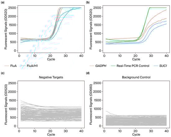
Figure 1.
Example amplification plots of a patient sample as tested by POCm. Fluorescence signals of the 40 pathogen targets and 3 quality controls were obtained in a single run. The data were separated into four graphs for interpretation. (a) The sample tested positive for Flu A/H1. Positive amplification signals were detected for Flu A generic assay (red line) and H1 subtype specific assay (green line). The qPCR curves showed typical sigmoidal shape. The real-time PCR assays were performed in triplicate. (b) Amplification plots for internal control (GAPDH) (red line), external control (SUC1) (blue line) and real-time PCR control (green line). Duplicated real-time PCR reactions were performed for each control assay. (c) Negative result was obtained for all of the 38 non-target assays (grey line), indicating their assay specificities. (d) Atto 665 fluorescent signals (grey line) for background monitoring throughout real-time PCR.
2.5. Manually Performed RT-PCR Tests
The detection assays were optimized by conventional manual methods using bench-top instruments before adopting POCm. Bench-top manual nucleic acid extraction was performed using QIAamp MinElute Kit (Qiagen, Valencia, CA, USA; Cat# 57704). First-round RT-PCR and second-round real-time PCR were carried out in Veriti 96-Well Thermal Cycler and QuantStudio 7 Flex Real-Time PCR System (both from Thermo Fisher Scientific, Waltham, MA, USA), respectively, with conditions as described above.
2.6. Preparation of Reference Controls
To evaluate the test performance, pathogen-derived genomic DNA and RNA (ATCC, Manassas, VA, USA), PCR controls (Vircell, Granada, Spain) or plasmids with inserts of target sequences (GenScript, Nanjing, China) were used for constructing reference control plasmids (Supplementary Table S3). The plasmids were used for evaluation tests of all bacterial and fungal targets, as well as DNA viruses including AdV and BoV. For RNA viruses, in vitro transcriptions were further performed to generate reference control RNAs that contained the RT-PCR amplicon sequences.
Twelve mixtures of reference controls were further prepared (Supplementary Table S4). For 11 of the mixtures, three to four pathogen controls were spiked in a background of human RNA extracted from HeLa cells. The 12th mixture served as a negative control with human RNA only.
2.7. Evaluation with Clinical Samples
The POCm assay was further evaluated using archived clinical specimens retrieved by the Department of Microbiology, Queen Mary Hospital, from January 2020 to December 2021. A total of 283 samples were tested. The samples included nasopharyngeal aspirate (NPA) (n = 141), nasopharyngeal swab (NPS) (n = 136), combined nasopharyngeal and throat swab (n = 1), endotracheal aspirate (n = 1), and saliva (n = 4) from patients suffering from influenza-like signs and symptoms (Supplementary Table S5). They were previously tested with “standard-of-care” tests, which included immunofluorescent antibody assays (IFA) (D3 Ultra 8 DFA Respiratory Virus Screening and ID Kit, QuidelOrtho, San Diego, CA, USA; Cat# I-01-110000), conventional PCR, Xpert Xpress Flu/RSV (Cepheid, Sunnyvale, CA, USA; Cat# GXFLU/RSV-CE-10) and/or FilmArray Respiratory Panel (bioMérieux, Marcy-l’Étoile, France; Cat# RFIT-ASY-0124). For pathogens not covered by IFA and Xpert Xpress Flu/RSV, further tests were conducted by the reference laboratory (Public Health Laboratory Centre in Hong Kong) using in-house multiplex assays targeting respiratory viruses. Details on the standard-of-care tests for each pathogen are listed in Supplementary Table S6.
2.8. Statistical Analysis
Statistical analyses were conducted using SigmaPlot 13 (Systat Software, Inc., San Jose, CA, USA) and R (version 4.3.1) (The R Foundation, Ames, IA, USA).
3. Results and Discussion
3.1. Selection of Respiratory Pathogens for Multiplex Assay Panel
The selection of respiratory pathogens for inclusion in our assay panel (Supplementary Table S1) was guided by both clinical relevance and epidemiological significance. Definite pathogens, including all the targeted respiratory viruses, Mycobacterium tuberculosis (MTB), and Burkholderia pseudomallei, etc., were included due to their established roles as causative agents. Notably, MTB was incorporated based on increasing evidence supporting the use of upper respiratory tract specimens for diagnosis of tuberculosis, with a sensitivity reaching 81.8% [23]. In addition, Pneumocystis jirovecii (PJ) was included in the panel, despite potentially indicating colonization, as its detection in NPA specimens may guide further testing with bronchoalveolar lavage in patients who are suspected to have Pneumocystis pneumonia [24]. Furthermore, our panel also encompassed bacteria such as Streptococcus pneumoniae, Staphylococcus aureus, and Streptococcus pyogenes. Although their detection in the upper respiratory tract specimens may represent asymptomatic colonization, this information can be relevant to decisions on infection control or prevention measures. Clinicians can use clinical information for treatment decisions, including the use of antibiotics. Our broadened panel covering these additional pathogens would be able to enhance diagnostic yield and inform more comprehensive patient management.
3.2. Analytical Detectability
The microfluidics layout of the POCm cartridge was designed in order to elevate the multiplexing capability. Firstly, the starting amount of nucleic acids is an important factor for detection sensitivity. Inside the POCm cartridge, the whole extracted volume of nucleic acids (16 μL) was channeled for the first-round RT-PCR. On the other hand, only 1/20 volume of extracted nucleic acids (5 μL out of 100 μL) could be used for the bench-top approach. Secondly, for the second-round real-time PCR detection, single-plex reactions were individually carried out in 120 physically separated chambers in the POCm cartridge. This design could reduce cross-reactivity among different pathogen assays and therefore enhance detection specificity. Owing to the high-multiplexing capacity, more than one PCR target can be designed for a particular pathogen. This increases detection reliability for epidemiologically or genetically complex pathogens. To evaluate the benefit of the POCm platform over the bench-top approach for high-order multiplexing reactions, their analytical performance was compared.
The analytical detectability of the targeted pathogens was compared between POCm and the manual approach by using the limiting dilution method. Reference controls of individual pathogens (Supplementary Table S3) were serially diluted and were tested in triplicate at each concentration. The lowest concentration at which all of the triplicated reactions showed positive results was referred to as the minimum detectable concentration (MDC), which was expressed as copies per milliliter of sample (copies/mL).
Multiplex assays commonly showed decreased detection sensitivities when compared to their single-plex counterparts. This phenomenon was also observed in our respiratory pathogen assays that were carried out using the manual bench-top platform. When switching from manual-single-plex to manual-40-plex reactions, the detectability of the 40 pathogen targets dropped. Their MDCs were significantly increased for a median of 11-fold (p < 0.05, Wilcoxon signed-rank test) (Table 1). On the other hand, we showed that this shortcoming of multiplexing was resolved by the POCm platform. For all 40 pathogen targets, the detectability of the automatic-POCm assay was restored to that of the manual-single-plex counterparts with no statistically significant difference in MDCs (p = 0.562, Wilcoxon signed-rank test). The MDCs of 53 to 5300 copies per milliliter of sample (copies/mL) were obtained for all pathogen targets except hCoV-NL63 (53,000 copies/mL) (Table 1 and Figure 2).

Table 1.
Comparison of minimum detectable concentrations and corresponding Ct values between the bench-top and POCm methods.
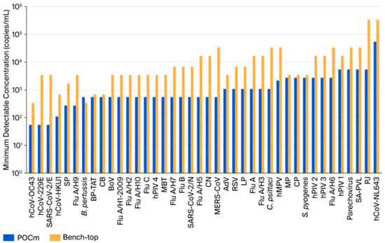
Figure 2.
Comparison of analytical sensitivities between POCm and bench-top methods. POCm (blue bar) showed similar or better analytical sensitivities (lower minimum detectable concentrations) than the bench-top method (orange bar) for all pathogen targets except B pertussis.
To inspect cross-reactivity of the POCm 40-plex reaction, we analyzed the reference control mixtures (Supplementary Table S4) at concentrations 1000-fold higher than the corresponding MDCs. Negative results were obtained for all non-targeted pathogens. Hence, no cross-reactivity among the pathogen targets was observed for POCm.
3.3. Linearity
The capability of the POCm platform in quantification of respiratory pathogens was evaluated. Linearity panels of the 40 pathogen targets were generated using the reference control mixtures (Supplementary Table S4). The mixtures were serially log10 diluted from 1000- to 1-fold of MDCs of the corresponding pathogen targets and analyzed by POCm. We found statistically significant correlations between Ct values and input concentrations for all of the 40 pathogen targets (p < 0.05, ANOVA for linear regression). The coefficients of correlation (r) ranged from 0.792 to 0.994, with 60% (24 of 40) of the targets showing r > 0.9 (Figure 3). The result hence demonstrated that POCm can potentially be used for pathogen load estimation.
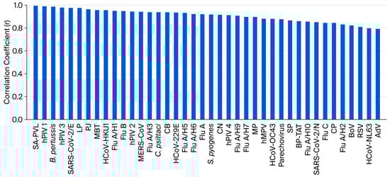
Figure 3.
Evaluation of quantitative performance of POCm. For all of the 40 pathogen targets, linear relationships were obtained between Ct values and log (concentrations in copies/reaction) as measured by POCm (p < 0.05). Coefficients of correlation (r) ranged from 0.792 to 0.994. Twenty-four of the pathogen targets showed r > 0.9.
3.4. Clinical Sample Evaluation
The clinical evaluation of POCm was carried out in the laboratory of the Department of Microbiology, Queen Mary Hospital. A total of 283 clinical samples were tested and included for clinical performance analysis. This study involved POCm assays of two initial versions (11-plex and 18-plex assays) (Supplementary Table S5) and a final 40-plex assay (Supplementary Table S1). The results from all of the POCm assay versions were combined for analysis.
Of the 39 pathogens that the POCm assay targeted, 13 pathogens tested positive in the “standard-of-care” tests on at least 1 out of the 283 samples (Supplementary Table S7). Eleven of the pathogens that were tested on 200 clinical samples were further evaluated for testing performance (Table 2). In general, for 10 of the 11 (90.91%) pathogens, the results of POCm highly agreed with those diagnosed by standard-of-care tests, with agreements and Cohen’s κ coefficients achieving 98.2–100% and 94.6–100%, respectively. The detection of Mycoplasma pneumoniae (MP) was sub-optimal since POCm could only detect one of the two samples that tested positive by the standard-of-care test (Table 2). Further evaluation of the MP detection performance with additional positive samples is required.
Table 2.
Clinical performance of POCm in detecting respiratory pathogens.
We further evaluated the clinical accuracy of the POCm platform. The sample sizes of all of the 11 pathogens were sufficient for specificity calculation [25]. The specificities were 100% for all of the assays (Table 2). For sensitivity evaluation, a minimum sample size of 200 with at least 20 positive samples has been recommended for diseases with 5–10% prevalence [25,26]. We therefore selected Flu A (Matrix), Flu A (H1-pdm09) and SARS-CoV2, which had sufficient sample sizes, for sensitivity calculation. The detection sensitivities were 89.8%, 95.2% and 91.7% for Flu A (Matrix), Flu A (H1-pdm09) and SARS-CoV2, respectively (Table 2). Despite no definite threshold requirement, the detection sensitivities should be further improved in order to have better utilization in frontline diagnostics. Potential assay optimization strategies include improving binding buffer and elution buffer formulation to recover more templates during nucleic acid extraction, increasing the amount of input in the RT-PCR step, constantly reviewing any mutations in the gene targets that may affect the detection and redesign primers/probe if necessary.
In addition to the use of standard-of-care testing results as a reference, we also compared the performance of POCm with another POC system manufactured by FilmArray [27], which is one of the multiplexing POC platforms available on the market. FilmArray provides qualitative detection of 23 pathogen strains with its Respiratory Panel (BioFire FilmArray Panel, bioMérieux, Marcy-l’Étoile, France). We tested ten positive clinical samples, as well as three negative samples collected from healthy individuals in parallel using both POCm and FilmArray. As shown in Table 3, the results obtained from both POC systems were fully concordant.
Table 3.
Concordant clinical result of 13 samples as determined by FilmArray and the POCm system.
In summary, the validation results of clinical samples demonstrated that the performance of POCm concurred with those of standard-of-care testing methods as well as other POC platforms on the market. Due to the limiting sample size, we could not fully evaluate the clinical performance of all of the 39 pathogens. In addition, for some of the pathogens, the studied clinical samples were not the representative sample types when clinically indicated, which might influence result accuracy. Nonetheless, the promising results of this study will promote the future expansion of clinical studies in terms of sample sizes and testing sites.
3.5. Pathogen Co-Infection Detection
One advantage of the multiplexed detection platform is pathogen co-infection identification [28,29]. During the period of this study, one clinical sample, 20QM0123, was initially diagnosed as single-pathogen infection with SARS-CoV-2. With the use of POCm, we identified additional co-infected pathogen hMPV in these samples (Table 4), which was subsequently confirmed by standard-of-care tests. We also detected co-infection in one sample (20QM0113), which was concordant with the initial co-infection diagnostic result (Table 4).
Table 4.
POCm results of co-infected samples.
Two additional samples, 20QM0119 and 21QM0003, were diagnosed with SARS-CoV-2 and RSV, respectively, but Streptococcus pneumoniae (SP) was also detected. SP may usually be considered as normal colonization in the upper respiratory tract. Hence, we demonstrated that POCm could provide a more comprehensive pathogen screening, as well as the potential of our assays to detect colonization such as SP in NPS/NPA samples, which may assist in more informative targeted patient care [30].
3.6. Measurement Agreement of Ct Values
We further assessed the performance of POCm among the clinical samples. A total of 114 clinical samples that tested positive using POCm were evaluated using the bench-top approach in parallel, and the measurement agreement of Ct values was analyzed. We found a statistically significant correlation of overall Ct values between POCm and the bench-top tests (p < 0.05, r = 0.70, Pearson’s correlation test) (Figure 4a). Bland–Altman analysis showed systematically higher Ct values for POCm when compared with the corresponding bench-top result (bias = 3.64) (Figure 4b). Despite the bias, 95% of the data points fell within the limits of agreement (−3.37–10.65) (Figure 4b). The result hence confirmed that nucleic acid measurement by POCm was comparable to that of the reference bench-top approach. In practice, whether the bias would have any clinical implications, such as affecting the detection sensitivity in particular for samples with low pathogen loads, still warrants further investigation.
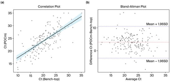
Figure 4.
Ct value agreement between POCm and bench-top methods. (a) Scatter plot of Ct values as measured by POCm and bench-top methods. The Ct values obtained by both methods were significantly correlated (p < 0.05, r = 0.70, Pearson’s correlation test). (b) Bland–Altman plot was used to evaluate the Ct value agreement between POCm and the bench-top method. There were systematically higher Ct values for POCm when compared with the corresponding bench-top result (bias = 3.64) (red dashed line). The blue dashed lines represent 95% limits of agreement.
3.7. Mutation Genotyping
Mutations acquired by emerging pathogens are valuable biomarkers for differentiating virulent pathogen strains and tracing epidemic transmission events. We therefore assessed the feasibility of mutation genotyping by POCm. As a feasibility evaluation, a TaqMan allelic discrimination assay was designed to genotype del:21765:6 mutation in the gene coding spike protein of SARS-CoV-2, a deletion found in the alpha variant [31,32]. The assay involved a pair of TaqMan probes that were 5′-labeled with Cy5 and FAM to discriminate wild-type and mutant alleles, respectively (Integrated DNA Technologies, Coralville, IA, USA). Synthetic oligonucleotides with amplicon sequences of the wild-type and mutated pathogen strains (Integrated DNA Technologies, Coralville, IA, USA) were used for testing. Figure 5 shows the amplification result of POCm, with genotypes of the wild-type and mutant control oligonucleotides being correctly detected. Since the POCm cartridge can simultaneously perform up to 120 real-time PCRs in individual mini-chambers, a large panel of mutations can be genotyped in a single automated test. This is favorable for pathogens with abundant variants such as SARS-CoV-2. Moreover, assay design and evaluation are straightforward, which allows rapid adaptation to newly emerging variants. This would facilitate the development of comprehensive assays for both detection and sub-typing of fast-evolving pathogens.
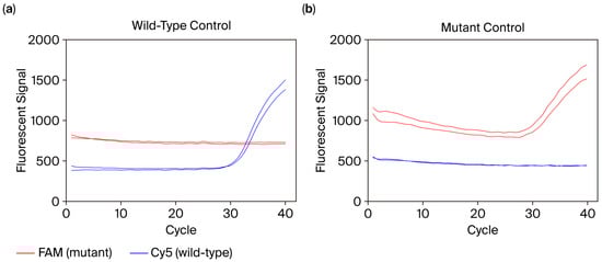
Figure 5.
Example amplification plots of allelic discrimination assay for the mutation del:21765:6 of SARS-CoV-2. The allelic discriminative TaqMan probes for the mutant and wild-type alleles were labeled with FAM and Cy5 fluorescent dyes, respectively. The performance of allelic discrimination was confirmed by using synthetic oligonucleotides containing the (a) wild-type and (b) mutant alleles.
4. Conclusions
The POCm system that we previously developed offers a major advantage to infectious disease diagnostics, as it can detect a large panel of pathogen infections in a single automated test. In this study, we demonstrated its practicality in respiratory tract infections, including influenza and other concurrent pathogens that share similar clinical symptoms but require distinct disease treatment and management [5].
In this study, we developed a 40-plex real-time RT-PCR assay that targeted 39 pathogens. The order of multiplexing is, to our knowledge, the largest among the POC platforms available on the market [3,33]. The assay was demonstrated to be efficiently automated in the POCm system, with analytical performance comparable to that of the bench-top method. The fully integrated POCm system with automated nucleic acid extraction and multiplex real-time RT-PCR was evaluated with clinical samples. The detection results were highly concordant with those of standard diagnostic methods, as well as a commercially available RT-PCR-based POC platform. With the advantage of high-order multiplexing, POCm was able to detect a co-infection that had been missed by standard diagnostic methods. This result hence highlights the benefit of POCm in the comprehensive identification and exclusion of pathogens, as well as in the detection of mixed-pathogen infections [28,34,35]. In addition, the demonstrated semi-quantitative capability of POCm suggests a potential application for the detection and complementary quantification of pathogen loads, which has been suggested to be associated with disease severity and patient outcomes [36,37].
The current POCm test allowed for subtyping of common pathogens, such as influenza A, by including both a generic assay and various assays specific to the subtypes. POCm could potentially add another level of pathogen characterization by genotyping mutations acquired by rapidly emerging strains [31,32]. As a feasibility demonstration, we were able to genotype an alpha-variant mutation in SARS-CoV-2 by using a POCm allelic-discriminative assay. Along with rapidly sequenced pathogen genomes, POCm would allow prompt development of molecular assays that match the pathogen strains responsible for the outbreak [7,8,9].
In conclusion, POCm is both robust and reliable for high-order multiplexed pathogen detection. It could aid infection diagnosis when utilized in conjunction with clinical history and epidemiological information. With straightforward real-time RT-PCR primer design, the detection targets of POC assays can be promptly updated to accommodate both existing and newly emerging pathogens in one test [38]. Combined with the relative simplicity and low production cost of the single-use cartridge, the POCm system has the potential to streamline clinical diagnostics as well as public health surveillance in the fight against recurrent and upcoming unforeseen epidemics. Further large-scale clinical studies will be valuable in order to evaluate various performance parameters, including clinical sensitivities, specificities and predictive values. Performance studies under different environmental conditions are worthwhile to assess the robustness of the POCm system outside well-equipped laboratories. In addition, guidelines for system operation and training should be established, and usability tests should be conducted to evaluate suitability in community and field use.
Supplementary Materials
The following supporting information can be downloaded at: https://www.mdpi.com/article/10.3390/diagnostics15192445/s1, Figure S1: POCm cartridge and microfluidic analyzer; Figure S2: Schematic diagram of the current study; Table S1: Pathogen targets of the respiratory infectious disease assay panel; Table S2: Content of real-time PCR mini-chambers in POCm cartridge for respiratory disease pathogen detection; Table S3: Construction of reference controls for evaluation tests; Table S4: Reference control mixtures constructed from reference controls of different pathogens; Table S5: Information of pre-clinical study of POCm in Department of Microbiology, Queen Mary Hospital, Hong Kong; Table S6: Standard-of-care tests of the 39 selected pathogens in Department of Microbiology, Queen Mary Hospital, Hong Kong; Table S7: Clinical evaluation of the 39 pathogen targets separated by the three phases.
Author Contributions
Conceptualization, K.K.W.T., K.-Y.Y., M.F., J.Y.N.L. and L.T.L.; methodology, L.C.W.L. and C.C.Y.Y.; software, P.Y.L.T. and S.L.H.C.; validation, D.M.S.T., C.T.L. and B.K.W.L.; formal analysis, A.Y. and N.B.Y.T.; investigation, L.C.W.L., D.M.S.T., A.Y., P.Y.L.T., S.L.H.C., C.T.L., B.K.W.L., W.W., F.T.K., P.Y.M.L., T.C., C.C.Y.Y., J.H.K.C. and R.W.S.P.; resources, P.Y.L.T., S.L.H.C. and K.-Y.Y.; data curation, D.M.S.T., W.W., F.T.K. and P.Y.M.L.; writing—original draft preparation, L.C.W.L., A.Y., N.B.Y.T., J.Y.N.L. and L.T.L.; writing—review and editing, L.C.W.L., A.Y., N.B.Y.T., C.C.Y.Y., K.K.W.T., K.-Y.Y. and L.T.L.; supervision, K.K.W.T., K.-Y.Y., M.F., J.Y.N.L. and L.T.L.; funding acquisition, L.T.L. All authors have read and agreed to the published version of the manuscript.
Funding
This work was supported by the grants from Midstream Research Programme for Universities (MRP/040/20X), Public Sector Trial Scheme (SST/031/20GP) and Health@InnoHK, all were from Innovation and Technology Commission, The Government of Hong Kong Special Administrative Region, RGC-TRS (T11-709/21-N), The Government of Hong Kong Special Administrative Region, and private donations to The Hong Kong Polytechnic University directed to this project (ZJMA).
Institutional Review Board Statement
The study was approved by Human Subjects Ethics Committee of The Hong Kong Polytechnic University (HSEARS20200806001, dated 26 August 2020). All experiments were performed in accordance with relevant guidelines and regulations.
Informed Consent Statement
Informed consent was waived due to using archived specimens.
Data Availability Statement
The data presented in this study are available on request from the corresponding author.
Acknowledgments
The authors would like to thank the support from the Hong Kong Baptist University and its Wu Jieh Yee Institute of Translational Chinese Medicine Research.
Conflicts of Interest
Libby C. W. Li., Deborah M. S. Tai, Nancy B. Y. Tsui, Parker Y. L. Tsang, Sunny L. H. Chu, C. T. Leung, Winston Wong, Manson Fok, Johnson Y. N. Lau and Lok Ting Lau are employees/stakeholders of Emerging Viral Diagnostics (HK) Limited, Hong Kong. Kwok-Yung Yuen and Kelvin K.W. To are affiliated with Centre for Virology, Vaccinology and Therapeutics, The University of Hong Kong, Hong Kong. Emerging Viral Diagnostics (HK) Limited and Centre for Virology, Vaccinology and Therapeutics (CVVT) have entered into an agreement to co-develop multiplex respiratory panel and CVVT may have financial returns from the final product(s).
References
- Kozel, T.R.; Burnham-Marusich, A.R. Point-of-Care Testing for Infectious Diseases: Past, Present, and Future. J. Clin. Microbiol. 2017, 55, 2313–2320. [Google Scholar] [CrossRef]
- Niemz, A.; Ferguson, T.M.; Boyle, D.S. Point-of-care nucleic acid testing for infectious diseases. Trends Biotechnol. 2011, 29, 240–250. [Google Scholar] [CrossRef]
- Nelson, P.P.; Rath, B.A.; Fragkou, P.C.; Antalis, E.; Tsiodras, S.; Skevaki, C. Current and Future Point-of-Care Tests for Emerging and New Respiratory Viruses and Future Perspectives. Front Cell Infect Microbiol. 2020, 10, 181. [Google Scholar] [CrossRef]
- Easley, C.J.; Karlinsey, J.M.; Bienvenue, J.M.; Legendre, L.A.; Roper, M.G.; Feldman, S.H.; Hughes, M.A.; Hewlett, E.L.; Merkel, T.J.; Ferrance, J.P.; et al. A fully integrated microfluidic genetic analysis system with sample-in-answer-out capability. Proc. Natl. Acad. Sci. USA 2006, 103, 19272–19277. [Google Scholar] [CrossRef]
- Charlton, C.L.; Babady, E.; Ginocchio, C.C.; Hatchette, T.F.; Jerris, R.C.; Li, Y.; Loeffelholz, M.; McCarter, Y.S.; Miller, M.B.; Novak-Weekley, S.; et al. Practical Guidance for Clinical Microbiology Laboratories: Viruses Causing Acute Respiratory Tract Infections. Clin. Microbiol. Rev. 2019, 32, 1110–1128. [Google Scholar] [CrossRef] [PubMed]
- Tsang, P.Y.L.; Chu, S.L.H.; Li, L.C.W.; Tai, D.M.S.; Cheung, B.K.C.; Kebede, F.T.; Leung, P.Y.M.; Wong, W.; Chung, T.; Yip, C.C.Y.; et al. Automated System for Multiplexing Detection of COVID-19 and Other Respiratory Pathogens. IEEE J. Transl. Eng. Health Med. 2023, 11, 424–434. [Google Scholar] [CrossRef]
- Khoury, M.J.; Iademarco, M.F.; Riley, W.T. Precision Public Health for the Era of Precision Medicine. Am. J. Prev. Med. 2016, 50, 398–401. [Google Scholar] [CrossRef]
- Gardy, J.L.; Loman, N.J. Towards a genomics-informed, real-time, global pathogen surveillance system. Nat. Rev. Genet. 2018, 19, 9–20. [Google Scholar] [CrossRef]
- Sozhamannan, S.; Holland, M.Y.; Hall, A.T.; Negron, D.A.; Ivancich, M.; Koehler, J.W.; Minogue, T.D.; Campbell, C.E.; Berger, W.J.; Christopher, G.W.; et al. Evaluation of Signature Erosion in Ebola Virus Due to Genomic Drift and Its Impact on the Performance of Diagnostic Assays. Viruses 2015, 7, 3130–3154. [Google Scholar] [CrossRef]
- Schmitz, J.E.; Stratton, C.W.; Persing, D.H.; Tang, Y.W. Forty Years of Molecular Diagnostics for Infectious Diseases. J. Clin. Microbiol. 2022, 60, e0244621. [Google Scholar] [CrossRef]
- Hayden, M.K.; Hanson, K.E.; Englund, J.A.; Lee, M.J.; Loeb, M.; Lee, F.; Morgan, D.J.; Patel, R.; El Mikati, I.K.; Iqneibi, S.; et al. The Infectious Diseases Society of America Guidelines on the Diagnosis of COVID-19: Molecular Diagnostic Testing (December 2023). Clin. Infect. Dis. 2024, 78, e385–e415. [Google Scholar] [CrossRef]
- Uyeki, T.M.; Bernstein, H.H.; Bradley, J.S.; Englund, J.A.; File, T.M.; Fry, A.M.; Gravenstein, S.; Hayden, F.G.; Harper, S.A.; Hirshon, J.M.; et al. Clinical Practice Guidelines by the Infectious Diseases Society of America: 2018 Update on Diagnosis, Treatment, Chemoprophylaxis, and Institutional Outbreak Management of Seasonal Influenzaa. Clin. Infect. Dis. 2019, 68, 895–902. [Google Scholar] [CrossRef]
- Force, U.S.P.S.T.; Davidson, K.W.; Barry, M.J.; Mangione, C.M.; Cabana, M.; Caughey, A.B.; Davis, E.M.; Donahue, K.E.; Doubeni, C.A.; Krist, A.H.; et al. Screening for Chlamydia and Gonorrhea: US Preventive Services Task Force Recommendation Statement. JAMA 2021, 326, 949–956. [Google Scholar] [CrossRef]
- Shane, A.L.; Mody, R.K.; Crump, J.A.; Tarr, P.I.; Steiner, T.S.; Kotloff, K.; Langley, J.M.; Wanke, C.; Warren, C.A.; Cheng, A.C.; et al. 2017 Infectious Diseases Society of America Clinical Practice Guidelines for the Diagnosis and Management of Infectious Diarrhea. Clin. Infect. Dis. 2017, 65, 1963–1973. [Google Scholar] [CrossRef] [PubMed]
- Walker, A.M.; Timbrook, T.T.; Hommel, B.; Prinzi, A.M. Breaking Boundaries in Pneumonia Diagnostics: Transitioning from Tradition to Molecular Frontiers with Multiplex PCR. Diagnostics 2024, 14, 752. [Google Scholar] [CrossRef]
- Samuel, L. Direct-from-Blood Detection of Pathogens: A Review of Technology and Challenges. J. Clin. Microbiol. 2023, 61, e0023121. [Google Scholar] [CrossRef]
- Ji, M.; Xia, Y.; Loo, J.F.; Li, L.; Ho, H.P.; He, J.; Gu, D. Automated multiplex nucleic acid tests for rapid detection of SARS-CoV-2, influenza A and B infection with direct reverse-transcription quantitative PCR (dirRT-qPCR) assay in a centrifugal microfluidic platform. RSC Adv. 2020, 10, 34088–34098. [Google Scholar] [CrossRef] [PubMed]
- Fang, Y.; Wang, Y.; Su, X.; Liu, H.; Chen, H.; Chen, Z.; Jin, L.; He, N. A miniaturized and integrated dual-channel fluorescence module for multiplex real-time PCR in the portable nucleic acid detection system. Front Bioeng. Biotechnol. 2022, 10, 996456. [Google Scholar] [CrossRef]
- Shrestha, K.; Kim, S.; Han, J.; Florez, G.M.; Truong, H.; Hoang, T.; Parajuli, S.; Am, T.; Kim, B.; Jung, Y.; et al. Mobile Efficient Diagnostics of Infectious Diseases via On-Chip RT-qPCR: MEDIC-PCR. Adv. Sci. 2023, 10, e2302072. [Google Scholar] [CrossRef]
- Nguyen, P.Q.M.; Wang, M.; Ann Maria, N.; Li, A.Y.; Tan, H.Y.; Xiong, G.M.; Tan, M.M.; Bhagat, A.A.S.; Ong, C.W.M.; Lim, C.T. Modular micro-PCR system for the onsite rapid diagnosis of COVID-19. Microsyst. Nanoeng. 2022, 8, 82. [Google Scholar] [CrossRef]
- Zowawi, H.M.; Alenazi, T.H.; AlOmaim, W.S.; Wazzan, A.; Alsufayan, A.; Hasanain, R.A.; Aldibasi, O.S.; Althawadi, S.; Altamimi, S.A.; Mutabagani, M.; et al. Portable RT-PCR System: A Rapid and Scalable Diagnostic Tool for COVID-19 Testing. J. Clin. Microbiol. 2021, 59, e03004-20. [Google Scholar] [CrossRef]
- Gradisteanu Pircalabioru, G.; Iliescu, F.S.; Mihaescu, G.; Cucu, A.I.; Ionescu, O.N.; Popescu, M.; Simion, M.; Burlibasa, L.; Tica, M.; Chifiriuc, M.C.; et al. Advances in the Rapid Diagnostic of Viral Respiratory Tract Infections. Front Cell Infect. Microbiol. 2022, 12, 807253. [Google Scholar] [CrossRef]
- Costales, C.; Crump, J.A.; Mremi, A.R.; Amsi, P.T.; Kalengo, N.H.; Kilonzo, K.G.; Kinabo, G.; Lwezaula, B.F.; Lyamuya, F.; Marandu, A.; et al. Performance of Xpert Ultra nasopharyngeal swab for identification of tuberculosis deaths in northern Tanzania. Clin. Microbiol. Infect. 2022, 28, 1150.E1–1150.E6. [Google Scholar] [CrossRef]
- To, K.K.; Wong, S.C.; Xu, T.; Poon, R.W.; Mok, K.Y.; Chan, J.F.; Cheng, V.C.; Chan, K.H.; Hung, I.F.; Yuen, K.Y. Use of nasopharyngeal aspirate for diagnosis of pneumocystis pneumonia. J. Clin. Microbiol. 2013, 51, 1570–1574. [Google Scholar] [CrossRef]
- Bujang, M.A.; Adnan, T.H. Requirements for Minimum Sample Size for Sensitivity and Specificity Analysis. J. Clin. Diagn. Res. 2016, 10, YE01–YE06. [Google Scholar] [CrossRef]
- Chan, W.S.; Yau, S.K.; To, M.Y.; Leung, S.M.; Wong, K.P.; Lai, K.C.; Wong, C.Y.; Leung, C.P.; Au, C.H.; Wan, T.S.; et al. The Seasonality of Respiratory Viruses in a Hong Kong Hospital, 2014-2023. Viruses 2023, 15, 1820. [Google Scholar] [CrossRef]
- Poritz, M.A.; Blaschke, A.J.; Byington, C.L.; Meyers, L.; Nilsson, K.; Jones, D.E.; Thatcher, S.A.; Robbins, T.; Lingenfelter, B.; Amiott, E.; et al. FilmArray, an automated nested multiplex PCR system for multi-pathogen detection: Development and application to respiratory tract infection. PLoS ONE 2011, 6, e26047. [Google Scholar] [CrossRef]
- Kim, D.; Quinn, J.; Pinsky, B.; Shah, N.H.; Brown, I. Rates of Co-infection Between SARS-CoV-2 and Other Respiratory Pathogens. JAMA 2020, 323, 2085–2086. [Google Scholar] [CrossRef]
- Musuuza, J.S.; Watson, L.; Parmasad, V.; Putman-Buehler, N.; Christensen, L.; Safdar, N. Prevalence and outcomes of co-infection and superinfection with SARS-CoV-2 and other pathogens: A systematic review and meta-analysis. PLoS ONE 2021, 16, e0251170. [Google Scholar] [CrossRef] [PubMed]
- Liu, Y.; Ling, L.; Wong, S.H.; Wang, M.H.; Fitzgerald, J.R.; Zou, X.; Fang, S.; Liu, X.; Wang, X.; Hu, W.; et al. Outcomes of respiratory viral-bacterial co-infection in adult hospitalized patients. EClinicalMedicine 2021, 37, 100955. [Google Scholar] [CrossRef] [PubMed]
- Vasireddy, D.; Vanaparthy, R.; Mohan, G.; Malayala, S.V.; Atluri, P. Review of COVID-19 Variants and COVID-19 Vaccine Efficacy: What the Clinician Should Know? J. Clin. Med. Res. 2021, 13, 317–325. [Google Scholar] [CrossRef] [PubMed]
- Tao, K.; Tzou, P.L.; Nouhin, J.; Gupta, R.K.; de Oliveira, T.; Kosakovsky Pond, S.L.; Fera, D.; Shafer, R.W. The biological and clinical significance of emerging SARS-CoV-2 variants. Nat. Rev. Genet. 2021, 22, 757–773. [Google Scholar] [CrossRef]
- Zhang, J.Y.; Bender, A.T.; Boyle, D.S.; Drain, P.K.; Posner, J.D. Current state of commercial point-of-care nucleic acid tests for infectious diseases. Analyst 2021, 146, 2449–2462. [Google Scholar] [CrossRef] [PubMed]
- Lansbury, L.; Lim, B.; Baskaran, V.; Lim, W.S. Co-infections in people with COVID-19: A systematic review and meta-analysis. J. Infect. 2020, 81, 266–275. [Google Scholar] [CrossRef]
- Lai, C.C.; Wang, C.Y.; Hsueh, P.R. Co-infections among patients with COVID-19: The need for combination therapy with non-anti-SARS-CoV-2 agents? J. Microbiol. Immunol. Infect. 2020, 53, 505–512. [Google Scholar] [CrossRef] [PubMed]
- Clark, T.W.; Ewings, S.; Medina, M.J.; Batham, S.; Curran, M.D.; Parmar, S.; Nicholson, K.G. Viral load is strongly associated with length of stay in adults hospitalised with viral acute respiratory illness. J. Infect. 2016, 73, 598–606. [Google Scholar] [CrossRef]
- Gu, L.; Qu, J.; Sun, B.; Yu, X.; Li, H.; Cao, B. Sustained Viremia and High Viral Load in Respiratory Tract Secretions Are Predictors for Death in Immunocompetent Adults with Adenovirus Pneumonia. PLoS ONE 2016, 11, e0160777. [Google Scholar] [CrossRef]
- Nickbakhsh, S.; Thorburn, F.; Mc, M.J.; Gunson, R.N.; Murcia, P.R. Extensive multiplex PCR diagnostics reveal new insights into the epidemiology of viral respiratory infections. Epidemiol. Infect. 2016, 144, 2064–2076. [Google Scholar] [CrossRef]


Disclaimer/Publisher’s Note: The statements, opinions and data contained in all publications are solely those of the individual author(s) and contributor(s) and not of MDPI and/or the editor(s). MDPI and/or the editor(s) disclaim responsibility for any injury to people or property resulting from any ideas, methods, instructions or products referred to in the content. |
© 2025 by the authors. Licensee MDPI, Basel, Switzerland. This article is an open access article distributed under the terms and conditions of the Creative Commons Attribution (CC BY) license (https://creativecommons.org/licenses/by/4.0/).