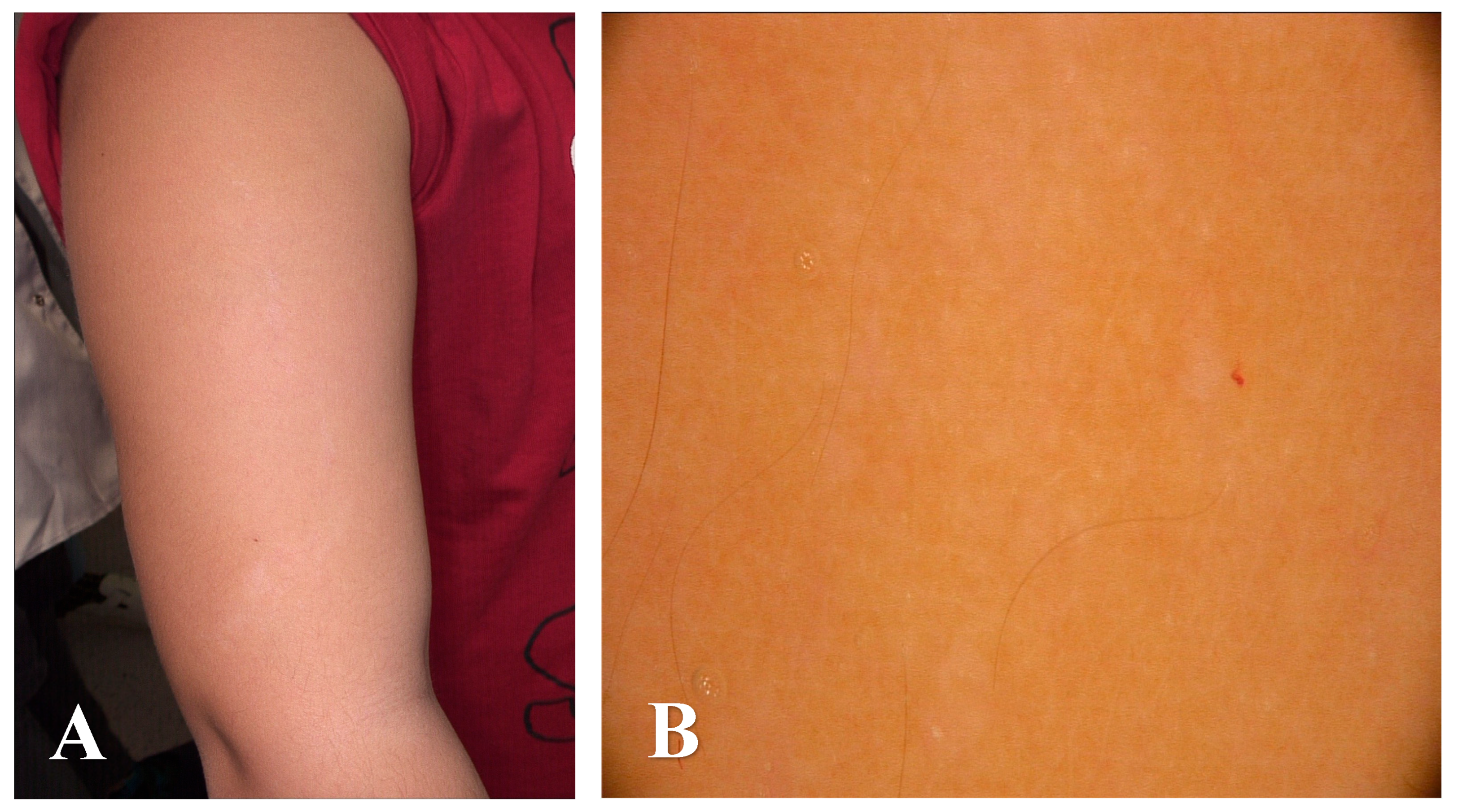
Lichen Striatus Albus–A Case Report of a Rare Disease Entity
Abstract


| Lichen Striatus | Blaschkitis | ILVEN | Linear Lichen Planus | Linear Scleroderma | Hypomelanosis of Ito | |
|---|---|---|---|---|---|---|
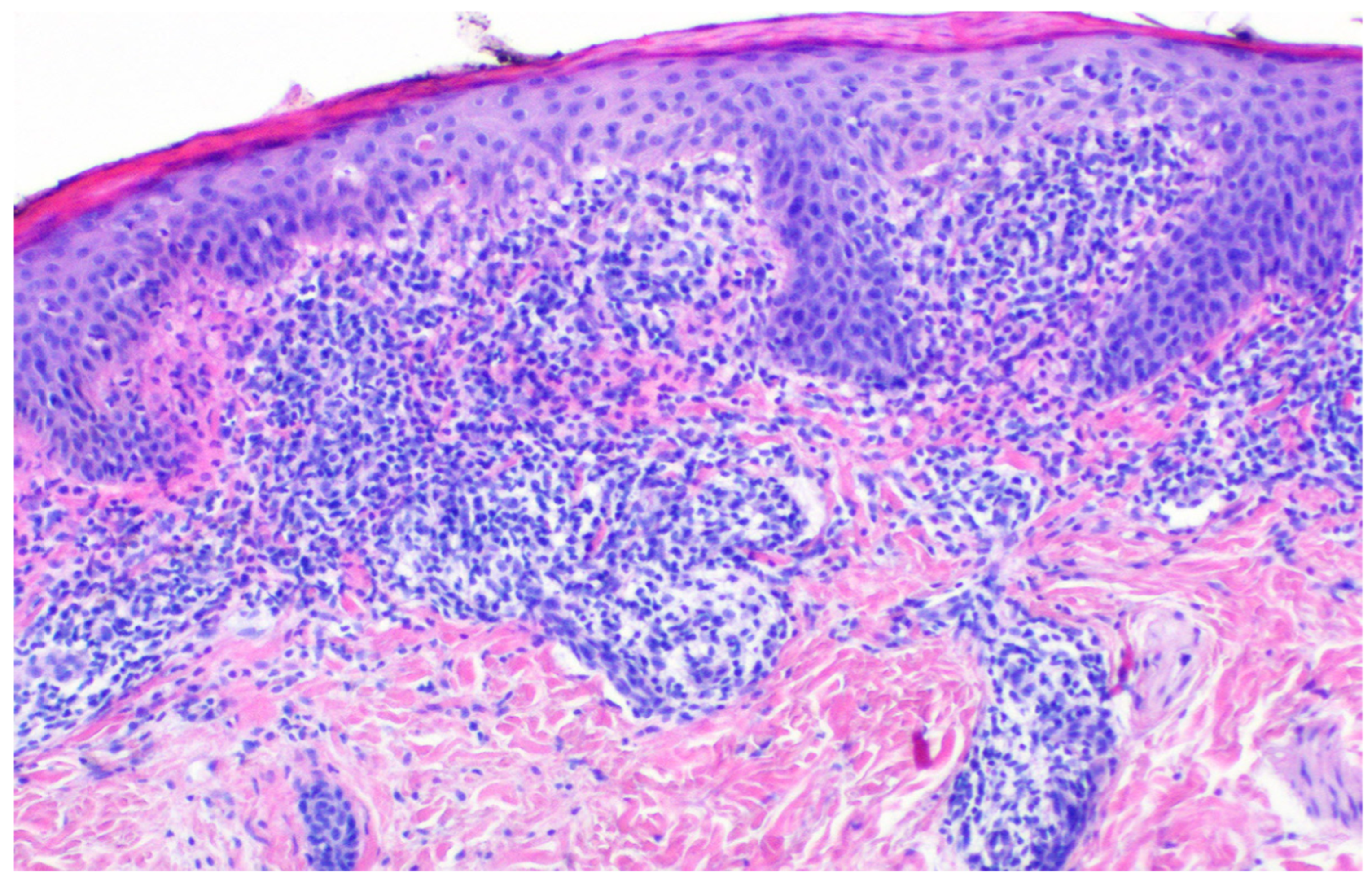
| Epidermis | Mild acanthosis, focal parakeratosis, hyperkeratosis; basal vacuolization and Civatte bodies may be present [3]. | Spongiosis (more pronounced compared to LS); mild acanthosis; parakeratosis possible [8,9]. | Alternating areas of hypergranulosis with orthokeratosis and agranulosis with parakeratosis (“checkerboard” pattern); psoriasiform hyperplasia [10,11]. | Hypergranulosis, wedge-shaped hyperkeratosis, irregular acanthosis, saw-tooth rete ridges; Civatte bodies may be present [12]. | Usually unremarkable or mild atrophy [13]. | Reduced melanocyte density in the basal epidermis. Decreased number and size of basal melanosomes. Selective reduction in eumelanin. Increased Langerhans cell presence in depigmented areas [14]. |
| Dermis | Dense, band-like lichenoid infiltrate at the dermoepidermal junction (DEJ) (similar to LP) associated with perivascular and periappendagealinflammatory infiltrate–involvement of eccrine glands and ducts distinguishes LS from other lichenoid dermatosies [3]. | Less lichenoid pattern; minimal eccrine involvement [8,9]. | Mild superficial perivascular infiltrate; less prominent than in LS [10,11]. | Dense, band-like lichenoid infiltrate along the DEJ; involvement limited to the upper dermis [12,15]. | Thickened, hyalinized collagen bundles; loss of adnexal structures; perivascular lymphocytes [13]. | Absence of extracellular melanin (no signs of pigment leakage). No histological signs of inflammatory infiltration [14]. |
Author Contributions
Funding
Institutional Review Board Statement
Informed Consent Statement
Data Availability Statement
Conflicts of Interest
References
- Peramiquel, L.; Baselga, E.; Dalmau, J.; Roé, E.; del Mar Campos, M.; Alomar, A. Lichen striatus: Clinical and epidemiological review of 23 cases. Eur. J. Pediatr. 2006, 165, 267–269. [Google Scholar] [CrossRef] [PubMed]
- Patrizi, A.; Neri, I.; Fiorentini, C.; Bonci, A.; Ricci, G. Lichen striatus: Clinical and laboratory features of 115 children. Pediatr. Dermatol. 2004, 21, 197–204. [Google Scholar] [CrossRef] [PubMed]
- Syed, H.A.; Jamil, R.T.; Ramphul, K. Lichen Striatus. In StatPearls; StatPearls Publishing: Treasure Island, FL, USA. Available online: https://www.ncbi.nlm.nih.gov/books/NBK507830/ (accessed on 22 January 2025).
- Miller, R.C.; Lipner, S.R. Lichen Striatus. J. Cutan. Med. Surg. 2022, 26, 649. [Google Scholar] [CrossRef] [PubMed]
- Nair, J.V.; Guntreddi, G.; Nirujogi, S. An Unusual Rash in a Five-Year-Old Girl: Blaschkoid Distribution Is the Key to the Diagnosis. Cureus 2020, 12, e12124. [Google Scholar] [CrossRef]
- Leung, A.K.; Lam, J.M.; Barankin, B.; Leong, K.F. Lichen Striatus: An Updated Review. Curr. Pediatr. Rev. 2024, 21, 233–244. [Google Scholar] [CrossRef] [PubMed]
- Herzum, A.; Viglizzo, G.; Occella, C.; Gariazzo, L.; Vellone, V.G.; Orsi, S.M.; Ciccarese, G. Lichen striatus: A review. Ital. J. Dermatol. Venereol. 2024, 160, 242–247. [Google Scholar] [CrossRef] [PubMed]
- Keegan, B.R.; Kamino, H.; Fangman, W.; Shin, H.T.; Orlow, S.J.; Schaffer, J.V. “Pediatric blaschkitis”: Expanding the spectrum of childhood acquired Blaschko-linear dermatoses. Pediatr. Dermatol. 2007, 24, 621–627. [Google Scholar] [CrossRef] [PubMed]
- Al-Balbeesi, A. Adult Blaschkitis with Lichenoid Features and Blood Eosinophilia. Cureus 2021, 13, e16846. [Google Scholar] [CrossRef] [PubMed]
- Dupre, A.; Christol, B. Inflammatory linear verrucous epidermal nevus: A new epidermal nevus. Bull. Soc. Fr. Dermatol. Syphiligr. 1977, 84, 403–406. [Google Scholar]
- Kaddu, S.; Soyer, H.P.; Hödl, S.; Kerl, H. ILVEN and linear psoriasis: A histopathologic study of 13 cases. J. Cutan. Pathol. 1998, 25, 436–442. [Google Scholar]
- Boyd, A.S.; Neldner, K.H. Lichen planus. J. Am. Acad. Dermatol. 1991, 25, 593–619. [Google Scholar] [CrossRef] [PubMed]
- Walker, D.; Susa, J.S.; Currimbhoy, S.; Jacobe, H. Histopathological changes in morphea and their clinical correlates: Results from the Morphea in Adults and Children Cohort V. J. Am. Acad. Dermatol. 2017, 76, 1124–1130. [Google Scholar] [CrossRef] [PubMed]
- Chamli, A.; Litaiem, N. Hypomelanosis of Ito. In StatPearls; StatPearls Publishing: Treasure Island, FL, USA, 2025. Available online: https://www.ncbi.nlm.nih.gov/books/NBK538268/ (accessed on 20 June 2023).
- Boch, K.; Langan, E.A.; Kridin, K.; Zillikens, D.; Ludwig, R.J.; Bieber, K. Lichen Planus. Front. Med. 2021, 8, 737813. [Google Scholar] [CrossRef] [PubMed]
Disclaimer/Publisher’s Note: The statements, opinions and data contained in all publications are solely those of the individual author(s) and contributor(s) and not of MDPI and/or the editor(s). MDPI and/or the editor(s) disclaim responsibility for any injury to people or property resulting from any ideas, methods, instructions or products referred to in the content. |
© 2025 by the authors. Licensee MDPI, Basel, Switzerland. This article is an open access article distributed under the terms and conditions of the Creative Commons Attribution (CC BY) license (https://creativecommons.org/licenses/by/4.0/).
Share and Cite
Zagórska, B.; Żółkiewicz, J.; Biernat, W.; Sobjanek, M.; Sławińska, M. Lichen Striatus Albus–A Case Report of a Rare Disease Entity. Diagnostics 2025, 15, 2367. https://doi.org/10.3390/diagnostics15182367
Zagórska B, Żółkiewicz J, Biernat W, Sobjanek M, Sławińska M. Lichen Striatus Albus–A Case Report of a Rare Disease Entity. Diagnostics. 2025; 15(18):2367. https://doi.org/10.3390/diagnostics15182367
Chicago/Turabian StyleZagórska, Beata, Jakub Żółkiewicz, Wojciech Biernat, Michał Sobjanek, and Martyna Sławińska. 2025. "Lichen Striatus Albus–A Case Report of a Rare Disease Entity" Diagnostics 15, no. 18: 2367. https://doi.org/10.3390/diagnostics15182367
APA StyleZagórska, B., Żółkiewicz, J., Biernat, W., Sobjanek, M., & Sławińska, M. (2025). Lichen Striatus Albus–A Case Report of a Rare Disease Entity. Diagnostics, 15(18), 2367. https://doi.org/10.3390/diagnostics15182367


