Sella Turcica and Cranial Base Symmetry in Anterior Synostotic Plagiocephaly Patients: A Retrospective Case–Control Study
Abstract
1. Introduction
2. Materials and Methods
2.1. Study Design
2.2. Sample Size Calculation
2.3. Patient Selection
- -
- Patients with anterior synostotic plagiocephaly;
- -
- Adult patients of Caucasian ethnicity between 20 and 40 years of age;
- -
- Patients with CT examinations of the facial massif.
- -
- Patients with a history of orthodontic treatment or orthognathic surgery;
- -
- Patients with a history of trauma and neoplasms in the craniofacial region;
- -
- Patients with a long-term history of drug use that may have affected bone development.
2.4. Protocol and Measurements for 2D Point-Based Quantitative Analysis
- -
- CT orientation: The CT scans were reoriented using the axial, coronal, and sagittal reference planes;
- -
- CT segmentation: The following software tools were used in the following order: “Thresholding” was used to identify a “mask” with the cranial base through manual selection of pixel intensity values; “Crop mask” allowed for outlining the region of interest; and “Calculate 3D” enabled the generation of a three-dimensional model from the contours of the previously segmented mask.
2.5. Two-Dimensional Point-Based Quantitative Analysis: Sella Turcica
- -
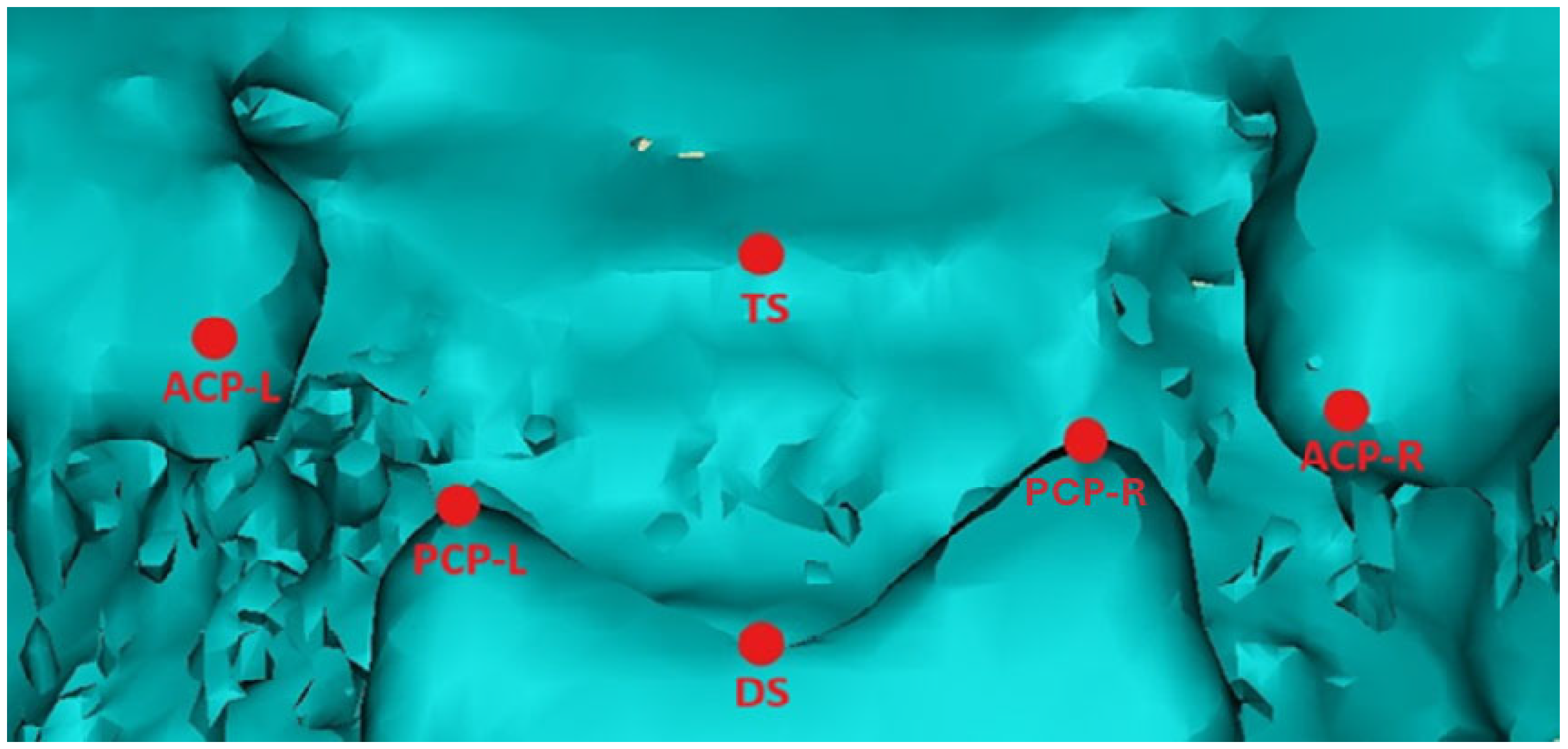
- Anterior clinoid distance: The distance between the right and left anterior clinoid processes (ACPs).
- -
- Posterior clinoid distance: The distance between the right and left posterior clinoid processes (PCPs).
- -
- Left sella length: The distance between the left (L) anterior and left posterior clinoid processes.
- -
- Right sella length: The distance between the right (R) anterior and right posterior clinoid processes.
- -
- Median sella length: The distance between the tuberculum sellae (TS) and the dorsum sellae (DS).
2.6. Two-Dimensional Point-Based Quantitative Analysis: Skull Base
- -
- The CSX angle for the anterior cranial fossa, between the crista galli (C), sella turcica (S), and xiphoid process of the lesser wing of the sphenoid (X);
- -
- The XSM angle for the middle cranial fossa, between the xiphoid process (X), sella turcica (S), and internal acoustic meatus (M);
- -
- The MSO angle for the posterior cranial fossa, between the internal acoustic meatus (M), sella turcica (S), and opisthion (O).
- -
- CX length for the anterior cranial fossa between the crista galli (C) and xiphoid process of the lesser wing of the sphenoid (X);
- -
- XM length for the middle cranial fossa between the xiphoid process (X) and the internal acoustic meatus (M);
- -
- MO length for the posterior cranial fossa between the internal acoustic meatus (M) and opisthion (O).
2.7. Qualitative 3D Surface-Based Analysis: Sella Turcica
- -
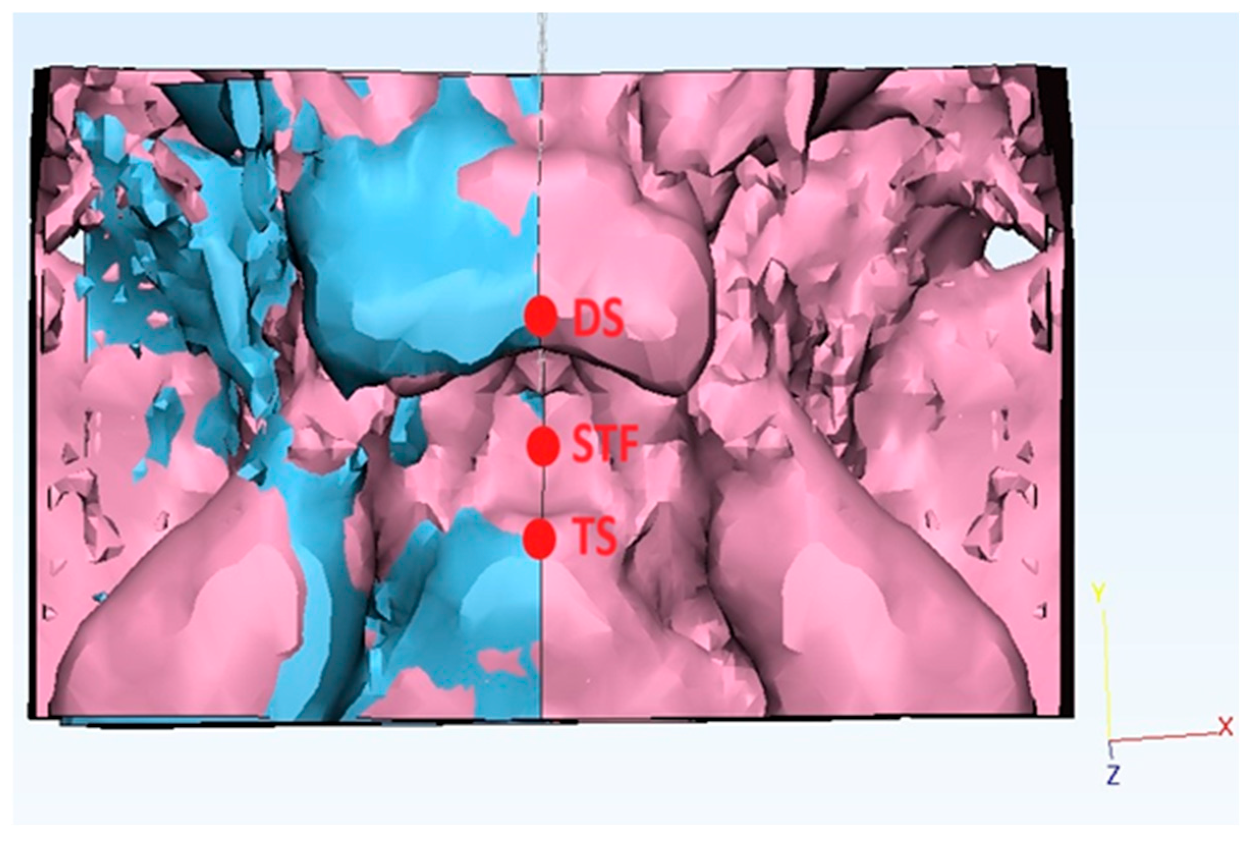
- Firstly, the “new sketch” tool was used to generate a cutting plane passing by a selection of three median landmarks (Figure 3): the tuberculum sellae (TS), the sella turcica floor (STF) (lowest point of the pituitary fossa of the sphenoid), and the dorsum sellae (DS);
- -
- Secondly, the “Cut” function was performed to split the sella turcica along the abovementioned plane into two hemi-portions: the “unhealthy side” (ipsilateral to the ASP) from the “healthy side” (contralateral to the ASP);
- -
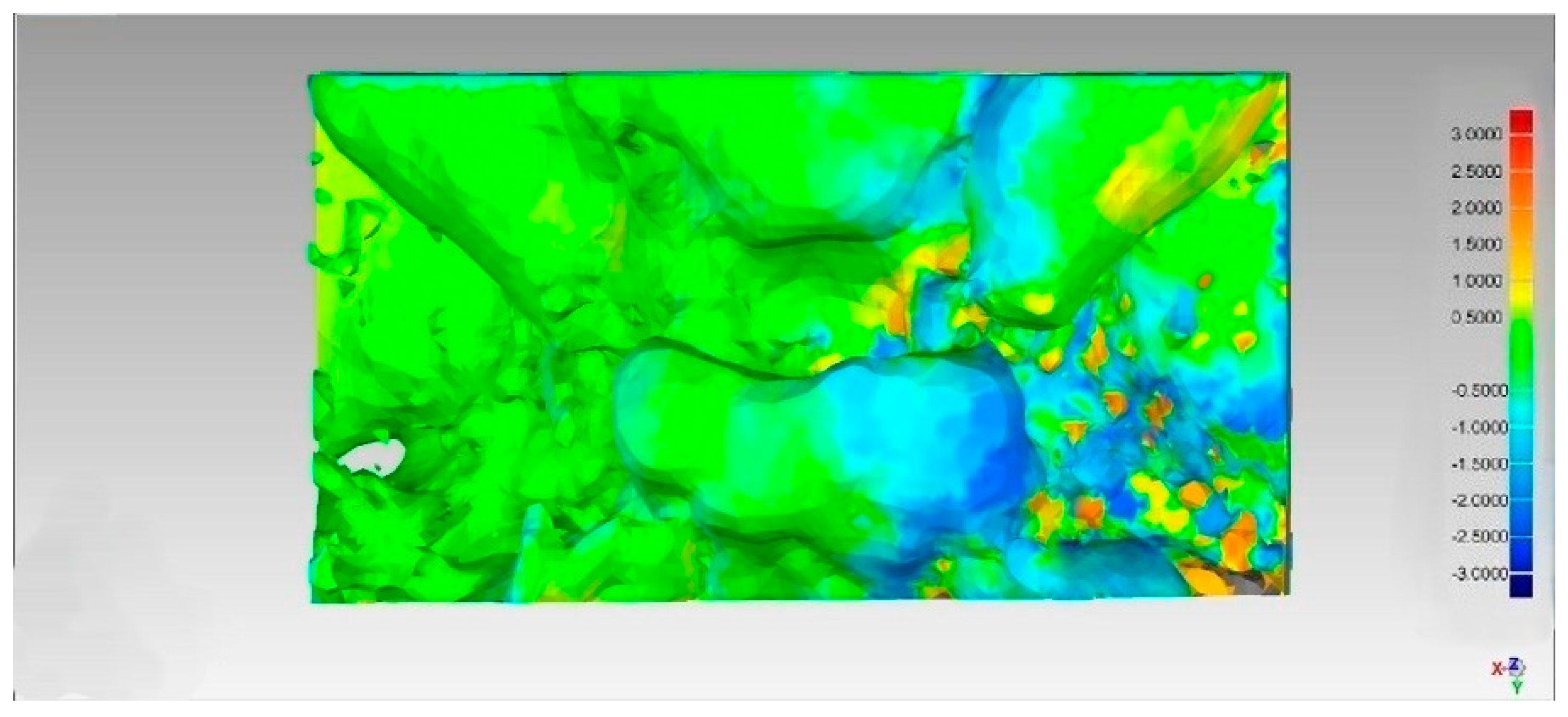
- Thirdly, the “Mirror” function was used to create a mirror copy of the “unhealthy side”.
2.8. Statistical Analysis
Affected side)) × 100
3. Results
3.1. Two-Dimensional Asymmetry
- -
- Sella length (defined as the comparison between the right and left sella lengths), with asymmetry index values of 5.94 (study) vs. 1.62 (control);
- -
- The anterior cranial fossa length measurements (defined as the comparison between the right and left CX lengths) were 39.62 ± (4.89) for the ill side and 46.50 ± (5.56) for the healthy side in the study group vs. 41.98 ± (3.22) for the ill side and 41.96 ± (3.23) for the healthy side, with asymmetry index values of 7.96 (study) vs. 0.02 (control);
- -
- The anterior cranial fossa angle widths (defined as the comparison between the right and left CSX angles) were 48.23 ± (6.80) for the ill side and 58.25 ± (7.43) for the healthy side in the study group vs. 51.84 ± (2.42) for the ill side and the 52.43 ± (3.76) for healthy side, with asymmetry index values of 7.96 (study) vs. 0.02 (control) and asymmetry index values of 9.42 (study) vs. 0.51 (control).
3.2. Three-Dimensional Asymmetry
3.3. Reliability
4. Discussion
4.1. Discussion of the Results of the Two-Dimensional Point-Based Quantitative Analysis
4.2. Discussion of the Results of the Three-Dimensional Surface-Based Qualitative Analysis
4.3. Limitations of This Study
5. Conclusions
Supplementary Materials
Author Contributions
Funding
Institutional Review Board Statement
Informed Consent Statement
Data Availability Statement
Acknowledgments
Conflicts of Interest
Abbreviations
| ASP | Anterior Synostotic Plagiocephaly |
| CT | Computed Tomography |
| 2D | Two-Dimensional |
| 3D | Three-Dimensional |
| CBCT | Cone Beam Computed Tomography |
| AI | Asymmetry Index |
| IRCCS | Scientific Institute for Research Hospitalization and Healthcare |
| ID | Identification Number |
| STROBE | Strengthening the Reporting of Observational Studies in Epidemiology |
| DICOM | Digital Imaging and Communications in Medicine |
| ACP | Anterior Clinoid Process |
| PCP | Posterior Clinoid Process |
| TS | Tuberculum Sellae |
| DS | Dorsum Sellae |
| C | Crista Galli |
| S | Sella Turcica |
| O | Opisthion |
| X | Xiphoid Process of the Lesser Wing of the Sphenoid |
| M | Internal Acoustic Meatus |
| CSO angle | Crista Galli Sella Opisthion Angle |
| CSX angle | Crista Galli Sella Xiphoid Angle |
| XSM angle | Xiphoid Sella Meatus Angle |
| MSO angle | Meatus Sella Opisthion Angle |
| CX length | Crista Galli Xiphoid Length |
| XM length | Xiphoid Meatus Length |
| MO length | Meatus Opisthion Length |
| STF | Sella Turcica Floor |
| STL | Standard Tessellation Language |
| RMS | Root Mean Square |
| p-value | Probability Value |
References
- Losee, J.E.; Mason, A.C. Deformational plagiocephaly: Diagnosis, prevention, and treatment. Clin. Plast. Surg. 2005, 32, 53–64. [Google Scholar] [CrossRef]
- Di Rocco, C.; Paternoster, G.; Caldarelli, M.; Massimi, L.; Tamburrini, G. Anterior plagiocephaly: Epidemiology, clinical findings, diagnosis, and classification. A review. Childs Nerv. Syst. 2012, 28, 1413–1422. [Google Scholar] [CrossRef] [PubMed]
- Gasparini, G.; Saponaro, G.; Moro, A.; De Angelis, P.; Pelo, S. Early surgical treatment in anterior synostotic plagiocephaly: Is. this the best choice? J. Craniofac. Surg. 2018, 29, 2166–2172. [Google Scholar] [CrossRef] [PubMed]
- Cucchi, A.; Vignudelli, E.; Napolitano, A.; Marchetti, C.; Corinaldesi, G. Evaluation of complication rates and vertical bone gain after guided bone regeneration with non-resorbable membranes versus titanium meshes and resorbable membranes. A randomized clinical trial. Clin. Implant. Dent. Relat. Res. 2017, 19, 821–832. [Google Scholar] [CrossRef] [PubMed]
- Gasparini, G.; Saponaro, G.; Marianetti, T.M.; Tamburrini, G.; Moro, A.; Di Rocco, C.; Pelo, S. Mandibular alterations and facial lower third asymmetries in unicoronal synostosis. Childs Nerv. Syst. 2013, 29, 665–671. [Google Scholar] [CrossRef] [PubMed]
- Corridore, D.; Saccucci, M.; Zumbo, G.; Fontana, E.; Lamazza, L.; Stamegna, C.; Di Carlo, G.; Vozza, I.; Guerra, F. Impact of Stress on Periodontal Health: Literature Revision. Healthcare 2023, 11, 1516. [Google Scholar] [CrossRef]
- Di Spirito, F.; Pisano, M.; Di Palo, M.P.; Franci, G.; Rupe, A.; Fiorino, A.; Rengo, C. Peri-Implantitis-Associated Microbiota before and after Peri-Implantitis Treatment, the Biofilm “Competitive Balancing” Effect: A Systematic Review of Randomized Controlled Trials. Microorganisms 2024, 12, 1965. [Google Scholar] [CrossRef]
- Di Carlo, G.; Gurani, S.F.; Pinholt, E.M.; Cattaneo, P.M. A new simple three-dimensional method to characterize upper airway in orthognathic surgery patient. Dentomaxillofac Radiol. 2017, 46, 20170042. [Google Scholar] [CrossRef]
- Saponaro, G.; Pelo, S.; Gasparini, G.; Torado, M.; Cerbelli, E.; Moro, A.; Doneddu, P. Approach to fronto-orbital sequelae in anterior synostotic plagiocephaly: Our flow chart based on Di Rocco classification. J. Craniofac Surg. 2021, 32, 1986–1989. [Google Scholar] [CrossRef]
- Kim, Y.H.; Sato, K.; Mitani, H.; Shimizu, Y.; Kikuchi, M. Asymmetry of the sphenoid bone and its suitability as a reference for analyzing craniofacial asymmetry. Am. J. Orthod. Dentofacial Orthop. 2003, 124, 656–662. [Google Scholar] [CrossRef]
- Azzi, L.; Moretto, P.; Vinci, R.; Croveri, F.; Boggio, A.; Silvestre-Rangil, J.; Tettamanti, L.; Tagliabue, A.; Passi, A. Human β2-defensin in oral lichen planus expresses the degree of inflammation. J. Biol. Regul. Homeost. Agents. 2017, 31 (Suppl. S1), 77–87. [Google Scholar] [PubMed]
- Pelo, S.; Tamburrini, G.; Marianetti, T.M.; Saponaro, G.; Moro, A.; Gasparini, G.; Di Rocco, C. Correlations between the abnormal development of the skull base and facial skeleton growth in anterior synostotic plagiocephaly: The predictive value of a classification based on CT scan examination. Childs Nerv. Syst. 2011, 27, 1431–1443. [Google Scholar] [CrossRef] [PubMed]
- Luzzi, V.; Mazur, M.; Guaragna, M.; Di Carlo, G.; Cotticelli, L.; Magliulo, G.; Marasca, B.; Pirro, V.; Di Giorgio, G.; Ndokaj, A.; et al. Correlations of Obstructive Sleep Apnea Syndrome and Daytime Sleepiness with the Risk of Car Accidents in Adult Working Population: A Systematic Review and Meta-Analysis with a Gender-Based Approach. J. Clin. Med. 2022, 11, 3971. [Google Scholar] [CrossRef]
- Blasi, A.; Nucera, R.; Ronsivalle, V.; Candida, E.; Grippaudo, C. Asymmetry Index for the photogrammetric assessment of facial asymmetry. Am. J. Orthod. Dentofacial. Orthop. 2022, 162, 394–402. [Google Scholar] [CrossRef] [PubMed]
- Alqattan, M.; Djordjevic, J.; Zhurov, A.I.; Richmond, S. Comparison between landmark and surface-based three-dimensional analyses of facial asymmetry in adults. Eur. J. Orthod. 2015, 37, 1–12. [Google Scholar] [CrossRef] [PubMed]
- Calandrelli, R.; Pilato, F.; Massimi, L.; Panfili, M.; Di Rocco, C.; Colosimo, C. Quantitative analysis of cranial-orbital changes in infants with anterior synostotic plagiocephaly. Childs Nerv. Syst. 2018, 34, 1725–1733. [Google Scholar] [CrossRef]
- Ortiz, P.M.; Tabbaa, S.; Flores-Mir, C.; Al-Jewair, T. A CBCT investigation of the association between sella-turcica bridging and maxillary palatal canine impaction. Biomed. Res. Int. 2018, 2018, 4329050. [Google Scholar] [CrossRef]
- Ugurlu, M.; Bayrakdar, I.S.; Kahraman, F.; Oksayan, R.; Dagsuyu, I.M. Evaluation of the relationship between impacted canines and three-dimensional sella morphology. Surg. Radiol. Anat. 2020, 42, 23–29. [Google Scholar] [CrossRef]
- Yan, S.; Huang, S.; Wu, Z.; Liu, Y.; Men, Y.; Nie, X.; Guo, J. A CBCT investigation of the sella turcica dimension and sella turcica bridging in different vertical growth patterns. J. Clin. Med. 2023, 12, 1890. [Google Scholar] [CrossRef]
- Captier, G.; Leboucq, N.; Bigorre, M.; Canovas, F.; Bonnel, F.; Bonnafe, A.; Montoya, P. Plagiocephaly: Morphometry of skull base asymmetry. Surg. Radiol. Anat. 2003, 25, 226–233. [Google Scholar] [CrossRef]
- Springate, S.D. The effect of sample size and bias on the reliability of estimates of error: A comparative study of Dahlberg’s formula. Eur. J. Orthod. 2012, 34, 158–163. [Google Scholar] [CrossRef] [PubMed]
- Türker, G.; Öztürk Yaşar, M. Evaluation of associations between condylar morphology, ramus height, and mandibular plane angle in various vertical skeletal patterns: A digital radiographic study. BMC Oral. Health. 2022, 22, 330. [Google Scholar] [CrossRef]
- Son, K.; Lee, W.S.; Lee, K.B. Effect of different software programs on the accuracy of dental scanner using three-dimensional analysis. Int. J. Environ. Res. Public. Health. 2021, 18, 8449. [Google Scholar] [CrossRef]
- Marsh, J.L.; Gado, M.H.; Vannier, M.W.; Stevens, W.G. Osseous anatomy of unilateral coronal synostosis. Cleft Palate J. 1986, 23, 87–95. [Google Scholar] [PubMed]
- Paduano, S.; Rongo, R.; Lucchese, A.; Aiello, D.; Michelotti, A.; Grippaudo, C. Late-Developing Supernumerary Premolars: Analysis of Different Therapeutic Approaches. Case Rep. Dent. 2016, 2016, 2020489. [Google Scholar] [CrossRef]
- Farmer, D.T.; Mlcochova, H.; Zhou, Y.; Koelling, N.; Wang, G.; Ashley, N.; Bugacov, H.; Chen, H.J.; Parvez, R.; Tseng, K.C.; et al. The developing mouse coronal suture at single-cell resolution. Nat. Commun. 2021, 12, 4797. [Google Scholar] [CrossRef] [PubMed]


| Landmark | Sagittal Plane Description | Coronal Plane Description |
|---|---|---|
| ACP-R (anterior clinoid process—right side) | Most anterior and superior point of the right anterior clinoid process apex. | Most lateral and superior projection of the right anterior clinoid process. |
| ACP-L (anterior clinoid process—left side) | Most anterior and superior point of the left anterior clinoid process apex. | Most lateral and superior projection of the left anterior clinoid process. |
| PCP-R (posterior clinoid process—right side) | Most posterior and superior point of the right posterior clinoid process apex. | Most medial and superior projection of the right posterior clinoid process. |
| PCP-L (left posterior clinoid process—left side) | Most posterior and superior point of the left posterior clinoid process apex. | Most medial and superior projection of the left posterior clinoid process. |
| DS (Dorsum sellae point) | Most posterior and superior point on the midline of the dorsum sellae upper margin. | Central point on the upper margin of the dorsum sellae, equidistant from PCP-R and PCP-L. |
| TS (Tuberculum sellae) | Most anterior and superior point on the midline of the tuberculum sellae margin. | Central point on the anterior margin of the sella turcica, equidistant from ACP-R and ACP-L. |
| Linear Measurements | Study Group | Control Group | p-Value |
|---|---|---|---|
| Anterior Clinoid Distance | 23.20 ± (2.82) | 24.28 ± (2.67) | 0.41 |
| Posterior Clinoid Distance | 13.88 ± (3.02) | 12.13 ± (2.44) | 0.19 |
| Median sella Length | 9.29 ± (1.31) | 9.53 ± (1.35) | 0.71 |
| Diseased-side sella length | 7.21 ± (1.68) | 7.57 ± (1.83) | 0.67 |
| Healthy-side sella length | 8.16 ± (1.97) | 7.29 ± (1.64) | 0.33 |
| A.I. Sella length | 5.94 | 1.62 | 0.02* |
| Linear Measurements | Study Group | Control Group | p-Value |
|---|---|---|---|
| Ill-side anterior cranial fossa length | 39.62 ± (4.89) | 41.98 ± (3.22) | 0.24 |
| Healthy-side anterior cranial fossa length | 46.50 ± (5.56) | 41.96 ± (3.23) | 0.05 |
| A.I. anterior cranial fossa length | 7.96 | 0.02 | 0.01* |
| Ill-side median cranial fossa length | 53.16 ± (3.46) | 56.89 ± (6.08) | 0.12 |
| Healthy-side median cranial fossa length | 56.27 ± (3.22) | 56.23 ± (6.21) | 0.98 |
| A.I. middle cranial fossa length | 2.86 | 0.62 | 0.00* |
| Ill-side posterior cranial fossa length | 46.03 ± (4.39) | 44.32 ± (6.37) | 0.51 |
| Healthy-side posterior cranial fossa length | 45.85 ± (5.02) | 44.16 ± (5.67) | 0.51 |
| A.I. posterior cranial fossa length | 0.25 | 0.05 | 0.79 |
| Angular Widths | Study Group | Control Group | p-Value |
|---|---|---|---|
| Ill-side anterior cranial fossa angle | 48.23 ± (6.80) | 51.84 ± (2.42) | 0.15 |
| Healthy-side anterior cranial fossa angle | 58.25 ± (7.43) | 52.43 ± (3.76) | 0.05 |
| A.I. anterior cranial fossa angle | 9.42 | 0.51 | 0.02 * |
| Ill-side middle cranial fossa angle | 84.27 ± (4.52) | 87.82 ± (8.64) | 0.29 |
| Healthy-side middle cranial fossa angle | 88.69 ± (3.87) | 89.00 ± (8.79) | 0.92 |
| A.I. middle cranial fossa angle | 2.57 | 0.65 | 0.09 |
| Ill-side posterior cranial fossa angle | 40.6 ± (2.57) | 40.48 ± (7.32) | 0.96 |
| Healthy-side posterior cranial fossa angle | 40.05 ± (3.53) | 40.26 ± (6.78) | 0.93 |
| A.I. posterior cranial fossa angle | 0.76 | 1.77 | 0.62 |
Disclaimer/Publisher’s Note: The statements, opinions and data contained in all publications are solely those of the individual author(s) and contributor(s) and not of MDPI and/or the editor(s). MDPI and/or the editor(s) disclaim responsibility for any injury to people or property resulting from any ideas, methods, instructions or products referred to in the content. |
© 2025 by the authors. Licensee MDPI, Basel, Switzerland. This article is an open access article distributed under the terms and conditions of the Creative Commons Attribution (CC BY) license (https://creativecommons.org/licenses/by/4.0/).
Share and Cite
Staderini, E.; Guerrieri, D.; Tepedino, M.; Saponaro, G.; Moro, A.; Gasparini, G.; Gallenzi, P.; Cordaro, M. Sella Turcica and Cranial Base Symmetry in Anterior Synostotic Plagiocephaly Patients: A Retrospective Case–Control Study. Diagnostics 2025, 15, 2199. https://doi.org/10.3390/diagnostics15172199
Staderini E, Guerrieri D, Tepedino M, Saponaro G, Moro A, Gasparini G, Gallenzi P, Cordaro M. Sella Turcica and Cranial Base Symmetry in Anterior Synostotic Plagiocephaly Patients: A Retrospective Case–Control Study. Diagnostics. 2025; 15(17):2199. https://doi.org/10.3390/diagnostics15172199
Chicago/Turabian StyleStaderini, Edoardo, Davide Guerrieri, Michele Tepedino, Gianmarco Saponaro, Alessandro Moro, Giulio Gasparini, Patrizia Gallenzi, and Massimo Cordaro. 2025. "Sella Turcica and Cranial Base Symmetry in Anterior Synostotic Plagiocephaly Patients: A Retrospective Case–Control Study" Diagnostics 15, no. 17: 2199. https://doi.org/10.3390/diagnostics15172199
APA StyleStaderini, E., Guerrieri, D., Tepedino, M., Saponaro, G., Moro, A., Gasparini, G., Gallenzi, P., & Cordaro, M. (2025). Sella Turcica and Cranial Base Symmetry in Anterior Synostotic Plagiocephaly Patients: A Retrospective Case–Control Study. Diagnostics, 15(17), 2199. https://doi.org/10.3390/diagnostics15172199


